Biochemical Factors Influencing Measurement of Cardiac Troponin I ...
Biochemical Factors Influencing Measurement of Cardiac Troponin I ...
Biochemical Factors Influencing Measurement of Cardiac Troponin I ...

You also want an ePaper? Increase the reach of your titles
YUMPU automatically turns print PDFs into web optimized ePapers that Google loves.
Clin Chem Lab Med 1999; 37(11/12):1091–1095 © 1999 by Walter de Gruyter · Berlin · New York<strong>Biochemical</strong> <strong>Factors</strong> <strong>Influencing</strong> <strong>Measurement</strong> <strong>of</strong> <strong>Cardiac</strong> <strong>Troponin</strong> Iin SerumA. Katrukha 1 , A. Bereznikova 1 , V. Filatov 2 andT. Esakova 31HyTest Ltd., Turku, Finland2 Department <strong>of</strong> Bioorganic Chemistry, Moscow StateUniversity, Moscow, Russia3Moscow Institute <strong>of</strong> Medical Ecology, Moscow, Russia<strong>Troponin</strong> I (cTnI), a sensitive and reliable marker <strong>of</strong>damaged cardiac tissue, is now widely used in clinics.But the existence <strong>of</strong> different cTnI assays with a widevariety <strong>of</strong> cut-<strong>of</strong>f values and discrepancies betweenthe results <strong>of</strong> measurements <strong>of</strong> one and the same sampleby different assays is puzzling for clinicians. Themost urgent issue at the moment is the development<strong>of</strong> the international standard, which can be used forthe calibration <strong>of</strong> different assays, thus decreasing betweenassay biases. But another important item,which should be considered by manufacturers, is thestandardisation <strong>of</strong> the epitopes <strong>of</strong> the antibodies usedfor the assay development. The importance <strong>of</strong> suchstandardisation originates from the complicated biochemicalnature <strong>of</strong> cTnI. Here we briefly try to analysethe main factors that can influence antigen recognitionby different antibodies and formulate principles <strong>of</strong> antibodyselection for assay development.Key words: <strong>Cardiac</strong> troponin I; <strong>Measurement</strong>; Standardisation;Immunodetection; Acute myocardial infarction;<strong>Cardiac</strong> marker.IntroductionAntibody recognition <strong>of</strong> a protein can be affected byconformational changes <strong>of</strong> the antigen, a wide variety <strong>of</strong>post-translational modifications, or epitope blocking bydifferent macromolecules. The complicated biochemistry<strong>of</strong> cTnI in the living cell and long-term incubation(≥3 hours) in necrotic cardiac tissue after acute myocardialinfarction (AMI) can result in significant changes inp r i m a r y, secondary and ternary structure <strong>of</strong> the cTnImolecule and we can suppose that protein in the patients’blood is different from newly synthesised or recombinantprotein. To understand what factors can beimportant for accurate cTnI immunodetection, we willtry to analyse some biochemical features <strong>of</strong> this protein.Complex FormationcTnI is a component <strong>of</strong> the troponin complex, responsiblefor the regulation <strong>of</strong> muscle contraction. The troponincomplex consists <strong>of</strong> three different molecules:troponin C (TnC) with molecular weight 18 kDa and pI4.1, cTnI, with molecular weight 23 kDa and pI 9.9, andtroponin T (cTnT), with molecular weight 34 kDa and pI5.1. All three components interact with each other withdifferent strengths. The strongest interaction wasdemonstrated for cTnI – cTnT complex (K A = 4.4 x 10 –5M –1 ) and especially for cTnI – TnC complex (in the presence<strong>of</strong> Ca 2+ ) K A = 1.5 x 10 –8 M –1 ) (1). Complex formationbetween cTnI and TnC and possibly between cTnI andcTnT can make some epitopes unrecognisable bysome anti-cTnI antibodies.Recently it was demonstrated (2, 3) that formation <strong>of</strong>the cTnI-TnC binary complex is actually very importantfor the recognition <strong>of</strong> the antigen by some monoclonalantibodies (MAbs) and for the immunodetection <strong>of</strong> thecTnI in AMI blood samples. Bodor et al. (2), studying apanel <strong>of</strong> cTnI-specific MAbs, noted that some MAbsbetter recognise the complexed than the free form <strong>of</strong>the antigen. Katrukha et al. (3) noticed that amongtested monoclonal antibodies one MAb was unable torecognise the complexed form <strong>of</strong> the antigen whereasseveral MAbs better recognised the free form <strong>of</strong> theprotein; others were not affected by complex formationat all. In the same article, the authors demonstratedthat the main part <strong>of</strong> cTnI in the blood <strong>of</strong> AMI patients iscomplexed with TnC and only 5–10% <strong>of</strong> the antigen existsas a free molecule. This finding was confirmed laterby other scientists (4, 5). If the assay utilises antibodiesspecific to the free form <strong>of</strong> the antigen and not recognisingcTnI in binary complex with troponin C, it willmiss the major part <strong>of</strong> cTnI in the patient’s blood sample.Wu et al. (4) described the effect <strong>of</strong> cTnI-cTnT complexformation on several commercially available assays.Some <strong>of</strong> the tested assays better recognised thefree form <strong>of</strong> the antigen, others, the complexed; forsome <strong>of</strong> them, there was no difference. Segura et al.,studying the effect <strong>of</strong> chelating agents on three commerciallyavailable assays (6), conclude that the Accessassay better recognises the free form <strong>of</strong> the proteincomparing with the complexed.Two different assays, one <strong>of</strong> which better recognisesthe free form <strong>of</strong> the protein and another, the complexed,will give different results with one and thesame patient’s sample, thus confusing clinicians.So it can be concluded that cTnI-TnC complex formationis a very important factor influencing immunochemicaldetection <strong>of</strong> cTnI. For precise cTnI immunodetectionin patient’s blood, the antibodies used in theassay should be insensitive to cTnI-TnC binary complexformation. To decrease the discrepancy between existingassays, it is a matter <strong>of</strong> great importance to calibrateassays against the standard representing the naturalform <strong>of</strong> the antigen. As was demonstrated recently (7),
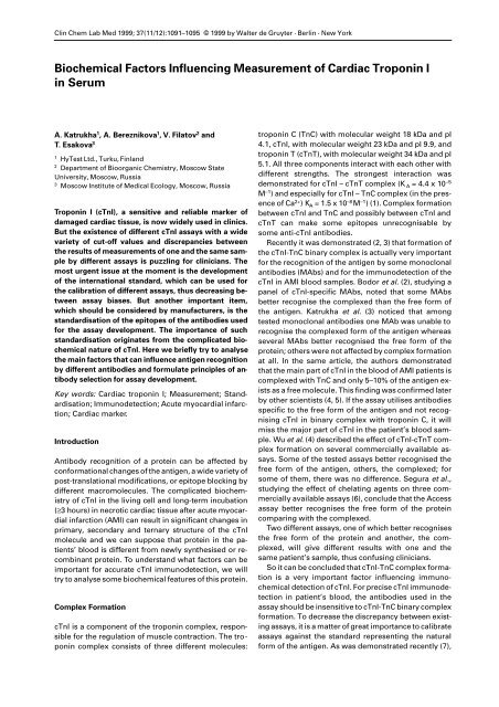
1092 Katrukha et al.: <strong>Measurement</strong> <strong>of</strong> cTnI in serumthe best results can be achieved with ternary cTnI-cTnT-TnC complex purified from human cardiac tissue.In cardiac muscle, cTnI may form a complex withcTnT but with significantly lower affinity comparedwith the cTnI-TnC binary complex. For the detection <strong>of</strong>the cTnI-cTnT binary complex in patient samples, itwas suggested to use a ”mixed“ sandwich immunoassay(5). Capture MAb in such an assay is specific tocTnI; detection, to cTnT. <strong>Measurement</strong>s <strong>of</strong> the binarycomplex in AMI sera demonstrated (8) that only 50% <strong>of</strong>patients sera contain detectable amounts <strong>of</strong> such complexand that the concentration <strong>of</strong> it in these serumsamples does not exceed 5% <strong>of</strong> the total amount <strong>of</strong> thecTnI. Comparable results were obtained by a gel filtrationmethod (4). There is no available informationabout anti-cTnI MAbs that are affected by cTnI – cTnTcomplex formation. So we can conclude that the cTnIcTnTcomplex in some cases can be detected in patient’ssamples, but obviously is not as important forcTnI immunodetection as the binary complex with TnC.Stable and Unstable Parts <strong>of</strong> the MoleculeAnother very important factor for cTnI measurementsis the stability <strong>of</strong> the cTnI molecule. cTnI is known to bea very unstable protein sensitive to proteolytic degradation.During infarction, cells are suffering from severeischemia and if circulation is not restored, diewithin 30–60 minutes. In necrotic tissue, internal aswell as external cell membranes, are destroyed and differentproteases are released from the lysosomes to intracellularmatrix. It means that 30–60 minutes after onset<strong>of</strong> the symptoms and up to its release into the bloodstream, cTnI is incubated in a protease cocktail. It is obviousthat in such conditions cTnI should be (at leastpartially) cleaved by proteases.In in vitro experiments with necrotic human cardiactissue (9), cTnI rapidly undergoes proteolytic degradation.Only 1–3% <strong>of</strong> the protein remained intact in the tissuesamples after 20 hours <strong>of</strong> experimental necrosis.Different parts <strong>of</strong> the cTnI molecule revealed differentstability. N- and C- terminal parts <strong>of</strong> the molecule wererapidly cleaved by proteases. The central part <strong>of</strong> cTnIlocated between amino acid residues 28 and 110 hadsignificantly better stability, possibly because <strong>of</strong> theprotection by troponin C. Extracts <strong>of</strong> human cardiac tissuethat were incubated for 20 hours at 37°C weretested by several sandwich immunoassays utilisingMAbs with different epitope specificity. Only 2–5% <strong>of</strong>initial immunological activity was observed in suchsamples if the epitopes <strong>of</strong> MAbs were located at the N-and C-terminal parts <strong>of</strong> the molecule, whereas morethan 40% <strong>of</strong> immunological activity was detected in thecase <strong>of</strong> the central location <strong>of</strong> the epitopes.Morjana (10) reported isolation <strong>of</strong> two main cTnIpeptides with molecular weights 18 and 14 kDa frompatient’s blood. The 18 kDa peptide is considered to bea product <strong>of</strong> cTnI’s proteolytic degradation from the C-terminal region whereas its subsequent degradationfrom the N-terminal region results in the appearance <strong>of</strong>the 14 kDa fragment. So results presented by Morjanaconfirm the assumption about better stability <strong>of</strong> thecentral part <strong>of</strong> the molecule.The epitope location can be also very important forbetter sensitivity <strong>of</strong> the assays and for the standardisation<strong>of</strong> cTnI assays (9). Serial blood samples from AMIpatients were tested by two different assays, one withMAbs recognising the stable part <strong>of</strong> the molecule andanother with MAbs specific to the N- and C- terminalparts <strong>of</strong> the molecule. Within 24 hours after onset <strong>of</strong> thechest pain, the ratio <strong>of</strong> concentrations measured by thesecond and first assay was close to one, but 2–3 dayslater it was close to 3 or even more. Four to five daysafter infarction, in some cases there was still no detectablecTnI in the second assay, but significantamounts <strong>of</strong> the antigen were measured by the assaywith MAbs recognising the stable part <strong>of</strong> the molecule.We have got the same results with two commerciallyavailable assays, one <strong>of</strong> which is specific to the stablepart <strong>of</strong> the molecule, the other to the unstable part (ourunpublished data).So we can conclude that for the better sensitivity andreproducibility <strong>of</strong> measurements <strong>of</strong> one and the samesample by different assays, antibodies used in assaysshould recognise the stable central part <strong>of</strong> the cTnImolecule.Phosphorylation by Protein Kinase AThe cTnI molecule contains two serines in the 22 and23 positions. Both amino acid residues can be phosphorylatedin vivo by protein kinase A (11), so fourforms <strong>of</strong> protein, one dephospho-, two monophospho-,and one biphospho- can coexist in the cell and theoreticallyall these forms can be released into the patient’sblood after infarction. Phosphorylation changes thestructure and conformation <strong>of</strong> the cTnI molecule and asa result can affect the interaction <strong>of</strong> some antibodieswith the antigen (12).We studied the effect <strong>of</strong> phosphorylation on the interaction<strong>of</strong> the antigen with anti-cTnI MAbs fromHyTest (Turku, Finland) by Western blotting and insandwich immunoassay. Some MAbs from the anticTnIpanel (22B11, 16F1, 10G4, 17H6) recognise part <strong>of</strong>the molecule, containing sites <strong>of</strong> phosphorylation – Ser22 and Ser 23. In Figure 1 results <strong>of</strong> Western blottingFig. 1 Monoclonal antibodies 22B11(A), 10B11(B) and 8E10(C), recognising dephospho- and bisphospho- forms <strong>of</strong> humancardiac TnI in Western blotting.
Katrukha et al.: <strong>Measurement</strong> <strong>of</strong> cTnI in serum 1093with dephosphorylated and phosphorylated forms <strong>of</strong>the protein are presented. cTnI bands were visualisedby three monoclonal antibodies: 22B11 ( 20 RRSSN 24 ),10B11 ( 16 APIRR 20 ) and 8E10 ( 87 LGFAE 91 ) (13). PhosphorylatedcTnI is not recognised by 22B11, whereas thereis no difference in recognition <strong>of</strong> the two forms in thecase <strong>of</strong> 10B11 and 8E10 MAbs. Using these three MAbs,we developed two sandwich immunoassays with captureMAb 8E10 and MAbs 22B11 and 10B11 as detection.Both forms <strong>of</strong> the antigen – phosphorylated anddephosphorylated – were tested by such assays. As followsfrom Figure 2, phosphorylation had no effect onthe signal level in the case <strong>of</strong> the 8E10–10B11 pair, and,on the contrary, the phosphorylated form was not detectedby the assay utilising 22B11 antibodies. So weconcluded that the first assay can be used for the detection<strong>of</strong> both – phosphorylated and dephosphorylated– forms <strong>of</strong> the antigen and the second assay onlyfor the detection <strong>of</strong> dephosphorylated cTnI.Fig. 3 Two sandwich immunoassays recognising native(black columns) and dephosphorylated (white columns) cTnIfrom human cardiac tissue.Fig. 2 Two sandwich immunoassays recognising dephospho-(black columns) and bisphospho- (white columns) forms<strong>of</strong> cTnI.Fig. 4 cTnI measurements by two experimental assays –10B11–8E10 (––) and 22B11–8E10 (––) in serum samplesfrom representative AMI patients.By both assays, we checked in what form cTnI existsin human cardiac tissue. Antigen was purified from thetissue obtained from several different donors 5–8 hpostmortem. Two forms, native and dephosphorylatedby phosphatase from E.coli, were tested. There was nodifference in recognising both forms by the assay utilising10B11 MAbs (Figure 3), but in the assay sensitiveto phosphorylation, the signal level increased up to300%. We concluded that even in several hours postmortemtissue, cTnI is still phosphorylated. So it isquite possible that the phosphorylated form <strong>of</strong> the proteincan be found in the patients’ blood. To verify thisassumption, both assays were calibrated against thedephosphorylated form <strong>of</strong> the antigen and cTnI concentrationswere measured in patients’ samples. In alltested samples, the concentration <strong>of</strong> cTnI determinedby the assay sensitive to phosphorylation was half asmuch compared with the results we have got with theassay that does not discriminate phosphorylated anddephosphorylated forms (Figure 4). Thus our resultssuggest that a significant part <strong>of</strong> the protein in the patients’blood is phosphorylated.Unfortunately, because <strong>of</strong> the indirect method weused in our study described above, results should beconsidered only as preliminary. To be more precise,MAbs recognising only the phosphorylated form <strong>of</strong> theantigen should be used instead <strong>of</strong> 22B11 antibodies.Effect <strong>of</strong> HeparinThe cTnI molecule has a very high positive charge (pI9.87) and because <strong>of</strong> it forms complexes with negativelycharged molecules. Such complexes can interferewith the antibody-antigen interaction. Heparin,widely used in clinical practice, is a mixture <strong>of</strong> moleculeswith negative charge. Heparin forms complexeswith cTnI thus changing antigen – antibody interaction.We studied the effect <strong>of</strong> heparin at the concentration <strong>of</strong>50 and 200 units per ml on the recognition <strong>of</strong> the antigenby several sandwich immunoassays. As followsfrom Figure 5, in some cases heparin had no effect onthe antigen-antibody interaction, but in two immunoassaysthe signal level in the presence <strong>of</strong> heparinwas significantly reduced.
1094 Katrukha et al.: <strong>Measurement</strong> <strong>of</strong> cTnI in serumThe clinical importance <strong>of</strong> the oxidation – reduction<strong>of</strong> the cysteines and existence <strong>of</strong> cTnI-specific autoantibodiesshould be clarified. But if both forms – oxidisedand reduced – are found in patient’s samples and existence<strong>of</strong> auto-antibodies will be confirmed, these tw<strong>of</strong>actors also should be considered by assay manufacturers.ReferencesFig. 5 Effect <strong>of</strong> heparin on cTnI recognition by different sandwichimmunoassays. Black columns: samples without heparin,white columns: heparin 50 U/ml, grey columns: heparin200 U/ml.AutoantibodiesSeveral years ago, Bohner (14) reported a 69-year-oldCABG patient with diffuse three-vessel disease whowas measured as by Dade’s cTnI assay but positivewith troponin T and CKMB assays. The authors clearlydemonstrated that cTnI in the samples was not detectedbecause <strong>of</strong> the presence <strong>of</strong> auto anti-TnI antibodies.Unfortunately, the authors did not check theepitope specificity <strong>of</strong> the autoantibodies, so we do notknow what sites are important for the generation <strong>of</strong>such antibodies.This case is the only one described in the literature <strong>of</strong>the presence <strong>of</strong> the autoantibodies in patients’ samples,so we can conclude that appearance <strong>of</strong> autoantibodiesto cTnI is a very rare phenomenon and possiblyhas no practical significance.Oxidation-ReductioncTnI has two cysteine residues, Cys 79 and Cys 96 (15)which can be oxidised or reduced (16) and this oxidationand reduction change the structure and the conformation<strong>of</strong> the antigen. Wu et al. (4) demonstratedthe effect <strong>of</strong> oxidation – reduction on the recognition <strong>of</strong>the antigen by different commercially available assays.Some assays better recognised the oxidised form <strong>of</strong>the antigen, but for the majority <strong>of</strong> assays there was nodifference what form <strong>of</strong> the protein was tested. Unfortunately,we do not know in what form oxidised or reducedcTnI is present in the blood stream <strong>of</strong> the patients,it is difficult to estimate the importance <strong>of</strong>troponin oxidation-reduction for immunodetection.But in any case, preferably MAbs used in assays shouldrecognise sites different from the sites that can bechanged by oxidation-reduction.To summarise: monoclonal or polyclonal antibodiesused for the development <strong>of</strong> reliable cTnI assayspreferably should have epitopes that are not affectedby: cTnI – TnC complex formation, phosphorylation,molecules with low pI. Epitopes should be located inthe stable part <strong>of</strong> the cTnI molecule.1. Reiffert S, Jaquet K, Heilmeyer L, Herberg F. Stepwisesubunit interaction changes by mono- and bisphosphorylation<strong>of</strong> cardiac troponin I. Biochemistry 1998; 37;13516–5.2. Bodor G, Porter S, Landt Y, Ladenson J. Development <strong>of</strong>monoclonal antibodies for an assay <strong>of</strong> cardiac troponin -Iand preliminary results in suspected cases <strong>of</strong> myocardialinfarction. Clin Chem 1992; 38:2203–14.3. Katrukha A, Bereznikova A, Esakova T, Pettersson K, LovgrenT, Severina M, et al. <strong>Troponin</strong> I is released in bloodstream<strong>of</strong> patients with acute myocardial infarction not infree form but as complex. ClinChem 1997; 43:1379–85.4. Wu A, Feng Y-J, Moore R, Apple F, McPherson P, BuechlerK, et al. Characterization <strong>of</strong> cardiac troponin subunit releaseinto serum after acute myocardial infarction andcomparison <strong>of</strong> assays for troponin T and I. Clin Chem 1998;44:1198–208.5. Giuliani I, Bertinchant J-P, Granier C, Laprade M, ChocronS, Toubin G, et al. Determination <strong>of</strong> cardiac troponin Iforms in the blood <strong>of</strong> patients with acute myocardial infarctionand patients receiving crystalloid or cold bloodcardioplegia. Clin Chem 1999; 45: 213–22.6. Segura R, Varela E, Marti R, Vidal E, Gonzalez C, Figueras J,et al. Comparison <strong>of</strong> three assays for cardiac troponin Imeasurement: effect <strong>of</strong> chelating agents. Clin Chem LabMed 1999; 37 (Special Suppl):457.7. Katrukha A, Bereznikova A, Pettersson K. New approach tostandardisation <strong>of</strong> human cardiac troponin I (cTnI). ScandJ Clin Lab Invest 1999; 59 (Suppl 230):124–7.8. Katrukha A, Bereznikova A, Filatov V, Pettersson K,Esakova T, Kolosova O, et al. Binary troponin I-troponin Tcomplex in serum <strong>of</strong> patients with myocardial infarction.Clin Chem Lab Med 1999; 37 (Special Suppl):449.9. Katrukha A, Bereznikova A, Filatov V, Esakova T, KolosovaO, Pettersson K, et al. Degradation <strong>of</strong> cardiac troponin I:implication for reliable immunodetection. Clin Chem 1998;44:2433–40.10. Morjana N. Degradation <strong>of</strong> human cardiac troponin I aftermyocardial infarction. Biotechnol Appl Biochem 1998; 28(part 2):105–1111. Solaro RJ. Protein phosphorylation and the cardiac my<strong>of</strong>ilaments.In: Solaro RJ, ed. Protein phosphorylation in theheart muscle. Boca Raton: CRC Press Inc, 1986:129–56.12. Hillawi E, Chilton D, Trayer I, Cummins P. Phosphorylationspecificantibodies for human cardiac troponin-I. Eur JBiochem 1998; 256: 535–40.13. Filatov V, Katrukha A, Bereznikova AV, Esakova T, BularginaT, Severina M, et al. Epitope mapping <strong>of</strong> anti-TnImonoclonal antibodies. Biochem Mol Biol Intern 1998;45:1179–87.14. Bohner J, Pape K-W, Hannes W, Stegmann T. False-negativeimmunoassay results for cardiac troponin I probablydue to circulating troponin I autoantibodies. Clin Chem1996; 42:2046.15. Vallins W, Brand N, Dabhade N, Butler-Browne G, Yacoub
Katrukha et al.: <strong>Measurement</strong> <strong>of</strong> cTnI in serum 1095M, Barton P. Molecular cloning <strong>of</strong> human cardiac troponinI using polymerase chain reaction. FEBS Lett 1990;270:57–61.16. Ingraham R, Hodges R. Effects <strong>of</strong> Ca 2+ and subunit interactionon surface accessibility <strong>of</strong> cysteine residues <strong>of</strong> cardiactroponin. Biochemistry 1988; 27: 5891–8.Received 15 July 1999; accepted November 1999Corresponding author: Dr. A. Katrukha, Hytest Ltd.,Tykistökatu 2B, Euro-City, 20520 Turku, FinlandTel.: +358-2-512-0900, Fax: Tel.: +358-2-512-0909Email: hytest@hytestfinland.com