![Anatomic distribution of [18F] fluorodeoxyglucose-avid lymph nodes ...](https://img.yumpu.com/51788410/4/500x640/anatomic-distribution-of-18f-fluorodeoxyglucose-avid-lymph-nodes-.jpg)
Anatomic distribution of [18F] fluorodeoxyglucose-avid lymph nodes ...
Anatomic distribution of [18F] fluorodeoxyglucose-avid lymph nodes ...
Anatomic distribution of [18F] fluorodeoxyglucose-avid lymph nodes ...

You also want an ePaper? Increase the reach of your titles
YUMPU automatically turns print PDFs into web optimized ePapers that Google loves.
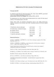
48 H.P. Fontanilla et al Practical Radiation Oncology: January-March 2013Figure 1 “Atlas” <strong>of</strong> positron emission tomography (PET)-positive LNs. Representative axial images (superior to inferior) showingthe location <strong>of</strong> all 190 identified PET-positive <strong>lymph</strong> <strong>nodes</strong>. All <strong>nodes</strong> from each patient are contoured in the same color; each coloris used for 2 patients.We identified 190 FDG-<strong>avid</strong> LNs; 122 in group 1 (the41 consecutive initially identified patients) and 68 in group2 (the additional 9 patients with positive paraortic LNs).The median number <strong>of</strong> positive LNs per patient was 3(range, 1-6) in group 1 and 5 (range, 3-17) in group 2. The<strong>distribution</strong> <strong>of</strong> FDG-<strong>avid</strong> LNs by disease stage is shownin Table 1.<strong>Anatomic</strong> <strong>distribution</strong> <strong>of</strong> FDG-<strong>avid</strong> LNsAxial images depicting the anatomic <strong>distribution</strong> <strong>of</strong> all190 FDG-<strong>avid</strong> LNs (from groups 1 and 2) are shown inFig 1. There were 94 external iliac <strong>lymph</strong> <strong>nodes</strong>, 40common iliac <strong>lymph</strong> <strong>nodes</strong>, and 2 parametrial <strong>lymph</strong><strong>nodes</strong> in groups 1 and 2 combined. Our mapping analysisrevealed that most PET-positive <strong>nodes</strong> were locatedaround major vessels, between the psoas muscle and thevascular bundle as seen in Fig 1. The FDG-<strong>avid</strong> externaliliac <strong>nodes</strong> were generally posterior to the external iliacvessels and extended laterally to the pelvic musculature orbones. Several FDG-<strong>avid</strong> LNs were located posterior andcaudal to the distal external iliac vessels, classically definedas the medial external iliac <strong>nodes</strong>; these LNs extendedjust inferior to the level <strong>of</strong> the superior femoralheads. Two FDG-<strong>avid</strong> parametrial <strong>nodes</strong> were noted andthese were primarily lateral, most likely because <strong>of</strong> thedifficulty <strong>of</strong> identifying distinct nodal volumes in thevicinity <strong>of</strong> the primary tumor. There were also multiplepositive common iliac <strong>nodes</strong> located between the commoniliac vein and psoas muscle, extending posteriorly betweenthe psoas muscle and the sacrum. Positive common iliac<strong>nodes</strong> were also noted lateral to the vessels and anterior tothe psoas muscle.The anatomic <strong>distribution</strong> <strong>of</strong> positive LNs in group 1 issummarized in Table 2 and Fig 2. The most commonlocations <strong>of</strong> positive LNs were the external iliac region(63.9% <strong>of</strong> positive LNs) and the common iliac region(17.2%). In group 1, all 41 patients had at least 1 positivedistal pelvic LN (ie, at or distal to the bifurcation <strong>of</strong> thecommon iliac vessels). Sixteen <strong>of</strong> the 41 patients (39%)