Structure and Function of the Ductuli Efferentes - University of Illinois ...
Structure and Function of the Ductuli Efferentes - University of Illinois ...
Structure and Function of the Ductuli Efferentes - University of Illinois ...

You also want an ePaper? Increase the reach of your titles
YUMPU automatically turns print PDFs into web optimized ePapers that Google loves.
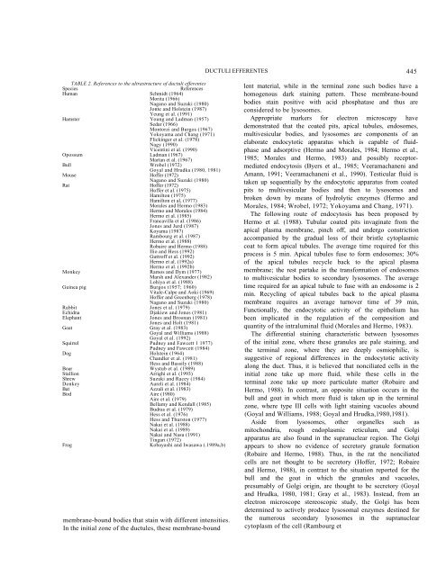
DUCTULI EFFERENTES445TABLE 2. References to <strong>the</strong> ultrastructure <strong>of</strong> ductuli efferentesSpeciesReferencesHuman Schmidt (1964)Morita (1966)Nagano <strong>and</strong> Suzuki (1980)Jonte <strong>and</strong> Holstein (1987)Yeung et al. (1991)Hamster Young <strong>and</strong> Ladman (1957)Sedar (1966)Montorzi <strong>and</strong> Burgos (1967)Yokoyama <strong>and</strong> Chang (1971)Flickinger et al. (1978)Nagy (1990)Vicentini et al. (1990)Opossum Ladman (1967)Martan et al. (1967)Bull Wrobel (1972)Goyal <strong>and</strong> Hrudka (1980, 1981)Mouse H<strong>of</strong>fer (1972)Nagano <strong>and</strong> Suzuki (1980)Rat H<strong>of</strong>fer (1972)H<strong>of</strong>fer et al. (1975)Hamilton (1975)Hamilton et al, (1977)Morales <strong>and</strong> Hermo (1983)Hermo <strong>and</strong> Morales (1984)Hermo et al. (1985)Francavilla et al. (1986)Jones <strong>and</strong> Jurd (1987)Koyama (1987)Rambourg et al. (1987)Hermo et al. (1988)Robaire <strong>and</strong> Hermo (1988)Ilio <strong>and</strong> Hess (1992)Guttr<strong>of</strong>f et al. (1992)Hermo et al. (1992a)Hermo et al. (1992b)Monkey Ramos <strong>and</strong> Dym (1977)Marsh <strong>and</strong> Alex<strong>and</strong>er (1982)Lohiya et al. (1988)Guinea pig Burgos t1957; 1960)Vitale-Calpe <strong>and</strong> Aoki (1969)H<strong>of</strong>fer <strong>and</strong> Greenberg (1978)Nagano <strong>and</strong> Suzuki (1980)Rabbit Jones et al. (1979)Echidna Djakiew <strong>and</strong> Jones (1981)Elephant Jones <strong>and</strong> Brosnan (1981)Jones <strong>and</strong> Holt (1981)Goat Gray et al. (1983)Goyal <strong>and</strong> Williams (1988)Goyal et al. (1992)Squirrel Pudney <strong>and</strong> Fawcett 1 1977)Pudney <strong>and</strong> Fawcett (1984)Dog Holstein (1964)Ch<strong>and</strong>ler et al. (1981)Hess <strong>and</strong> Bassily (1988)Boar Wystub et al. (1989)Stallion Arrighi et al. (1993)Shrew Suzuki <strong>and</strong> Racey (1984)Donkey Aureli et al. (1984)Bat Azzali et al. (1983)Bird Aire (1980)Aire et al. (1979)Bellamy <strong>and</strong> Kendall (1985)Budras et al. (1979)Hess et al. (1976)Hess <strong>and</strong> Thurston (1977)Nakai et al. (1988)Nakai et al. (1989)Nakai <strong>and</strong> Nasu (1991)Tingari (1972)FrogKobayashi <strong>and</strong> Iwasawa (.1989a,b)membrane-bound bodies that stain with different intensities.In <strong>the</strong> initial zone <strong>of</strong> <strong>the</strong> ductules, <strong>the</strong>se membrane-boundlent material, while in <strong>the</strong> terminal zone such bodies have ahomogenous dark staining pattern. These membrane-boundbodies stain positive with acid phosphatase <strong>and</strong> thus areconsidered to be lysosomes.Appropriate markers for electron microscopy havedemonstrated that <strong>the</strong> coated pits, apical tubules, endosomes,multivesicular bodies, <strong>and</strong> lysosomes are components <strong>of</strong> anelaborate endocytotic apparatus which is capable <strong>of</strong> fluidphase<strong>and</strong> adsorptive (Hermo <strong>and</strong> Morales, 1984; Hermo et al.,1985; Morales <strong>and</strong> Hermo, 1983) <strong>and</strong> possibly receptormediatedendocytosis (Byers et al., 1985; Veeramachaneni <strong>and</strong>Amann, 1991; Veeramachaneni et al., 1990). Testicular fluid istaken up sequentially by <strong>the</strong> endocytotic apparatus from coatedpits to multivesicular bodies <strong>and</strong> <strong>the</strong>n to lysosomes <strong>and</strong>broken down by means <strong>of</strong> hydrolytic enzymes (Hermo <strong>and</strong>Morales, 1984; Wrobel, 1972; Yokoyama <strong>and</strong> Chang, 1971).The following route <strong>of</strong> endocytosis has been proposed byHermo et al. (1988). Tubular coated pits invaginate from <strong>the</strong>apical plasma membrane, pinch <strong>of</strong>f, <strong>and</strong> undergo constrictionaccompanied by <strong>the</strong> gradual loss <strong>of</strong> <strong>the</strong>ir bristle cytoplasmiccoat to form apical tubules. The average time required for thisprocess is 5 min. Apical tubules fuse to form endosomes; 30%<strong>of</strong> <strong>the</strong> apical tubules recycle back to <strong>the</strong> apical plasmamembrane; <strong>the</strong> rest partake in <strong>the</strong> transformation <strong>of</strong> endosomesto multivesicular bodies to secondary lysosomes. The averagetime required for an apical tubule to fuse with an endosome is 2min. Recycling <strong>of</strong> apical tubules back to <strong>the</strong> apical plasmamembrane requires an average turnover time <strong>of</strong> 39 min,<strong>Function</strong>ally, <strong>the</strong> endocytotic activity <strong>of</strong> <strong>the</strong> epi<strong>the</strong>lium hasbeen implicated in <strong>the</strong> regulation <strong>of</strong> <strong>the</strong> composition <strong>and</strong>quantity <strong>of</strong> <strong>the</strong> intraluminal fluid (Morales <strong>and</strong> Hermo, 1983).The differential staining characteristic between lysosomes<strong>of</strong> <strong>the</strong> initial zone, where <strong>the</strong>se granules are pale staining, <strong>and</strong><strong>the</strong> terminal zone, where <strong>the</strong>y are deeply osmiophilic, issuggestive <strong>of</strong> regional differences in <strong>the</strong> endocytotic activityalong <strong>the</strong> duct. Thus, it is believed that nonciliated cells in <strong>the</strong>initial zone take up more fluid, while <strong>the</strong>se cells in <strong>the</strong>terminal zone take up more particulate matter (Robaire <strong>and</strong>Hermo, 1988). In contrast, an opposite situation occurs in <strong>the</strong>bull <strong>and</strong> goat in which more fluid is taken up in <strong>the</strong> terminalzone, where type III cells with light staining vacuoles abound(Goyal <strong>and</strong> Williams, 1988; Goyal <strong>and</strong> Hrudka,1980,1981).Aside from lysosomes, o<strong>the</strong>r organelles such asmitochondria, rough endoplasmic reticulum, <strong>and</strong> Golgiapparatus are also found in <strong>the</strong> supranuclear region. The Golgiappears to show no evidence <strong>of</strong> secretory granule formation(Robaire <strong>and</strong> Hermo, 1988). Thus, in <strong>the</strong> rat <strong>the</strong> nonciliatedcells are not thought to be secretory (H<strong>of</strong>fer, 1972; Robaire<strong>and</strong> Hermo, 1988), in contrast to <strong>the</strong> situation reported for <strong>the</strong>bull <strong>and</strong> <strong>the</strong> goat in which <strong>the</strong> granules <strong>and</strong> vacuoles,presumably <strong>of</strong> Golgi origin, are thought to be secretory (Goyal<strong>and</strong> Hrudka, 1980, 1981; Gray et al., 1983). Instead, from anelectron microscope stereoscopic study, <strong>the</strong> Golgi has beendetermined to actively produce lysosomal enzymes destined for<strong>the</strong> numerous secondary lysosomes in <strong>the</strong> supranuclearcytoplasm <strong>of</strong> <strong>the</strong> cell (Rambourg et