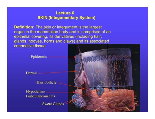
Lecture 6 SKIN (Integumentary System) Definition: The skin or ...
Lecture 6 SKIN (Integumentary System) Definition: The skin or ...
Lecture 6 SKIN (Integumentary System) Definition: The skin or ...

You also want an ePaper? Increase the reach of your titles
YUMPU automatically turns print PDFs into web optimized ePapers that Google loves.
<strong>Lecture</strong> 6<br />
<strong>SKIN</strong> (<strong>Integumentary</strong> <strong>System</strong>)<br />
<strong>Definition</strong>: <strong>The</strong> <strong>skin</strong> <strong>or</strong> integument is the largest<br />
<strong>or</strong>gan in the mammalian body and is comprised of an<br />
epithelial covering, its derivatives (including hair,<br />
glands, hooves, h<strong>or</strong>ns and claws) and its associated<br />
connective tissue<br />
Epidermis<br />
Dermis<br />
Hair Follicle<br />
Hypodermis<br />
(subcutaneous fat)<br />
Sweat Glands
II. Functional Characteristics:<br />
1. Protection: <strong>The</strong> most imp<strong>or</strong>tant function of the <strong>skin</strong><br />
is its effectiveness as a barrier between the internal<br />
and external environments (guards against injury,<br />
bacterial invasion, UV damage and desiccation).<br />
2. Regulation of body temperature: m ediated by t he<br />
hair coat, cutaneous blood supply and in some<br />
animals, sweat glands.<br />
3. Secretion: from sweat, sebaceous and mammary<br />
glands<br />
4. Sens<strong>or</strong>y Organ: innervation of the <strong>skin</strong> provides<br />
pain, touch, pressure and temperature sensation.<br />
5. Communication: <strong>The</strong> <strong>skin</strong> is an imp<strong>or</strong>tant <strong>or</strong>gan in<br />
the social life of animals because it gives off od<strong>or</strong>s<br />
that govern sexual behavi<strong>or</strong> and helps animals<br />
identify each other and their territ<strong>or</strong>ies<br />
6.<br />
Reflects the physiological condition of the animal: <strong>skin</strong> and coat<br />
condition are good indicat<strong>or</strong>s of overall health and alterations may<br />
reflect a variety of external and internal disease processes (endocrine<br />
dis<strong>or</strong>ders, nutritional problems; i.e. Vitamin A deficiency is<br />
characterized by very dry, hardened <strong>skin</strong>, dry lack-luster hair and hair<br />
loss.)
III. Organization of Skin: Epidermis, Dermis and Hypodermis<br />
Epidermis<br />
Hair<br />
follicle<br />
= loose<br />
fascia
1. Epidermis: stratified squamous epithelium of ectodermal <strong>or</strong>igin<br />
Dermal<br />
Papillae<br />
Epidermal Pegs
A. Epidermal Cell Types:<br />
1) Keratinocytes: represent the maj<strong>or</strong>ity of cells<br />
2) Melanocytes: derived from Neural Crest, “octopus-like” cells<br />
that produce melanin.<br />
3) Langerhans Cells: dendritic cells<br />
located in the stratum spinosum<br />
4) Merkel Cells: ubiquitous cells in<br />
the <strong>skin</strong> that couple with axon<br />
terminals to f<strong>or</strong>m mechan<strong>or</strong>ecept<strong>or</strong>s
A. Epidermal Cell Types:<br />
1) Keratinocytes: represent the maj<strong>or</strong>ity of cells<br />
2) Melanocytes: derived from Neural Crest, “octopus-like” cells<br />
Keratinocytes<br />
Epidermal Melanocyte
1) Keratinocytes: represent the maj<strong>or</strong>ity of <strong>skin</strong> cells and are<br />
arranged in 5 layers (discussed later)<br />
keratinocyte<br />
dermis
2) Melanocytes: octopus like cells that produce the pigment, melanin<br />
<strong>The</strong>y don’t retain melanin but pass it on to neighb<strong>or</strong>ing Keratinocytes<br />
Keratinocytes<br />
Melanocyte<br />
Melanin granule
Melanin Granules in Keratinocytes<br />
Melanin Granules
Melanocytes in <strong>skin</strong><br />
Clinical Note: A Melanoma is a type of very aggressive and metastatic<br />
cancer that arises from the uncontrolled mitosis and migration<br />
of these cells. It commonly occurs in dogs with pigmented <strong>skin</strong>.<br />
Melanomas can occur in areas of haired <strong>skin</strong> (usually benign),<br />
where they usually f<strong>or</strong>m small, dark (brown to black) lumps,<br />
but can also appear as large flat wrinkled masses. <strong>The</strong>y can also occur in<br />
the mouth, toes <strong>or</strong> behind the eye (these tend to be malignant).
3) Langrhans cells: dendritic (immune) cells located in the stratum<br />
spinosum that play a pivotal role in induction of cutaneous immune<br />
responses (allergic reactions). Migrate to draining lymph node->T-cells.<br />
Cell process<br />
Langerhans cell<br />
Future Development<br />
of <strong>skin</strong> patches<br />
containing vaccines<br />
that will stimulate<br />
Langerhans cells<br />
Keratinocyte
4) Merkel cells: found throughout <strong>skin</strong>, not visible with n<strong>or</strong>mal<br />
stains; couple with axon terminals to f<strong>or</strong>m slowly adapting<br />
mechan<strong>or</strong>ecept<strong>or</strong>s. <strong>The</strong>y can f<strong>or</strong>m malignant tum<strong>or</strong>s in cats.<br />
Immunostained Merkel cells in <strong>skin</strong><br />
Microvilli on surface<br />
Merkel Cell Tum<strong>or</strong>
1. Epidermis<br />
B. Layers<br />
1) Stratum<br />
Germinativum<br />
a) Stratum Basale:<br />
b) Stratum<br />
Spinosum<br />
2) Stratum Granulosum<br />
3) Stratum Lucidum<br />
4) Stratum C<strong>or</strong>neumoutermost<br />
keratinized<br />
layer of flattened,<br />
dead cells (squames)<br />
Single layer of cells in contact with basement membrane<br />
Stratum<br />
Germinativum
1. Epidermis<br />
B. Layers<br />
1) Stratum<br />
Germinativum<br />
a) Stratum Basale<br />
b) Stratum<br />
Spinosum<br />
2) Stratum<br />
Granulosum<br />
3) Stratum Lucidum<br />
4) Stratum C<strong>or</strong>neumoutermost<br />
keratinized<br />
layer of flattened,<br />
dead, anuclear cells:<br />
(squames) have no<br />
distnict cytoplasmic<br />
boundaries
Layers of the Epidermis (thick <strong>skin</strong>)<br />
(Stratum Lucidum)<br />
(stratum basale)<br />
(Stratum c<strong>or</strong>neum)
Three Layers of the upper epidermis<br />
Duct<br />
Stratum C<strong>or</strong>neum<br />
Stratum granulosum
C.<br />
A. Keratinization: process involving the f<strong>or</strong>mation of<br />
keratin<br />
1) Keratin Proteins: Keratin is a structural<br />
protein that f<strong>or</strong>ms the cytoskeleton of all<br />
keratinocytes. Keratins are a principle part of<br />
the cells in the epidermis, hair, nails, feathers,<br />
hooves, and t he enamel of teeth. <strong>The</strong>re are<br />
several subtypes of keratin proteins, some are<br />
called "soft" keratins and others are "hard"<br />
keratins.<br />
a) Soft keratin – elastic, desquamates<br />
(example: <strong>skin</strong>)<br />
b) Hard keratin – contains m<strong>or</strong>e sulfur than<br />
soft keratin; less elastic; m<strong>or</strong>e permanent;<br />
resistant to degradation, does not<br />
desquamate (examples: nails, h<strong>or</strong>ns, hoof)
2) F<strong>or</strong>mation of Keratin:<br />
a) Synthesis of filaments begins in<br />
stratum basale (makes 2 of the 4<br />
types of keratin-other 2 in spinosum)<br />
b) Aggregation of filaments occurs in the<br />
superficial cells of the stratum spinosum<br />
c) Cells of st. spinosum also f<strong>or</strong>m “membrane<br />
coating granules”, MCG, that later release their<br />
lipid-rich contents into intracellular space--><br />
f<strong>or</strong>m water proof permeability barrier.<br />
d) Keratohyalin granules (non-membrane<br />
bound) appear in close association with the<br />
filaments in the stratum granulosum and nonkeratin<br />
proteins released by these granules<br />
cause keratin filaments to associate into<br />
thicker bundles.<br />
e) Degradation of the nucleus and loss of cell<br />
<strong>or</strong>ganelles in the most superficial layer of<br />
stratum granulosum-->completed in stratum<br />
lucidum and stratum c<strong>or</strong>neum<br />
f) F<strong>or</strong>mation of keratin filament matrix<br />
complex occurs in stratum c<strong>or</strong>neum
Melanin and keratohyalin granules
2. Dermis (c<strong>or</strong>ium):layer of <strong>skin</strong> immediately deep to the epidermis<br />
derived from mesoderm and comprised of connective tissue.<br />
embedded in the dermis<br />
(Reticular layer)<br />
Dermis<br />
Papillary layer
2. Dermis: consists of 2 layers:<br />
A. Papillary Layer: subepithelial loose connective tissue<br />
B. Reticular Layer: dense connective tissue deep to papillary layer;<br />
contains epidermally derived hair, sweat and sebaceous glands<br />
Reticular layer/dermis<br />
elastic fibers
3. Hypodermis: subcutaneous layer (superficial fascia), technically not<br />
part of the <strong>skin</strong>.<br />
A. Loose and irregular connective tissue that anch<strong>or</strong>s dermis<br />
B. In healthy animals, it has large depots of fat in it
Clinical Considerations:<br />
4. Clinical Considerations:<br />
Hyperkeratosis: hypertrophy of the stratum c<strong>or</strong>neum<br />
[nasodigital hyperkeratosis - an ailment affecting either the<br />
nose <strong>or</strong> foot pads (<strong>or</strong> both) of older dogs. In<br />
hyperkeratosis, keratin - the tough, fibrous outer covering<br />
of foot pads - grows excessively. Often, the hard, cracked<br />
pads appear to have "keratin feathers" around their<br />
edges.]<br />
Squamous Cell Carcinoma: neoplasia of cells of the<br />
stratum spinosum [Squamous cell carcinoma (SCC) is a<br />
common tum<strong>or</strong> involving the <strong>skin</strong> and a ccounts f<strong>or</strong><br />
approximately 15% of cutaneous tum<strong>or</strong>s in the cat and 5%<br />
of those in the dog. SCCs are usually found in<br />
unpigmented <strong>or</strong> lightly pigmented <strong>skin</strong>.]<br />
. Melanoma: Melanocytic tum<strong>or</strong>s represent 4 to 7% of a ll<br />
canine neoplasms and are the most common malignant<br />
tum<strong>or</strong> of the canine <strong>or</strong>al cavity and digits [Melanoma<br />
tum<strong>or</strong>s in dogs, m<strong>or</strong>e than most cancers, demand<br />
immediate attention since early recognition can lead to<br />
m<strong>or</strong>e successful attempts at removal and identification of<br />
the grade <strong>or</strong> stage of cancer. Malignant melanomas can<br />
metastasize (spread) to any area of the body especially<br />
the lymph nodes and lungs and present very challenging<br />
and dangerous prospects f<strong>or</strong> the dog. Cats seem much<br />
less susceptible to melanoma tum<strong>or</strong>s than dogs]<br />
1.
IV. Access<strong>or</strong>y Structures of the Skin: Access<strong>or</strong>y structures of the <strong>skin</strong> include<br />
hairs and two types of glands: sweat glands and sebaceous glands. Although<br />
these structures are anch<strong>or</strong>ed in the dermis <strong>or</strong> hypodermis, they are in fact<br />
derived from the epidermis.<br />
1. Hair: hair itself is dead, but it’s produced by living keratinocytes at the<br />
base of the hair (hair follicles) and the pigmentation in hair comes<br />
from melanocytes (just like <strong>skin</strong> pigmentation)<br />
A. Functions: Hair serves several functions: serves as insulation;<br />
provides camouflage; sex recognition; social purposes.<br />
B. Types of hair follicles:<br />
1) Single (simple) follicle – one hair emerges from a single opening;<br />
found in h<strong>or</strong>se, cattle, pig and sheep (face, ear, distal p<strong>or</strong>tion of<br />
limbs)<br />
2) Compound follicle - several hairs emerge from a single opening.<br />
Found in cat, dog, sheep (wool growing areas). Consists of a<br />
long principal (guard) hair and a n umber of smaller auxillary<br />
(wool) hairs.<br />
Hair follicles: <strong>Definition</strong>: Cylindrical depressions in the epidermis, lined with<br />
stratified squamous epithelium; hair fibers are located in the lumen of<br />
the follicle.
Auxillary Hairs<br />
Compound Hair Follicle - Cat<br />
Principal<br />
Hair
C. Structure of hair follicles:<br />
1. Hair shaft: part of hair above surface of the <strong>skin</strong>; 3 parts<br />
a) Outer Cuticle-single layer of flat keratinized cells<br />
b) C<strong>or</strong>tex-compact dead cell layer under the cuticle<br />
c) Medulla-central region of the shaft, cuboidal <strong>or</strong> flat cells<br />
2. Hair Root-part of the hair below the surface<br />
of the <strong>skin</strong>; similar structure to hair shaft<br />
3. Hair bulb: conical mass at the base of the<br />
hair root, covered by stratified squamous<br />
epithelium (Germinal Matrix). Epithelium is<br />
indented at the base of the bulb by dermal<br />
conn. tissue the<br />
Dermal Papilla<br />
Hair Root<br />
3.<br />
1.
Hair Follicle: <strong>The</strong> structure from which the hair grows. Parts: Internal<br />
Root Sheath, External Root sheath and at the base the Dermal Papilla and<br />
Germinal matrix<br />
Actively<br />
produces<br />
the hair<br />
Hair Follicle<br />
Root sheath<br />
Hair<br />
Follicle
Hair Follicle:<br />
a) Internal Root<br />
Sheath<br />
b) External Root<br />
Sheath
4) Hair Follicle:<br />
a) Internal Root<br />
Sheath<br />
b) External Root<br />
Sheath<br />
Inner<br />
Root sheath<br />
Longitudinal section through hair shaft<br />
C<strong>or</strong>tex
Hair with epithelial root sheaths (ORS & IRS)<br />
C<strong>or</strong>tex<br />
External (outer) Root Sheath<br />
Dermal<br />
Sheath
5) Arrect<strong>or</strong> Pili Muscle: smooth muscle with <strong>or</strong>igin at the epidermal/dermal junction<br />
it inserts outside the follicle and when it contracts it functions to erect the hair<br />
= Piloerection, in response to cold, anger <strong>or</strong> fear.
D. Cyclic activity of hair:<br />
1) Anagen (growth period): the active phase of hair production<br />
when cells of the hair bulb are mitotically active and the hair<br />
grows in length (see fig. 5).<br />
2) Catagen (period of involution): transit<strong>or</strong>y period during which<br />
cellular proliferation slowly decreases and finally ceases; hair<br />
bulb becomes a solid mass of keratinized cells resembling a<br />
club (club hair) and the hair detaches from the underlying<br />
matrix and is easily removed (that’s where the hairs embedded<br />
in the bristles of your hairbrush come from every m<strong>or</strong>ning).<br />
3) Telogen (resting phase): transitional stage of the cycle where<br />
hair bulb atrophies; chemicals released from the dermal papilla<br />
wakens the fo llicle from its d<strong>or</strong>mancy and it begins to renew<br />
itself f<strong>or</strong> activity; it then changes back to t he active anagen<br />
stage again.<br />
Clinical Note: Hair loss (alopecia) is a common side effect of radiation & Chemotherapy
2. Glands of Skin:<br />
A. Sweat (sud<strong>or</strong>iferous) glands: simple, coiled tubular glands seen as<br />
hollow m<strong>or</strong>e <strong>or</strong> less circular profiles in tissue sections with walls<br />
composed of low cuboidal epithelium. Two types:<br />
1) Eccrine Sweat Glands: small glands that are widely distributed<br />
and produce a watery secretion; they are mainly a mechanism<br />
f<strong>or</strong> cooling; restricted to f oot pads of carniv<strong>or</strong>es, frog of<br />
ungulates and nasolabial region of ruminants and swine.<br />
2) Apocrine Sweat Glands: larger glands with cuboidal epithelium<br />
that produce oily and foamy secretions; most common in the<br />
groin, axilla and scrotum of dogs and cats; most numerous and<br />
extensive in h<strong>or</strong>ses. <strong>The</strong>se are the most common type found in<br />
domestic animals.<br />
B. Sebaceous Glands: simple, (often branched) acinar glands that<br />
open into a ha ir follicle about halfway up the shaft; they secrete<br />
sebum, a sec retion consisting of lysed cells and accumulated<br />
lipids containing precurs<strong>or</strong>s of vitamin D. Sebum gives hair its<br />
“sheen”. <strong>The</strong> oily sebum also acts as a lubricant f<strong>or</strong> the <strong>skin</strong> and<br />
hair.
Apocrine<br />
Sweat<br />
glands
Compound Hair Follicle with sebaceous glands (SbG) and sweat glands (SG)<br />
Sweat Glands
B. Sebaceous Gland
Cross Section of a Hair with a Sebaceous Gland<br />
Sebaceous gland