Craniosynostosis & Craniofacial Surgery
Craniosynostosis & Craniofacial Surgery Parent's Guide
Craniosynostosis & Craniofacial Surgery Parent's Guide
- No tags were found...

Create successful ePaper yourself
Turn your PDF publications into a flip-book with our unique Google optimized e-Paper software.
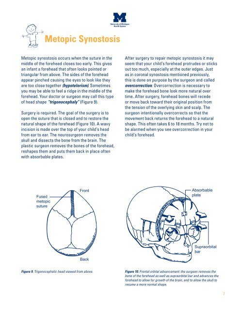
Metopic Synostosis<br />
Metopic synostosis occurs when the suture in the<br />
middle of the forehead closes too early. This gives<br />
an infant a forehead that often looks pointed or<br />
triangular from above. The sides of the forehead<br />
appear pinched causing the eyes to look like they<br />
are too close together (hypotelorism). Sometimes<br />
you may be able to feel a ridge in the middle of the<br />
forehead. Your doctor or surgeon may call this type<br />
of head shape “trigonocephaly” (Figure 9).<br />
<strong>Surgery</strong> is required. The goal of the surgery is to<br />
open the suture that is closed and to restore the<br />
natural shape of the forehead (Figure 10). A wavy<br />
incision is made over the top of your child’s head<br />
from ear to ear. The neurosurgeon removes the<br />
skull and dissects the bone from the brain. The<br />
plastic surgeon removes the bones of the forehead,<br />
reshapes them and puts them back in place often<br />
with absorbable plates.<br />
After surgery to repair metopic synostosis it may<br />
seem that your child’s forehead protrudes or sticks<br />
out too much, especially at the outer edges. Just<br />
as in coronal synostosis mentioned previously,<br />
this is done on purpose by the surgeon and called<br />
overcorrection. Overcorrection is necessary to<br />
make the forehead bone look more natural over<br />
time. After surgery, forehead bones will recede<br />
or move back toward their original position from<br />
the tension of the overlying skin and scalp. The<br />
surgeon intentionally overcorrects so that the<br />
movement back returns the forehead to a natural<br />
shape. This often takes 6 to 18 months. Try not to<br />
be alarmed when you see overcorrection in your<br />
child’s forehead.<br />
Fused<br />
metopic<br />
suture<br />
Front<br />
Absorbable<br />
plate<br />
Back<br />
Supraorbital<br />
bar<br />
Figure 9. Trigonocephalic head viewed from above.<br />
Figure 10. Frontal orbital advancement: the surgeon removes the<br />
bone of the forehead as well as supraorbital bar and advances the<br />
forehead to allow for growth of the brain, and to allow the skull to<br />
resume a more normal shape.<br />
7