Comparison of treatments with the Forsus fatigue resistant device in ...
Comparison of treatments with the Forsus fatigue resistant device in ...
Comparison of treatments with the Forsus fatigue resistant device in ...

Create successful ePaper yourself
Turn your PDF publications into a flip-book with our unique Google optimized e-Paper software.
Aras et al 617<br />
changes produced by semirigid fixed functional appliances<br />
might be different from those produced <strong>with</strong> rigid<br />
<strong>device</strong>s. In treatment <strong>with</strong> <strong>the</strong> Jasper jumper, which is<br />
a semirigid fixed functional appliance, Nalbantgil<br />
et al 13 found no significant effects <strong>in</strong> mandibular growth<br />
and <strong>in</strong> <strong>the</strong> degree <strong>of</strong> mandibular protrusion <strong>in</strong> late adolescents,<br />
but Stucki and Ingervall 14 reported that <strong>the</strong><br />
skeletal mandibular effects were <strong>the</strong> same <strong>in</strong> subjects allocated<br />
to 2 groups based on whe<strong>the</strong>r <strong>the</strong>y were younger<br />
or older than 15 years <strong>of</strong> age.<br />
The <strong>Forsus</strong> <strong>fatigue</strong> <strong>resistant</strong> <strong>device</strong> (3M Unitek, Monrovia,<br />
Calif) is a fairly new semirigid fixed functional<br />
appliance that is claimed by <strong>the</strong> manufacturer to be <strong>resistant</strong><br />
aga<strong>in</strong>st fracture and to exert consistent forces.<br />
The literature comprises only a few studies on its<br />
effects. 15,16 In treatment <strong>with</strong> <strong>the</strong> <strong>Forsus</strong> <strong>fatigue</strong><br />
<strong>resistant</strong> <strong>device</strong>, <strong>the</strong> <strong>in</strong>quiry is still unanswered<br />
concern<strong>in</strong>g whe<strong>the</strong>r <strong>the</strong> skeletal and dental changes <strong>in</strong><br />
subjects who have not passed <strong>the</strong> peak stage <strong>of</strong><br />
pubertal growth are different from those <strong>in</strong> subjects <strong>in</strong><br />
later maturation stages.<br />
Ano<strong>the</strong>r issue dur<strong>in</strong>g functional <strong>the</strong>rapy is its effect<br />
on <strong>the</strong> temporomandibular jo<strong>in</strong>t (TMJ). The general<br />
consensus is that <strong>the</strong> acceleration and peak phases <strong>of</strong><br />
<strong>the</strong> pubertal growth spurt have optimal conditions for<br />
TMJ adaptations to functional treatment. 6,17 This<br />
notion is supported by studies us<strong>in</strong>g magnetic<br />
resonance imag<strong>in</strong>g, 18-29 computerized tomography<br />
scans, 30 and horizontally corrected tomographies 31,32<br />
to evaluate <strong>the</strong> TMJ. These studies demonstrated that<br />
no adverse effects are encountered at <strong>the</strong> TMJs <strong>of</strong><br />
adolescents and patients at various ages from puberty<br />
to adulthood who are treated <strong>with</strong> functional<br />
appliances such as Herbst, functional mandibular<br />
advancer, Tw<strong>in</strong>-block, activator, bionator, and<br />
Fr€ankel. 18-32 However, no report has been published<br />
regard<strong>in</strong>g <strong>the</strong> effects <strong>of</strong> semirigid functional appliance<br />
<strong>the</strong>rapy on <strong>the</strong> TMJ. When unexpected TMJ responses<br />
to functional treatment are also taken <strong>in</strong>to account, it<br />
becomes apparent that new studies are needed. 33,34<br />
The purpose <strong>of</strong> this study was to compare <strong>the</strong> dentoskeletal<br />
changes by means <strong>of</strong> lateral cephalograms <strong>in</strong> subjects<br />
who were treated <strong>with</strong> <strong>the</strong> <strong>Forsus</strong> <strong>fatigue</strong> <strong>resistant</strong><br />
<strong>device</strong> at <strong>the</strong> peak and <strong>the</strong> end <strong>of</strong> <strong>the</strong> pubertal growth<br />
period. In addition, comparisons <strong>of</strong> <strong>the</strong> possible TMJ<br />
effects, <strong>in</strong>clud<strong>in</strong>g changes <strong>in</strong> condylar position and<br />
disc-condyle relationships, were made <strong>with</strong> magnetic<br />
resonance images <strong>of</strong> <strong>the</strong> TMJs.<br />
MATERIAL AND METHODS<br />
Patients <strong>with</strong> <strong>the</strong> follow<strong>in</strong>g characteristics were<br />
<strong>in</strong>cluded <strong>in</strong> this study: (1) Angle Class II Division 1 malocclusion<br />
<strong>in</strong> <strong>the</strong> permanent dentition, (2) ANB angle<br />
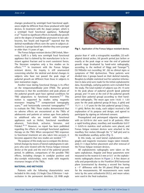
Fig 1. Application <strong>of</strong> <strong>the</strong> <strong>Forsus</strong> <strong>fatigue</strong> <strong>resistant</strong> <strong>device</strong>.<br />
greater than 4 <strong>with</strong> a retrognathic mandible, (3) wellaligned<br />
dental arches, (4) growth period just before or<br />
exactly at <strong>the</strong> peak stage or near <strong>the</strong> end <strong>of</strong> pubertal<br />
growth stage (evaluated by hand-wrist radiographs,<br />
accord<strong>in</strong>g to <strong>the</strong> method <strong>of</strong> H€agg and Taranger 35 ),<br />
(5) SNGoGn not exceed<strong>in</strong>g 38 , (6) no cl<strong>in</strong>ical signs or<br />
symptoms <strong>of</strong> TMJ dysfunction. These patients were<br />
divided <strong>in</strong>to 2 groups based on <strong>the</strong>ir skeletal maturity.<br />
Boxplots (a reliable statistical tool for variation <strong>in</strong>formation<br />
<strong>in</strong> data sets) were made for <strong>the</strong> <strong>in</strong>itial cephalometric<br />
data. Accord<strong>in</strong>gly, outly<strong>in</strong>g patients were excluded from<br />
<strong>the</strong> study. The total number <strong>of</strong> subjects was 29; 15 were<br />
<strong>in</strong> <strong>the</strong> peak phase <strong>of</strong> pubertal growth (peak pubertal<br />
group), and 14 were at <strong>the</strong> end <strong>of</strong> <strong>the</strong> pubertal growth<br />
period (late pubertal group) accord<strong>in</strong>g to <strong>the</strong>ir handwrist<br />
radiographs. The mean ages were 14.02 6 1.03<br />
years for <strong>the</strong> peak pubertal group (9 boys, 6 girls) and<br />
15.11 6 1.17 years for <strong>the</strong> late pubertal group (3 boys,<br />
11 girls). Before <strong>the</strong> study, each subject received a full<br />
explanation <strong>of</strong> its purpose and design, and agreed to<br />
participate by sign<strong>in</strong>g an <strong>in</strong>formed consent.<br />
Preangulated and pretorqued edgewise appliances<br />
<strong>with</strong> an 0.018-<strong>in</strong> slot were used <strong>in</strong> all patients. After<br />
a rapid level<strong>in</strong>g phase, maxillary and mandibular 0.017<br />
3 0.025-<strong>in</strong> sta<strong>in</strong>less steel archwires were placed, and<br />
<strong>Forsus</strong> <strong>fatigue</strong> <strong>resistant</strong> <strong>device</strong>s were attached to <strong>the</strong><br />
maxillary first molars (through <strong>the</strong> “L” ball p<strong>in</strong>) and to<br />
<strong>the</strong> mandibular auxillary arches (Fig 1).<br />
In this study, we used lateral cephalometric radiographs<br />
and magnetic resonance <strong>in</strong>ages taken immediately<br />
(1-3 days) before placement and after removal <strong>of</strong><br />
<strong>the</strong> <strong>Forsus</strong> <strong>fatigue</strong> <strong>resistant</strong> <strong>device</strong>s.<br />
All cephalometric radiographs were taken on <strong>the</strong><br />
same cephalostat. Angular and l<strong>in</strong>ear parameters were<br />
measured to <strong>the</strong> nearest 0.5 mm or 0.5 on <strong>the</strong> cephalometric<br />
radiographs shown <strong>in</strong> Figure 2. A l<strong>in</strong>e drawn at<br />
sella and perpendicular to <strong>the</strong> Frankfort (FH) horizontal<br />
plane (constructed by draw<strong>in</strong>g a second l<strong>in</strong>e hav<strong>in</strong>g<br />
a 7 difference <strong>with</strong> <strong>the</strong> SN plane) (FHp) was used<br />
for <strong>the</strong> measurements. All measurements were made<br />
twice by <strong>the</strong> same orthodontist (H.S.), and mean values<br />
were used <strong>in</strong> <strong>the</strong> f<strong>in</strong>al evaluation.<br />
American Journal <strong>of</strong> Orthodontics and Dent<strong>of</strong>acial Orthopedics November 2011 Vol 140 Issue 5