VAAFT: Video-Assisted Anal Fistula Treatment - Dr. Piercarlo Meinero
VAAFT: Video-Assisted Anal Fistula Treatment - Dr. Piercarlo Meinero
VAAFT: Video-Assisted Anal Fistula Treatment - Dr. Piercarlo Meinero

Create successful ePaper yourself
Turn your PDF publications into a flip-book with our unique Google optimized e-Paper software.
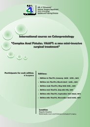
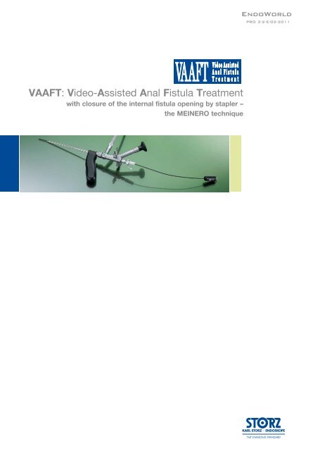
<strong>VAAFT</strong>: <strong>Video</strong>-<strong>Assisted</strong> <strong>Anal</strong> <strong>Fistula</strong> <strong>Treatment</strong><br />
with closure of the internal fistula opening by stapler –<br />
the MEINERO technique<br />
EndoWorld<br />
PRO 3-2-E/02-2011
Introduction<br />
The <strong>VAAFT</strong> technique is performed for the surgical treatment of complex anal<br />
fistulas and their recurrences. Key points are the correct localization of the internal<br />
fistula opening under vision, the fistula treatment from inside, and the hermetic<br />
closure of the internal opening. This technique comprises two phases: a diagnostic<br />
one and an operative one. There is no need to know the fistula classification which<br />
obviously saves time and money. Moreover, surgical wounds in the perianal region<br />
are prevented and the risk of faecal incontinence is avoided because no sphincter<br />
damages are provoked.<br />
Fig. 1<br />
piercarlo <strong>Meinero</strong>, M.D<br />
Chief of proctology<br />
Surgery Department - ASL 4 Chiavarese<br />
S. Margherita Ligure – Lavagna Hospital, Italy<br />
e-mail: pmeinero@asl4.liguria.it<br />
Tel.: 0039 320 4391395<br />
www.piercarlomeinero.it
Materials<br />
KARL STORZ Equipment is used (Fig. 1). The personal kit includes the MEINERO<br />
Fistuloscope (Fig. 2), a unipolar electrode (Fig. 3) connected to a high frequency<br />
unit, a fistula brush (Fig. 4) and a forceps (Fig. 5). Moreover, a semicircular or<br />
linear stapler and 0,5 ml of synthetic cyanoacrylate with a tiny catheter are used<br />
as well.<br />
The fistuloscope is equipped with an optical channel, a working channel and an irrigation<br />
channel. The working length adds up to 18 cm; the use of a handle reduces<br />
it to an effective length of 14 cm.<br />
Fig. 2<br />
Fig. 4<br />
Fig. 3<br />
Fig. 5<br />
2 3
The Technique<br />
The optimal patient positioning is the lithotomic position. Spinal anaesthesia is<br />
required. The fistuloscope is connected to the KARL STORZ equipment and to<br />
the washing solution bag (5000 cc glycine and mannitol 1% solution). Fig. 6<br />
shows an example of an anal fistula with its external (E.O.) and internal (I.O.)<br />
openings.<br />
E.O.<br />
Fig. 6<br />
The technique comprises a diagnostic phase and an operative phase.<br />
1. The diagnostic phase<br />
I.O.<br />
The purpose of the diagnostic phase is the correct localization of the internal fistula<br />
opening.<br />
The fistuloscope is inserted through the external fistula opening with the washing<br />
solution (glycine 1% and mannitol 1%) already running; providing a clear view of<br />
the fistula pathway which appears on the screen (Fig. 7). Blocking tissue can be<br />
removed using the 2 mm forceps to facilitate the insertion of the fistuloscope. The<br />
direction of the telescope is correct when the obturator appears in the lower part<br />
of the screen (Fig. 8).<br />
Fig. 7 Fig. 8<br />
Obturator
The surgeon follows the fistula pathway using slow left-right and up-down<br />
movements. Since the fistuloscope is rigid it helps to guide it using a transanally<br />
inserted finger. These maneuvers are favored by the complete relaxation of the<br />
surrounding tissue induced by the spinal anesthesia.<br />
The continous flow of the glycine-mannitol solution allows for an optimal view of<br />
the fistula's inside up to the internal opening.<br />
The assistant can insert an anal retractor in order to localize the internal fistula<br />
opening by looking for the light of the telescope in the rectum or anal canal.<br />
Dimming the lights in the operating theatre enables an easy localization of the<br />
fistuloscope light in the rectum.<br />
When the fistuloscope exits through the internal opening the rectal mucosa clearly<br />
appears on the screen. In some cases the internal opening might be very narrow;<br />
in this case, its location is suspected viewing the fistuloscope light behind the<br />
rectal mucosa. (Fig. 9–11).<br />
Fig. 9<br />
Fig. 10 Fig. 11<br />
Internal<br />
opening<br />
location<br />
4 5<br />
4 5
The Technique<br />
At this point, we put two or three stitches in two opposite points of the internal<br />
opening margin in order to isolate and, above all, not to loose it. We must be sure<br />
to capture sufficient tissue thickness (Fig. 12–14)<br />
Fig. 12<br />
Fig. 13<br />
Fig. 14<br />
Internal<br />
opening<br />
isolation
2. The operative phase<br />
purpose of this phase is the destruction of the fistula from the inside. As a next<br />
step, the fistula canal is cleaned / the waste material removed and its internal<br />
opening is then closed hermetically. We start destroying the fistula under vision<br />
using a unipolar electrode which can be passed through the operative channel of<br />
the fistuloscope and is connected to the electrosurgical power unit (Fig. 15–18).<br />
Fig. 15<br />
Fig. 16<br />
Fig. 18<br />
Fig. 17<br />
6 7<br />
6 7<br />
Starting at the internal fistula opening, all<br />
fragments of the whitish material adhering<br />
to the fistula wall and all granulation<br />
tissue are coagulated. We complete this<br />
phase of the operation, centimeter by<br />
centimeter, from the internal opening to<br />
the external opening not forgetting any<br />
abscessual cavity.
The Technique<br />
The necrotic material is removed under vision using the fistula brush (Fig. 19–21).<br />
Until that time, the isolated internal fistula opening remains open to allow the leakage<br />
of waste and washing material into the rectum.<br />
Fig. 20<br />
Fig. 21<br />
Fig. 19
At this point we completely remove the fistuloscope. The assistant stretches the<br />
threads towards the internal rectal space or rather the anal canal using a straight<br />
forceps in order to lift the internal fistula opening at least 2 cm into the shape of<br />
a volcano. Subsequently, we insert a stapler (e.g. CCS30 Transtar Contour from<br />
ETHICON EndoSurgery) at the volcano’s base (Fig. 22–25) and complete the<br />
mechanical cutting and suturing.<br />
Fig. 22<br />
Fig. 23<br />
Fig. 24 Fig. 25<br />
Fig.24<br />
8 9<br />
8 9
The Technique<br />
The hermetic closure of the internal fistula opening can also be accomplished by<br />
using a linear stapler (Fig. 26–28). This also depends on the internal opening<br />
position.<br />
Fig. 25 26 Fig. Fig. 26<br />
27<br />
Fig. 28<br />
Using a semicircular stapler, the suture will be horizontal. Using a linear stapler,<br />
the suture will be vertical (Fig. 29–31)<br />
Fig. 28 Fig. 29<br />
Fig. 29 Fig. 30<br />
Fig. 31
When the tissue in the area of the internal<br />
opening is not sclerotic and allows<br />
to form a good "volcano", the stapler<br />
can be used, however if the tissue<br />
around the internal opening is too rigid<br />
and sclerotic, the use of the stapler<br />
might be difficult. In this case a cutaneous<br />
mucosal flap would be preferred<br />
Fig. 32).<br />
As a last step we insert 0,5 ml of synthetic<br />
cyanoacrylate right after the<br />
suture / staple line via the fistula pathway<br />
to further reinforce the suture.<br />
Fig. 32<br />
So the use of the synthetic cyanoacrylate behind the suture line or behind the<br />
flap assures the perfect opening closure. It is essential to keep in mind that not<br />
the whole fistula tract is filled up with the synthetic cyanoacrylate; only a small<br />
amount is inserted directly below the suture line. That's why the fistula pathway<br />
has to stay open to allow the passage of secretions. (Fig. 33–36).<br />
Fig. 33 Fig. 34<br />
Fig. 35<br />
Fig. 36<br />
This procedure assures a perfect excision and a hermetic closure of the<br />
internal fistula opening, excluding the risk of stool passage. Since the suture<br />
is situated tangential to the sphincter, the postoperative pain is low even if<br />
the suture falls both in the anal canal and the rectum.<br />
10 11<br />
10 11
Conclusion<br />
Conclusion<br />
The advantages of the <strong>VAAFT</strong> technique are evident: no surgical wounds on the<br />
buttocks or in the perianal region are provoked, there is complete certainty in the<br />
localization of the internal fistula opening (a key point in all fistula surgical treatments),<br />
and the fistula can be completely destroyed from the inside. There is no<br />
requirement to know if the fistula is transphincteric, extrasphincteric or above<br />
sphincteric because operating from the inside no damage is caused to the anal<br />
sphincters. Therefore, no preoperative examination is necessary. The risk of<br />
postoperative faecel incontinence is excluded. Moreover, the patient doesn’t need<br />
any medications and he can start working again after a few days since the <strong>VAAFT</strong><br />
technique can be performed in day surgery.
Instrument Set<br />
<strong>VAAFT</strong> Instrument Set for <strong>Video</strong>-<strong>Assisted</strong> <strong>Anal</strong> <strong>Fistula</strong><br />
<strong>Treatment</strong> acc. to MEINERO<br />
24511 Fistulectomy Set,<br />
consisting of:<br />
24511 AA Telescope 8°, angled eyepiece,<br />
O.D. 3.3 × 4.7 mm, working length 18 cm,<br />
autoclavable, with straight instrument channel for<br />
instruments diameter 2.5 mm, fiber optic light<br />
transmission incorporated, colour code green,<br />
39501 xp Sterilization Tray<br />
24513 Obturator<br />
24511AA Telescope 8°, angled eyepiece,<br />
O.D. 3.3 × 4.7 mm, working length 18 cm, autoclavable, with<br />
straight instrument channel for instruments diameter 2.5 mm,<br />
fiber optic light transmission incorporated, colour code green,<br />
24512 Handle<br />
24513 Obturator for the endoscope<br />
24515 Coagulating Electrode, 7 Fr., for the Fistulectomy<br />
12 13
Instrument Set<br />
24514 <strong>Fistula</strong> Brush, with handle<br />
consisting of:<br />
3-Ring Handle,<br />
Outer Sheath,<br />
Three different <strong>Fistula</strong> Brush Inserts: with 4,0 mm; 4,5 mm and<br />
5,0 mm outer diameter.<br />
30221 KJ c REDDICK-OLSEN Grasping Forceps, rotating,<br />
size 2 mm, length 30 cm, with connector pin for unipolar<br />
coagulation, double action jaws, with LUER lock adaptor<br />
for cleaning,<br />
consisting of:<br />
33121 Plastic Handle, without ratchet<br />
30210 KJ Outer Tube with insert, insulated<br />
24981 AUCKLAND EASI <strong>Anal</strong> Distending Speculum,<br />
for anal examinations, with 3 blades, outer diameter 27 mm,<br />
working length 6 cm, with obturator 24981 O, with ratchet<br />
39501 xp Wire Basket Tray for cleaning, sterilization and storage.<br />
Including cleaning adaptor for washer-disinfector. With lid,<br />
spare parts basket 39501 xS and silicone telescope holders.<br />
External dimensions (w x d x h): 460 x 150 x 80 mm.<br />
For instruments with up to 27 cm working length.
Cold Light Fountain XENON 300 SCB ®<br />
20133101-1 Cold Light Fountain XENON 300 SCB ®<br />
with integrated KARL STORZ SCB ® , integrated Anti-Fog pump,<br />
one 300 Watt xenon Lamp and one KARL STORZ light outlet,<br />
power supply: 100–125/220–240 VAC, 50/60 Hz<br />
consisting of:<br />
20 1331 20-1 XENON 300<br />
400 A Mains Cord<br />
610 A FT Silicone Tubing Set, length 250 cm<br />
20 0901 70 SCB-Connecting Cable, length 100 cm<br />
201330 27 Xenon-Spare-Lamp-Module,<br />
300 Watt, 15 Volt<br />
201330 28 XENON Spare Lamp, only, 300 Watt, 15 Volt<br />
495 NL Fiber Optic Light Cable, size 3.5 mm, length 180 cm<br />
495 NA Fiber Optic Light Cable, size 3.5 mm, length 230 cm<br />
14 15
Additional Instrumentation<br />
IMAGE 1 HD<br />
HD hub Camera Control Unit<br />
• Maximum resolution and the consistent use of<br />
the 16:9 aspect ratio guarantee FULL HD<br />
• Endoscopic camera systems have to be equipped<br />
with three-CCD chips that support the 16:9<br />
input format as well as capturing images with a<br />
resolution of 1920 x 1080 pixels<br />
Specifications:<br />
22 2010 20-1xx<br />
Signal-to-noise ratio AGC <strong>Video</strong> output Input<br />
IMAGE 1 HUB HD<br />
Three-chip camera systems M 60 dB<br />
Control output /input<br />
Micro -<br />
processor-<br />
controlled<br />
- KARL STORZ-SCB ® at 6-pin Mini DIN socket (2x)<br />
- 3.5 mm stereo jack plug (ACC 1, ACC 2),<br />
- Serial port at RJ-11<br />
- USB port (only IMAGE 1 HUB HD with ICM) (2x)<br />
22 2010 11U102 IMAGE 1 HUB HD Camera Control Unit (CCU)<br />
with SDI Module<br />
for use with IMAGE 1 HD and standard one- and three-chip camera heads,<br />
max. resolution 1920 x 1080 pixel, with integrated KARL STORZ SCB ® and<br />
integrated digital Image processing Module, color systems pAL/NTSC, power<br />
supply 100 – 240 VAC, 50/60 Hz<br />
consisting of:<br />
22 2010 20-102 IMAGE 1 HUB HD (with SDI) Camera Control Unit<br />
400 A Mains Cord<br />
3 x 536 MK BNC/BNC <strong>Video</strong> Cable, length 180 cm<br />
547 S S-<strong>Video</strong> (Y/C) Connecting Cable, length 180 cm<br />
20 2032 70 Special RGB Connecting Cable<br />
2x 20 2210 70 Connecting Cable, for controlling peripheral units,<br />
length 180 cm<br />
20 0400 86 DVI Connecting Cable, length 180 cm<br />
20 0901 70 SCB Connecting Cable, length 100 cm<br />
20 2001 30U Keyboard, with English character set<br />
- Composite signal to BNC socket<br />
- S-<strong>Video</strong> signal to 4-pin Mini DIN socket (2x)<br />
- RGBS signal to D-Sub socket<br />
-- SDI signal to BNC socket (only IMAGE 1 HUB HD with SDI<br />
module)(2x)<br />
- HDTV signal to DVI-D socket (2x)<br />
Dimensions<br />
w x h x d (mm)<br />
The benefits of High Definition Technology (HD) for<br />
medical applications are<br />
• Up to 6 times* higher input resolution of the<br />
camera delivers more detail and depth of focus<br />
• Using 16:9 format during image acquisition<br />
enlarges the field of vision and supports ergonomic<br />
viewing<br />
• The brilliance of color enables optimal diagnosis<br />
• Lateral view is enhanced by 32% when the<br />
endoscope is withdrawn slightly, providing<br />
the same image enhancement as a standard<br />
system. Any vertical information loss is restored<br />
and the lens remains clean<br />
Keyboard for title generator,<br />
5-pin DIN socket<br />
Weight (kg) Power supply Certified to:<br />
305 x 89 x 335 2.95 100-240 VAC,<br />
50/60 Hz<br />
IEC 601-1, 601-2-18, CSA 22.2<br />
No. 601, UL 2601-1 and CE acc. to<br />
MDD, protection class 1/CF<br />
SDI – Serial Digital Interface: optimized to display medical images on Flat Screens, Routing with OR1 and<br />
digital recording with AIDA-DVD-M<br />
ICM: USB-connector for recording video streams and stills on USB storage media or for connection of USB<br />
printers for direct printing of the recorded stills
IMAGE 1 HD<br />
HD Camera Head<br />
22 2200 55-3<br />
KARL STORZ<br />
HD Flat<br />
Screens<br />
Color systems<br />
PAL/NTSC<br />
Version<br />
Wall mounted<br />
with VESA<br />
100-adaption<br />
Desktop with<br />
pedestal<br />
The following accessories are included:<br />
400 A Mains Cord<br />
9523 pS External 24VDC Power Supply<br />
9419 NSF Pedestal<br />
22 2200 55-3 50 Hz IMAGE 1 H3-Z,<br />
60 Hz <strong>Dr</strong>ei-Chip HD Kamerakopf<br />
max. resolution 1920 x 1080 pixels, progressive scan, soakable, gas<br />
and plasma sterilizable, with integrated parfocal Zoom Lens, focal length<br />
f = 15 – 31 mm (2x), 2 freely programmable camera head buttons,<br />
for use with color system pAL/NTSC<br />
Specifications:<br />
Order<br />
No.<br />
9524 NB<br />
9526 NB<br />
9524 N<br />
9526 N<br />
Image sensor<br />
pixel output signall H x V<br />
Dimensions<br />
Weight<br />
Min. sensitivity<br />
Lens<br />
Grip mechanism<br />
Cable<br />
Cable length<br />
Screen<br />
diagonal<br />
24"<br />
26"<br />
24"<br />
26"<br />
Max. screen<br />
resolution<br />
1920 x 1200<br />
l<br />
3x 1 /3" CCD-Chip<br />
1920 x 1080<br />
Diameter 32-44 mm, length 114 mm<br />
246 g<br />
F 1,4/1,17 Lux<br />
Integrated parfocal Zoom Lens,<br />
f = 15-31 mm<br />
Standard eyepiece detector,<br />
non-detachable<br />
300 cm<br />
Composite signal<br />
to BNC socket<br />
l<br />
S-<strong>Video</strong> to 4-pin<br />
Mini DIN socket<br />
RGB to<br />
5x BNC socket<br />
<strong>Video</strong> input<br />
VGA to 15-pin<br />
HD-D-Sub socket<br />
SDI to<br />
HD-SDI to<br />
BNC socket<br />
BNC socket<br />
l l l l l<br />
DVI to<br />
l<br />
16 17<br />
DVI-D socket
Additional Instrumentation<br />
Data Management and Documentation<br />
KARL STORZ AIDA ® compact NEO (HD/SD)<br />
Brilliance in documentation continues!<br />
AIDA compact NEO from KARL STORZ combines all the required functions for integrated and precise<br />
documentation of endoscopic procedures and open surgeries in a single system.<br />
AIDA compact NEO:<br />
Voice control<br />
AIDA compact NEO:<br />
Review screen<br />
AIDA compact NEO: Automatic<br />
creation of standard reports<br />
AIDA compact NEO:<br />
Effi cient archiving<br />
Data acquisition<br />
Still images, video sequences and audio comments can be recorded<br />
easily during an examination or intervention on command by either<br />
pressing the on screen button, voice control, foot switch or pressing the<br />
camera head button. All captured images will be displayed on the right<br />
hand side as a “thumbnail” preview to ensure the still image has been<br />
generated.<br />
The patient data can be entered by the on-screen keyboard or by a<br />
standard keyboard.<br />
Flexible post editing and data storage<br />
Captured still images or video files can be previewed before final storage<br />
or can be edited and deleted easily in the edit screen.<br />
Reliable storage of data<br />
� Digital saving of all image, video and audio fi les on DVD, CD-ROM,<br />
USB stick, external/internal hard-drive or to the central hospital storage<br />
possibilities over DICOM/HL7<br />
� Buffering ensures data backup if saving is temporarily not possible<br />
� Continuous availability of created image, video and sound material for<br />
procedure documentation and for research and teaching purposes.<br />
Efficient data archiving<br />
After a procedure has been completed, KARL STORZ AIDA ® compact<br />
HD/SD saves all captured data efficiently on DVD, CD-ROM, USB stick,<br />
external hard-drive, internal hard-drive and/or the respective network on<br />
the FTp server. Furthermore the possibility exists to store the data directly<br />
on the pACS respective HIS server, over the interface package AIDA<br />
communication HL7/DICOM.<br />
Data that could not be archived successfully remains in a special buffered<br />
procedure until it is finally saved. A two-line report header and a logo can<br />
be used by the user to meet his or her needs.<br />
Multi-session and Multi-patient<br />
Efficient data archiving is assured as several treatments can be saved<br />
on a DVD, CD-ROM or a USB stick.
Features and Benefits<br />
� Digital storage of still images with a resolution of 1920 x 1080 pixels, video sequences in 720p and audio fi les with<br />
AIDA compact NEO HD<br />
� Optional interface package DICOM/HL7<br />
� Sterile, ergonomic operation via touch screen, voice control, camera head buttons and/or foot switches<br />
� Auto detection of the connected camera system on HD-SDI/SD-SDI input<br />
� Effi cient archiving on DVD, CD-ROM or USB stick, multi-session and multi-patient<br />
� Network saving<br />
� Automatic generation of standard reports<br />
� Approved use of computers and monitors in the OR environment as per EN 60601-1<br />
� Compatibility with the KARL STORZ Communication Bus (SCB) and with the KARL STORZ OR1 AV NEO<br />
� KARL STORZ AIDA ® compact NEO HD/SD is an attractive, digital alternative to video printers, video recorders and<br />
dictaphones.<br />
Specifications:<br />
<strong>Video</strong> Systems<br />
Signal Inputs<br />
Image Formats<br />
- pAL<br />
- NTSC<br />
- S-<strong>Video</strong> (Y/C)<br />
- Composite<br />
- RGBS<br />
- SDI<br />
- HD-SDI<br />
- DVI<br />
- JpG<br />
- BMp<br />
20 0409 10 KARL STORZ AIDA ® compact NEO SD<br />
Communication, documentation system<br />
for digital storage of still images,<br />
video sequences and audio files,<br />
power supply 115/230 VAC, 50/60 Hz<br />
20 0409 11 KARL STORZ AIDA ® compact NEO HD<br />
Communication, documentation system<br />
for digital storage of still images,<br />
video sequences and audio files,<br />
power supply 115/230 VAC, 50/60 Hz<br />
20 0406 10 KARL STORZ AIDA ® compact NEO SD,<br />
documentation system for digital storage of<br />
still images, video sequences and audio files,<br />
power supply 115/230 VAC, 50/60 Hz<br />
20 0406 11 KARL STORZ AIDA ® compact NEO HD,<br />
documentation system for digital storage of<br />
still images, video sequences and audio files,<br />
power supply 115/230 VAC, 50/60 Hz<br />
<strong>Video</strong> Formats<br />
Audio Formats<br />
Storage Media<br />
- MpEG2<br />
- WAV<br />
- DVD+R<br />
- DVD+RW<br />
- DVD-R<br />
- DVD-RW<br />
- CD-R<br />
- CD-RW<br />
- USB stick<br />
18 19
Additional Instrumentation<br />
AUTOCON ® II 400 HF surgical unit<br />
for interdisciplinary high-frequency surgery<br />
• Can be used across a wide range of surgical disciplines<br />
• The world’s only HF unit with a 6.5" touch screen for easy, safe, ergonomic operation and cleaning<br />
• Preprogrammed settings available for certain procedures; settings for procedures can be additionally<br />
programmed easily and quickly<br />
Necessary accessories:<br />
20 5352 01-115 KARL STORZ AUTOCON ® II 400 High End<br />
power supply: 230 VAC, 50/60 Hz<br />
HF Connecting Sockets: Bipolar Standard, Bipolar multifunction,<br />
Unipolar 3-pin + Erbe, Neutral electrode 6.3 mm jack,<br />
consists of:<br />
20 5352 20-115 AUTOCON ® II 400 with KARL STORZ SCB ®<br />
400 A Mains Cord<br />
20 0901 70 SCB Connection Cable, length 100 cm<br />
20 0138 30 Double Pedal Footswitch<br />
for 205352 01-11x<br />
26176 LE Bipolar High Frequency Cord for<br />
KARL STORZ AUTOCON ® system (50,<br />
200, 350), AUTOCON ® II 400 (Tpe 111,<br />
113, 115) and Erbe-Coagulator, T- and<br />
ICC-row, length 300 cm<br />
26005 M Unipolar High Frequency Cord<br />
with 5 mm plug for HF unit, models<br />
KARL STORZ AUTOCON ® system (50,<br />
200, 350) AUTOCON ® II 400 (Tpe 111,<br />
115) and Erbe type ICC; length 300 cm<br />
27805 Neutral Electrode made of conductive<br />
silicone, with 2 rubber straps for fastening,<br />
for use with<br />
KARL STORZ AUTOCON ® (type 200,<br />
350), AUTOCON ® II 400 (type 111, 115)<br />
A= 500 cm² surface area<br />
27806 Connecting Cord, for connecting<br />
the neutral electrodes 27805 and<br />
860021 E, length 400 cm
Notes<br />
20 21
Notes
Notes<br />
22 23
www.karlstorz.com<br />
KARL STORZ GmbH & Co. KG<br />
Mittelstraße 8, 78532 Tuttlingen, Germany<br />
postbox 230, 78503 Tuttlingen, Germany<br />
phone: +49 (0)7461 708-0<br />
Fax: +49 (0)7461 708-105<br />
E-Mail: info@karlstorz.de<br />
www.karlstorz.com<br />
KARL STORZ Endoscopy-America, Inc.<br />
2151 E. Grand Avenue<br />
El Segundo, CA 90245-5017, USA<br />
phone: +1 424 218-8100<br />
phone toll free: 800 421-0837 (US only)<br />
Fax: +1 424 218-8525<br />
Fax toll free: 800 321-1304 (US only)<br />
E-Mail: info@ksea.com<br />
EndoWorld ®<br />
EW pRO 3-2-E/02-2011