1 Fuchs Endothelial Dystrophy
1 Fuchs Endothelial Dystrophy
1 Fuchs Endothelial Dystrophy

You also want an ePaper? Increase the reach of your titles
YUMPU automatically turns print PDFs into web optimized ePapers that Google loves.
126 Herpes Simplex Keratitis and Related Syndromes<br />
7<br />
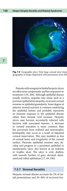
Fig. 7.4 Geographic ulcer. Note large corneal ulcer stained with fluorescein that is<br />
geographic in shape (Reprinted with permission from [80])<br />
Patientswithmarginalorlimbalherpeticulcers<br />
are often more symptomatic and less responsive to<br />
treatment [139, 206]. Although epithelial disease<br />
usually resolves, sequelae may ensue such as a<br />
punctate epithelial keratopathy, recurrent corneal<br />
erosions or epithelial granularity. Some degree of<br />
anterior stromal reaction is common underlying<br />
the epithelial lesions and probably represents<br />
an immune response to the epithelial disease<br />
rather than stromal viral invasion. Herpetic<br />
ulcers may become secondarily infected with<br />
bacteria with associated features. A decrease<br />
in corneal sensation is more common than<br />
has previously been realized and neurotrophic<br />
keratopathy may occur as a result of impaired<br />
corneal innervation. This may manifest as loss<br />
of corneal luster and irregularity of the corneal<br />
surface. Punctate epithelial erosions may develop<br />
and progress to a persistent epithelial or<br />
metaherpetic ulcer, also known as an indolent<br />
or trophic ulcer. The ulcer is oval, shallow,<br />
with smooth borders of grey, elevated, thickened<br />
and rolled epithelium [17, 69, 100].<br />
7.5.7 Stromal Keratitis<br />
Herpetic stromal disease accounts for 2% of initial<br />
presentations and 20–48% of recurrent her-<br />
petic disease [42, 99, 128]. It results from viral<br />
invasion of the stroma, either from reactivation<br />
of latent virus in the supplying sensory nerves or<br />
from within the cornea itself.<br />
The associated immune response contributes<br />
to the type of stromal disease seen. There may be<br />
no history of previous infectious epithelial keratitis<br />
and the epithelium is usually intact. The main<br />
characteristic is stromal inflammation, which<br />
may be focal, multifocal or diffuse and may be associated<br />
with anterior uveitis [205]. The inflammation<br />
may be chronic, recurrent or recrudescent<br />
leading to stromal scarring, thinning, neovascularization,<br />
and lipid deposition (Fig. 7.5).<br />
Occasionally, a partial or complete immune<br />
ring, similar to a Wessely ring, is seen in the central<br />
or paracentral mid-stroma [115]. As with<br />
epithelial disease, marginal keratitis or limbitis<br />
tends to be associated with a greater inflammatory<br />
or immune response. Stromal neovascularization<br />
can be sectoral or diffuse and frequently<br />
occurs in several layers of the cornea, a condition<br />
described as “interstitial stromal HSV keratitis”<br />
[100]. Necrotizing stromal keratitis may occur.<br />
There is necrosis, ulceration, and dense infiltration<br />
of the stroma, usually with an overlying<br />
epithelial defect. Grayish white homogeneous<br />
abscesses with edema, keratic precipitates, and<br />
iridocyclitis are seen.