cytokine
cytokine BULLETIN
cytokine BULLETIN
- No tags were found...

Create successful ePaper yourself
Turn your PDF publications into a flip-book with our unique Google optimized e-Paper software.
Regulation of VE-Cadherin and VEGF R2 by VEGF<br />
VE-Cadherin and VEGF R2 are transmembrane<br />
glycoproteins that are expressed in the adherens<br />
junctions between vascular endothelial<br />
cells (EC). VE-Cadherin interacts homophilically<br />
between neighboring cells to<br />
provide strength to the endothelium. VE-<br />
Cadherin adhesion is reduced during angiogenesis<br />
and leukocyte extravasation, two<br />
processes that require decreased EC attachment.<br />
It is well established that adherens<br />
junction integrity is regulated by VE-Cadherin<br />
phosphorylation and internalization in<br />
response to VEGF stimulation. 1,2 Several recent<br />
publications provide additional details<br />
about the molecular mechanism directing<br />
this process.<br />
In adherens junctions between quiescent<br />
vascular EC, VEGF R2 is maintained in an inactive<br />
state by protein tyrosine phosphatases.<br />
2 VEGF binding activates the tyrosine<br />
kinase domain of VEGF R2, initiating the<br />
sequential activation of the Src-Vav2-Rac1-<br />
PAK pathway, which results in the phosphorylation<br />
of VE-Cadherin at Ser665 by<br />
PAK. 3 The subsequent binding of b-arrestin2<br />
to serine-phosphorylated VE-Cadherin promotes<br />
the internalization of VE-Cadherin<br />
into clathrin-coated pits. 3 This disrupts the<br />
architecture of endothelial junctions and allows<br />
for the passage of molecules and cells.<br />
In addition, phosphorylation of the VE-<br />
Cadherin complexes by Src at Tyr685 may<br />
contribute to the disassembly of adherens<br />
junctions. 4<br />
It was recently reported that VEGF R2 signaling<br />
is enhanced by endocytosis. Interestingly,<br />
VE-Cadherin was shown to play an inhibitory<br />
role in this process. By binding to VEGF<br />
R2, VE-Cadherin prevents VEGF R2 endocytosis.<br />
This favors inactivation by a junction-associated,<br />
transmembrane tyrosine<br />
phosphatase, called DEP-1. 5 When clathrindependent<br />
internalization transports phosphorylated<br />
VEGF R2 to endosomal vesicles,<br />
uninterrupted VEGF R2 signaling is made<br />
possible by phosphorylated Tyr1175 inaccessible<br />
to cell surface DEP-1. 5 Phosphorylated<br />
Tyr1175 is required for the binding and activation<br />
of PLCg, a primary mediator of VEGF<br />
R2-promoted cell proliferation. 6 Notably, cell<br />
surface VEGF R2-mediated clathrin-dependent<br />
internalization delivers VE-Cadherin to<br />
compartments distinct from intracellular<br />
VEGF R2 compartments. 3<br />
Other molecules influence these processes<br />
as well, although specifically how they are<br />
integrated into the above pathways is not<br />
clear. The scaffolding protein IQGAP1, which<br />
is bound to VE-Cadherin both at the adherens<br />
junction and after internalization, is necessary<br />
for VE-Cadherin localization at cellcell<br />
contacts. 7 It potentially mediates the<br />
interaction of VE-Cadherin and VEGF R2, and<br />
also tethers VE-Cadherin to the cytoskeleton.<br />
1,7 In addition, VEGF stimulation of<br />
quiescent microvascular EC induces the<br />
transport of intracellular JAM-C to adherens<br />
junctions, where it disrupts VE-Cadherin<br />
adhesion. 8 TNF-a also promotes vascular<br />
permeability by inducing Fyn-dependent<br />
tyrosine phosphorylation of VE-Cadherin. 9<br />
Lastly, exposure of endothelium to oxidized<br />
LDL promotes VE-Cadherin internalization<br />
and increased monocyte transendothelial<br />
migration. 10<br />
These advances in the understanding of the<br />
interactions between VE-Cadherin and VEGF<br />
R2 clarify how the adherens junction is<br />
regulated in response to VEGF stimulation.<br />
Prolonged VEGF R2 signaling enables the<br />
continued proliferation and migration of<br />
vascular EC during angiogenesis. Internalization<br />
of VE-Cadherin weakens adherens<br />
junction adhesion, permitting increased EC<br />
mobility, vascular permeability, and leukocyte<br />
extravasation.<br />
References<br />
1. Wallez, Y. et al. (2006) Trends Cardiovasc. Med. 16:55.<br />
2. Mukherjee, S. et al. (2006) Circ. Res. 98:743.<br />
3. Gavard, J. & J.S. Gutkind (2006) Nat. Cell Biol. 8:1223.<br />
4. Wallez, Y. et al. (2007) Oncogene 26:1067.<br />
5. Lampugnani, M.G. et al. (2006) J. Cell Biol. 174:593.<br />
6. Takahashi, T. et al. (2001) EMBO J. 20:2768.<br />
7. Yamaoka-Tojo, M. et al. (2006) Arterioscler.<br />
Thromb. Vasc. Biol. 26:1991.<br />
8. Orlova, V.V. et al. (2006) J. Exp. Med. 203:2703.<br />
9. Angelini, D.J. et al. (2006) Am. J. Physiol. Lung Cell Mol.<br />
Physiol. 291:L1232.<br />
10. Hashimoto, K. et al. (2006) Atherosclerosis, Dec. 26<br />
[EPub ahead of print].<br />
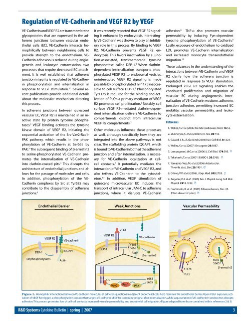
Endothelial Barrier Weak Junctions Vascular Permeability<br />
VEGF<br />
DEP-1<br />
VEGF R2<br />
Dephosphorylates<br />
Actin<br />
VE-cadherin<br />
IQGAP1<br />
VEGF R2<br />
DEP-1<br />
Y685<br />
S665<br />
Actin<br />
VE-cadherin<br />
β-arrestin2<br />
Y1175<br />
PLCγ<br />
Y685<br />
S665<br />
β-arrestin2<br />
src<br />
Vav2<br />
Rac1<br />
PAK<br />
Y685<br />
S665<br />
Figure 1. Homophilic interactions between VE-cadherin molecules at adherens junctions in adjacent endothelial cells help maintain the endothelial barrier. Upon VEGF exposure, activation<br />
of VEGF R2 triggers a phosphorylation cascade that targets VE-cadherin. VEGF R2 continues to signal after internalization, while sequestration of VE-cadherin in endosomes disrupts<br />
adhesion. This process promotes loss of cell-cell contacts, increased vascular permeability, and endothelial cell migration. (Figure adapted from those contained within references 2 & 3)<br />
R&D Systems Cytokine Bulletin | spring | 2007