MEDICINE
download a copy - University of British Columbia Faculty of Medicine
download a copy - University of British Columbia Faculty of Medicine
- No tags were found...

Create successful ePaper yourself
Turn your PDF publications into a flip-book with our unique Google optimized e-Paper software.
18 UBC <strong>MEDICINE</strong><br />
research<br />
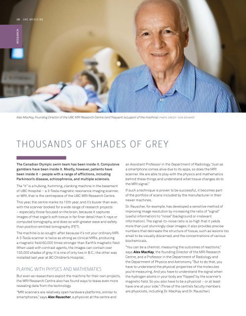
Alex MacKay, Founding Director of the UBC MRI Research Centre (and frequent occupant of the machine). Photo credit: Don Erhardt<br />
Thousands of shades of grey<br />
The Canadian Olympic swim team has been inside it. Compulsive<br />
gamblers have been inside it. Mostly, however, patients have<br />
been inside it – people with a range of afflictions, including<br />
Parkinson’s disease, schizophrenia, and multiple sclerosis.<br />
The “it” is a hulking, humming, clanking machine in the basement<br />
of UBC Hospital – a 3-Tesla magnetic resonance imaging scanner,<br />
or MRI, that is the centrepiece of the UBC MRI Research Centre.<br />
This year, the centre marks its 10th year, and it’s busier than ever,<br />
with the scanner booked for a wide range of research projects<br />
– especially those focused on the brain, because it captures<br />
images of that organ’s soft tissue in far finer detail than X-rays or<br />
computed tomography, and does so with greater ease and safety<br />
than positron emitted tomography (PET).<br />
The machine is so sought-after because it’s not your ordinary MRI.<br />
A 3-Tesla scanner is twice as strong as clinical MRIs, producing<br />
a magnetic field 60,000 times stronger than Earth’s magnetic field.<br />
When used with contrast agents, the images can contain over<br />
100,000 shades of grey. It is one of only two in B.C.; the other was<br />
installed last year at BC Children’s Hospital.<br />
Playing with physics and mathematics<br />
But even as researchers exploit the machine for their own projects,<br />
the MRI Research Centre also has found ways to tease even more<br />
revealing data from the technology.<br />
“MRI scanners are relatively open hardware platforms, similar to<br />
smartphones,” says Alex Rauscher, a physicist at the centre and<br />
an Assistant Professor in the Department of Radiology. “Just as<br />
a smartphone comes alive due to its apps, so does the MRI<br />
scanner. We are able to play with the physics and mathematics<br />
behind these things and understand what tissue changes do to<br />
the MRI signal.”<br />
If such a technique is proven to be successful, it becomes part<br />
of the portfolio of scans included by the manufacturer in their<br />
newer machines.<br />
Dr. Rauscher, for example, has developed a sensitive method of<br />
improving image resolution by increasing the ratio of “signal”<br />
(useful information) to “noise” (background or irrelevant<br />
information). The signal-to-noise ratio is so high that it yields<br />
more than just stunningly clear images; it also provides precise<br />
numbers that delineate the structure of tissue, such as lesions too<br />
small to be visually discerned, and the concentrations of various<br />
biochemicals.<br />
“You can be a chemist, measuring the outcomes of reactions,”<br />
says Alex MacKay, the founding Director of the MRI Research<br />
Centre, and a Professor in the Department of Radiology and<br />
the Department of Physics and Astronomy. “But to do that, you<br />
have to understand the physical properties of the molecules<br />
you’re measuring. And you have to understand the signal when<br />
the hydrogen atoms in your body are ‘flipped’ by the scanner’s<br />
magnetic field. So you also have to be a physicist – or at least<br />
have one at your side.” (Three of the centre’s faculty members<br />
are physicists, including Dr. MacKay and Dr. Rauscher.)