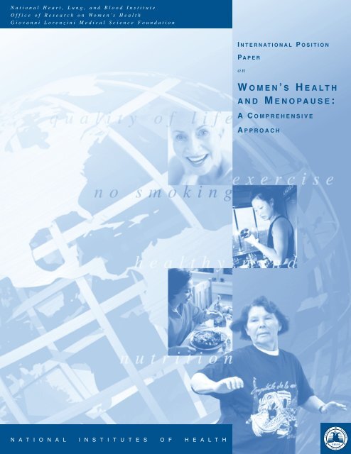
WOMEN 'S HEALTH AND MENOPAUSE : - National Heart, Lung ...
WOMEN 'S HEALTH AND MENOPAUSE : - National Heart, Lung ...
WOMEN 'S HEALTH AND MENOPAUSE : - National Heart, Lung ...

You also want an ePaper? Increase the reach of your titles
YUMPU automatically turns print PDFs into web optimized ePapers that Google loves.
<strong>National</strong> <strong>Heart</strong>, <strong>Lung</strong>, and Blood Institute<br />
Office of Research on Women’s Health<br />
Giovanni Lorenzini Medical Science Foundation<br />
N A T I O N A L I N S T I T U T E S O F H E A L T H<br />
I NTERNATIONAL P OSITION<br />
P APER<br />
on<br />
W OMEN’ S H EALTH<br />
<strong>AND</strong> M ENOPAUSE:<br />
A COMPREHENSIVE<br />
A PPROACH
I NTERNATIONAL<br />
P OSITION P APER<br />
on<br />
W OMEN’ S H EALTH<br />
<strong>AND</strong> M ENOPAUSE:<br />
A COMPREHENSIVE<br />
A PPROACH<br />
NATIONAL HEART, LUNG,<br />
<strong>AND</strong> BLOOD INSTITUTE<br />
OFFICE OF RESEARCH ON<br />
<strong>WOMEN</strong>’S <strong>HEALTH</strong><br />
NATIONAL INSTITUTES OF <strong>HEALTH</strong><br />
<strong>AND</strong><br />
GIOVANNI LORENZINI MEDICAL<br />
SCIENCE FOUNDATION<br />
NIH PUBLICATION NO. 02-3284<br />
JULY 2002
CHAIRS <strong>AND</strong> EDITORS<br />
Chair and Editor<br />
Nanette K. Wenger, M.D.<br />
Professor of Medicine (Cardiology)<br />
Emory University School of Medicine<br />
Atlanta, GA, U.S.A.<br />
Cochair and Coeditor<br />
Rodolfo Paoletti, M.D.<br />
Professor of Pharmacology<br />
Director, Department of Pharmacological Sciences<br />
University of Milan<br />
Milan, Italy<br />
PANEL MEMBERS<br />
Elizabeth Barrett-Connor, M.D.<br />
Professor and Chief<br />
Division of Epidemiology<br />
Department of Family and Preventive Medicine<br />
University of California, San Diego School of<br />
Medicine<br />
La Jolla, CA, U.S.A.<br />
Martin H. Birkhäuser, M.D.<br />
Professor<br />
Division of Gynecological Endocrinology<br />
Department of Obstetrics and Gynecology<br />
University of Bern<br />
Bern, Switzerland<br />
II<br />
Cochair and Coeditor<br />
Claude J. M. Lenfant, M.D.<br />
Director<br />
<strong>National</strong> <strong>Heart</strong>, <strong>Lung</strong>, and Blood Institute<br />
<strong>National</strong> Institutes of Health<br />
Bethesda, MD, U.S.A.<br />
Cochair and Coeditor<br />
Vivian W. Pinn, M.D.<br />
Associate Director for Research on<br />
Women’s Health<br />
Director, Office of Research on Women’s Health<br />
<strong>National</strong> Institutes of Health<br />
Bethesda, MD, U.S.A.<br />
Louise A. Brinton, Ph.D.<br />
Chief, Environmental Epidemiology Branch<br />
<strong>National</strong> Cancer Institute<br />
<strong>National</strong> Institutes of Health<br />
Bethesda, MD, U.S.A.<br />
Aila Collins, Ph.D.<br />
Associate Professor<br />
Psychology Section<br />
Department of Clinical Neuroscience<br />
Karolinska Institute<br />
Stockholm, Sweden
Peter Collins, M.D.<br />
Reader and Honorary Consultant Cardiologist<br />
Imperial College School of Medicine and Royal<br />
Brompton Hospital<br />
London, United Kingdom<br />
Pier Giorgio Crosignani, M.D.<br />
Professor and Chairman<br />
First Department of Obstetrics and Gynecology<br />
University of Milan<br />
Milan, Italy<br />
Lorraine Dennerstein, A.O., M.B.B.S., Ph.D.,<br />
D.P.M., F.R.A.N.Z.C.P.<br />
Professor and Director<br />
Office for Gender and Health<br />
Department of Psychiatry<br />
University of Melbourne<br />
Royal Melbourne Hospital<br />
Parkville, VIC, Australia<br />
Bruce Ettinger, M.D.<br />
Senior Investigator<br />
Division of Research<br />
Kaiser Permanente Medical Care Program<br />
Oakland, CA, U.S.A.<br />
Jan-Åke Gustafsson, M.D., Ph.D.<br />
Professor and Chairman<br />
Department of Medical Nutrition<br />
Chairman, Department of Biosciences<br />
Karolinska Institute<br />
Novum, Huddinge University Hospital<br />
Huddinge, Sweden<br />
Victor W. Henderson, M.D., M.S.<br />
Professor<br />
Departments of Geriatrics and Neurology<br />
Donald W. Reynolds Center on Aging<br />
Alzheimer’s Disease Center<br />
University of Arkansas for Medical Sciences<br />
Little Rock, AR, U.S.A.<br />
Susan Hendrix, D.O.<br />
Associate Professor<br />
Division of Gynecology<br />
Department of Obstetrics and Gynecology<br />
Wayne State University School of Medicine<br />
Hutzel Hospital<br />
Detroit, MI, U.S.A.<br />
Carlo La Vecchia, M.D.<br />
Head, Laboratory of Epidemiology<br />
Mario Negri Institute for Pharmacological<br />
Research<br />
Professor of Epidemiology<br />
University of Milan<br />
Milan, Italy<br />
Robert Lindsay, M.D., Ph.D.<br />
Chairman of Medicine<br />
Helen Hayes Hospital<br />
Professor of Clinical Medicine<br />
College of Physicians and Surgeons<br />
Columbia University<br />
New York, NY, U.S.A.<br />
Adriana Maggi, Ph.D.<br />
Professor of Biotechnology and Pharmacology<br />
Director, Molecular Pharmacology Laboratory<br />
Department of Pharmacological Sciences<br />
University of Milan<br />
Milan, Italy<br />
Joan A. McGowan, Ph.D.<br />
Director, Musculoskeletal Diseases Branch<br />
<strong>National</strong> Institute of Arthritis and Musculoskeletal<br />
and Skin Diseases<br />
<strong>National</strong> Institutes of Health<br />
Bethesda, MD, U.S.A.<br />
III
Anne McTiernan, M.D., Ph.D.<br />
Associate Member<br />
Public Health Sciences Division<br />
Fred Hutchinson Cancer Research Center<br />
Research Associate Professor<br />
Department of Epidemiology<br />
University of Washington Schools of Medicine and<br />
Public Health and Community Medicine<br />
Seattle, WA, U.S.A.<br />
Maryann Redford, D.D.S., M.P.H.<br />
Director, Clinical Trials and Epidemiologic<br />
Studies Program<br />
<strong>National</strong> Institute of Dental and Craniofacial<br />
Research<br />
<strong>National</strong> Institutes of Health<br />
Bethesda, MD, U.S.A.<br />
NONPANEL COAUTHORS<br />
Janet R. Guthrie, Ph.D., M.Sc.<br />
Research Fellow<br />
Office for Gender and Health<br />
Department of Psychiatry<br />
Faculty of Medicine, Dentistry, and<br />
Health Sciences<br />
The University of Melbourne<br />
Parkville, VIC, Australia<br />
Barbara E. K. Klein, M.D., M.P.H.<br />
Professor of Ophthalmology and Visual Sciences<br />
Ocular Epidemiology<br />
University of Wisconsin-Madison<br />
Madison, WI, U.S.A.<br />
IV<br />
Susan M. Resnick, Ph.D.<br />
Investigator<br />
Cognition Section<br />
Laboratory of Personality and Cognition<br />
<strong>National</strong> Institute on Aging<br />
<strong>National</strong> Institutes of Health<br />
Baltimore, MD, U.S.A.<br />
Jacques E. Rossouw, M.D.<br />
Acting Director, Women’s Health Initiative<br />
<strong>National</strong> <strong>Heart</strong>, <strong>Lung</strong>, and Blood Institute<br />
<strong>National</strong> Institutes of Health<br />
Bethesda, MD, U.S.A.<br />
Sherry Sherman, Ph.D.<br />
Director, Clinical Endocrinology and<br />
Osteoporosis Research<br />
<strong>National</strong> Institute on Aging<br />
<strong>National</strong> Institutes of Health<br />
Bethesda, MD, U.S.A.<br />
Stefan Nilsson, Dr. Med. Sci.<br />
Associate Professor<br />
Affiliated to Department of Medical Nutrition<br />
Karolinska Institute<br />
Novum, Huddinge University Hospital<br />
Huddinge, Sweden<br />
Nanette Santoro, M.D.<br />
Division Director<br />
Division of Reproductive Endocrinology<br />
Department of Obstetrics, Gynecology, and<br />
Women’s Health<br />
Albert Einstein College of Medicine<br />
Bronx, NY, U.S.A.
WITH APPRECIATION FOR THE EXPERTISE OF THE FOLLOWING, WHO GRACIOUSLY REVIEWED <strong>AND</strong><br />
COMMENTED ON THE PRELIMINARY INDIVIDUAL CHAPTERS:<br />
Beth Alder, Ph.D.<br />
Professor and Director of Research<br />
Faculty of Health and Life Sciences<br />
Napier University<br />
Edinburgh, United Kingdom<br />
Gloria A. Bachmann, M.D.<br />
Professor<br />
Department of Obstetrics and Gynecology<br />
Robert Wood Johnson Medical School<br />
University of Medicine and Dentistry<br />
of New Jersey<br />
New Brunswick, NJ, U.S.A.<br />
Linda Cardozo, M.D.<br />
Professor of Urogynecology<br />
Department of Obstetrics and Gynecology<br />
King’s College Hospital<br />
London, United Kingdom<br />
Sir Richard Doll<br />
Emeritus Professor of Medicine<br />
Clinical Trial Service Unit and Epidemiology<br />
Study Unit (CTSU)<br />
University of Oxford<br />
Harkness Laboratory<br />
Oxford, United Kingdom<br />
Timothy M. M. Farley, Ph.D.<br />
Epidemiological Research<br />
Special Programme of Research<br />
Development and Research Training in Human<br />
Reproduction<br />
Department of Reproductive Health and Research<br />
World Health Organization<br />
Geneva, Switzerland<br />
Marcha Flint, Ph.D.<br />
Department of Anthropology<br />
Montclair State University<br />
Upper Montclair, NJ, U.S.A.<br />
Silvia Franceschi, M.D.<br />
Chief, Unit of Field and Intervention Studies<br />
International Agency for Research on Cancer<br />
Lyon Cedex, France<br />
Ulysses J. Gaspard, M.D., Ph.D.<br />
Professor and Chief<br />
Service of Gynecology<br />
University Center of Liege<br />
Liege, Belgium<br />
Anna Glasier, M.D., Ph.D.<br />
Consultant and Director<br />
Family Planning and Well Woman Services<br />
Lothian Primary Care<br />
Edinburgh, United Kingdom<br />
Elina Hemminki, M.D., Ph.D.<br />
Research Professor<br />
<strong>National</strong> Research and Development Centre for<br />
Welfare and Health, STAKES<br />
Helsinki, Finland<br />
Myra S. Hunter, Ph.D.<br />
Department of Psychology<br />
St. Thomas Hospital<br />
London, United Kingdom<br />
Won-Whe Kim, M.D.<br />
Professor<br />
Department of Obstetrics and Gynecology<br />
Pusan <strong>National</strong> University School of Medicine<br />
Pusan, Korea<br />
Britt-Marie Landgren, M.D., Ph.D.<br />
Professor<br />
Department of Obstetrics and Gynecology<br />
Huddinge University Hospital<br />
Stockholm, Sweden<br />
V
Lenore Manderson, Ph.D.<br />
Director and Professor<br />
Key Centre for Women’s Health in Society<br />
Faculty of Medicine, Dentistry, and<br />
Health Sciences<br />
The University of Melbourne<br />
Carlton, VIC, Australia<br />
Olav Meirik, M.D., Ph.D.<br />
Senior Research Associate<br />
Chilean Institute of Reproductive Medicine<br />
Providencia, Santiago, Chile<br />
Daniel R. Mishell, Jr., M.D.<br />
Lyle G. McNeile Professor and Chairman<br />
Department of Obstetrics and Gynecology<br />
Keck School of Medicine<br />
University of Southern California<br />
Los Angeles, CA, U.S.A.<br />
Bert O’Malley, M.D.<br />
Department of Molecular and Cellular Biology<br />
Baylor College of Medicine<br />
Houston, TX, U.S.A.<br />
Bent Ottesen, M.D.<br />
Department of Obstetrics and Gynecology<br />
Hvidovre Hospital<br />
University of Copenhagen<br />
Copenhagen, Denmark<br />
David Purdie, M.D.<br />
Centre for Metabolic Bone Disease<br />
Hull Royal Infirmary<br />
Hull, United Kingdom<br />
Lawrence G. Raisz, M.D.<br />
Program Director<br />
General Clinical Research Center<br />
University of Connecticut Health Center<br />
Farmington, CT, U.S.A.<br />
VI<br />
Henri Rochefort, M.D., Ph.D.<br />
Hormones and Cancer Unit<br />
INSERM U540<br />
Montpellier, France<br />
Giuseppe Rosano, M.D., Ph.D.<br />
Institute of Cardiology<br />
San Raffaele del Monte Tabor Hospital<br />
Rome, Italy<br />
Raymond Rosen, Ph.D.<br />
Robert Wood Johnson Medical School<br />
Department of Psychiatry<br />
Piscataway, NJ, U.S.A.<br />
Karin Schenck-Gustafsson, M.D., Ph.D.<br />
Associate Professor, Department of Cardiology<br />
Director, Women’s Health Center<br />
Karolinska Institute<br />
Stockholm, Sweden<br />
Peter Schmidt, M.D.<br />
<strong>National</strong> Institute of Mental Health<br />
<strong>National</strong> Institutes of Health<br />
Bethesda, MD, U.S.A.<br />
Ethel Siris, M.D.<br />
Madeline C. Stabile Professor of<br />
Clinical Medicine<br />
Director of the Toni Stabile Osteoporosis Center<br />
Columbia University<br />
New York, NY, U.S.A.<br />
Sven O. Skouby, M.D.<br />
Professor, D.M.Sc.<br />
Department of Obstetrics and Gynecology<br />
Frederiksberg University Hospital<br />
Copenhagen, Denmark<br />
Leon Speroff, M.D.<br />
Department of Obstetrics and Gynecology<br />
Oregon Health Sciences University<br />
Portland, OR, U.S.A.
John C. Stevenson, M.B.B.S.<br />
Endocrinology and Metabolic Medicine<br />
Mint Wing, St. Mary’s Hospital<br />
London, United Kingdom<br />
David W. Sturdee, M.D.<br />
Consultant, Obstetrician and Gynecologist<br />
Department of Obstetrics and Gynecology<br />
Solihull Hospital<br />
Senior Clinical Lecturer<br />
Birmingham University<br />
West Midlands, United Kingdom<br />
Cornelia Van Duijn, M.D.<br />
Department of Epidemiology and Biostatistics<br />
Erasmus University Medical School<br />
Rotterdam, The Netherlands<br />
Nancy F. Woods, Ph.D., R.N.<br />
Dean, School of Nursing<br />
University of Washington<br />
Seattle, WA, U.S.A.<br />
WITH APPRECIATION FOR THE EXPERTISE OF THE FOLLOWING, WHO GRACIOUSLY REVIEWED <strong>AND</strong><br />
COMMENTED ON THE PRELIMINARY COMPOSITE MANUSCRIPT:<br />
Takeshi Aso, M.D., Ph.D.<br />
Professor and Chairman<br />
Comprehensive Reproductive Medicine<br />
Department of Obstetrics and Gynecology<br />
Graduate School, Tokyo Medical and<br />
Dental University<br />
Tokyo, Japan<br />
Giuseppe Benagiano, M.D.<br />
Professor and Past Director<br />
<strong>National</strong> Institute of Health<br />
Rome, Italy<br />
Henry Burger, M.D., Ph.D.<br />
Professor, Prince Henry’s Institute of<br />
Medical Research<br />
Monash Medical Center<br />
Clayton, VIC, Australia<br />
Desmond Julian, M.D.<br />
Emeritus Professor, Department of Cardiology<br />
Medical School University of Newcastle upon Tyne<br />
London, United Kingdom<br />
Peter Kenemans, M.D., Ph.D.<br />
Professor and Chairman<br />
Department of Obstetrics and Gynecology<br />
Director Menopause Project “Ageing Women”<br />
Vrije University Medical Centre<br />
Amsterdam, The Netherlands<br />
Luella Klein, M.D.<br />
Charles Howard Candler Professor<br />
Department of Gynecology and Obstetrics<br />
Emory University School of Medicine<br />
Director, Maternal and Infant Care Project<br />
Grady Memorial Hospital<br />
Atlanta, GA, U.S.A.<br />
Rogerio A. Lobo, M.D.<br />
Willard C. Rappleye Professor of Obstetrics and<br />
Gynecology<br />
Chairman of the Department<br />
College of Physicians and Surgeons of<br />
Columbia University<br />
Director, Sloane Hospital for Women<br />
Columbia-Presbyterian Medical Center<br />
New York, NY, U.S.A.<br />
VII
Michael Mendelsohn, M.D.<br />
Director, Molecular Cardiac Research Institute<br />
New England Medical Center<br />
Professor of Medicine and Physiology<br />
Tufts University School of Medicine<br />
Boston, MA, U.S.A.<br />
Henri Rozenbaum, M.D.<br />
President<br />
French Association for the Study of<br />
Menopause (AFEM)<br />
Federation of European Menopause<br />
Societies (FEMS)<br />
Paris, France<br />
Goran Samsioe, M.D.<br />
Professor<br />
Department of Obstetrics and Gynecology<br />
Lund University Hospital<br />
President-Elect<br />
European Menopause and Andropause Society<br />
Lund, Sweden<br />
SPECIAL ACKNOWLEDGMENT<br />
Suzanne S. Hurd, Ph.D.<br />
Scientific Director<br />
Medical Communications Resources, Inc.<br />
Gaithersburg, MD, U.S.A.<br />
VIII<br />
Isaac Schiff, M.D.<br />
Joe Vincent Meigs Professor of Gynecology,<br />
Harvard Medical School<br />
Chief of Vincent Memorial, Obstetrics and<br />
Gynecology Service<br />
The Women’s Care Division of the Massachusetts<br />
General Hospital<br />
Boston, MA, U.S.A.<br />
Piero Sismondi, M.D., Ph.D.<br />
Professor of Gynecologic Oncology<br />
Director, Department of Obstetrics and<br />
Gynecology<br />
Umberto I Mauriziano Hospital<br />
Institute for Cancer Research and Treatment<br />
(I.R.C.C.)<br />
Candiolo (Turin), Italy<br />
Wulf H. Utian, M.D.<br />
Chairman<br />
The North American Menopause Society<br />
Mayfield Heights, OH, U.S.A.<br />
Nelson Watts, M.D.<br />
Director, University of Cincinnati<br />
Osteoporosis Center<br />
Cincinnati, OH, U.S.A.<br />
Ruth Johnsson Hegyeli, M.D.<br />
Associate Director for International Programs<br />
<strong>National</strong> <strong>Heart</strong>, <strong>Lung</strong>, and Blood Institute<br />
<strong>National</strong> Institutes of Health<br />
Bethesda, MD, U.S.A.
OTHER ACKNOWLEDGMENTS<br />
Silvia Belcredito<br />
Scientific Editor<br />
Department of Pharmacological Sciences<br />
University of Milan<br />
Milan, Italy<br />
Cristina Bolsi<br />
Giovanni Lorenzini Medical Science Foundation<br />
Milan, Italy<br />
Andrea Cignarella, Ph.D.<br />
Department of Pharmacological Sciences<br />
University of Milan<br />
Milan, Italy<br />
Anna Cozzi<br />
Giovanni Lorenzini Medical Science Foundation<br />
Milan, Italy<br />
Emanuela Folco<br />
Giovanni Lorenzini Medical Science Foundation<br />
Milan, Italy<br />
Ann S. Jackson<br />
Giovanni Lorenzini Medical Science Foundation<br />
Houston, TX, U.S.A.<br />
Anna Miniotti<br />
Giovanni Lorenzini Medical Science Foundation<br />
Milan, Italy<br />
Nancy Morris<br />
Women’s Health Initiative<br />
<strong>National</strong> <strong>Heart</strong>, <strong>Lung</strong>, and Blood Institute<br />
<strong>National</strong> Institutes of Health<br />
Bethesda, MD, U.S.A.<br />
Nora Iris Rivera<br />
Office of International Programs<br />
<strong>National</strong> <strong>Heart</strong>, <strong>Lung</strong>, and Blood Institute<br />
<strong>National</strong> Institutes of Health<br />
Bethesda, MD, U.S.A.<br />
IX
TABLE OF CONTENTS<br />
Preface XIII<br />
1 Executive Summary 1<br />
2 The Menopause and Aging 23<br />
3 Symptoms and the Menopause 43<br />
4 Sociocultural Issues in Menopause 65<br />
5 Physiological Role of Estrogen and Estrogen Receptors 77<br />
6 The Pharmacologic Modulation of Estrogen Receptor Activity 103<br />
7 Sexuality 121<br />
8 Cardiovascular and Pulmonary Disease 141<br />
9 Osteoporosis and Oral Bone Loss in Aging Women: Risks and Therapy 181<br />
10 Gynecologic and Urinary Aspects of Menopause 203<br />
11 Hormone Replacement Therapy, Related Therapies, and Cancer 223<br />
12 Menopause and Disorders of Neurologic Function, Mental Health, and the Eye 251<br />
13 Best Clinical Practices: A Comprehensive Approach 271<br />
14 List of Abbreviations 295<br />
XI
XII
PREFACE<br />
Women’s health and menopause is a rapidly<br />
expanding field of medical practice and scientific<br />
investigation. It is a field of great social importance<br />
and impact, nationally and globally, in<br />
developed as well as developing countries.<br />
Menopause is a normal event in a woman’s life.<br />
Some women view it as a positive and liberating<br />
experience. Others think of it as a negative event.<br />
Today, most women live long enough to become<br />
postmenopausal. In the developed world, the percentage<br />
of women over 50 years of age has tripled<br />
in the last 100 years. During this period, women’s<br />
life expectancy in the United States has increased<br />
from 50 to 81.7 years, meaning that more than one<br />
third of life will be lived in postmenopause.<br />
This “International Position Paper on Women’s<br />
Health and Menopause: A Comprehensive<br />
Approach” is based on extensive international<br />
review and evaluation of the scientific evidence for<br />
current clinical practices as presented in the published<br />
literature. The purpose of this international<br />
and multidisciplinary monograph is to enhance the<br />
composite health of menopausal and postmenopausal<br />
women on a global basis, with consideration of<br />
sociocultural concerns and economic issues.<br />
The monograph represents the culmination of 7<br />
years of cooperation between the <strong>National</strong> <strong>Heart</strong>,<br />
<strong>Lung</strong>, and Blood Institute (NHLBI) and the<br />
Giovanni Lorenzini Medical Science Foundation<br />
(Milan, Italy and Houston, TX) in a public/private<br />
partnership in the development and cosponsorship<br />
of four international conferences on Women’s Health<br />
and Menopause since the mid-1990s. The first three<br />
conferences were held in Italy, and the most recent<br />
one was held in May 2001 in Washington, DC. The<br />
last two conferences were also cosponsored by the<br />
Office of Research on Women’s Health (ORWH),<br />
<strong>National</strong> Institutes of Health (NIH). These conferences<br />
have addressed not only cardiovascular disease,<br />
but also other health problems, such as cancer,<br />
osteoporosis, and Alzheimer’s disease, as well as<br />
the use and impact of hormone replacement therapy.<br />
Menopause offers the primary care health provider<br />
an opportunity to assess a woman’s health, her<br />
concerns, and her needs for health promotion and<br />
disease prevention measures worldwide. Given<br />
the multifactorial approaches needed for women<br />
during their middle and older years, the NHLBI,<br />
the ORWH, and the Giovanni Lorenzini Medical<br />
Science Foundation, in a cooperative venture<br />
assembled an international panel of experts on<br />
menopausal health.<br />
The individual chapters of the International<br />
Position Paper, prepared by panel members and<br />
invited authors, evaluate published research studies<br />
to establish relevant background information and<br />
compile strategies for management. These were<br />
reviewed by internationally acknowledged leaders<br />
in their fields. The volume describes and references<br />
relevant clinical information and provides<br />
evidence-based recommendations for best clinical<br />
practices as well as recommendations for future<br />
research. Importantly, the goal of this monograph<br />
is that the materials be reproduced and translated<br />
in individual countries for optimal global dissemination,<br />
which will be furthered by presentations at<br />
topic-related scientific meetings.<br />
XIII
On behalf of the NHLBI, I would like to thank<br />
Nanette K. Wenger, M.D., chair of the Executive<br />
Committee of the International Position Paper;<br />
Rodolfo Paoletti, M.D., cochair; Vivian W. Pinn,<br />
M.D., cochair; the panel members; the nonpanel<br />
coauthors; and the experts who reviewed the preliminary<br />
versions of the individual chapters and of<br />
the composite document for their valuable scientific<br />
contributions and dedication during this 4-year<br />
effort to critically review and evaluate extensive<br />
international databases on women’s health and<br />
menopause. I hope that the readers will come away<br />
with a sense of appreciation for what biomedical<br />
research has accomplished in this important field<br />
and anticipation of the many opportunities and<br />
needs for future research.<br />
Claude Lenfant, M.D.<br />
Director, <strong>National</strong> <strong>Heart</strong>, <strong>Lung</strong>, and Blood Institute<br />
and Cochair, Executive Committee of the<br />
“International Position Paper on Women’s Health<br />
and Menopause: A Comprehensive Approach.”<br />
<strong>National</strong> Institutes of Health<br />
XIV
CHAPTER 1: EXECUTIVE SUMMARY<br />
Nanette K. Wenger, M.D., Chair; Rodolfo Paoletti, M.D., Cochair; Claude J. M. Lenfant, M.D., Cochair; Vivian W. Pinn,<br />
M.D., Cochair, Elizabeth Barrett-Connor, M.D.; Martin H. Birkhäuser, M.D.; Louise A. Brinton, Ph.D.; Aila Collins,<br />
Ph.D.; Peter Collins, M.D.; PierGiorgio Crosignani, M.D.; Lorraine Dennerstein, A.O., M.B.B.S., Ph.D., D.P.M.,<br />
F.R.A.N.Z.C.P.; Bruce Ettinger, M.D.; Jan-Åke Gustafsson, M.D., Ph.D.; Janet Guthrie, Ph.D., M.Sc.; Victor W. Henderson,<br />
M.D., M.S.; Susan Hendrix, D.O.; Barbara E. K. Klein, M.D., M.P.H.; Carlo La Vecchia, M.D.; Robert Lindsay, M.D.,<br />
Ph.D.; Adriana Maggi, Ph.D.; Joan A. McGowan, Ph.D.; Anne McTiernan, M.D., Ph.D.; Stefan Nilsson, Dr. Med. Sci.;<br />
Maryann Redford, D.D.S., M.P.H.; Susan M. Resnick, Ph.D; Jacques E. Rossouw, M.D.; Nanette Santoro, M.D.; and Sheryl<br />
S. Sherman, Ph.D.<br />
1. INTRODUCTION<br />
Today, most women live long enough to become<br />
menopausal. In the developed world, the percentage<br />
of women over 50 years of age has tripled in<br />
the last 100 years. Mean female life expectancy<br />
has increased from 50 to 81.7 years, meaning that<br />
more than one-third of life will be lived in postmenopause.<br />
The absolute and relative numbers of<br />
older people in both developed and developing<br />
countries substantially increased during the 20th<br />
century, and the mean age of the population of the<br />
world will increase much faster in the next half<br />
century. It is projected that there will be almost<br />
2 billion (1,970 million) older persons in 2050,<br />
compared with 580 million in 1998; the majority<br />
of these elderly are women. The quality of life of<br />
older women in the aging population will depend<br />
in large measure on the ability of societies to cope<br />
with the economic, social, and medical challenges<br />
of the postmenopausal years.<br />
Menopause is not a disease, but rather a normal<br />
physiologic event in a woman’s life. It can be associated,<br />
however, with health complaints, a decrease<br />
in quality of life, and an increase in risk for illnesses,<br />
Menopause is a time in<br />
a woman’s life when<br />
such as osteoporosis the primary health care<br />
and coronary heart provider should assess<br />
disease (CHD). Studies<br />
a woman’s health and<br />
of menopause are<br />
numerous but largely her need for health<br />
recent. Although many promotion and disease<br />
clinically relevant<br />
questions remain unan-<br />
prevention measures.<br />
swered, women seeking<br />
advice about menopause currently have more options<br />
and better interventions for healthy menopausal<br />
years than ever before. Menopause is a time in a<br />
woman’s life when the primary health care provider<br />
should assess a woman’s health and her need for<br />
health promotion and disease prevention measures.<br />
Given the multifactorial approaches needed for<br />
women during their middle and older years, the<br />
<strong>National</strong> <strong>Heart</strong>, <strong>Lung</strong>, and Blood Institute<br />
(NHLBI) and the Office of Research on Women’s<br />
Health of the <strong>National</strong> Institutes of Health (NIH)<br />
in Bethesda, MD and the Giovanni Lorenzini<br />
Medical Science Foundation of Milan, Italy, and<br />
Houston, TX, in a cooperative venture assembled<br />
1
an international panel of experts on menopausal<br />
and postmenopausal health. Through a collaboration<br />
that included a series of meetings from<br />
November 1998 to July 2000, the Executive<br />
Committee and Panel members evaluated published<br />
information to determine management<br />
strategies that would constitute evidence-based<br />
recommendations on menopause for best clinical<br />
practice. Evidence statements and recommendations<br />
were categorized by a level of evidence ranging<br />
from A to D as shown in table 1–1 and indicated<br />
within square brackets in the text. Additional participants<br />
in document development were referees and<br />
reviewers, selected by the Executive Committee in<br />
cooperation with the panel members. The resulting<br />
multichapter monograph addresses a spectrum of<br />
evidence to provide a multidisciplinary approach<br />
to the enhancement of menopausal and postmenopausal<br />
health globally. Although the evidence<br />
reviewed is disease specific, the monograph is<br />
designed to enhance the composite health of<br />
menopausal and postmenopausal women and is not<br />
intended to medicalize menopause.<br />
The terms “estrogen replace-<br />
Menopause is ment therapy (ERT)” and<br />
defined as the<br />
“hormone replacement therapy<br />
(HRT),” along with their<br />
permanent cessation initialisms ERT and HRT, are<br />
of menstruation well established; nevertheless,<br />
the panel concurs with critics<br />
resulting from the<br />
who view replacement as<br />
loss of ovarian suggesting that menopause is a<br />
follicular activity.<br />
disease state and that hormonal<br />
status should be restored to<br />
that of the reproductive years.<br />
In the absence of scientific consensus regarding a<br />
more appropriate term, the panel has decided to<br />
use the term HRT for the present monograph, since<br />
HRT is now the most common in use by the medical<br />
profession and familiar to the general public.<br />
Possible benefits and risks of HRT are summarized<br />
in table 1–2.<br />
2<br />
2. <strong>MENOPAUSE</strong> <strong>AND</strong> AGING<br />
• Menopause is defined as the permanent cessation<br />
of menstruation resulting from the loss of<br />
ovarian follicular activity. Natural menopause is<br />
recognized to have occurred after 12 consecutive<br />
months of amenorrhea for which there is no<br />
other obvious pathologic or physiologic cause. At<br />
present, it can be recognized only retrospectively.<br />
• Endocrine changes will have begun years earlier.<br />
Changes in serum concentrations of folliclestimulating<br />
hormone (FSH) and estradiol are<br />
maximal in the year of the final menstrual<br />
period (FMP). FSH elevation, while a harbinger<br />
of menopause, is a poor predictor of age at<br />
menopause; the clinician cannot draw any<br />
conclusions about the timing of an individual<br />
woman’s menopause on the basis of the<br />
presence or degree of FSH elevation.<br />
• The endocrine changes of menopause do not<br />
include any acute decrease in androgens. After<br />
menopause, estrone, rather than the more potent<br />
estradiol, is the major circulating estrogen. It is<br />
produced primarily by peripheral aromatization<br />
of androgens, so that fat cells become the major<br />
source of endogenous estrogen after menopause.<br />
• There is considerable individual and racial/<br />
ethnic variation in age at natural menopause, in<br />
climacteric signs and symptoms, and in what<br />
may be considered menopause-related sequelae.<br />
• There is a lack of consensus as to whether changes<br />
in health occurring during the climacteric or<br />
presenting later in life are attributable to menopause<br />
and reduced ovarian function or to aging.
TABLE 1–1<br />
Evidence Categories<br />
Evidence Category<br />
A<br />
B<br />
C<br />
D<br />
Sources of Evidence<br />
Randomized, controlled trials<br />
(rich body of data)<br />
Randomized, controlled trials<br />
(limited body of data)<br />
Nonrandomized trials and/or<br />
observational studies<br />
Expert judgment<br />
Definition<br />
Evidence is from endpoints of well-defined RCTs<br />
(or trials that depart only minimally from randomization)<br />
that provide a consistent pattern of findings<br />
in the population for which the recommendation is<br />
made. Category A, therefore, requires substantial<br />
numbers of studies involving substantial numbers<br />
of participants.<br />
Evidence is from endpoints of intervention studies<br />
that include only a limited number of RCTs, post<br />
hoc or subgroup analysis of RCTs or meta-analysis<br />
of RCTs, controlled trials. In general, category B<br />
pertains when few randomized trials exist, they<br />
are small in size, and the trial results are somewhat<br />
inconsistent, or the trials were undertaken in a<br />
population that differs from the target population<br />
of the recommendation.<br />
Evidence is from outcomes of uncontrolled or<br />
nonrandomized trials or from observational studies.<br />
Expert judgment is based on the authors’ synthesis<br />
of evidence from research described in the literature<br />
that does not meet the above-listed criteria, taking<br />
into consideration critical advice by other members<br />
of the international panel of experts, external<br />
referees, and external reviewers. The category is<br />
used only in cases in which the provision of some<br />
guidance was deemed valuable but an adequately<br />
compelling clinical literature addressing the subject<br />
of the recommendation was deemed insufficient to<br />
justify placement in one of the other categories<br />
(A through C).<br />
Source: Adapted from the NIH. Clinical Guidelines on the Identification, Evaluation, and Treatment of Overweight and Obesity in Adults. The Evidence<br />
Report. Bethesda, MD, 1998; NIH publication no. 98–4083.<br />
3
TABLE 1–2<br />
Possible Benefits and Risks of Hormone Replacement Therapy,<br />
With Evidence Categories<br />
4<br />
Possible Benefits<br />
Hot flushes<br />
Symptoms of<br />
vulvovaginal atrophy<br />
Decreased sexual<br />
function*<br />
Urinary flow problems<br />
Urinary tract infection<br />
Osteoporosis<br />
Oral bone loss*<br />
Regimen<br />
Estrogen Alone Estrogen Plus Progestin<br />
Significant reductions [A]<br />
Improvements, with topical as well as<br />
systemic administration [A/C]<br />
Variable success; data inconclusive [B]<br />
Alleviation in many cases of urgency,<br />
urge incontinence, frequency [B],<br />
nocturia, and dysuria [D]; may worsen<br />
genuine stress incontinence [A]<br />
Reduction in frequency, with local as<br />
well as systemic administration [D]<br />
Maintenance of bone density and favorable<br />
effects on markers of bone resorption<br />
[A]; marked reduction in risk for<br />
vertebral fracture [B]; non-vertebral<br />
fracture [C]<br />
Possible benefit [C/D]<br />
Same [A]<br />
Improvements with systemic preparation<br />
(only available preparation) [A/C]<br />
Estrogen and androgen: same [B]<br />
Same<br />
Same [D]<br />
Same<br />
Same [C/D]<br />
Note: Evidence categories are shown in square brackets. A = randomized clinical trials (rich body of data); B = randomized clinical trials (limited data);<br />
C = nonrandomized trials and observational epidemiologic studies; D = Panel expert judgment. (See also table 1–1.)<br />
* Because clinical data are sparse or inconclusive, consideration of potential benefit would ordinarily be overridden by the extent to which benefits and<br />
risks of HRT are well characterized for other organ systems or disorders.<br />
† All findings belong to evidence category C, as they address side effects rather than interventions. This should not weaken the significance of the results.
TABLE 1–2 (continued)<br />
Possible Benefits<br />
Neurologic function and<br />
mental health*<br />
Eye*<br />
Colorectal cancer*†<br />
Possible Risks<br />
Period-like symptoms,<br />
including vaginal<br />
bleeding<br />
Mastalgia<br />
CHD events<br />
Stroke<br />
Regimen<br />
Estrogen Alone Estrogen Plus Progestin<br />
Possible preservation of certain cognitive<br />
skills during the period immediately<br />
after induced menopause [B] and during<br />
the aging process [C]; possible reduction<br />
in risk for Alzheimer’s disease with<br />
replacement therapy begun after<br />
menopause [C], with no effect when<br />
begun after the onset of dementia symptoms<br />
[B]; possible benefit in certain<br />
sleep disorders occurring during the climacteric<br />
[C]; possible positive effect on<br />
mood [B]<br />
Little evidence of an effect on age-related<br />
maculopathy, cataract, and dry eye<br />
[C]<br />
Possible reduction in risk<br />
Vaginal bleeding may occur [A]<br />
Significant increase in breast tenderness<br />
[A]<br />
Apparent increase in risk in the first 1 or<br />
2 years of therapy [A]; no definitive evidence-based<br />
rationale to recommend for<br />
prevention of disease<br />
Conflicting results; no overall effect on<br />
stroke risk [B] or possible increase in<br />
risk and possible decrease in stroke<br />
mortality [C]<br />
Possible preservation of certain cognitive<br />
skills during the aging process [C]; possible<br />
reduction in risk for Alzheimer’s disease<br />
with therapy begun after menopause<br />
[C]; possible benefit in certain sleep disorders<br />
occurring during the climacteric [C];<br />
possible positive effect on mood [C]<br />
Same [C]<br />
Same, but less information available<br />
May improve, remain the same, or worsen<br />
depending on the specific formulation<br />
(e.g., MPA versus norethindrone acetate),<br />
dose, or schedule (sequential versus combined<br />
continuous) of the progestin [A]<br />
Same or increased discomfort [A]<br />
Same<br />
Same<br />
5
TABLE 1–2 (continued)<br />
• Estimates of the median age of menopause range<br />
from 45 to 55 years worldwide. Understanding<br />
of the factors that influence age at menopause is<br />
limited. Familial or hereditary factors appear to be<br />
the most predictive. Of other variables studied, the<br />
most consistent relation is for cigarette smoking,<br />
which advances menopause by 1 to 2 years. The<br />
timing of menopause may substantially influence<br />
subsequent morbidity and mortality.<br />
• Contraception is still needed during the<br />
menopausal transition.<br />
6<br />
Possible Risks<br />
Venous thromboembolic<br />
events in legs and lungs<br />
Breast cancer*†<br />
Endometrial cancer*†<br />
Ovarian cancer*†<br />
Gallbladder disease<br />
Asthma<br />
Regimen<br />
Estrogen Alone Estrogen Plus Progestin<br />
Increase in risk, perhaps fourfold<br />
increase in RR initially, with a persistent<br />
twofold increase thereafter [A]<br />
No appreciable risk association with<br />
short-term (< 5 years’) use; moderate<br />
excess risk with longer use for current<br />
users but not for former users<br />
Significant increase in risk<br />
Possible increase in risk<br />
Apparent increase in risk [B/C]<br />
Possible increase in risk with CEEs [C]<br />
Similar [A]<br />
Possible higher risk compared with<br />
unopposed estrogen<br />
Not related to a major excess risk when<br />
progestins are given for more than<br />
10–14 days per cycle<br />
No adequate information<br />
Apparent increase in risk [B/C]<br />
No data<br />
3. SYMPTOMS <strong>AND</strong> THE <strong>MENOPAUSE</strong><br />
• The climacteric* is sometimes but not always<br />
associated with symptoms. There is debate as<br />
to whether the term “symptoms” should be used<br />
when referring to events of the climacteric.<br />
The term is used here to refer to those bodily<br />
perceptions presented as complaints by the<br />
individual woman. The presence of occasional<br />
symptoms does not indicate their impact on the<br />
woman and may not be clinically relevant or<br />
indicative of treatment needs.<br />
• Conflicting findings as to the causes of symptoms<br />
in midlife reflect some of the methodologic<br />
difficulties inherent in menopause research as<br />
well as specific issues pertaining to the measurement<br />
of symptoms. General methodologic<br />
issues relate to sample selection, validity of
symptom measures, cultural factors, determination<br />
of menopausal phase, lack of systematic<br />
hormonal level assessment, age at baseline and<br />
length of followup, separation of the effects of<br />
natural menopause transition from those of<br />
induced menopause, and statistical and experimental<br />
design. A number of studies suggests<br />
that symptom experience is likely to be worse<br />
when women have undergone surgical<br />
menopause. There is a risk that stereotypes will<br />
become operative in menopause research when<br />
subjects know the topic of the research.<br />
• Individual women may view menopause as negative<br />
and troublesome or positive and liberating.<br />
Importantly, the knowledge base on menopause<br />
is narrow in that most studies have been carried<br />
out on white women of northern European<br />
ancestry; relatively little is known about the<br />
range of climacteric experiences in women of<br />
other racial/ethnic groups. Only studies of<br />
women derived randomly from a general population<br />
provide findings that can be confidently<br />
generalized to be the experience of most women<br />
of that particular culture and geographic location.<br />
• The following generalizations about climacteric<br />
symptoms can be made.<br />
• When symptom checklists are used,<br />
middle-aged women are highly symptomatic.<br />
• Age-related symptoms have to be differentiated<br />
from those related to the menopausal phase.<br />
• It is important to consider whether reported<br />
symptoms reflect a change relative to a<br />
baseline level.<br />
• Only vasomotor symptoms, vulvovaginal<br />
atrophic symptoms, and breast tenderness<br />
consistently vary with the phase of the<br />
climacteric and are significantly affected [A]<br />
by the administration of hormones.<br />
• Other symptoms, such as insomnia and<br />
changes in mood, may be affected by the<br />
presence of bothersome vasomotor symptoms.<br />
• Symptoms are influenced by psychosocial<br />
and lifestyle factors.<br />
3.1 Vasomotor Symptoms<br />
In North America and Europe, most women have<br />
at least some menopausal hot flushes (also called<br />
hot flashes). While menopausal hot flushes have<br />
been described in a limited number of studies in a<br />
variety of other cultures, the prevalence varies<br />
widely. There is consensus about the marked temporal<br />
relation of vasomotor symptoms to the climacteric.<br />
They begin to increase in the menopausal<br />
transition, peak 1 to 2 years after the FMP, and may<br />
remain increased for several years. A number of<br />
studies have shown a statistical relation between hot<br />
flushes and night sweats, and<br />
some show a relation between<br />
The knowledge base<br />
those vasomotor symptoms and<br />
insomnia. The mechanism of on menopause is<br />
menopausal flushing remains narrow in that most<br />
unclear. Core body temperature<br />
elevations precede the<br />
studies have been<br />
menopausal hot flush and serve carried out on white<br />
as one trigger of the heat loss<br />
women of northern<br />
phenomenon, but what is<br />
responsible for the core tem- European ancestry.<br />
perature elevation is uncertain.<br />
• Hormone replacement therapy. Estrogen<br />
therapy is effective in reducing hot flushes [A].<br />
The use of continuous or sequential progestins<br />
with estrogen does not reduce the efficacy of<br />
estrogen in the reduction of hot flushes.<br />
• Other pharmacologic agents. Other agents<br />
reported to be more effective than placebo in<br />
decreasing hot flushes include megestrol<br />
acetate, veralipride, opipramol, venlafaxine,<br />
sertraline, paroxetine, and tibolone [B].<br />
* Perimenopause comprises the period of time immediately before menopause (when the endocrinologic, biologic, and clinical features of approaching<br />
menopause commence) and the first year after menopause. The climacteric incorporates perimenopause by extending for a longer, variable period of<br />
time before and after it.<br />
7
• Physical activity. Evidence is conflicting as to<br />
whether increased physical activity affects<br />
menopausal symptoms.<br />
• Foods and beverages. Avoidance of hot beverages,<br />
foods containing nitrites or sulfites, spicy<br />
foods, and alcohol may help limit hot flushes.<br />
• Phytoestrogens. Although some evidence suggests<br />
that dietary supplementation with phytoestrogens<br />
yields improvements<br />
in hot flushes, the issue<br />
Estrogen therapy is remains unclear because of<br />
effective in reducing methodologic limitations of the<br />
studies.<br />
hot flushes.<br />
8<br />
• Gamma-linolenic acid.<br />
Gamma-linolenic acid provided<br />
in evening primrose oil, a popular alternative<br />
therapy, appears to offer no benefit over placebo<br />
in the treatment of vasomotor symptoms [B].<br />
3.2 Vulvovaginal Atrophic Symptoms<br />
Atrophic changes of the vulva, vagina, and lower<br />
urinary tract are common causes of complaints<br />
among menopausal women. Only vulvovaginal<br />
atrophic changes can be clearly related to<br />
menopause; findings as to whether disorders of<br />
urinary tract atrophy are menopause or age related<br />
are conflicting. (See also “Lower Genital and<br />
Urinary Tract Atrophy” in “Gynecologic and<br />
Urinary Aspects” below.) With estrogen loss, the<br />
vagina shortens and narrows, and its walls become<br />
thinner. Decreased production of lactic and acetic<br />
acids alters the normal low vaginal pH, to create a<br />
milieu that does not favor continued growth of the<br />
normal flora. A decrease in estrogen-supported<br />
lubrication causes vaginal dryness, which can lead<br />
to vaginitis, vaginismus, and dyspareunia.<br />
Cystocele and rectocele are also common problems<br />
in postmenopausal women.<br />
• Hormone replacement therapy. Estrogen therapy<br />
is effective in relieving vulvovaginal atrophic<br />
symptoms, and local estrogen preparations are as<br />
effective as systemic ones [A]. In observational<br />
studies, ERT reduces the frequency of urinary<br />
tract infections in the menopausal years [D].<br />
3.3 Mastalgia (Breast Soreness/Tenderness)<br />
In clinical trials, mastalgia has been related to<br />
estrogen and progestin concentrations [A].<br />
Mastalgia that is related to the menstrual period<br />
often resolves with menopause. Compliance with<br />
HRT can be limited by the side effect of mastalgia.<br />
4. SOCIOCULTURAL ISSUES<br />
• Attitudes toward and beliefs about menopause<br />
vary historically and among cultures [C].<br />
• Cross-cultural comparisons demonstrate that<br />
reported symptoms can vary significantly among<br />
countries and among ethnic groups within<br />
countries in type (e.g., vasomotor, psychologic)<br />
and in the degree of distress caused [C].<br />
• Difficulties in integrating findings from crosscultural<br />
studies stem from a number of limitations.<br />
Among these are differences among cultures<br />
in language used to describe symptoms,<br />
use of different methodologies in study design<br />
and in instruments used to measure symptoms,<br />
and differences in diet and other lifestyle factors<br />
that make it difficult to establish cultural versus<br />
biologic causes of symptom expression.<br />
• Abetter appreciation of cross-cultural differences<br />
in the experience of menopause may<br />
derive from an emerging interdisciplinary<br />
model in which symptoms are seen as a result<br />
of increased vulnerability due to hormonal<br />
changes in interaction with psychologic and<br />
sociocultural factors.
5. PHYSIOLOGICAL ROLE OF ESTROGEN<br />
<strong>AND</strong> ESTROGEN RECEPTORS <strong>AND</strong><br />
PHARMACOLOGIC MODULATION OF<br />
ESTROGEN RECEPTOR ACTIVITY<br />
• The two known estrogen receptor (ER) subtypes,<br />
ERα and ERβ, mediate many biologic<br />
effects of estrogens and antiestrogens.<br />
• Different ligands induce different ER<br />
conformations.<br />
• Different mechanisms of target gene regulation<br />
affect the agonist/antagonist profile of a ligand.<br />
Selective ER modulators (SERMs) have a tissueand<br />
gene-specific mixed agonist/antagonist effect.<br />
• Of interest are the SERMs (third-generation<br />
HRT), nonsteroidal agents that behave as agonists<br />
in target tissues such as bone and liver and<br />
as antagonists or partial agonists in reproductive<br />
tissues [A/B].<br />
• Both subtypes are also important for normal ovarian<br />
follicular development and female fertility.<br />
• Available data suggest that ERα plays an<br />
important role in bone maturation and homeostasis<br />
in both women and men, and that ERβ<br />
has a specific role in bone physiology in women.<br />
• ERα and ERβ are expressed in vascular<br />
endothelial cells, vascular smooth muscle cells,<br />
and myocardial cells. Beneficial effects of estrogens<br />
on cardiovascular function and reactivity<br />
stem from direct effects on cells in the vascular<br />
system and also from effects on liver and on<br />
circulating monocytes/macrophages.<br />
• In the central nervous system (CNS), estrogen is<br />
linked to a variety of functions, including<br />
learning, memory, awareness, fine motor skills,<br />
temperature regulation, mood, reproductive<br />
functions, and depression. The predominance<br />
of expression and localization of ERβ in rat<br />
neocortex, hippocampus, and nuclei of the basal<br />
forebrain suggests an important role for ERβ in<br />
learning and memory.<br />
• Estrogen and inhibins produced by the ovaries<br />
are important feedback regulators of the<br />
hypothalamic-pituitary axis and serum concentrations<br />
of luteinizing hormone (LH) and FSH.<br />
ERα seems to be more involved than ERβ in<br />
the LH, FSH feedback loop.<br />
• Increased knowledge of the structure of ERs<br />
and of the mechanisms of the receptors’<br />
synthesis and their interaction with key elements<br />
of the transcription apparatus is facilitating<br />
the synthesis of new pharmacologically active<br />
molecules.<br />
• ERα- and ERβ-selective SERMs in development<br />
might provide improved therapy. Since<br />
both ER subtypes are expressed in human breast<br />
cancer, measurements of both ERα and ERβ<br />
may help in the selection of appropriate breast<br />
cancer therapy.<br />
5.1 Hormone Replacement Therapy and<br />
Related Therapies<br />
The regimens most commonly<br />
used to treat climacteric symp- The two known<br />
toms and to intervene against<br />
ER subtypes, ERα<br />
menopausal and postmenopausal<br />
health risks are and ERβ, mediate<br />
17β-estradiol, esterified estro- many biologic<br />
gens, and conjugated equine<br />
effects of estrogens<br />
estrogens (CEEs) in combination<br />
with a progestin, for and antiestrogens.<br />
example, medroxyprogesterone<br />
acetate (MPA). The awareness<br />
of undesired effects and serious health risks (breast<br />
cancer, endometrial cancer, and venous thromboembolism)<br />
with existing HRT call for alternatives<br />
with improved safety profiles.<br />
Alternative regimens for women who do not wish<br />
to take estrogen exist. Non-ER subtype-selective<br />
SERMs display tissue-selective estrogen agonism.<br />
They do not increase the risk of breast and<br />
endometrial cancer but aggravate hot flushes and<br />
increase the risk of venous thromboembolism. The<br />
existence of two ER subtypes provides the oppor-<br />
9
tunity to develop ER subtype-selective ligands;<br />
such agents will likely have improved therapeutic<br />
profiles. Novel synthetic steroidal ER agonists<br />
hold promise because of agonist activity for progesterone<br />
and androgen receptors.<br />
6. SEXUALITY<br />
Multiple population- Multiple population-based<br />
based studies imply studies imply a decrease in<br />
female sexual function associ-<br />
a decrease in female<br />
ated with the midlife years,<br />
sexual function and there is growing evidence<br />
associated with the that the decrease reflects hormonal<br />
changes of the<br />
midlife years.<br />
menopausal transition rather<br />
than increasing age. Hormonal<br />
change is only one of many factors that affect sexual<br />
function. Other factors include presence of a<br />
sexual partner, partner’s age and health, length of<br />
the relationship, feelings toward the partner, level<br />
of past sexual function, social class, educational<br />
level, experience of physical or psychologic ill<br />
health, stressors, employment, personality factors,<br />
and negative attitudes toward menopause.<br />
• Declining sexual function is common but<br />
not universal with aging. There may be an<br />
additional decrement associated with the<br />
menopausal transition.<br />
• The causes of decreased sexual activity are<br />
multiple and include physiologic, psychological,<br />
and social factors.<br />
• Definitions and Classification of Female Sexual<br />
Dysfunction given by the consensus panel of<br />
the Sexual Function Health Council of the<br />
American Foundation for Urologic Disease provide<br />
a standardized system for clinical diagnosis<br />
and treatment and are recommended for use by<br />
health care professionals [D].<br />
10<br />
• Sexual interest, behavior, and activity should be<br />
routinely assessed at office visits on a regular<br />
basis, and a plan should be developed to address<br />
the woman’s concerns.<br />
• HRT (estrogen or estrogen plus androgen) and<br />
behavioral therapy have had variable success in<br />
the treatment of sexual dysfunction [B] but<br />
should be considered in patients who desire<br />
treatment.<br />
• Hormone replacement therapy. Although<br />
estrogen is effective in relieving vulvovaginal<br />
atrophic symptoms, including increasing<br />
vaginal lubrication, HRT has not been consistently<br />
shown to increase sexual desire or<br />
activity [B].<br />
7. CARDIOVASCULAR <strong>AND</strong><br />
PULMONARY DISEASE<br />
• Cardiovascular disease (CVD) remains the commonest<br />
single cause of female mortality and<br />
morbidity in the western world [C]. Despite the<br />
protection apparently offered by endogenous<br />
sex hormones in their premenopausal years, the<br />
longevity of women exposes them to a lifetime<br />
risk for coronary and other vascular diseases<br />
similar to that of men. There is a wide variation<br />
in CHD incidence among countries. In countries<br />
in which the incidence is high in men, it is also<br />
high in women; likewise, the incidence is low in<br />
women and men in countries with low rates of<br />
CHD. Because CVD tends to develop at a later<br />
age in women than in men, women are more<br />
likely to have complicating comorbidities, such<br />
as hypertension and diabetes mellitus, which<br />
contribute to poorer short-term outcomes after<br />
coronary events or revascularization.<br />
7.1 Coronary <strong>Heart</strong> Disease<br />
• Major modifiable risk factors for atherosclerotic<br />
CHD are similar in women and men and include<br />
dyslipidemia, hypertension, diabetes mellitus,<br />
cigarette smoking, lack of physical activity, and<br />
obesity (especially abdominal obesity) [C].
The atherogenic risk profile of older women is<br />
appreciably more adverse than that of younger<br />
women, although it is uncertain whether age or<br />
hormone status is the primary determinant of<br />
the evolution of the adverse risk profile.<br />
• Large randomized, placebo-controlled clinical<br />
trials have shown that beta-blockers, aspirin,<br />
3-hydroxy-3-methylglutaryl-coenzyme A<br />
(HMG-CoA) reductase inhibitors (statins),<br />
and angiotensin-converting enzyme (ACE)<br />
inhibitors reduce risk for CHD events in women<br />
as well as in men [A]. For some of these<br />
therapies, the evidence derives largely from<br />
secondary prevention trials; in general, therapies<br />
that work in secondary prevention will work<br />
in primary prevention as well. Treatment effects<br />
appear to be similar in women and men. For<br />
example, meta-analysis of data from several<br />
major lipid-lowering statin trials showed a<br />
29-percent reduction in risk for major CHD<br />
events in women, similar to the 31-percent<br />
reduction observed in men.<br />
• HRT has consistently been shown to improve<br />
the blood lipoprotein profile markedly, and<br />
many large observational studies found that<br />
menopausal women who chose to use hormone<br />
therapy had a 35- to 50-percent lower<br />
risk for CHD than nonusers. In contrast, no<br />
hormone benefit on hard cardiovascular outcomes,<br />
such as myocardial infarction (MI) or<br />
cardiac death, has been demonstrated in clinical<br />
trials [B]. In fact, there appears to be an<br />
excess risk for cardiovascular events in the<br />
first year or two of treatment [A], although<br />
coronary benefit over the long term remains<br />
possible [B].<br />
In the <strong>Heart</strong> and Estrogen/progestin<br />
Replacement Study (HERS), the first published<br />
large trial conducted in postmenopausal<br />
U.S. women with CHD, those<br />
assigned to daily oral CEE plus MPA had<br />
an increased relative risk (RR) versus<br />
placebo for nonfatal MI and coronary<br />
death during the first year and did not<br />
have coronary benefit across the average<br />
followup of 4.1 years. Also, more women<br />
in the hormone replacement group experienced<br />
venous thromboembolic events and<br />
gallbladder disease [A].<br />
The large Women’s Health Initiative (WHI)<br />
trial of HRT in the United States includes<br />
predominantly women without prior CVD<br />
and includes women treated with daily<br />
CEEs alone or daily CEEs with MPA,<br />
versus placebo. All WHI participants were<br />
informed of an increased risk associated<br />
with active treatment for heart attack and<br />
stroke, during the first 2 years after enrollment<br />
(www.nhlbi.nih.gov/whi/hrt-en.htm).<br />
The majority of participants did not have<br />
prior CVD, and the subgroup with prior<br />
disease did not account alone for these<br />
findings. The trial is continuing to assess<br />
long-term benefits and risks of HRT [B].<br />
Major modifiable risk factors for<br />
atherosclerotic CHD are similar in women and<br />
men and include dyslipidemia, hypertension,<br />
diabetes mellitus, cigarette smoking, lack of<br />
physical activity, and obesity.<br />
In the angiographic Estrogen Replacement<br />
and Atherosclerosis (ERA) study, there<br />
was no coronary angiographic lesion benefit<br />
from either estrogen or estrogen plus<br />
progestin replacement therapy compared<br />
with placebo [A].<br />
Preliminary results from the PHASE trial<br />
of transdermal HRT for secondary prevention<br />
have not shown cardioprotective<br />
benefit to postmenopausal women taking<br />
estrogen or estrogen plus a progestin<br />
compared to the placebo group [B]. 1<br />
11
A lack of benefit may be due to countervailing<br />
adverse changes in coagulation or inflammatory<br />
mechanisms. In view of sex differences in atherosclerotic<br />
plaque and the vascular remodeling<br />
effects of estrogen and progesterone, other doses,<br />
preparations, or routes of administration may<br />
prove to have an important role in the prevention<br />
of CVD in women.<br />
• Hormone replacement therapy. There is<br />
no definitive evidence-based rationale to recommend<br />
HRT for the prevention of CHD [A].<br />
At present, prevention of CHD should rely<br />
on identifying and treating the classic risk<br />
factors, such as dyslipidemia,<br />
hypertension,<br />
There is no definitive<br />
diabetes mellitus,<br />
evidence-based rationale smoking, obesity, and<br />
to recommend HRT for the sedentary lifestyle [A].<br />
Vigilant management of<br />
prevention of CHD.<br />
risk for CHD in women<br />
is imperative.<br />
Instead of HRT, HMG-CoA reductase<br />
inhibitors (statins), beta-blockers, ACE<br />
inhibitors, and aspirin should be recommended<br />
to all eligible women with CHD<br />
or diabetes mellitus [A].<br />
12<br />
• Phytoestrogens and selective estrogen<br />
receptor modulators. There are insufficient<br />
data to make recommendations regarding the<br />
use of soy phytoestrogens or SERMs for<br />
prevention of CHD [C].<br />
7.2 Stroke<br />
Despite similar stroke rates, women are more likely<br />
than men to die of stroke. The main risk factors<br />
for stroke are not gender dependent. Although<br />
strokes are more closely related to hypertension<br />
(which is probably their most important risk<br />
factor) than to hypercholesterolemia, HMG-CoA<br />
reductase inhibitors (statins) reduce risk for stroke,<br />
as do antihypertensive medications.<br />
• Hormone replacement therapy. HRT has<br />
not been consistently linked to stroke protection.<br />
In the HERS trial of continuous CEEs<br />
combined with MPA in women with prior<br />
CHD, HRT was not significantly associated<br />
with risk of nonfatal or fatal stroke or transient<br />
ischemic attack [B]. Results of the first<br />
clinical trial of HRT in women with prior<br />
stroke indicate no reduction in the risk for<br />
recurrent stroke and death and suggest<br />
increased risk for more severe strokes in the<br />
first few months after initiation of HRT [B]. 2<br />
7.3 Peripheral Vascular Disease<br />
Peripheral vascular disease occurs fairly commonly<br />
in women, and, as in all atherothrombotic CVD<br />
incidence increases with age in women. Smoking<br />
is the most prevalent risk factor for peripheral vascular<br />
disease, as it is in men. Peripheral vascular<br />
disease carries with it an increased risk for CHD,<br />
which is not gender dependent.<br />
• Hormone replacement therapy. The effect<br />
of HRT on peripheral vascular disease is<br />
unknown.<br />
7.4 Venous Thromboembolism<br />
Modifiable risk factors for venous thromboembolism<br />
include the presence of hemostatic<br />
disorders, immobilization, and perhaps obesity.<br />
Although most cases of venous thrombosis are not<br />
fatal, death from pulmonary embolism can occur,<br />
and postthrombotic syndrome occurs in as many as<br />
one-fourth of patients with deep venous thrombosis.<br />
Venous thromboembolism remains a major cause of<br />
morbidity and mortality after gynecologic surgery.<br />
1 Clarke S, Kelleher J, Lloyd-Jones H, et al. Transdermal hormone replacement therapy for secondary prevention of coronary artery disease in postmenopausal<br />
women. Eur <strong>Heart</strong> J 2000;21(Abstract Supplment):212.<br />
2 Viscoli CM, Brass LM, Kernan WN, et al. A clinical trial of estrogen-replacement therapy after ischemic stroke. N Engl J Med 2001;345:1243–9.
• Hormone replacement therapy:<br />
Findings from recent observational studies<br />
and data from clinical trials show a consistent<br />
increase in risk for venous thromboembolic<br />
events in women taking estrogen<br />
compared with those that do not [A].<br />
These studies indicate that there may be a<br />
fourfold increase in RR initially, with a<br />
persistent twofold increase in risk thereafter.<br />
The increased risk for venous thromboembolism<br />
was similar in women using<br />
an estrogen plus progestin [B]. Similar risk<br />
is associated with the SERM raloxifene. 3<br />
7.5 Pulmonary Disease<br />
Estrogen may play a role in the pathophysiology of<br />
asthma. Observational studies suggest a positive<br />
dose response for use of CEEs and risk for asthma.<br />
Otherwise, there appears to be little impact of<br />
menopause or HRT on the pulmonary system,<br />
although further research is warranted.<br />
8. OSTEOPOROSIS <strong>AND</strong> ORAL BONE<br />
LOSS: RISKS <strong>AND</strong> THERAPY<br />
8.1 Osteoporosis<br />
• Osteoporosis affects a large proportion of the<br />
population of elderly women throughout the<br />
world. More women than men are affected. The<br />
overall lifetime risk for fractures in women in<br />
the United States and most European countries<br />
is from 30 to 40 percent, but there is clear variability<br />
across cultures. Worldwide for women<br />
and men, about 1.26 million hip fractures occur<br />
each year, a number expected to double by 2025.<br />
• Rates of osteoporosis and related bone fractures<br />
increase with age. Low bone mass at<br />
menopause can be due to insufficient bone<br />
acquisition during growth or bone loss during<br />
adulthood. Ovarian failure heralds dramatic<br />
changes in skeletal homeostasis. Bone loss<br />
accelerates for a few years after natural<br />
menopause or oophorectomy, and continues at a<br />
lower rate for the remainder of life. The mechanism<br />
of how loss of estrogen at menopause contributes<br />
significantly to skeletal bone loss is not<br />
completely understood. Postmenopausal bone<br />
loss may be exacerbated by low levels of physical<br />
activity and poor nutrition, especially low<br />
calcium intake. Severe bone loss and fractures<br />
are not natural consequences of aging and can<br />
be prevented or substantially delayed.<br />
• The principal method for making the diagnosis<br />
of osteoporosis is evaluation of the skeleton<br />
by using a noninvasive measurement of bone<br />
mineral density (BMD). Fracture risk is the<br />
most important determinant in patient selection<br />
for treatment or intervention for osteoporosis,<br />
although bone density is<br />
only one of many risk factors<br />
that contribute to risk Osteoporosis affects<br />
for fracture. Although there a large proportion<br />
has been major progress in<br />
of the population<br />
methods for assessing risk<br />
for osteoporotic fracture, of elderly women<br />
identifying individuals at throughout the world.<br />
greatest need for treatment<br />
remains a problem.<br />
8.1.1 Nonpharmacologic Interventions<br />
Adequate nutrition—in particular, but not exclusively,<br />
from intake of calcium and vitamin D—and<br />
adequate physical activity are requisite preventive<br />
efforts against osteoporosis throughout life.<br />
Avoidance of tobacco use and moderation in<br />
alcohol intake are obvious.<br />
• Calcium. Adequate calcium intake in older<br />
adults can retard bone loss and reduce risk<br />
for fracture. While it is recommended that<br />
calcium be obtained from the diet, not all<br />
3 Cummings SR, Eckert S, Krueger KA, et al. The effect of raloxifene on risk of breast cancer in postmenopausal women: results from the MORE randomized<br />
trial. Multiple Outcomes of Raloxifene Evaluation [published erratum appears in JAMA 1999;282:2124]. JAMA 1999 281:2189-97.<br />
13
14<br />
individuals are able to increase calcium<br />
intake in this way. In such individuals,<br />
supplementation may be encouraged. In the<br />
United States, it is recommended that an<br />
average intake of about 1,200 mg of calcium<br />
per day should be achieved by adults ≥ 51<br />
years of age [Dietary Reference Intake (DRI)].<br />
• Vitamin D. Because it is inexpensive to provide<br />
vitamin D and because many of the controlled<br />
studies of calcium also used vitamin D<br />
supplementation, supplements<br />
of vitamin D are recommended<br />
ERT maintains bone<br />
for at-risk populations, especial-<br />
density and favorably ly those 65 years of age and<br />
influences markers older. For at-risk populations,<br />
700 to 800 IU (international<br />
of bone resorption.<br />
units) of vitamin D per day<br />
may be sufficient.<br />
• Physical activity. Bone density responses to<br />
increased physical activity in adults have<br />
been fairly modest. The type of exercise that<br />
promotes bone response may be different<br />
from the type recommended for aerobic fitness:<br />
Muscle building, weight bearing, resistance<br />
exercise is required to alter bone density<br />
[A]. Where not medically contraindicated,<br />
increased physical activity should be encouraged<br />
for all.<br />
8.1.2 Pharmacologic Interventions<br />
A decade ago estrogen and injectable calcitonin<br />
were the only available pharmacologic therapies<br />
for menopausal women with osteoporosis. Now<br />
there are new bone-specific drugs (e.g., bisphosphonates<br />
and parathyroid hormone (PTH)) and<br />
broad-spectrum drugs (e.g., SERMs) that combat<br />
osteoporosis and have potentially beneficial effects<br />
in other organ systems, as well as calcitonin delivered<br />
as an intranasal spray.<br />
• Hormone replacement therapy. ERT maintains<br />
bone density and favorably influences<br />
markers of bone resorption [A]. 4 Long-term<br />
and continuing use of estrogen markedly<br />
reduces risk for fracture; discontinuation<br />
allows bone loss to occur, and fracture protection<br />
wanes [B].<br />
• Selective estrogen receptor modulators.<br />
The SERM raloxifene exerts effects similar<br />
to those of estrogen in the skeleton and has<br />
been shown to prevent vertebral fractures [A].<br />
• Bisphosphonate therapy. The first-generation<br />
bisphosphonate, etidronate, reduces vertebral<br />
but not nonvertebral fractures. Newer<br />
and more potent bisphosphonates have been<br />
shown to reduce risk for vertebral fracture by<br />
approximately 45 percent and to reduce risk<br />
for nonspine fracture to a lesser but clinically<br />
important degree [A].<br />
• Salmon calcitonin. Salmon calcitonin can<br />
reduce resorption and help preserve bone<br />
mass. Vertebral fracture rates may be reduced<br />
[A/B]. Benefit in peripheral fracture risk has<br />
been suggested, but data are not conclusive.<br />
Choosing among estrogen preparations, raloxifene,<br />
bisphosphonates, and calcitonin is challenging for<br />
individual patients. Considerations are the need for<br />
prevention versus treatment, the need for<br />
bone-specific versus broad-spectrum effects,<br />
patient acceptability and tolerance, and cost. The<br />
long-term effects of many of the newer agents<br />
(SERMs and bisphosphonates) are not known.<br />
8.2 Oral Bone Loss<br />
Oral bone, like the rest of the skeleton, comprises<br />
both trabecular and cortical bone and undergoes<br />
formation and resorption throughout the lifespan.<br />
When oral bone loss exceeds gain, it manifests as<br />
either loss of tooth-anchoring support or a diminu-<br />
4 Torgerson DJ, Bell-Syer SE. Hormone replacement therapy and prevention of nonvertebral fractures: a meta-analysis of randomized trials. JAMA<br />
2001;285(22):2891–7.
tion of the remaining ridge in areas of partial or<br />
complete tooth loss.<br />
The prevalence of oral bone loss is significant<br />
among adult populations worldwide and increases<br />
with age for both sexes. Oral bone loss and attendant<br />
tooth loss are associated with estrogen deficiency<br />
and osteoporosis [C]. As a consequence,<br />
women’s experiences with postmenopausal<br />
osteopenia may affect the need for, and outcome of,<br />
a variety of periodontal and prosthetic procedures,<br />
including guided tissue regeneration and tooth<br />
implantation [D]. Furthermore, it is possible that<br />
oral examination and radiographic findings may be<br />
useful signs of extraoral bone diminution [C].<br />
8.2.1 Interventions<br />
Nonpharmacologic approaches to preserving oral<br />
bone include oral hygiene self-care behaviors, such<br />
as brushing and flossing; professional dental services,<br />
including oral examination, tooth scaling, and polishing;<br />
and smoking cessation. Calcium and vitamin<br />
D supplementation and pharmacologic therapies<br />
for osteoporosis, including HRT and bisphosphonates,<br />
may yield positive oral bone effects [C].<br />
9. GYNECOLOGIC <strong>AND</strong> URINARY ASPECTS<br />
Atrophic changes of the vulva and vagina are<br />
discussed in “Vulvovaginal Atrophic Symptoms”<br />
above (“Symptoms and the Menopause”). The<br />
present section highlights the assessment of uterine<br />
bleeding in older women, the occurrence of lower<br />
urinary tract atrophy, and abnormalities of pelvic<br />
floor and urinary tract.<br />
9.1 Assessment of Uterine Bleeding<br />
• During the menopausal transition, women<br />
request consultation for gynecologic evaluation<br />
when cycle irregularities start or when hot<br />
flushes and other complaints related to hypoestrogenemia<br />
occur. The gynecologist may be the<br />
only medical contact for healthy women.<br />
• Different patterns of uterine bleeding can be<br />
confusing when they occur in older women, and<br />
physicians must be alert to the possibility of<br />
genital tract pathology. Endometrial bleeding<br />
can be linked to endometrial pathology (atrophy,<br />
polyps, submucosal leiomyoma, hyperplasia,<br />
adenocarcinoma) or to general pathology, dysfunctional<br />
conditions, or drugs. Dysfunctional<br />
uterine bleeding is common between ages 40<br />
and 50 years. The associated endometrial histology<br />
is highly variable. In some patients with<br />
bleeding, the endometrial histologic findings<br />
appear out of phase with endocrine events.<br />
In many, the endometrium will be hyperplastic<br />
and may be secretory until the year before<br />
menopause. In postmenopausal women,<br />
endometrial atrophy is the most common histologic<br />
finding.<br />
• Cancer is not the<br />
most common Different patterns of uterine<br />
cause of abnormal<br />
bleeding can be confusing<br />
bleeding in perimenopausal<br />
when they occur in older<br />
women, but women, and physicians must<br />
abnormal uterine<br />
bleeding occur-<br />
be alert to the possibility of<br />
ring during peri- genital tract pathology.<br />
menopause should<br />
be considered secondary<br />
to malignancy until proven otherwise.<br />
The most important risk factors for endometrial<br />
cancer are obesity and menopausal use of unopposed<br />
estrogen, even at low dosages. See “HRT,<br />
Related Therapies, and Cancer Epidemiology”<br />
below, for a discussion of HRT and risk for<br />
endometrial cancer. Endometrial hyperplasia is a<br />
premalignant lesion, particularly when atypia is<br />
present. Endometrial hyperplasia but not atypia<br />
can be reversed by the administration of progestin.<br />
• Bleeding during the estrogen-only phase of<br />
sequential combined HRT is much more likely<br />
to be associated with endometrial pathology<br />
than bleeding during the progestin phase. No<br />
15
available hormone formulation suits all women.<br />
Prolonged and/or heavy cyclical bleeding<br />
may be due to excess estrogen or insufficient<br />
progestin in the sequential formula-<br />
In observational<br />
tion or to endometrial pathology.<br />
Breakthrough bleeding is associated<br />
studies, ERT with a hyperplastic endometrium<br />
reduces the but may also occur with an atrophic<br />
endometrium. Continuous adminis-<br />
frequency of<br />
tration of progestins with estrogens<br />
urinary tract has been suggested to prevent the<br />
infections in the cyclical withdrawal bleeding<br />
associated with hormone regimens.<br />
postmenopausal<br />
A high incidence of irregular bleed-<br />
years.<br />
ing episodes (50 percent) has been<br />
observed during the first year.<br />
• Transvaginal ultrasonographic measurement of<br />
endometrial thickness provides a noninvasive<br />
clinical indicator of endometrial status.<br />
Endometrial thickness < 4 mm usually corresponds<br />
to histologically atrophic endometrium;<br />
thickness greater than 4 to 7 mm correlates with<br />
increased incidence of endometrial pathology<br />
in postmenopausal women. The exact level is<br />
uncertain in women receiving HRT whose<br />
endometrium is often thicker than in untreated<br />
postmenopausal women. Present-day ultrasound<br />
scanning cannot replace histopathologic assessment<br />
of the endometrium in women receiving<br />
HRT. Management of bleeding during HRT<br />
includes observation, surgery, and specific<br />
changes in the treatment regimen.<br />
9.2 Lower Genital and Urinary Tract Atrophy<br />
• The epithelium of the inner layer of the vagina<br />
has high levels of ERs and undergoes progressive<br />
loss of cells during menopause due to<br />
estrogen depletion. Estrogen-dependent secretions<br />
decrease, leading to vaginal dryness and,<br />
in some women, vaginitis, vaginismus, and dispareunia.<br />
Local estrogen proved as effective as<br />
systemic in treatment for vaginal dryness [A].<br />
• Loss of glycogen-producing cells, a consequence<br />
16<br />
of vaginal and urethral atrophy, causes decreased<br />
production of lactic acid and an environment<br />
that favors vaginal and urethral infection. It is<br />
important to identify and treat patients with<br />
recurrent infections to prevent significant morbidity,<br />
which includes risk for renal impairment.<br />
• In observational studies, ERT reduces the frequency<br />
of urinary tract infections in the postmenopausal<br />
years [D]. Its beneficial effects can<br />
partially be explained through its support of<br />
normal vaginal flora.<br />
9.3 Pelvic Floor and Urinary Incontinence<br />
• All four functional layers of the urethra—<br />
epithelium, connective tissue, vascular tissue,<br />
and muscle—are affected by estrogen status.<br />
Estrogen deficiency causes atrophic changes of<br />
the urethral epithelium and of the submucosa.<br />
• Urinary incontinence (UI) is defined by the<br />
International Continence Society as involuntary<br />
loss of urine that is objectively demonstrable<br />
and is a social or hygienic problem.<br />
• The relationship between menopause and UI<br />
is unknown and not well studied. Limited data<br />
are available to support the hypothesis that<br />
menopause is a major risk factor for incontinence,<br />
especially for stress and urge incontinence.<br />
• Established UI can usually be divided as follows:<br />
stress incontinence, which occurs in the absence<br />
of detrusor activity; urge incontinence, when<br />
detrusor muscle contracts during the filling<br />
phase of the bladder; mixed incontinence, which<br />
is a combination of both stress and urge incontinence;<br />
and overflow incontinence, which is the<br />
result of bladder obstruction or injury. Other<br />
factors that can cause UI include decreased<br />
mobility, cognitive impairment, or medications.<br />
• Evaluation and treatment for incontinence is<br />
dependent on the type of incontinence and<br />
the person’s age, medical history, and desire<br />
for therapy.
9.3.1 Interventions<br />
• In many cases, urine leakage can be prevented<br />
or improved by improving pelvic muscle tone<br />
through different kinds of exercises. Pelvic floor<br />
exercises (including Kegel exercises), vaginal<br />
weight training, and pelvic floor electrical stimulation<br />
significantly reduce incontinence in<br />
RCTs [A].<br />
• Behavioral therapies can help patients regain<br />
control of bladder function. Bladder training<br />
teaches people to resist the urge to void and<br />
gradually expand the intervals between voiding.<br />
Toilet assistance uses routine or scheduled<br />
toileting, habit training schedules, and prompted<br />
voiding to empty the bladder regularly to<br />
prevent leaking.<br />
• Pharmacological therapies for the treatment of<br />
UI vary accordingly to the kind of incontinence<br />
that needs to be treated.<br />
• Muscarinic receptor antagonists. Both<br />
tolterodine tatrate and oxybutinine significantly<br />
increased volume-voided/micturition and<br />
decreased micturition and incontinence episodes<br />
per 24 hours compared to placebo, but<br />
tolterodine only was significantly better than<br />
placebo in reducing micturition frequency [A].<br />
• Hormone replacement therapy.<br />
Uncontrolled trials showed subjective<br />
improvement of incontinence upon estrogen<br />
treatment, while no objective improvement in<br />
measures of urine loss was found in RCTs<br />
[A]. HERS found HRT to be associated with<br />
worsening of stress UI [A]. By reducing<br />
potential afferent stimuli from the bladder,<br />
such as lower urinary tract infections, estrogen<br />
may benefit urge incontinence [C].<br />
• Bulking injections. Periurethral injection of<br />
collagen in women with genuine stress<br />
incontinence and intrinsic sphincter deficiency<br />
has a low short-term cure rate and has not<br />
been shown to improve stress incontinence<br />
long-term [A].<br />
• Surgical treatment. This intervention can<br />
be very effective in improving or curing<br />
stress incontinence.<br />
10. MENOPAUSAL THERAPIES<br />
<strong>AND</strong> CANCER<br />
Experimental, clinical, and epidemiological data<br />
support an important role for reproductive hormones<br />
in the aetiology of some human cancers,<br />
including breast, endometrium, and ovary.<br />
Whereas for common adult cancers, such as lung<br />
and colon cancers, incidence rises continuously<br />
and progressively with age, the slope of increase<br />
slows around the time of menopause for most hormone-dependent<br />
cancers. Worldwide, breast cancer<br />
is by far the most frequent invasive cancer in<br />
women and the leading cause of cancer death in<br />
women, accounting for more than 300,000 deaths<br />
each year. Ovarian cancer adds another 100,000<br />
deaths each year, and cancer of the corpus uteri<br />
adds 40,000. The issue of the effect of menopausal<br />
therapies on risk for cancer is a critical one.<br />
10.1 Hormone Replacement Therapy<br />
Most of the potential favourable and adverse<br />
effects of HRT on cancer risks are restricted to<br />
current users.<br />
• Breast cancer. In observational<br />
studies, there is no<br />
appreciable association<br />
between less than 5 years of<br />
use of HRT and risk for<br />
breast cancer. Longer use is<br />
associated with a small but<br />
significant excess breast<br />
cancer risk for current users<br />
but not for former users.<br />
Combined HRT may be<br />
associated with higher breast<br />
cancer risk compared with<br />
unopposed estrogen.<br />
Worldwide,<br />
breast cancer is<br />
by far the most<br />
frequent invasive<br />
cancer in women<br />
and the leading<br />
cause of cancer<br />
death in women.<br />
17
18<br />
• Endometrial cancer. Estrogen use is strongly<br />
related to increased risk for endometrial cancer.<br />
When progestins are given for 10 days or<br />
more per cycle, combined HRT is not related<br />
to a major excess risk for endometrial cancer.<br />
• Ovarian cancer. Results of observational<br />
studies of HRT and ovarian cancer have been<br />
inconsistent. Available findings include the<br />
possibility that HRT increases risk for cancer<br />
of the ovary.<br />
• Colorectal cancer. Observational studies suggest<br />
that HRT may reduce risk for colorectal<br />
cancer. No clinical trial data are available.<br />
• Other cancers. There is no consistent<br />
relationship between the use of HRT and<br />
liver cancer, other digestive neoplasms, or<br />
melanoma.<br />
10.2 Selective Estrogen Receptor Modulators<br />
Given the recognized adverse effects of HRT,<br />
much recent attention has focused on assessing<br />
alternative approaches to treating menopause,<br />
including use of tamoxifen and other SERMs.<br />
SERMs may offer many of the advantages of HRT<br />
while eliminating some of the disadvantages.<br />
• Breast cancer. SERMs may reduce risk for<br />
breast cancer. Tamoxifen has been shown to<br />
reduce the risk for breast cancer by almost 50<br />
percent. One clinical study of raloxifene showed<br />
a strikingly reduced risk. 3 Further studies of<br />
tamoxifen and raloxifene are in progress.<br />
11. NEUROLOGIC FUNCTION, MENTAL<br />
<strong>HEALTH</strong>, <strong>AND</strong> EYE<br />
The CNS and eye are among the many tissues<br />
thought to be affected by hormonal changes around<br />
the time of menopause. In the brain and eye, as in<br />
other target organ systems, estrogen interacts with<br />
specific intranuclear receptors and putative membrane<br />
receptors to regulate intracellular processes.<br />
HRT with estrogen or other sex steroids has the<br />
potential to influence brain and eye functions.<br />
Few clinical characteristics or diagnostic procedures<br />
identify subgroups of women who can benefit<br />
from HRT for the prevention or treatment of<br />
disorders of neurologic function, mental health, or<br />
the eye [D]. Despite a strong biologic rationale,<br />
clinical data are sparse. Thus, recommendations<br />
regarding HRT to prevent or ameliorate the disorders<br />
are necessarily limited.<br />
11.1 Cognitive Decline<br />
Memory and other cognitive abilities change over<br />
time during adult life. Changes that represent<br />
«usual» or «normal» accompaniments of aging are<br />
not viewed as pathologic. Modest cognitive decrements<br />
initially detectable in middle age are accentuated<br />
at elderly age. Many studies suggest that sex<br />
hormones influence brain function throughout life,<br />
but there is little evidence that menopause per se<br />
initiates cognitive deterioration, and serum estrogen<br />
concentrations in postmenopausal women do<br />
not appear to be closely related to cognitive skills.<br />
• Hormone replacement therapy. ERT may<br />
help preserve specific cognitive skills (e.g.,<br />
verbal memory) immediately after induced<br />
menopause [B]. Clinical trial data of cognitive<br />
function with HRT during normal aging<br />
are limited and inconsistent [C].<br />
11.2 Alzheimer’s Disease and Other<br />
Neurologic Disorders<br />
A number of neurologic conditions are associated<br />
with aging.<br />
• Hormone replacement therapy<br />
Alzheimer’s disease. HRT begun after<br />
menopause may reduce risk of Alzheimer’s<br />
disease [C]. In contrast, ERT begun after<br />
the onset of dementia does not improve<br />
Alzheimer symptoms [B].<br />
Stroke. HRT does not appear to modify<br />
substantially the risk of stroke in older<br />
healthy women [A].<br />
Other neurologic disorders. For many<br />
neurologic disorders, including epilepsy,<br />
migraine headache, multiple sclerosis, and
Parkinson’s disease, no overall positive<br />
or negative impact of menopause or HRT<br />
on neurologic symptoms or disability has<br />
been described [C].<br />
Sleep disorders. Some sleep disturbances<br />
occurring during the climacteric may<br />
benefit from HRT [C].<br />
11.3 Mental Health<br />
Women of all ages have higher rates of depression<br />
than men, and geriatric depression is a particularly<br />
important public health concern. Hot flushes and<br />
other climacteric symptoms clearly affect the quality<br />
of a woman’s life. The menopausal transition<br />
does not appear to represent a time of heightened<br />
vulnerability to affective disorders.<br />
• Hormone replacement therapy<br />
Mood. Hormonal changes associated with<br />
menopause have little direct impact on<br />
mood [C]. Limited data from studies of<br />
women without clinical depression suggest<br />
a beneficial effect of estrogen on mood [B].<br />
11.4 Eye<br />
Increasing age is often accompanied by visual loss<br />
or blindness, and diminished visual acuity among<br />
older persons affects women more often than men.<br />
Some observational studies suggest the potential<br />
relevance of estrogen in eye disease.<br />
• Hormone replacement therapy<br />
Maculopathy, cataract, and dry eye. There<br />
is little evidence that HRT alters risk for<br />
certain types of age-related maculopathy,<br />
cataract, or dry eye [C].<br />
12. FUTURE NEEDS<br />
As life expectancy continues to increase, a<br />
challenge for the future will be to maintain and<br />
improve the quality of life in women as they age<br />
through better management of menopausal symptoms<br />
and health risks associated with menopause.<br />
Despite exciting new research in the field of<br />
menopause, including the availability of more<br />
choices for intervention and major breakthroughs<br />
in the understanding of ER-mediated effects, much<br />
work remains to be done. The increasing number<br />
of postmenopausal women and their increasing<br />
longevity highlight the importance of women’s<br />
health and well-being.<br />
Key needs for the field of menopause in the near<br />
future are outlined in table 1–3.<br />
Note:<br />
Limited data from<br />
The Women’s Health<br />
studies of women<br />
Initiative, NHLBI, NIH, is<br />
without clinical<br />
referred to throughout this<br />
International Position Paper. depression suggest<br />
As this document went to<br />
a beneficial effect of<br />
press, the NHLBI stopped an<br />
important component of the estrogen on mood.<br />
WHI on the basis of recommendations<br />
by the study’s Data and Safety<br />
Monitoring Board (DSMB), an independent advisory<br />
committee. The reasons for stopping this major<br />
clinical trial of estrogen plus progestin early were<br />
due to an increased risk of invasive breast cancer<br />
as well as increases in coronary heart disease,<br />
stroke, and pulmonary embolism in participants on<br />
estrogen plus progestin compared to women taking<br />
placebo pills. There were noteworthy benefits of<br />
estrogen plus progestin, including fewer cases of<br />
hip fractures and colon cancer, but on balance<br />
the harm was greater than the benefits. The study,<br />
which was scheduled to run until 2005, was<br />
stopped after an average follow-up of 5.2 years.<br />
However, because the balance of risks and benefits<br />
of estrogen alone is still uncertain, the DSMB<br />
recommended that that component of the WHI be<br />
continued unchanged.<br />
Because of the importance of the information<br />
from the estrogen plus progestin study, the results<br />
were released early in an expedited article on<br />
July 9, 2002 on the JAMA Website. Links to the<br />
JAMA article and a related editorial, can be found<br />
at http://www.nhlbi.nih.gov/whi/hrtupd/.<br />
19
TABLE 1–3<br />
Women’s Health and Menopause: Future Needs<br />
20<br />
Menopause and Aging<br />
• Conduct more research on the biologic and psychosocial processes of menopause.<br />
• Conduct additional longitudinal studies using prospective observational designs and large cohorts on the natural<br />
history of menopause representing women from a broad array of racial/ethnic and socioeconomic backgrounds.<br />
• Undertake additional menstrual diary research with concurrent hormone measures, to establish biomarkers of<br />
women’s proximity to menopause.<br />
• Undertake additional research on fertility and contraception in the perimenopause.<br />
• Further understanding is needed about the reciprocal influences of lifestyle, decision to use HRT, and quality of life.<br />
• Providing women and their families with balanced information about menopause, fostering positive attitudes<br />
towards aging and menopause, and encouraging healthy lifestyles may improve their health and quality of life<br />
related to menopause.<br />
Symptoms and the Menopause<br />
• Better document the natural menopausal transition through prospective investigations to distinguish<br />
menopause-related changes from those of aging or disease.<br />
• Conduct questionnaire studies validating phytoestrogen intake against metabolic measures of metabolites in<br />
different cultural settings.<br />
• Conduct a larger RCT of phytoestrogen supplementation, including metabolic measures of metabolite levels.<br />
Sociocultural Issues<br />
• Undertake better controlled population-based studies using standardized instruments adapted to the culture studied.<br />
• Develop an interactive psycho-bio-cultural model of menopause.<br />
• Disseminate research results within cultures under study, so that women can make their own decisions about the<br />
need for and choice of interventions and treatment strategies.<br />
• Conduct more interdisciplinary research for a better understanding of interactive factors.<br />
Physiological Role of Estrogen and Estrogen Receptors and Pharmacologic Modulation<br />
of Estrogen Receptor Activity<br />
• Further characterize a possible antiproliferative role of ERβ in the uterus and mammary gland.<br />
• Determine the physiologic functions of ERβ in the ovary (role in polycystic ovarian syndrome?), bladder, urethra,<br />
bone, cardiovascular system, immune system, and CNS.<br />
• Develop ERα- and ERβ-specific agonists and antagonists for experimental and therapeutic purposes.<br />
• Attain different profiles of action for exogenous estradiol through use of different formulations.<br />
• Obtain higher specificity of action by identification of new target molecules involved in gene transcription.<br />
• Increase knowledge of the mechanisms involved in ER activation through membrane receptors to develop new<br />
pharmacologic compounds acting along these pathways.
TABLE 1–3 (continued)<br />
Sexuality<br />
• Improve understanding of the natural hormone changes that occur with aging and menopause, and of the roles of<br />
endogenous estrogens and androgens in women’s sexuality.<br />
• Develop standardized methods to measure libido in women.<br />
• Better define the determinants of sexual health, including sexual desire and arousal, in postmenopausal women.<br />
• Increase knowledge about the effects of medications on female sexuality in postmenopause, including the role of<br />
therapeutic hormonal and nonhormonal agents in the treatment of sexual dysfunction.<br />
• Improve the transmission of information to postmenopausal women about sexual health.<br />
Cardiovascular and Pulmonary Disease<br />
• Conduct urgently needed randomized controlled clinical trials to investigate the potential benefits and risks of<br />
different hormone preparations in women with and without prior CHD.<br />
• Investigate low dosages of oral estrogen, nonoral preparations, SERMs, and androgens in randomized trials with<br />
clinical outcomes.<br />
• Except for asthma, very little data exist on the effect of menopause or HRT on the respiratory system, and<br />
investigation of the effects on important disease entities should be considered.<br />
Osteoporosis and Oral Bone Loss: Risks and Therapy<br />
• Develop noninvasive tools to measure bone quality or bone strength inexpensively.<br />
• Develop pharmacologic agents that will stimulate bone formation and restore lost bone. All currently approved<br />
drugs are antiresorptive.<br />
• Develop new pharmacologic agents for osteoporosis that are bone-specific and that can be used indefinitely.<br />
• Improve knowledge of the association between oral bone and the rest of the skeleton, in particular as related to<br />
therapeutic benefit.<br />
Gynecologic and Urinary Aspects<br />
• Obtain additional data on the determinants of endometrial function and the specific effects of ovarian hormones<br />
on skin and different urogenital mucosae.<br />
• Develop new ERα and ERβ agonists and antagonists as well as new progestins.<br />
• Develop sensitive methods for early diagnosis at the molecular level of estrogen defects in various tissues.<br />
• Develop noninvasive methods of endometrial testing.<br />
• Improve knowledge of the relationship between HRT and the pelvic floor, including UI.<br />
• Develop reliable, easy-to-use diagnostic indexes for pelvic floor and urinary syndromes.<br />
• Develop new pharmacologic agents for the treatment of UI.<br />
• Design clinical trials to properly assess the relationship between SERMs and pelvic organ prolapse.<br />
21
TABLE 1–3 (continued)<br />
22<br />
Menopausal Replacement Therapies and Cancer Epidemiology<br />
• Further quantify the breast cancer risk of estrogen and estrogen-progestin regimens.<br />
• Improve understanding of the relation between the use of HRT and risk for breast cancer according to age.<br />
• Further investigate the (potentially favorable) biologic effects of HRT on the biologic characteristics of<br />
breast tumors.<br />
• Undertake additional studies on the use of HRT in women with a diagnosis of breast cancer.<br />
• Better quantify risk for endometrial cancer with combined HRT.<br />
• Obtain additional data on the use of HRT and risks for cancers of the ovary, colon and rectum, lung, liver,<br />
and melanoma.<br />
• Conduct additional research regarding cancer risks of the use of tamoxifen and other SERMs, as well as so-called<br />
natural therapies for postmenopause.<br />
• Conduct RCTs to obtain data for the effects of SERMs on risks of various cancers.<br />
Neurologic Function, Mental Health, and the Eye<br />
• Explore the possibility that SERMs may act as estrogen antagonists as well as agonists in the brain or eye.<br />
• Conduct cohort studies from representative populations and randomized controlled clinical trials to assess potential<br />
hormonal effects on age-associated cognitive decline, risk for Alzheimer’s disease and vascular dementia, mood,<br />
macular degeneration, and other disorders of neurologic function, mental health, and eye.<br />
• Evaluate the effectiveness of combination replacement therapy with estrogen plus a cholinomimetic drug in RCTs<br />
for women with Alzheimer’s disease.<br />
• Evaluate in RCTs the potential effects of HRT on primary prevention of Parkinson’s disease and on symptoms of<br />
Parkinson’s disease.<br />
• Determine in RCTs whether estrogen combined with antidepressants or antipsychotic drugs might enhance effects<br />
of these medications in depressive disorders and schizophrenia, respectively.<br />
• Determine in long-term RCTs whether HRT might reduce incidence of age–associated maculopathy, cataract,<br />
or dry eye.<br />
• If beneficial effects of HRT are confirmed, undertake basic and clinical research to define mechanisms of action,<br />
optimal choice, timing of therapy and duration of usage of estrogen or SERMs, and potential modifying effects of<br />
progestin cotherapy.
CHAPTER 2: THE <strong>MENOPAUSE</strong> <strong>AND</strong> AGING<br />
Martin H. Birkhäuser, M.D.;* Lorraine Dennerstein, A.O., M.B.B.S., Ph.D., D.P.M.,<br />
F.R.A.N.Z.C.P.; † Sherry Sherman, Ph.D.; ‡ Nanette Santoro, M.D. §<br />
KEY POINTS a<br />
1. Dramatic increases in life expectancy have led to women expecting to live more than a<br />
third of their lives after menopause.<br />
2. The onset of menopause may affect progressive age-related changes in function and<br />
structure of body tissues and systems.<br />
3. There is a lack of consensus as to whether the biological and psychological changes<br />
occurring during the menopause transition or presenting later in life are attributable to menopause and<br />
reduced ovarian function or to aging.<br />
4. The timing of menopause may substantially influence subsequent morbidity and mortality.<br />
5. Menopause cannot be said to have occurred until there have been 12 months of amenorrhea for which there<br />
is no other obvious pathological or physiological cause. Contraception is still needed during the menopause<br />
transition.<br />
6. Endocrine changes begin years earlier. FSH and estradiol changes are maximal in the year of the FMP.<br />
7. The endocrine changes of menopause do not include any acute or sudden decrease in androgens.<br />
8. Menopause research has to address identified methodologic difficulties.<br />
9. There is considerable individual and racial/ethnic variation among women in the age of menopause and in<br />
their manifestation of perimenopausal signs, symptoms, and menopause-related sequelae.<br />
10. Influences of the menopause on health may affect a woman’s quality of life.<br />
* From the Division of Gynaecological Endocrinology. Department of Obstetrics and Gynaecology, University of Bern, Switzerland.<br />
† From the Office for Gender and Health, Department of Psychiatry, University of Melbourne, Royal Melbourne Hospital, Parkville, Victoria, Australia.<br />
‡ From Clinical Endocrinology and Osteoporosis Research, <strong>National</strong> Institute of Aging, NIH, Bethesda, MD, U.S.A.<br />
Dramatic increases<br />
in life expectancy<br />
have led to women<br />
expecting to live<br />
more than a third<br />
of their lives after<br />
menopause.<br />
§ From the Division of Reproductive Endocrinology, Department of Obstetrics, Gynecology, and Women’s Health, Albert Einstein College of Medicine,<br />
Bronx, NY, U.S.A.<br />
a Evidence categories are given in square brackets. A = randomized controlled trials (rich body of data); B = randomized controlled trials (limited data);<br />
C = nonrandomized trials and observational epidemiologic studies; D = Panel expert judgement. (See also table 1–1.)<br />
23
1. INTRODUCTION<br />
The third millennium begins with vast potential<br />
from unprecedented advances in medicine, technology,<br />
and public health.<br />
Major achievements over<br />
the past century in the conquest<br />
of infectious and parasitic<br />
diseases, progress in<br />
nutrition and education,<br />
reductions in maternal and<br />
infant mortality, although<br />
occurring unevenly on a<br />
global scale, have resulted<br />
in dramatic increases in life<br />
expectancy and burgeoning numbers of older individuals<br />
(figs. 2–1 and 2–2). 1 …Remarkable increase<br />
in the proportion of<br />
women over fifty in the<br />
population, which has<br />
tripled since the turn<br />
of the 19th century.<br />
Although the world’s<br />
FIGURE 2–1<br />
An Aging Population: Population Aged 65 and Above, 1996<br />
Modified with permission from WHO, 1998 1<br />
24<br />
elderly have been increasing in number for some<br />
time, the pace of population aging has accelerated<br />
during the past century, resulting in the “graying” of<br />
societies across the world as striking reductions in<br />
mortality have combined with declines in fertility<br />
to produce a rate of expansion of the older population<br />
that has outpaced total population growth. 2<br />
In the industrialized nations of Europe, Asia, and<br />
North America, this phenomenon has been a<br />
prominent issue. However, accelerated population<br />
aging in less-developed countries, where increases<br />
in the rate of growth of the older population are<br />
surpassing those in more-developed nations, has<br />
been underappreciated. Accommodating increases<br />
in the proportion of older individuals may be especially<br />
challenging to countries that are less-devel-<br />
Proportion of population<br />
< 5%<br />
5–9%<br />
10–14%<br />
15–19%<br />
No data available
oped and are less prepared to address the economic,<br />
social, cultural, and medical uncertainties, competing<br />
priorities vis-à-vis enhanced life expectancy,<br />
and the desire to maintain quality of life into the<br />
very late years of life. 2<br />
In the developed world, mean life expectancy for<br />
women since 1900 has increased from 50 to 81.7<br />
years. Particularly striking is the remarkable<br />
increase in the proportion of women over fifty in<br />
the population, which has tripled since the turn of<br />
the 19th century. 2 Population projections estimated<br />
approximately 467 million women in the world to<br />
be aged 50 years and older in 1990. By the year<br />
2030, this number is expected to grow dramatically<br />
to 1,200 million. The numbers of postmenopausal<br />
women in the developing world are anticipated to<br />
FIGURE 2–2<br />
An Aging Population: Population Aged 65 and Above, 2020<br />
Modified with permission from WHO, 1998 1<br />
increase much more rapidly than those in the<br />
industrialized world. From 1990 to 2030, the rate of<br />
growth of the postmenopausal population will<br />
decrease from 1.5 to 1.0 percent in the industrialized<br />
world, while averaging between 2 and 3.5 percent<br />
in less-developed countries. Therefore, during this<br />
period, the proportion of postmenopausal women<br />
in more developed countries is expected to decline<br />
from 40 to 24 percent, whereas it will increase<br />
from 60 to 76 percent in less-developed countries.<br />
Accelerated population aging has led to a major<br />
epidemiological transition in the leading causes of<br />
death from infections and acute diseases to the<br />
chronic and degenerative diseases of old age (such<br />
as malignant neoplasms (see ch. 11), cardiovascular<br />
and cerebrovascular diseases (see ch. 8), osteo-<br />
Proportion of population<br />
< 5%<br />
5–9%<br />
10–14%<br />
15–19%<br />
20%+<br />
No data available<br />
25
FIGURE 2–3<br />
Diabetes Mellitus. Estimated Prevalence Among Adults, 1997<br />
Modified with permission from WHO, 1998 1<br />
Approximately 467<br />
million women in<br />
the world … aged<br />
50 years and older<br />
in 1990. By the year<br />
2030, this number<br />
is expected to grow<br />
dramatically to<br />
1,200 million.<br />
26<br />
porosis (see ch. 9), and dementia<br />
(see ch. 12). The incidence of<br />
some metabolic diseases, such<br />
as diabetes mellitus, is increasing<br />
(figs. 2–3 and 2–4). 1<br />
Degenerative processes, such as<br />
macular degeneration or progressive<br />
lens opacity, have been<br />
linked to prolonged estrogen<br />
deficiency. 3 Another study shows<br />
that HRT has a positive influence<br />
on intraocular pressure<br />
increasing with age. 4 However,<br />
increasingly sophisticated<br />
research in aging since the mid-<br />
Number of persons (000)<br />
< 5<br />
5–74<br />
75–349<br />
350–1500<br />
>1500<br />
No data available<br />
1970s has led us to challenge the concept of an<br />
inevitable, inexorable, unified progression toward<br />
debility and infirmity before death. Now aging is<br />
appreciated as the heterogeneous product of a<br />
genetic disposition being revealed under variable<br />
environmental, behavioral, psychosocial, and economic<br />
conditions, many of which are amenable to<br />
profound change with existing as well as emerging<br />
new strategies.<br />
Aging is associated with progressive structural,<br />
functional, endocrine, and metabolic alterations in<br />
a variety of tissues and systems, many of which<br />
have been implicated in subsequent impairments in<br />
physiological, physical, psychosocial, and cogni-
FIGURE 2–4<br />
Diabetes Mellitus. Estimated Prevalence Among Adults, 2025<br />
Modified with permission from WHO, 1998 1<br />
tive functioning. A critical tenet in aging research,<br />
which is aimed at understanding, modifying, or<br />
preventing age-related morbidity, disability, and<br />
death, is the importance of differentiating changes<br />
due to disease and other pathologic processes from<br />
those attributable to aging per se. To better understand<br />
the etiology of age-related disease in women,<br />
it is important to additionally ascertain the independent<br />
role of menopause and its interaction with<br />
aging. There is little consensus as to the significance<br />
of menopause in healthy aging, and considerable<br />
controversy exists as to the scope of the<br />
physiological and psychological changes surrounding<br />
menopause or presenting later in life that are<br />
attributable to menopause and reduced ovarian<br />
Number of persons (000)<br />
< 5<br />
5–74<br />
75–349<br />
350–1500<br />
>1500<br />
No data available<br />
function. Future advances in preventing and managing<br />
diseases and disorders in middle-aged and<br />
older women will require more careful delineation<br />
of those diseases and disorders that are attributable<br />
to menopause and reduced ovarian hormone levels<br />
versus those diseases that are not. Such advances<br />
are critically needed to clarify ambiguities in the<br />
presentation of age-related disease, to improve<br />
diagnosis and treatment, and to constrain health<br />
care costs.<br />
The most profound and universal alteration in the<br />
mature aging endocrine system occurs in women<br />
and is due to menopause. However, while<br />
menopause is a universal phenomenon in women,<br />
27
there is considerable individual variation among<br />
women in the age of menopause and in the manifestation<br />
of perimenopausal signs and symptoms as<br />
well as what may be considered menopause-related<br />
sequelae. The perimenopausal and postmenopausal<br />
experience encompasses a complex interaction of<br />
sociocultural, psychological,<br />
and environmental factors<br />
as well as biological<br />
changes relating strictly to<br />
altered ovarian hormone<br />
status or deficiency. In the<br />
United States and many<br />
Western countries, the perimenopausal<br />
experience is<br />
usually perceived largely in negative terms—as a<br />
transitional phase dominated by disturbing physical<br />
and mental symptoms. It has been suggested that<br />
highly negative characterizations of menopause<br />
may be due to an over-sampling of clinic-based<br />
populations of perimenopausal women, who, seeking<br />
treatment for symptoms, were more readily<br />
available for study and whose experiences represented<br />
the extremes of a difficult transition.<br />
Importantly, our knowledge base on menopause is<br />
extremely narrow in that the majority of studies<br />
have been of white women (of northwest European<br />
ancestry); very little is known about the range of<br />
perimenopausal experiences in women of other<br />
racial/ethnic groups. The sociocultural and behavioral<br />
antecedents and consequences of menopause<br />
have also not been well addressed, and major gaps<br />
exist in understanding factors that may profoundly<br />
influence the perception of, and response to, perimenopausal<br />
symptoms and sequelae. (See ch. 4). 5<br />
Smoking is associated<br />
with a menopause<br />
which occurs 1 to 2<br />
years earlier than in<br />
nonsmokers.<br />
The age of menopause and the timing of the onset<br />
of changes in the endocrine milieu may have<br />
profound implications for subsequent morbidity<br />
and even mortality. Women reporting an earlier<br />
menopause are reported to be at greater risk of<br />
CVD disease 6 and osteoporosis 7 but at a reduced<br />
risk of breast cancer. 8 However, data are conflicting,<br />
and methodological difficulties in assessing<br />
the relationship between age at menopause and the<br />
28<br />
risk of subsequent conditions and diseases of aging<br />
abound in the literature base.<br />
Although estimates for the median age of<br />
menopause range from 45–55 years of age worldwide<br />
9 and between 50 and 52 years of age in white<br />
women from industrialized countries, our understanding<br />
of the determinants of, or factors which<br />
influence the age at, menopause is limited, with<br />
conflicting findings between studies commonly<br />
found. A prominent role appears to be played by<br />
race/ethnicity and lifestyle and sociocultural factors.<br />
10 Recent studies have suggested that compared<br />
to white women, menopause is experienced at a<br />
later age in Japanese women and at a younger age<br />
in African-American 11 and Latin American 12 women<br />
and in women living in less-developed countries.<br />
Of all the variables studied, the most consistent<br />
relationship has been for smoking, which is associated<br />
with a menopause which occurs 1 to 2 years<br />
earlier than in nonsmokers. 13 Reproductive variables,<br />
including later age at menarche, oral contraceptive<br />
use, longer menstrual cycle length, and parity, have<br />
been (albeit inconsistently from study to study)<br />
associated with an older age at natural menopause.<br />
Other studies have shown an earlier menopause to<br />
be associated with lower socioeconomic status,<br />
lower educational attainment, low body mass<br />
index (BMI), or being on a weight-reducing diet. 14<br />
2. AGING IN <strong>WOMEN</strong><br />
Aging is associated with profound changes in body<br />
composition. For reasons that are incompletely<br />
understood, as they age, both men and women lose<br />
bone and muscle and increase their proportion of<br />
fat mass. These changes are of great public health<br />
significance, particularly as their associated structural<br />
(e.g., diminished muscle and bone strength)<br />
and metabolic (e.g., glucose intolerance, hyperinsulinemia)<br />
sequelae have been implicated in the<br />
development of frailty and morbidity (including<br />
heart disease, hypertension, osteoporotic fractures,<br />
and osteoarthritis).
Sarcopenia, defined as reduced muscle mass, is a<br />
well-known consequence of aging and occurs in<br />
parallel with reductions in muscle strength and, to<br />
some extent, muscle quality. 15 Importantly,<br />
decreased muscle mass and strength can impair<br />
physical performance in the elderly and are associated<br />
with an increased risk of physical frailty,<br />
declines in functional capacity, impaired mobility,<br />
and falls. 16<br />
A recent study suggests that the menopause transition<br />
is associated with deleterious changes in body<br />
composition and fat distribution, promoting the<br />
selective accumulation of fat in the intra-abdominal<br />
compartment. However, while increases in total<br />
and central adiposity were observed, no differences<br />
in fat-free mass were noted, which suggests that<br />
menopause may have less impact on the processes<br />
promoting muscle loss. 17<br />
As both men and women age, collagen synthesis is<br />
reduced, and the skin becomes progressively thinner<br />
and wrinkled. The decrease in collagen synthesis<br />
is similar in other connective tissues and in<br />
tissues rich in collagen, such as the conjunctiva<br />
and articulation capsules. Although ERs are<br />
present in the skin, 18 thus defining this tissue as a<br />
target tissue—and it is widely believed that estrogen<br />
deficiency affects epithelial tissues—there is a<br />
dearth of prospective observational data using validated<br />
methods to assess skin quality in different<br />
racial/ethnic groups exposed to varying environmental<br />
factors. Importantly, the ability to differentiate<br />
the effects of menopause from those of aging<br />
is very limited as skin quality can be substantially<br />
influenced over time by genetic, smoking, and<br />
environmental influences, particularly ultraviolet<br />
exposure, which can cause premature aging of the<br />
skin. There is evidence from experimental studies<br />
that estrogen can stimulate collagen biosynthesis<br />
and maturation in animal models 19 and increase<br />
skin collagen content in humans. 20 Estrogen use in<br />
humans has been associated with greater collagen<br />
content, thickness, elasticity, and vascularization. 21<br />
Increases in facial hair, temporal hair loss, and<br />
deepening of the voice often observed in elderly<br />
women have been linked to the relative androgen<br />
dominance after menopause.<br />
Evidence suggests that estrogen<br />
administration can prevent<br />
hair loss, 18,22 and the frequent<br />
complaint of “dry eyes”<br />
(keratoconjunctivitis sicca). 23<br />
Recent studies have<br />
suggested that<br />
compared to white<br />
women, menopause<br />
However, there have been few<br />
RCTs demonstrating major is experienced at a<br />
benefits of HRT on the skin, later age in Japanese<br />
hair, or sensory organs, such<br />
women and at a<br />
as the eye.<br />
younger age in<br />
Because aging is associated<br />
with decrements in both skin African-American<br />
thickness and BMD, it has<br />
and Latin American<br />
often been postulated that<br />
measurement of the thickness women and in<br />
of skin can be used to predict women living in less-<br />
the risk of osteoporosis after<br />
developed countries.<br />
menopause. However, despite<br />
significant statistical correlations<br />
between skin thickness and bone mineral<br />
content (BMC), the risk for osteoporosis cannot be<br />
accurately deduced from skin thickness in an indi-<br />
24, 25<br />
vidual patient.<br />
3. DEFINITIONS<br />
The word “menopause” (“ménespausie”) was used<br />
for the first time in 1816 by Gardanne. 26 Initially,<br />
the phenomenon of menopause was explained as a<br />
deficiency of ganglionic regulatory functions. In<br />
1910, Marshall 27 recognized that the ovary should<br />
be classified as an endocrine organ. From the<br />
endocrine perspective, the menopause represents a<br />
primary ovarian insufficiency and has an inception<br />
between the ages of 40 and 56 years, with a mean<br />
age of 51 years. 28 From a scientific perspective,<br />
natural menopause coincides with the FMP, and<br />
this cannot be determined until there have been 12<br />
months of amenorrhea. 9 This definition is based on<br />
clinical epidemiological evidence that the probabil-<br />
29
ity of resumption of menstruation after 12 months<br />
of amenorrhea is vanishingly small. 29,30<br />
Much confusion has been caused by differing definitions<br />
used in relationship to changing ovarian<br />
status. Definitions were provided by the World<br />
Health Organization (WHO) Scientific Group on<br />
Research on the Menopause in the 1990s. 9 More<br />
recently, these definitions and others were considered<br />
by the Council of Affiliated Menopause Societies<br />
(CAMS) of the International Menopause Society<br />
(IMS). The only change recommended to the WHO<br />
definitions was the inclusion of the term “climacteric,”<br />
considered by many clinicians to be descriptive<br />
of this phase of life. The list of menopauserelated<br />
definitions given below was approved by<br />
the IMS in October 1999, in Yokohama, Japan. 31<br />
FIGURE 2–5<br />
30<br />
Climacteric<br />
Menopausal transition<br />
Perimenopause<br />
Mean age<br />
51 years<br />
1 year<br />
later<br />
Final menstrual period (FMP)<br />
Menopause<br />
TERMS SOURCE<br />
Menopause (natural menopause) WHO<br />
The term “natural menopause” is defined as the<br />
permanent cessation of menstruation resulting<br />
from the loss of ovarian follicular activity. Natural<br />
menopause is recognized to have occurred after<br />
12 consecutive months of amenorrhoea, for which<br />
there is no other obvious pathological or physiological<br />
cause. Menopause occurs with the FMP,<br />
which is known with certainty only in retrospect a<br />
year or more after the event. An adequate biological<br />
marker for the event does not exist.<br />
Relationships Between Different Time Periods Surrounding the Menopause<br />
Postmenopause
Perimenopause WHO<br />
The term “perimenopause” should include the<br />
period immediately prior to the menopause (when<br />
the endocrinological, biological, and clinical<br />
features of approaching menopause commence)<br />
and the first year after menopause.<br />
Menopausal transition WHO<br />
The term “menopausal transition” should be<br />
reserved for that period of time before the FMP<br />
when variability in the menstrual cycle is usually<br />
increased.<br />
Climacteric IMS<br />
This phase in the aging of women marks the transition<br />
from the reproductive phase to the nonreproductive<br />
state. This phase incorporates the perimenopause<br />
by extending for a longer variable<br />
period before and after the perimenopause.<br />
Climacteric syndrome IMS<br />
The climacteric is sometimes, but not necessarily<br />
always, associated with symptomatology. When<br />
this occurs, it may be termed the “climacteric syndrome.”<br />
Premenopause WHO<br />
The term “premenopause” is often used ambiguously<br />
to refer to the 1 or 2 years immediately<br />
before the menopause or to refer to the whole of<br />
the reproductive period prior to the menopause.<br />
The group recommended that the term be used<br />
consistently in the latter sense to encompass the<br />
entire reproductive period up to the FMP.<br />
Postmenopause WHO<br />
The term “postmenopause” is defined as dating<br />
from the FMP, regardless of whether the<br />
menopause was induced or spontaneous.<br />
Premature menopause WHO<br />
Ideally, premature menopause should be defined<br />
as menopause that occurs at an age more than<br />
two standard deviations below the mean estimated<br />
for the reference population. In practice, in the<br />
absence of reliable estimates of the distribution of<br />
age at natural menopause in populations in developing<br />
countries, the age of 40 years is frequently<br />
used as an arbitrary cutoff point, below which<br />
menopause is said to be premature.<br />
Induced menopause WHO<br />
The term “induced menopause” is defined as the<br />
cessation of menstruation, which follows either<br />
surgical removal of both ovaries (with or without<br />
hysterectomy) or iatrogenic ablation of ovarian<br />
function (e.g., by chemotherapy or radiation).<br />
Figure 2–5 shows the relationships between<br />
different time periods surrounding the menopause.<br />
4. PHYSIOLOGY<br />
The process of the menopausal transition appears to<br />
take about a decade. The earliest signs of this transition<br />
are (1) shorter menstrual cycles by 2–3 days and<br />
(2) infertility. After birth, the number of oocytes<br />
continuously decreases. At puberty, 1 million oocytes<br />
are left. 32 This number decreases to 0.3 million by<br />
the age of 20 years. 32 Menopause is marked by the<br />
exhaustion of the ovarian supply of oocytes. 33<br />
Although only approximately 400 follicles or less<br />
than 0.01 percent of all oocytes proceed through<br />
ovulation between menarche and menopause, 33,34<br />
long-standing amenorrhea or the prolonged intake of<br />
a contraceptive pill does not seem to postpone<br />
menopause. 13 Reduced fertility due to the aging<br />
process of the oocytes and to abnormal follicular<br />
maturation is the first sign of ovarian aging. After the<br />
age of 40, about 30–50 percent of all cycles show<br />
an abnormal basal temperature. 35,36 Two to eight years<br />
before menopause, the incidence of luteal insufficiency<br />
and anovulatory cycles increases, 37 resulting<br />
in a higher incidence of persisting follicles and dysfunctional<br />
bleeding. Shorter menstrual cycles appear<br />
to be detectable at about age 38–40. 38,39 The subtle<br />
but common shortening of the intermenstrual interval<br />
is clinically valuable, as it seems to be predictive of<br />
other perimenopausal changes.<br />
31
Subtle reproductive hormonal changes occur in the<br />
face of these minor cycle changes. FSH appears to<br />
rise throughout reproductive life, but the elevation<br />
becomes obvious in the late thirties/early forties in<br />
women. 40 Although it is elevated for most of the<br />
menstrual cycle, early follicular phase FSH concentrations<br />
are most easily discriminated from “normal”<br />
concentrations on cycle days 2–5. An elevated<br />
FSH is a harbinger of menopause, although it<br />
may still be many years away, and has clearly been<br />
shown to augur poorly for future fertility. 41 It is a<br />
poor predictor of age at menopause, however, and<br />
the clinician cannot make any conclusions on the<br />
timing of an individual woman’s menopause based<br />
upon the presence or degree of FSH elevation. 42<br />
On the other hand, as long as the active follicular<br />
phase permitting the maturation of healthy follicles<br />
remains stable and the luteal phase normal, fertility<br />
is maintained. Therefore, contraception is still<br />
needed during the menopause transition, despite<br />
moderately elevated FSH levels. An isolated elevated<br />
serum FSH level is not proof of the occurrence<br />
of menopause and is not sufficient to consider<br />
a perimenopausal woman infertile so that she<br />
could cease reliable contraception. 43<br />
4.1 Clinical Factors<br />
Environmental influences may alter the ovarian<br />
aging process. Smoking advances the age of<br />
menopause by about 2 years. 28 Recent studies suggest<br />
that high levels of galactose consumption may<br />
do the same. However, most of the determinants of<br />
menopause are innate. Familial and genetic factors<br />
appear to be the most predictive at present. 44 One<br />
recent study described an ERα polymorphism that<br />
is associated with a 1.1 year advancement in the<br />
age at menopause and a nearly threefold RR of<br />
hysterectomy for benign disease. 45 Ovarian surgery,<br />
adhesions, and pelvic endometriosis appear to be<br />
associated with poor ovarian stimulation for in<br />
vitro fertilization and perhaps are also risk factors<br />
for early age at menopause. 46<br />
32<br />
Menstrual cyclicity is currently the best indicator<br />
of menopausal status. The large variability in intermenstrual<br />
intervals that occurs at this time of life<br />
probably reflects a combination of short cycles 38<br />
and skipped cycles. Treloar et al. 37 reported a<br />
detailed analysis of intermenstrual intervals of<br />
women encompassing over 20,000 menstrual<br />
cycles. Variability of cycle length was enormous in<br />
both the perimenarcheal years and the years of<br />
menopausal transition. Cycle length shortening is<br />
probably due to elevated FSH levels in the early<br />
follicular phase/late luteal phase of the cycle.<br />
The classic characterization of the menopause transition<br />
was provided by Sherman and Korenman<br />
(1975). 47 Six women were followed in detail up to<br />
and including the actual last menstrual period.<br />
Their data described the key features of the<br />
menopause transition, which are still under investigation<br />
today, almost 20 years later: (1) a<br />
monotropic rise in FSH secretion, (2) continued<br />
folliculogenesis and evidence of ovulation up to the<br />
FMP, and (3) periods of hypoestrogenemia concomitant<br />
with large FSH rises. The loss of inhibin<br />
restraint was first hypothesized by this group.<br />
Metcalf followed perimenopausal women longitudinally<br />
throughout their forties and fifties. 48 This<br />
characterization of the perimenopause included the<br />
observation that the key feature of passage<br />
“through” menopause was the subsequent complete<br />
absence of luteal activity. Therefore, a permanent<br />
failure of ovulation is the cardinal observation.<br />
Within the first year after a woman’s FMP, variable<br />
estrogen excretion was observed; thereafter,<br />
estrogen excretion was abidingly low and basal. 48,49<br />
These results have been confirmed by others in<br />
serum studies.<br />
Using 6 years of prospective annual measures,<br />
Burger et al. reported that mean FSH levels began<br />
to increase from about 2 years before the FMP,<br />
increasing most rapidly about 10 months before<br />
the FMP, and had virtually plateaued by 2 years<br />
after the FMP. 50 Mean estradiol levels started to
decrease about 2 years before the FMP, decreased<br />
most rapidly around the time of the FMP, and had<br />
virtually plateaued by 2 years after the FMP.<br />
Klein et al. reported the endocrinology of follicles<br />
and peripheral hormones in women in their early<br />
forties. 51 Shortened follicular phases, apparent<br />
accelerated folliculogenesis, and a monotropic<br />
FSH rise were all confirmed in these women.<br />
Follicle fluid was aspirated and compared to<br />
younger controls. Follicle fluid in reproductively<br />
aged women contained more estradiol and less<br />
insulin-like growth factors than younger women,<br />
despite fewer granulosa cells per follicle. 51 Oocytes<br />
contained aberrant meiotic spindles in abundance,<br />
evidence for chromosomal damage to the oocyte<br />
either due to its age alone or to inappropriate<br />
paracrine/endocrine cues. 52 These data emphasize<br />
that reproductive aging begins earlier than previously<br />
believed (as early as age 40) and is happening<br />
in a very significant way before menstrual<br />
cycles become irregular and certainly before<br />
women notice any symptoms. This becomes particularly<br />
poignant in the office setting when healthy<br />
women in the older reproductive age group are<br />
informed of their poor fertility potential despite<br />
their robust, asymptomatic status.<br />
Santoro et al. 53 observed a small cohort of women<br />
in the mid-perimenopausal years when menstrual<br />
cyclicity was beginning to deteriorate. Compared<br />
to younger women, these perimenopausal women<br />
had evidence of greater estrogen excretion in conjunction<br />
with elevated FSH and LH concentrations<br />
and decreased luteal phase progesterone metabolite<br />
excretion. 53 Irregularities of menstrual cyclicity<br />
were characterized by occasionally dramatic excursions<br />
of estrogen well beyond the normal range for<br />
younger women. Thus, “skipped” cycles in the<br />
perimenopause may be due to either failure of folliculogenesis<br />
and hypoestrogenemia or accelerated<br />
and sustained estrogen secretion. These findings<br />
have recently been confirmed in a larger, epidemiologic<br />
sample of perimenopausal women. 54 The<br />
common gynecological problems of women in the<br />
perimenopause, such as dysfunctional uterine<br />
bleeding, growth of uterine leiomyomata, and the<br />
frequent utilization of dilatation and curettage<br />
(D&C) and hysterectomy for women in this age<br />
group, may be explained by the persistence of<br />
these hormonal patterns.<br />
4.2 FSH, Inhibins, and Reproductive Aging<br />
The monotropic rise in FSH that accompanies the<br />
onset of the menopause transition has<br />
been known since the 1970s. 47 At that<br />
time, the prevailing notion was that a<br />
lack of inhibin “restraint” of FSH<br />
caused the elevation, the so-called<br />
“inhibin hypothesis.” 47 Inhibins are<br />
molecules in the transforming growth<br />
factor-β (TGF) peptide superfamily.<br />
(See also ch. 5, sec. 6.) They are<br />
produced by the granulosa cells of the<br />
ovary. They are heterodimeric, consisting of a<br />
common alpha subunit and a specificity-providing<br />
beta subunit. Inhibin A appears to be expressed in<br />
large, dominant follicles and the corpus luteum<br />
while inhibin B appears to be a product of small<br />
follicles. 55 Although they were believed to act via<br />
specific binding to a cell surface receptor, inhibin<br />
receptors have only recently been identified. 56<br />
The available<br />
data shows<br />
that after<br />
hysterectomy<br />
menopause<br />
occurs earlier.<br />
Thus, two decades after it was proposed, the inhibin<br />
hypothesis has been confirmed by measurement.<br />
Diminished inhibin A and B have been reported in<br />
the circulation of older reproductive-aged<br />
women. 57–60 Elevated FSH in perimenopause may<br />
be more tightly linked to this loss of inhibitory<br />
tone, rather than to decreased estradiol production<br />
by the perimenopausal ovary. At the early stages of<br />
the menopause transition, women appear to be<br />
estrogen replete and do not demonstrate evidence<br />
of decreased estradiol until they are within several<br />
years of their FMP. 50–53 In fact, in some perimenopausal<br />
women, the elevated FSH may lead to<br />
“overshoot” and the consequent production of supraphysiological<br />
amounts of estradiol. 53–61 Follicular<br />
phase inhibin B appears to be detectably decreased<br />
33
in the early perimenopause, concomitant with the<br />
rise in FSH. 50 As the transition progresses, follicular<br />
phase inhibin A declines detectably as well,<br />
perhaps as a later event. 50<br />
In addition to these early changes in inhibin A and<br />
B, activin A has been shown to be elevated in perimenopausal<br />
women. 58,60 Activins are the beta<br />
homodimers of the inhibin molecules and exist as<br />
activin A and B. While they clearly play a local<br />
role in pituitary FSH secretion, their ability to<br />
act as endocrine factors influencing the production<br />
of FSH is not established.<br />
Considering the Moreover, activins circulate<br />
bound to follistatin, their serum-<br />
high incidence of<br />
binding protein. The relatively<br />
hysterectomy in high affinity of follistatin for<br />
some countries, activin, as well as its abundance<br />
in serum, suggests that activin<br />
this observation is<br />
may exert most of its effects in a<br />
clinically relevant.<br />
paracrine or autocrine, and not<br />
endocrine, fashion.<br />
4.2.1 Androgens in the Perimenopause<br />
Circulating androgens in women reflect contributions<br />
from adrenal, ovarian, and peripheral<br />
sources. When relatively stable adrenal hormones,<br />
such as dehydroepiandrosterone sulfate, are measured,<br />
a dramatic decline is observed across the<br />
adult lifespan. 62, 63 An independent effect of the<br />
process of menopause on circulating adrenal<br />
androgens has not been observed.<br />
Together with the age-related decline in adrenal<br />
androgen production, ovarian androgens, particularly<br />
testosterone and androstenedione, decrease<br />
throughout adult life as well. 64,65 Metabolites of<br />
dihydrotestosterone demonstrate the most precipitous<br />
declines between the ages of 20 to 40 years. 63<br />
While some studies suggest that a small<br />
menopause-associated decrease in testosterone<br />
occurs, 66,68 other longitudinal, prospective studies<br />
have not documented any acute decrease in testosterone<br />
or androstenedione associated with the<br />
menopause transition. 69,70 The modification of<br />
34<br />
testosterone bioavailability by sex hormone<br />
binding globulin (SHBG) may play a role in perimenopausal<br />
physiology; however, it is controversial<br />
as to which direction SHBG changes across<br />
menopause, with some studies observing a<br />
decline 67,70 and others an increase. 69 It seems clear<br />
that the major decline in circulating testosterone<br />
occurs well before the menopause transition. 65<br />
Based upon current well-designed studies and<br />
including observations about both adrenal and<br />
ovarian androgens, the medical “myth” that<br />
menopause is associated with an acute drop in<br />
androgens does not appear to be tenable any longer.<br />
4.2.2 Neuroendocrine Changes<br />
The loss of “positive feedback,” that is, the ability<br />
to respond to an estradiol challenge with an LH<br />
surge, appears to be another feature of the perimenopause<br />
which leads to potential morbidity for<br />
women. 71 It is unclear why this ability is lost, as it<br />
appears to be regained in the postmenopause,<br />
when LH surges in response to estrogen challenge<br />
have been well documented. 72,73 Perhaps the other<br />
concurrent abnormalities of the endocrine milieu<br />
predispose women to this temporary state. It may<br />
be the result of enhanced susceptibility to the negative<br />
feedback of estradiol. 60,74<br />
Whereas in young premenopausal women estradiol<br />
is the major circulating estrogen, estrone becomes<br />
the dominant estrogen after menopause. Estrone is<br />
produced primarily by peripheral aromatization of<br />
androgens. 75 Aromatization takes place in the adipose<br />
tissue, muscle, bone marrow, skin, brain, and<br />
other tissues. Estrone is less potent than estradiol.<br />
The postmenopausal concentration (20 to 60<br />
pg/mL) of estrone equals or surpasses the range<br />
observed in healthy premenopausal women. The<br />
conversion of androgens to estrogens (mostly<br />
estrone) increases from approximately 1.4 to 2.7<br />
percent in premenopausal compared to postmenopausal<br />
women.
4.3 Menstrually Defined Menopausal Status<br />
Most observational studies of changes in health<br />
outcomes related to menopause have tried to subcharacterize<br />
the perimenopausal years based on<br />
changes in menstrual status. 76 The Korpilampi<br />
Workshop in 1985 77 defined the boundary between<br />
the premenopausal and perimenopausal states by the<br />
reporting of changes in menstrual flow and/or regularity.<br />
They identified that the reliability of such<br />
definitions for predicting the further movement to<br />
postmenopause had not been sufficiently studied.<br />
Brambilla et al. 29 proposed a method for operationally<br />
defining the onset of perimenopause as (1)<br />
a self-report of 3 to 12 months of amenorrhea, and<br />
(2) for those without amenorrhea a self-report of<br />
increased menstrual irregularity. They stated that<br />
validation of this definition would require the<br />
examination of longitudinal hormonal changes<br />
with changes in the menstrual cycle.<br />
Using prospective data, the Melbourne Women’s<br />
Midlife Health Project 76 found that women who<br />
reported not having had a menstrual period in the<br />
last 3–1 months were older, had lower estradiol<br />
and inhibin levels and higher FSH, and were more<br />
likely to report hot flushes and to self-rate themselves<br />
as having started the menopausal transition,<br />
compared with women who had menstruated in the<br />
last 3 months. Subsequent analysis from the same<br />
project compared retrospective self-reports of menstrual<br />
status with prospectively kept menstrual<br />
diary data 78 for women reporting at least one menstrual<br />
period during the previous 3 months. This<br />
study found that no significant agreement exists<br />
between retrospective self-reports and prospective<br />
diary-based measures of change for cycle frequency<br />
nor for flow. Thus, retrospective self-reports at<br />
interview of changes in menstrual frequency and<br />
flow should not be regarded as reliable measures<br />
for the purpose of determining menopausal status.<br />
4.3.1 Hormone Measures<br />
Relatively few studies have undertaken any hormonal<br />
determinations. Endocrine change occurs<br />
for some years prior to the cessation of<br />
menopause, 50 so it is important to acquire measures<br />
while women are still menstruating regularly, some<br />
years before menses cease. Other issues involved<br />
in hormone measures have been those of the frequency<br />
of sampling (annual versus daily or weekly),<br />
type of sampling (plasma, urine, salivary), phase<br />
of the cycle sampled, and the presence of floor<br />
effects due to the lack of sensitivity of assays at<br />
the lower levels of estradiol and inhibin, which<br />
occur in the postmenopause.<br />
4.3.2 Age and Length of Followup<br />
Age at baseline and length of followup are important<br />
issues. Followup has often been only in the<br />
order of 3 years. 79–81 The Melbourne Women’s<br />
Midlife Health Project found that after 3 years of<br />
followup, only 12 percent<br />
of women had become naturally<br />
postmenopausal (12<br />
Whereas in young<br />
months of no bleeding premenopausal women<br />
after reaching their FMP).<br />
estradiol is the major<br />
At the end of 7 years of<br />
followup, only 39 percent circulating estrogen,<br />
of the women in this<br />
estrone becomes the<br />
cohort were naturally post-<br />
dominant estrogen after<br />
menopausal, reflecting<br />
splintering of the sample as menopause.<br />
well as length of followup.<br />
4.4 Natural Menopausal Transition Versus<br />
Induced Menopause<br />
A major problem in menopause research has been<br />
to establish the health experiences associated with<br />
natural menopause and how these may differ when<br />
menopause is induced. A number of studies suggest<br />
that symptom experience is likely to be worse<br />
when women have undergone surgical menopause. 82<br />
Documentation of medical treatment, which may<br />
impact ovarian functioning (surgery, chemotherapy,<br />
irradiation), and documentation of medication<br />
35
taken by women are needed so that these women<br />
may be treated separately in the analysis. The<br />
available data show that after hysterectomy<br />
menopause occurs earlier. The mean age of ovarian<br />
failure in the hysterectomized group was 45.4 ±<br />
4.0 years (standard deviation) and was significantly<br />
lower than the mean age of 49.5 ± 4.04 years in<br />
the nonhysterectomized control group (p < 0.001). 83<br />
Considering the high incidence of hysterectomy in<br />
some countries, this observation is clinically relevant.<br />
In the United States, one woman in three<br />
undergoes hysterectomy by age 65. The rate in the<br />
European Union nations ranges from 6 to 20 percent.<br />
84 In the 1980s, of a total sample of women in<br />
six European countries, 11.4 percent had undergone<br />
hysterectomy, the highest percentage being<br />
found in Italy (15.5 percent) and the lowest in<br />
France (8.5 percent). 85 In Europe, the prevalence of<br />
surgical procedures is higher in privately insured<br />
persons than in persons with only basic insurance.<br />
Among 25- to 74-year-old privately insured<br />
women, the lifetime prevalence of a hysterectomy<br />
is 30 percent with low educational status and 13<br />
percent with high educational status (p < 0.001). 86<br />
A European woman without education has a RR of<br />
2.2 (1.1–4.4) for hysterectomy compared to an<br />
educated woman. 87 Similar data are reported from<br />
the United States. 88 In the United States, hysterectomy<br />
rates increased with age, and rates for black<br />
women slightly exceeded the rates for whites. 89<br />
Hysterectomy with ovarian preservation is associated<br />
with increased risk of high diastolic blood<br />
pressure, diagnosis of hypertension, and increased<br />
BMI but is not associated with other heart diseases.<br />
90 Women who underwent hysterectomy<br />
reported more discomfort and frequent symptoms<br />
of urogenital atrophy. 91<br />
36<br />
4.5 Exogenous Hormones<br />
Exogenous hormones may mask the effects of<br />
changing ovarian function, so that women taking<br />
the oral contraceptive pill or any HRT must also be<br />
treated separately in analyses. In many countries, an<br />
increasing number of women are choosing to adopt<br />
HRT, and this may lead to a splintering of the sample<br />
and even to insufficient numbers to examine<br />
the effects of the natural menopausal transition.<br />
Holte reported that sample size in a longitudinal<br />
study over 5 years was reduced from 200 to 56. 92<br />
Prospective studies allow the profile of those who<br />
adopt HRT to be compared with that of nonusers,<br />
in order to elucidate any biases which are related<br />
to hormone selection and which may affect the<br />
endpoint. 93<br />
5. QUALTIY OF LIFE <strong>AND</strong> <strong>MENOPAUSE</strong><br />
Concepts of quality of life vary from measures of<br />
subjective well-being, symptoms, or other indicators<br />
of health status to that of functional status.<br />
Assessing the impact of a condition on quality of<br />
life is particularly relevant. A range of measurement<br />
tools are available for monitoring how climacteric<br />
symptoms affect patients with regard to<br />
their well-being, sleep disturbance, other somatic<br />
symptoms, and cognitive and sexual functioning.<br />
Only standardized and well-validated measurement<br />
tools should be used.<br />
The close association between lifestyle and health<br />
is generally recognized. Much is still to be learned<br />
about the reciprocal influences of lifestyle, decision<br />
to use HRT, and quality of life. The most relevant<br />
factors influencing a woman’s quality of life<br />
during the menopause transition appear to be her<br />
previous emotional and physical health, her social<br />
situation, her experience of stressful life events<br />
(particularly bereavements and separations), as<br />
well as her beliefs about menopause and aging.<br />
Those who seek medical help for menopausal<br />
problems tend to report more physical and psychological<br />
problems in general. They are more likely
to be under stress and to hold particular beliefs<br />
about the menopause. 94<br />
Health and quality of life related to menopause<br />
may be enhanced by providing: (1) balanced information<br />
about the menopause to women and to their<br />
families; (2) discussion of attitudes towards the<br />
menopause, with the promotion of positive attitudes<br />
towards aging and menopause; and (3) health promotion<br />
sessions focusing upon healthy lifestyles<br />
of balanced diet, daily exercise, and cessation of<br />
smoking. 94<br />
6. FUTURE NEEDS<br />
• Markers of a woman’s proximity to menopause<br />
are lacking. More menstrual diary research studies<br />
with concurrent hormone measures are needed<br />
to identify biomarkers of menopausal status.<br />
• More research is needed on fertility and contraception<br />
in the perimenopause.<br />
• Further understanding is needed about the<br />
reciprocal influences of lifestyle, decision to use<br />
HRT, and quality of life.<br />
• Providing women and their families with balanced<br />
information about menopause, fostering<br />
positive attitudes towards aging and menopause,<br />
and encouraging healthy lifestyles may improve<br />
their health and quality of life related to<br />
menopause.<br />
37
REFERENCES<br />
1 World Health Organization. The World Health Report<br />
1998. Geneva: World Heath Organization 1998.<br />
Available at: http://www.who.int/whr/1998/whren.htm.<br />
Last update 16 February 1999.<br />
2 U.S. Department of Commerce, Economic and<br />
Statistics Administration, Global Aging into the<br />
21st Century. Bureau of the Census. 1996.<br />
3 Smith W, Mitchell P, Wang JJ. Gender, oestrogen, hormone<br />
replacement and age-related macular degeneration:<br />
results from the Blue Mountains Eye<br />
Study. Aust N Z J Ophthalmol 1997;25(Suppl<br />
1):S13–5.<br />
4 Sator MO, Joura EA, Frigo P, Kurz C, Metka M,<br />
Hommer A, Huber JC. Hormone replacement<br />
therapy and intraocular pressure. Maturitas<br />
1997;28:55–8.<br />
5 NIH Guide, Menopause and health in aging women<br />
(RFA AG-94-002) 1993; 22(32).<br />
6 Derby CA, Cardiovascular Pathophysiology. In: Lobo<br />
RA, Kelsey J, Marcus R eds. Menopause, Biology<br />
and Pathobiology. Academic Press, 2000;230–3.<br />
7 Kritz-Silverstein D, Barrett-Connor E. Early<br />
menopause, number of reproductive years, and<br />
bone mineral density in postmenopausal women.<br />
Am J Public Health 1993;83:983–8.<br />
8 Karagas MR, Kelsey J, McGuire V. Cancers of the<br />
female reproductive system. In: Lobo RA, Kelsey J<br />
Marcus R eds. Menopause, Biology and<br />
Pathobiology. Academic Press, 2000;360–5.<br />
9 WHO Scientific Group on Research on the Menopause<br />
in the 1990’s; Research on the menopause, report<br />
of a WHO scientific group, Geneva, Switzerland.<br />
WHO Technical Report Series 866, 1996;1–107.<br />
10 Dos Santos Silva I, Beral V. Socioeconomic differences<br />
in reproductive behaviour (review). IARC Sci<br />
Publ 1997;138:285–308.<br />
38<br />
11 Bromberger JT, Matthews KA, Kuller LH, Wing RR,<br />
Meilahn EN, Plantinga P. Prospective study of the<br />
determinants of age at menopause. Am J Epidemiol<br />
1997;145:124–33.<br />
12 Beyene Y. Cultural significance and physiological<br />
manifestations of menopause. A biocultural analysis.<br />
Cult Med Psychiatry 1986;10(1):47–71.<br />
13 McKinlay SM, Brambilla DJ, Posner JG. The normal<br />
menopause transition. Maturitas 1992,14:103–15.<br />
14 Gold EB, Bromberger J, Crawford S, et al. Factors<br />
associated with age at natural menopause in a multiethnic<br />
sample of midlife women. Am J Epidemiol<br />
2001;153:865–74.<br />
15 Evans WJ. What is sarcopenia? J Gerontol A Biol Sci<br />
Med Sci 1995;50(Special Issue):5–8.<br />
16 Fiatarone MA, O’Neill EF, Ryan ND, Clements KM,<br />
Solares GR, Nelson ME, Roberts SB, Kehayias JJ,<br />
Lipsitz LA, Evans WJ. Exercise training and nutritional<br />
supplementation for physical frailty in very<br />
elderly people. N Eng J Med 1994;330:1769–75.<br />
17 Toth MJ, Tchernof A, Sites CK and Poehlman ET.<br />
Effect of menopausal status on body compostion<br />
and abdominal fat distribution. Int J Obes Relat<br />
Metab Disord 2000;24:226–31.<br />
18 Brincat M., Studd J. Skin and the Menopause. In:<br />
Mishell DR Jr ed. Menopause – Physiology and<br />
Pharmacology. Chicago, London: Year Book<br />
Medical Publ Inc, 1987;103.<br />
19 Henneman DH. Effect of estrogen on in vivo and in<br />
vitro collagen biosynthesis and maturation in old<br />
and young female guinea pigs. Endocrinology<br />
1968;83:678–90.<br />
20 Sauerbronn AV, Fonseca AM, Bagnoli VR, Saldiva<br />
PH, Pinotti JA. The effects of systemic hormonal<br />
replacement therapy on the skin of postmenopausal<br />
women. Int J Gynaecol Obstet 2000;68(1):35–41.
21 Brincat MP. Hormone replacement therapy and the<br />
skin. Maturitas 2000;35:107–17.<br />
22 Pierard GE, Letawe C, Dowlati A, Pierard-<br />
Franchimont C. Effect of hormone replacement<br />
therapy for menopause on the mechanical properties<br />
of skin. J Am Geriatr Soc 1995;43:662–5.<br />
23 Sator MO, Joura EA, Golaszewski T, Gruber D, Frigo<br />
P, Metka M, Hommer A, Huber JC. Treatment of<br />
menopausal keratoconjunctivitis sicca with topical<br />
oestradiol. Br J Obstet Gynaecol 1998;105:100–2.<br />
24 Castelo-Branco C, Pons F, Gratacos E, Fortuny A,<br />
Vanrell JA, Gonzalez-Merlo J. Relationship<br />
between skin collagen and bone changes during<br />
aging. Maturitas 1994;18:199–206.<br />
25 Holland EF, Studd JW, Mansell JP, Leather AT, Bailey<br />
AJ. Changes in collagen composition and crosslinks<br />
in bone and skin of osteoporotic postmenopausal<br />
women treated with percutaneous<br />
estradiol implants. Obstet Gynecol 1994;83:180–3.<br />
26 Wilbush J. La ménespausie—the birth of a syndrome.<br />
Maturitas 1979;1:145–51.<br />
27 Marshall FHA. The physiology of reproduction.<br />
London: Longmans, 1910.<br />
28 McKinlay SM, Bifano NL, McKinlay JB. Smoking<br />
and age at menopause in women. Ann Intern Med<br />
1985;103:350–6.<br />
29 Brambilla DJ, McKinlay SM, Johannes CB. Defining<br />
the perimenopause for application in epidemiologic<br />
investigations. Am J Epidemiol 1994;140:1091–5.<br />
30 W. Utian, press release of the Definition’s Group of<br />
the International Menopause Society. Members of<br />
the CAMS Definitions Committee: Dr. Utian, president<br />
(USA), Ronald Bossemeyer, M.D. (Brazil);<br />
Lorraine Dennerstein, Ph.D (Australia), Ulysse<br />
Gaspard, M.D., Ph.D (Belgium); Alkindar Soares,<br />
M.D. (Brazil), Regine Sitruk-Ware, M.D. (France;<br />
Andrea Genazzani, M.D., Ph.D (Italy); Hideao<br />
Honjo, M.D., Ph.D (Japan); Ko-En Huang, MF<br />
(Taiwan); Zephne van der Spuy, M.D. (South<br />
Africa); Bo von Schoultz, M.D. (Sweden); John<br />
Studd, M.D. (United Kingdom); Sherry Sherman,<br />
Ph.D (USA); and Ann Voda, RN, Ph.D, (USA),<br />
1999.<br />
31 Utian WH. The International Menopause Society<br />
menopause-related terminology definitions.<br />
Climacteric 1999;2:284–6.<br />
32 Baker TG: A quantitative and cytological study of<br />
germ cells in human ovaries. Proc Roy Soc Lond<br />
1963;158:417.<br />
33 Baker TG. Radiosensitivity of mammalian oocytes<br />
with particular reference to the human female.<br />
Am J Obstet Gynecol 1971;110:746–61.<br />
34 Gougeon A, Echochard R, Thalabard JC. Age-related<br />
changes of the population of human ovarian follicles:<br />
increase in the disappearance rate of nongrowing<br />
and early-growing follicles in aging<br />
women. Biol Reprod 1994;50:653–63.<br />
35 Döring GK. Üeber die relative Häufigkeit des<br />
anovulatorischen Cyclus im Leben der Frau. Arch<br />
Gynaekol 1963;199:115.<br />
36 Vollmann RF. The menstrual cycle. In: Friedman E.A<br />
ed. Major Problems in Obstetrics and Gynecology,<br />
vol 7. Philadelphia: WB Saunders, 1977;19.<br />
37 Treloar AE, Boynton RE, Behn BG, Brown BW.<br />
Variation of the human menstrual cycle through<br />
reproductive life. Int J Fertil 1970;12:77–126.<br />
38 Lenton EA, Landgren BM, Sexton L, Harper R.<br />
Normal variation in the length of the follicular<br />
phase of the menstrual cycle: effect of chronological<br />
age. Br J Obstet Gynaecol 1984;91:681–4.<br />
39 Klein NA, Battaglia DE, Fujimoto VY, Davis GS,<br />
Bremner WJ, Soules MR. Reproductive aging:<br />
accelerated ovarian follicular development associated<br />
with a monotropic follicle-stimulating hormone<br />
rise in normal older women. J Clin<br />
Endocrinol Metab 1996;81:1038–45.<br />
40 Ahmed-Ebbiary NA, Lenton EA, Cooke ID.<br />
Hypothalamic-pituitary ageing: progressive<br />
increases in FSH and LH concentrations throughout<br />
the reproductive life in regularly menstruating<br />
women. Clin Endocrinol (Oxf) 1994;41:199–206.<br />
41 Pearlstone AC, Fournet N, Gambone JC, Pang SC,<br />
Buyalos RP. Ovulation induction in women age 40<br />
and older: the importance of basal follicle-stimulating<br />
hormone level and chronological age. Fertil<br />
Steril 1992;58:674–9.<br />
42 Burger HG, Dudley EC, Hopper JL, Shelley JM,<br />
Green A, Smith A, Dennerstein L, Morse C. The<br />
endocrinology of the menopausal transition: a<br />
cross-sectional study of a population-based sample.<br />
J Clin Endocrinol Metab 1995;80:3537–45.<br />
39
43 Hee J, MacNaughton J, Bangah M, Burger HG.<br />
Perimenopausal patterns of gonadotrophins,<br />
immunoreactive inhibin, oestradiol and progesterone.<br />
Maturitas 1993;18(1):9–20.<br />
44 Cramer DW, Xu H, Harlow BL. Family history as<br />
a predictor of early menopause. Fertil Steril<br />
1995;64:740–5.<br />
45 Weel AE, Uitterlinden AG, Westendorp IC, Burger H,<br />
Schuit SC, Hofman A, Helmerhorst TJ, van<br />
Leeuwen JP, Pols HA. Estrogen receptor polymorphism<br />
predicts the onset of natural and surgical<br />
menopause. J Clin Endocrinol Metab<br />
1999;84:3146–50.<br />
46 Yanushpolsky EH, Best CL, Jackson KV, Clarke RN,<br />
Barbieri RL, Hornstein M.D.. Effects of endometriomas<br />
on oocyte quality, embryo quality and pregnancy<br />
rates in in vitro fertilization cycles: a<br />
prospective, case-controlled study. J Assist Reprod<br />
Genet 1998;15:193–7.<br />
47 Sherman BM, Korenman SG. Hormonal characteristics<br />
of the human menstrual cycle throughout<br />
reproductive life. J Clin Invest 1975;55:699–706.<br />
48 Metcalf MG. Incidence of ovulatory cycles in women<br />
approaching the menopause. J Biosoc Sci<br />
1979;11:39–48.<br />
49 Metcalf MG, Donald RA, Livesey JH. Pituitary-ovarian<br />
function before, during and after the menopause:<br />
a longitudinal study. Clin Endocrinol<br />
1982;17:489–94.<br />
50 Burger HG, Dudley EC, Hopper JL, Groome N,<br />
Guthrie JR, Green A, Dennerstein L. Prospectively<br />
measured levels of serum follicle-stimulating hormone,<br />
estradiol, and the dimeric inhibins during the<br />
menopausal transition in a population-based cohort<br />
of women. J Clin Endocrinol Metab<br />
1999:84;4025–30.<br />
51 Klein NA, Battaglia DE, Miller PB, Branigan EF,<br />
Giudice LC, Soules MR. Ovarian follicular development<br />
and the follicular fluid hormones and<br />
growth factors in normal women of advanced<br />
reproductive age. J Clin Endocrinol Metab<br />
1996;81:1946–51.<br />
52 Battaglia DE, Goodwin P, Klein NA, Soules MR.<br />
Influence of maternal age on meiotic spindle<br />
assembly in oocytes from naturally cycling women.<br />
Hum Reprod 1996;11:2217–22.<br />
40<br />
53 Santoro N, Brown JR, Adel T, Skurnick JH.<br />
Characterization of reproductive hormonal dynamics<br />
in the perimenopause. J Clin Endocrinol Metab<br />
1996;81:1495–501.<br />
54 Lasley BL, and the Study of Women’s Health Across<br />
the Nation (SWAN): The Daily Hormone Study.<br />
The 81st meeting of the Endocrine Society; 12–15<br />
June 1999, San Diego, California, Abstract S–35.<br />
55 Roberts VJ, Barth S, el-Roiey A, Yen SS. Expression<br />
of inhibin/activin subunits and follistatin messenger<br />
ribonucleic acids and proteins in ovarian follicles<br />
and the corpus luteum during the human menstrual<br />
cycle. J Clin Endocrinol Metab 1993;77:1402–10.<br />
56 Wang EY, Gitch J, Chong H, et al. The tissue distribution<br />
of a membrane-bound protein which interacts<br />
with inhibin. The 81st meeting of the Endocrine<br />
Society; 12–15 June 1999, San Diego, California,<br />
Abstract P3–489.<br />
57 Klein NA, Illingworth PJ, Groome NP, McNeilly AS,<br />
Battaglia DE, Soules MR. Decreased inhibin B<br />
secretion is associated with the monotropic FSH<br />
rise in older, ovulatory women: a study of serum<br />
and follicular fluid levels of dimeric inhibin A and<br />
B in spontaneous menstrual cycles. J Clin<br />
Endocrinol Metab 1996;81:2742–5.<br />
58 Reame NE, Wyman TL, Phillips DJ, de Kretser DM,<br />
Padmanabhan V. Net increase in stimulatory input<br />
resulting from a decrease in inhibin B and an<br />
increase in activin A may contribute in part to the<br />
rise in follicular phase follicle-stimulating hormone<br />
of aging cycling women. J Clin Endocrinol Metab<br />
1998;83:3302–7.<br />
59 Welt CK, McNicholl DJ, Taylor AE, Hall JE. Female<br />
reproductive aging is marked by decreased secretion<br />
of dimeric inhibin. J Clin Endocrinol Metab<br />
1999;84:105–11.<br />
60 Santoro N, Adel T, Skurnick JH. Decreased inhibin<br />
tone and increased activin A secretion characterize<br />
reproductive aging in women. Fertil Steril<br />
1999;71:658–62.<br />
61 Shideler SE, DeVane GW, Kalra PS, Benirschke K,<br />
Lasley BL. Ovarian-pituitary hormone interactions<br />
during the perimenopause. Maturitas<br />
1989;11:331–39.
62 Orentreich N, Brind JL, Rizer RL, Vogelman JH. Age<br />
changes and sex differences in serum dehydroepiandrosterone<br />
sulfate concentrations throughout<br />
adulthood. J Clin Endocrinol Metab<br />
1984;59:551–5.<br />
63 Labrie F, Belanger A, Cusan L, Gomez JL, Candas B.<br />
Marked decline in serum concentrations of adrenal<br />
C19 sex steroid precursors and conjugated androgen<br />
metabolites during aging. J Clin Endocrinol<br />
Metab 1997;82:2396–2402.<br />
64 Mushayandebvu T, Castracane VD, Gimpel T, Adel T,<br />
Santoro N. Evidence for diminished midcycle ovarian<br />
androgen production in older reproductive aged<br />
women. Fertil Steril 1996;65:721–3.<br />
65 Zumoff B, Strain GW, Miller LK, Rosner W. Twentyfour-hour<br />
mean plasma testosterone concentration<br />
declines with age in normal premenopausal<br />
women. J Clin Endocrinol Metab<br />
1995;80:1429–30.<br />
66 Rozenberg S, Bosson D, Peretz A, Caufriez A, Robyn<br />
C. Serum levels of gonadotrophins and steroid hormones<br />
in the post-menopause and later life.<br />
Maturitas 1988;10:215–24.<br />
67 Rannevik G, Jeppsson S, Johnell O, Bjerre B, Laurell-<br />
Borulf Y, Svanberg L. A longitudinal study of the<br />
perimenopausal transition: altered profiles of<br />
steroid and pituitary hormones, SHBG and bone<br />
mineral density. Maturitas 1995;21:103–13.<br />
68 Bancroft J, Cawood EH. Androgens and the<br />
menopause; a study of 40–60-year-old women.<br />
Clin Endocrinol (Oxf) 1996;45:577–87.<br />
69 Longcope C, Crawford S, McKinlay S. Endogenous<br />
estrogens: relationships between estrone, estradiol,<br />
non-protein bound estradiol and hot flashes and<br />
lipids. Menopause 1996;3:77–84.<br />
70 Burger HG, Dudley EC, Cui J, Dennerstein L, Hopper<br />
JL. A prospective longitudinal study of serum<br />
testosterone, dehydroepiandrosterone sulfate, and<br />
sex-hormone-binding globuline levels through the<br />
menopause transition. J Clin Endocrinol Metab<br />
2000;85:2832–38.<br />
71 van Look PF, Lothian H, Hunter WM, Michie EA,<br />
Baird DT. Hypothalamic-pituitary-ovarian function<br />
in perimenopausal women. Clin Endocrinol<br />
1977;7:13–31.<br />
72 Weiss G, Nachtigall LE, Ganguly M. Induction of an<br />
LH surge with estradiol benzoate. A clinical test of<br />
pituitary-hypothalamic axis competence. Obstet<br />
Gynecol 1976;47:415–18.<br />
73 Liu JH, Yen SS. Induction of midcycle gonadotopin<br />
surge by ovarian steroids in women: a critical evaluation.<br />
J Clin Endocrinol Metab 1983;57:797–802.<br />
74 Matt DW, Kauma SW, Pincus SM, Veldhuis JD,<br />
Evans WS. Characteristics of luteinizing hormone<br />
secretion in younger versus older premenopausal<br />
women. Am J Obstet Gynecol 1998;178:504–10.<br />
75 Maroulis GB, Abraham GE. Ovarian and adrenal contributions<br />
to peripheral steroid levels in postmenopausal<br />
women. Obstet Gynecol<br />
1976;48:150–4.<br />
76 Dudley EC, Hopper JL, Taffe J, Guthrie JR, Burger<br />
HG, Dennerstein L. Using longitudinal data to<br />
define the perimenopause by menstrual cycle characteristics.<br />
Climacteric 1998;1:18–25.<br />
77 Kaufert P, Lock M, McKinlay S, et al. Menopause<br />
research: the Korpilampi workshop. Soc Sci Med<br />
1986;22:1285–9.<br />
78 Taffe J, Dennerstein L. Retrospective self report compared<br />
with menstrual diary data prospectively kept<br />
during the menopausal transition. Climacteric<br />
2000;3:183–91.<br />
79 Kaufert PA, Gilbert P, Tate R. The Manitoba Project:<br />
a re-examination of the link between menopause<br />
and depression. Maturitas 1992;14:143-55.<br />
80 Bromberger JT, Matthews KA. A longitudinal study<br />
of the effects of pessimism, trait anxiety, and life<br />
stress on depressive symptoms in middle-aged<br />
women. Psychol Aging 1996;11:207–13.<br />
81 Woods NF, Mitchell ES. Patterns of depressed mood<br />
in midlife women; observations from the Seattle<br />
Midlife Women’s Health Study. Res Nurs Health<br />
1996;19:111–23.<br />
82 McKinlay JB, McKinlay SM, Brambilla DJ. Health<br />
status and utilization behavior associated with<br />
menopause. Am J Epidemiol 1987;125:110–21.<br />
83 Siddle N, Sarrel P, Whitehead M. The effect of hysterectomy<br />
on the age at ovarian failure: identification<br />
of a subgroup of women with premature loss<br />
of ovarian function and literature review (review).<br />
Fertil Steril 1987;47:94–100.<br />
41
84 Crosignani PG, Aimi G, Vercellini P, Meschia M.<br />
Hysterectomy for benign gynecologic disorders:<br />
when and why? (review) Postgrad Med<br />
1996;100:133–40.<br />
85 van Keep PA, Wildemeersch D, Lehert P.<br />
Hysterectomy in six European countries. Maturitas<br />
1983;5:69–75.<br />
86 Bisig B, Gutzwiller F, Domenighetti G. Die<br />
Haufigkeit von Operationen in der Schweiz nach<br />
Versicherungsstatus. Swiss Surg 1998;4:109–16;<br />
discussion 116–7.<br />
87 Settnes A, Jorgensen T. Hysterectomy in a Danish<br />
cohort. Prevalence, incidence and socio-demographic<br />
characteristics (review). Acta Obstet<br />
Gynecol Scand 1996;75:274–80.<br />
88 Brett KM, Marsh JV, Madans JH. Epidemiology of<br />
hysterectomy in the United States: demographic<br />
and reproductive factors in a nationally representative<br />
sample. J Womens Health 1997;6:309–16.<br />
89 Dicker RC, Scally MJ, Greenspan JR, et al.<br />
Hysterectomy among women of reproductive age.<br />
Trends in the United States, 1970–1978. JAMA<br />
1982;248:323–7.<br />
90 Luoto R, Kaprio J, Reunanen A, Rutanen EM.<br />
Cardiovascular morbidity in relation to ovarian<br />
function after hysterectomy. Obstet Gynecol<br />
1995;85:515–22.<br />
91 Hartmann BW, Kirchengast S, Albrecht A, Metka M,<br />
Huber JC. Hysterectomy increases the symptomatology<br />
of postmenopausal syndrome. Gynecol<br />
Endocrinol 1995;9:247–52.<br />
92 Holte A. Influences of natural menopause on health<br />
complaints: a prospective study of healthy<br />
Norwegian women. Maturitas 1992;14:127–41.<br />
93 Guthrie JR, Garamszegi CV, Dudley EC, et al.<br />
Hormone therapy use in Australian-born women: a<br />
longitudinal study. Med J Aust 1999;171:358–61.<br />
94 Hunter MS. Predictors of menopausal symptoms: psychosocial<br />
aspects. Baillières Clin Endocrinol<br />
Metab 1993;7:33–45.<br />
42
CHAPTER 3: SYMPTOMS <strong>AND</strong> THE <strong>MENOPAUSE</strong><br />
Lorraine Dennerstein, A.O., M.B.B.S., Ph.D., F.R.A.N.Z.C.P., D.P.M.;* Janet Guthrie, M.Sc., Dip. Ed. Ph.D.; † Martin<br />
Birkhäuser, M.D.; ‡ Sherry Sherman, Ph.D. §<br />
KEY POINTS a<br />
1. Conflicting findings regarding which symptoms are related to hormonal<br />
changes of menopause reflect different research methodologies and their<br />
limitations.<br />
2. When symptom checklists are used, middle-aged women are highly<br />
symptomatic.<br />
3. Age related symptoms may be differentiated from those related to the<br />
menopausal phase.<br />
4. Only vasomotor symptoms, vaginal atrophic symptoms, and breast tenderness<br />
consistently vary with the phase of the menopause transition and are<br />
significantly affected by the administration of hormones in double blind<br />
RCTs [A].<br />
5. Other symptoms, such as insomnia and mood, may be affected by the<br />
presence of bothersome vasomotor symptoms.<br />
6. Symptoms are influenced by psychosocial and lifestyle factors.<br />
* From the Office for Gender and Health, Department of Psychiatry, University of Melbourne, Royal Melbourne Hospital, Australia.<br />
† From the Office for Gender and Health, Department of Psychiatry, Faculty of Medicine, Dentistry, and Health Sciences, University of Melbourne,<br />
Australia.<br />
‡ From the Division of Gynaecological Endocrinology, Department of Obstetrics and Gynaecology, University of Bern, Switzerland.<br />
§ From the Clinical Endocrinology and Osteoporosis Research Branch, <strong>National</strong> Institute on Aging, NIH, Bethesda, MD U.S.A.<br />
Conflicting findings<br />
regarding which<br />
symptoms are related<br />
to hormonal changes<br />
of menopause reflect<br />
different research<br />
methodologies and<br />
their limitations.<br />
a Evidence categories are given in square brackets. A = randomized controlled trials (rich body of data); B = randomized controlled trials (limited data);<br />
C = nonrandomized trials and observational epidemiologic studies; D = Panel expert judgement. (See also table 1–1).<br />
43
1. INTRODUCTION<br />
A large number of symptoms have been variously<br />
linked with the menopause transition. Important<br />
clinical questions include the following:<br />
1. What are the most frequent symptoms experienced<br />
by middle-aged women?<br />
2. Which of these are related to the hormonal<br />
events reflected in the different phases of the<br />
menopausal transition, and which relate to aging?<br />
3. What is the role of other psychosocial and<br />
lifestyle factors in determining women’s experience<br />
of symptoms?<br />
4. What evidence is there for the effectiveness of<br />
treatment interventions for symptoms linked to<br />
the transition to menopause?<br />
2. QUALITY OF LIFE <strong>AND</strong> SYMPTOMS<br />
The term “quality of life” refers to<br />
a subjective perception on the part<br />
of both researcher and subject.<br />
Areas covered range from health<br />
status (SF–36), 1 life satisfaction, 2<br />
coping, 3 and depression (Center<br />
for Epidemiologic Studies<br />
Depression scale) 4 to scales measuring<br />
symptoms thought to be characteristic of<br />
specific states, such as menopause. This breadth of<br />
coverage and absence of a single, widely accepted<br />
definition may be a limiting feature of the concept<br />
of quality of life. 5 Symptoms are<br />
influenced by<br />
psychosocial and<br />
lifestyle factors.<br />
There are two different types of<br />
measures: global quality of life or aspects specific<br />
to a particular disease (such as osteoporosis) or a<br />
physiological state such as the hormonal changes<br />
underlying the menopause transition. Scales<br />
designed to measure the latter make the assumption<br />
that the more symptoms are present and the<br />
more severe those symptoms are, the lower is the<br />
ensuing quality of life. Yet there has been surprisingly<br />
little research linking symptom presence with<br />
the other more global aspects of quality of life<br />
described above.<br />
44<br />
There is debate as to whether the term “sign” or<br />
“symptom” should be used when referring to the<br />
events of the menopausal transition. The term<br />
“sign” is often used to refer to objective clinical<br />
manifestation of a disease, such as a lump or a<br />
bruit, whereas the term “symptoms” is used to<br />
refer to those bodily perceptions presented as<br />
complaints by the individual. This chapter uses the<br />
term “symptom” in this context.<br />
3. METHODOLOGICAL ASPECTS OF<br />
MEASURING SYMPTOMS<br />
Conflicting findings as to the etiology of symptoms<br />
in midlife reflect some of the methodological<br />
difficulties inherent in menopause research as well<br />
as specific issues pertaining to the measurement of<br />
symptoms, such as sample selection, validity of<br />
symptom measures, age at baseline and length of<br />
followup, separation of the effects of the natural<br />
menopausal transition from that of induced<br />
menopause, statistical and experimental design.<br />
The most obvious methodological issue is the<br />
potential to confuse studies evaluating the effects<br />
of estrogen or differences between estrogen users<br />
and nonusers on various outcomes as “studies of<br />
menopause.” In order to distinguish menopauserelated<br />
changes (due to changes from altered levels<br />
of estrogen and/or other sex hormones) from those<br />
of aging or disease, it is necessary to elucidate the<br />
processes of the transition from premenopause to<br />
postmenopause. Although there is an abundance<br />
of studies and findings on the effects of ERT on<br />
various physiological and psychological outcomes,<br />
the actual processes and mechanisms of follicular<br />
depletion which underlie the transition to<br />
menopause are poorly understood.<br />
3.1 Study Type<br />
Clearly a number of different research modes can<br />
be used to explore whether the menopause transition<br />
affects quality of life, varying from studies of<br />
primates (often involving extirpative surgery and<br />
then hormonal intervention), to clinical trials of
women who may have reached natural menopause<br />
or had menopause induced. However, clinical<br />
experience is based on a small proportion of selfselecting,<br />
predominantly ill women and may not<br />
be representative of most women’s experience of<br />
the menopause. 6,7 Population-based studies have<br />
demonstrated that women who seek treatment<br />
differ in systematic ways from those who do not. 6,7<br />
Patient-based samples are biased in terms of<br />
education, socioeconomic status, other health<br />
problems, and incidence of general depression. 8<br />
Clinical trial samples often included women who<br />
had undergone surgical menopause, and the hormones<br />
administered were usually synthetic, so<br />
these studies do not inform us of the relationship<br />
of symptoms to the natural menopause transition.<br />
Only studies of women derived randomly from<br />
the general population provide findings which<br />
can be confidently generalized to be the experience<br />
of most women of that particular culture and<br />
geographic location. Reliable transcultural comparisons<br />
are rare because rating scales and questionnaires<br />
cannot be easily translated to other languages:<br />
a specific term may not exist in the target language<br />
or may have a slightly different meaning. This<br />
problem exists even for translations within the<br />
group of western languages and points to the<br />
importance of validation.<br />
There are intercultural and intracultural differences<br />
in symptom reporting. Kaufert and Syrotuik 9<br />
describe how stereotypes held by differing social<br />
and cultural groups act as a framework within which<br />
an individual can select and organize and label<br />
experience. Moreover, in menopause research there<br />
is a risk that stereotypes will become operative<br />
whenever subjects know the topic of the research.<br />
3.2 Rating Scales<br />
Health outcomes and their determinants can be<br />
measured by validated rating scales. The failure to<br />
use adequately validated scales has been a major<br />
problem in menopause research. In the classical<br />
Kuppermann Menopausal Index, 10 a numerical<br />
summation of 11 menopausal complaints derived<br />
from clinical experience in New York in the 1950s,<br />
as well as in other rating scales, the importance of<br />
“neurovegetative symptoms” is overestimated,<br />
whereas other changes are neglected. This particularly<br />
applies to the measurement of sexuality and<br />
of symptom experience. For example, with regard<br />
to sexuality, relatively few of the population-based<br />
studies of the menopause transition have made any<br />
inquiry about sexual functioning. Differing measures<br />
of sexual functioning have been used but<br />
studies often fail to offer any data on the validity<br />
or reliability of these measures in their local population.<br />
The research process itself may result in<br />
response bias. This includes interviewer bias in the<br />
phrasing of questions and specification bias if the<br />
variable under study is not well specified to the full<br />
understanding of the subject.<br />
3.3 Symptom Measure<br />
A major methodological issue is that of the symptom<br />
measure utilized and its validity and reliability<br />
for the cultural group studied. The standard<br />
method used for collecting information on the<br />
prevalence and severity of symptoms has been a<br />
checklist of symptoms. But the checklist in itself<br />
introduces a number of biases, including the problem<br />
of elicitation. For example, Wright, 11 interviewing<br />
women of the Navajo tribe, found that virtually<br />
all respondents reported no bodily changes<br />
since menopause in relation to open-ended questions,<br />
but most responded positively to symptoms<br />
in the checklist. Holte 12 noted that the sounder the<br />
methodology, the lower the prevalence of symptoms.<br />
When frequency or bothersomeness of complaint<br />
are included, the reporting rate goes down<br />
further: irritability was reported by 57 percent of<br />
premenopausal women as being present occasionally,<br />
but only 10 percent of the same women<br />
reported that it was there frequently. 12 The presence<br />
of symptoms “occasionally” does not indicate their<br />
impact on the woman and may not be clinically<br />
relevant or indicative of treatment needs. Porter et<br />
al. 13 assessed the impact and prevalence of symp-<br />
45
toms in a Scottish postal survey of 6,096 women<br />
aged 45–54. Fifty-seven percent of the cohort had<br />
experienced a hot flush, but only 22 percent said<br />
that it had been a problem. Similar disparity existed<br />
for night sweats (55 percent and 24 percent)<br />
and dry vagina (34 percent and 14 percent). Only<br />
4 percent had experienced none of the symptoms.<br />
The most frequently used checklist has been based<br />
on a numerical summation of 11 menopausal complaints,<br />
the Kupperman Menopausal Index, derived<br />
from clinical experience in New York in the 1950s.<br />
The index was a combination of self-report and<br />
physician ratings. The index included 11 symptoms<br />
(vasomotor, paraesthesia, insomnia, nervousness,<br />
melancholia, vertigo, weakness (fatigue),<br />
arthralgia and myalgia, headaches, palpitations and<br />
formication) rated on a 4-point scale. In a critical<br />
review, Alder 14 noted that terms were ill defined,<br />
categories included overlapping scores, and scores<br />
were summed without being based on independent<br />
factors. Symptoms seemed to be arbitrarily selected;<br />
omitted were measures of vaginal dryness, dyspareunia,<br />
and breast tenderness. The following<br />
year Kupperman and coworkers, including Blatt, 15<br />
described a modification, which allowed for some<br />
symptoms to be weighted more than others.<br />
Weighting was used without statistical justification.<br />
Later, investigators, such as Neugarten and<br />
Kraines, 16 extended the list to 28 symptoms but<br />
found that only 9 of these distinguished<br />
menopausal women from those at other developmental<br />
phases. These authors, and many since,<br />
arbitrarily categorized groups of symptoms.<br />
Greene 17 was the first to use factor analysis as the<br />
basis for categorizing symptoms into three factors,<br />
vasomotor, somatic, and psychological. But<br />
Greene’s study contained a number of flaws.<br />
Although his 30-symptom list was constructed<br />
from the scale of Neugarten and Kraines, 16 he<br />
failed to include breast pain, somewhat curiously<br />
as Neugarten and Kraines had found this symptom<br />
to be associated with menopausal women. Nor did<br />
he include symptoms of vaginal atrophy. These<br />
46<br />
symptoms (mastalgia and atrophy) were still not<br />
included in a later amended 20-item list. 18<br />
Whether psychological complaints vary with the<br />
menopause transition has been a key concept, yet<br />
the capacity of most symptom checklists to adequately<br />
measure psychological morbidity is<br />
unknown. 9 In their Manitoba study, symptoms<br />
measuring psychological morbidity had to conform<br />
to scales used by psychological epidemiologists,<br />
and concurrent validity was sought. The symptom<br />
checklist was not restricted to items with an association<br />
with menopause but was embedded in an<br />
18-item general symptom list adapted from one<br />
used in a community health survey. The 11 symptoms<br />
forming the menopausal index were derived<br />
from the International Health Foundation studies<br />
(hot flush, night sweats, dizziness, rapid heart beat,<br />
pins and needles in hands and feet, tiredness, irritability,<br />
headaches, depression, nervous tension,<br />
and insomnia). Interestingly, the list did not<br />
include vaginal atrophy symptoms, yet the<br />
International Health Foundation list was supposedly<br />
chosen as a “succinct summary of the core<br />
symptoms as described in the clinical literature.”<br />
Four factors were found. Hot flushes and night<br />
sweats group together as a separate factor, and five<br />
symptoms burdened on a psychological factor.<br />
These were five of the six symptoms in the<br />
International Health Foundation arbitrary classification<br />
of “symptoms of the nervous system.” A<br />
strong association was found between the five psychological<br />
symptoms and the two standardized<br />
measures of psychological morbidity used for concurrent<br />
validity.<br />
Greene 18 compared the findings of seven factor<br />
analytic studies. Despite different methodologies<br />
and sampling, he found that vasomotor symptoms<br />
(hot flushes, night sweats) always formed a separate<br />
cluster, totally independent of other symptoms.<br />
There was also agreement that a number of symptoms<br />
cluster together to form a general somatic or<br />
perhaps psychosomatic factor: pressure or tightness<br />
in head or body, muscle and joint pains,
numb-tingling feelings, headaches, feeling dizzy or<br />
faint, breathing difficulties, and loss of feeling in<br />
hands or feet. A further group of symptoms cluster<br />
together to form a psychological factor, which in<br />
some studies can be subdivided into anxiety and<br />
depression.<br />
3.4 Cross-Sectional Versus<br />
Longitudinal Design<br />
As indicated above, observational studies of population-based<br />
samples are the best way to determine<br />
the symptom experience of women in relationship<br />
to the phases of the natural menopause transition.<br />
However, they often suffer from being cross-sectional<br />
in design 19 rather than having the power of<br />
longitudinal analysis of the same women through<br />
the menopause transition. 20 Cross-sectional studies<br />
can only indicate whether associations exist and<br />
are unable to determine causality. Cross-sectional<br />
studies have certain advantages. They are more<br />
convenient and less expensive to carry out.<br />
Subjects are only asked to participate on one occasion<br />
so that the response rate is likely to be higher<br />
than when subjects are asked to contribute time for<br />
assessments on a regular basis. Those who accept<br />
to participate in a longitudinal study may differ in<br />
certain systematic ways from those who decline<br />
and this may introduce bias into the sample. 21<br />
Thus, the sample participating in a cross-sectional<br />
study may be more similar to a general population<br />
sample than that of a longitudinal study sample.<br />
Splintering of the study population can continue<br />
for other reasons during the accrual process. 22<br />
From the target group, only those persons available<br />
to the investigator are potentially eligible for study.<br />
Further splintering occurs after applying inclusion<br />
and exclusion criteria. After being admitted to the<br />
study, the subject’s records must be properly filled<br />
in: missing data on some variables can lead to<br />
exclusion of the subject with some analytic techniques.<br />
If the reason for drop-out or premature<br />
early termination of the study is related to the studied<br />
endpoint, this can induce further bias. Another<br />
source of error is that of confounding, which<br />
necessitates the need for multivariate analytic<br />
methods to control for the influence of the various<br />
factors that can affect an outcome. The majority of<br />
community-based studies have been cross-sectional<br />
and thus limit researchers to inferring apparent<br />
associations. Cross-sectional studies cannot control<br />
for premenopausal characteristics nor separate the<br />
effects of aging from those of menopause.<br />
These studies are less satisfactory than longitudinal<br />
studies in which the same women are followed<br />
over time with the same instruments, so that what<br />
is being observed is change in the same population<br />
with time. Longitudinal cohort designs facilitate<br />
the identification of causal pathways and allow the<br />
effects of aging to be disentangled from those of<br />
menopause. 8 However, most longitudinal studies<br />
have used inadequate statistical methods, often<br />
resorting to a cross-sectional approach to data,<br />
which, as repeated measures, are no longer independent<br />
in nature. 23 Longitudinal collection of<br />
data reduces reliance on memory for long recall<br />
periods. The length of the recall period in crosssectional<br />
studies can lead to further inaccuracy of<br />
data. This is not only true for the studied endpoints,<br />
but also for possible covariates at the time<br />
of occurrence. In longitudinal studies, there is the<br />
opportunity for measures to be made prospectively<br />
(such as menstrual diaries) rather than relying on<br />
self-recall, which may be substantially less accurate.<br />
When change over time is the key concern, a<br />
prospective design is mandatory.<br />
3.5 Statistical Analysis<br />
Most studies have only utilized univariate analysis<br />
and thus been unable to take into account the role<br />
of confounding or interacting factors. The findings<br />
of these studies are thus often contradictory. A<br />
major problem in the longitudinal studies has been<br />
the lack of a sensitive enough statistical analysis,<br />
which would use a within-subject-repeated-measures<br />
method, allowing for the various factors which<br />
may affect the quality-of-life measure, changes in<br />
those factors, and interactions with the menopausal<br />
transition to be identified. 23<br />
47
The analysis of longitudinal studies becomes more<br />
complex as the temporal dimension is added to the<br />
other possible components of the study. Many statistical<br />
approaches are possible. A simple and powerful<br />
technique is to calculate mean values prior to<br />
and following an event such as the FMP. To allow<br />
for the influence of multiple factors, linear regression<br />
is preferred to logistic regression where continuous<br />
data are available. For more information<br />
about evolution in time, more complex techniques<br />
are needed. A suitable technique is repeated<br />
measures multivariate analysis of variance using<br />
a number of contrasts to estimate various effects.<br />
Simple split plot or randomized block designs<br />
cannot be recommended as they often violate compound<br />
symmetry assumptions. For series involving<br />
more than 100 observations for each subject, time<br />
series and spectral analysis techniques should be<br />
considered. Structural equation modeling is recommended<br />
for examination in detail of a range of<br />
factors that may influence the studied endpoint, the<br />
presence of feedback and of latent or nonmeasurable<br />
variables.<br />
In reviewing the extensive observational literature<br />
in this field accessible in Medline, we will concentrate<br />
on those studies which use adequate study<br />
design. 24 These include random sampling; describing<br />
the study as a general health survey, so that<br />
bias caused by emotional response to menopause is<br />
lessened; collecting information on current symptomatology,<br />
so that the problem of recall bias is<br />
minimized; utilizing an age range that encompasses<br />
the menopause transition, for example, 45–55<br />
years for cross-sectional studies or a younger<br />
(mean) age group for longitudinal studies of the<br />
menopause transition, to ensure that women are<br />
premenopausal at outset; longer followup in longitudinal<br />
studies; and collection of data on menstrual<br />
status, hormone usage, and induced menopause, so<br />
that the phase of the menopause transition can be<br />
adequately determined. Where there are methodological<br />
problems such as poor response rate, these<br />
are outlined.<br />
48<br />
4. PREVALENCE OF SYMPTOMS IN<br />
MIDDLE-AGED <strong>WOMEN</strong>: RELATIONSHIP<br />
TO HORMONAL EVENTS OF THE<br />
<strong>MENOPAUSE</strong> TRANSITION <strong>AND</strong> AGING<br />
A few studies have tried to address this question by<br />
examining different symptom experiences for women<br />
of different age groups and menopausal status.<br />
Two studies compared symptom checklist results<br />
for men and women of different age groups using<br />
lists from general practices. Results were presented<br />
by age groups rather than by menopausal status.<br />
Bungay et al., 25 in a United Kingdom postal survey,<br />
found that four different patterns occurred by age<br />
and sex. Peaks of prevalence of flushing and<br />
sweating were closely associated with the mean<br />
age of the menopause. Less impressive peaks of<br />
minor mental symptoms were associated with an<br />
age just preceding the mean age of menopause.<br />
Complaints about aching breasts, irritability, and<br />
low backache diminished after menopause. Male<br />
and female curves were parallel for loss of<br />
appetite, crawling or tingling sensations on skin,<br />
headaches, difficulty with intercourse, indigestion,<br />
constipation, diarrhea, shortness of breath, coldness<br />
of hands and feet, dryness of skin, dryness<br />
of hair, aching muscles, aching joints, feelings of<br />
panic, feelings of depression, and stinging on<br />
passing urine.<br />
A Dutch national study 26 of the symptoms in the<br />
Kupperman index experienced by men and women<br />
aged over 25 years, reported female/male ratios for<br />
each symptom. Only transpiration (excessive<br />
sweating) showed a significant increase at age<br />
45–54, compared to younger age groups and then<br />
remained raised. No other symptom showed a significant<br />
increase in the age group 45–54, including the<br />
General Health Questionnaire score of mental health.<br />
Most observational studies using symptom checklists<br />
find that middle-aged women are highly<br />
symptomatic. An Australian study 27 of women aged<br />
45–54 found the symptoms most commonly experienced<br />
in the prior 2 weeks to be very dry skin<br />
(68 percent), backache (49 percent), forgetfulness
(47 percent), problems sleeping (39 percent), irritability<br />
(37 percent), and mood swings (36 percent).<br />
Hot flushes were reported by 25 percent of women<br />
(rank order 10) and sweating attacks by 13 percent<br />
(rank order 19). Vaginal dryness and discomfort<br />
was reported by 16 percent (rank order 17).<br />
As noted earlier, there is consensus about the<br />
marked temporal relationship of vasomotor symptoms<br />
to menopause. 28 These begin to increase in<br />
perimenopause, reach a peak within 1–2 years of<br />
the FMP, 12 and remain elevated for up to 10<br />
years. 29–31 McKinlay et al., 32 in a followup study<br />
over 4 years of 1,178 premenopausal women,<br />
found increasing hot flushes—10 percent in early<br />
premenopause, 30 percent in early perimenopause<br />
increasing to 50 percent of women 1 year prior to<br />
the FMP—coinciding with late perimenopause,<br />
with reports of hot flushes starting to decline significantly<br />
2 years after FMP and reaching 20 percent<br />
of women 4 years after FMP. Thus, hot flushes<br />
are not the most frequent symptoms reported,<br />
nor are they pathognomonic of menopause, being<br />
reported by younger menstruating women. A number<br />
of studies have shown an association between<br />
hot flushes and night sweats, 33 and some show an<br />
association between these vasomotor symptoms<br />
and insomnia. 34 Women who had an artificially<br />
induced menopause were more likely to still report<br />
flushing than were naturally menopausal women<br />
and to report more symptoms. 34 Only a few studies<br />
included any reported measure of dryness of the<br />
vagina. Oldenhave reports that dry vagina<br />
increased in the perimenopause to postmenopause,<br />
with a slight decrease > 10 years postmenopause,<br />
which may be explained by lack of a partner. 31 This<br />
complaint is related to hot flush reporting. There<br />
was less consistency regarding other symptoms.<br />
A number of cross-sectional studies, including the<br />
two Ede studies, 29,31 report a small but transient<br />
increase in nonvasomotor symptoms in perimenopause.<br />
There was no attempt to differentiate<br />
whether any increase in such symptoms is due to<br />
distress caused by vasomotor symptoms. A<br />
Norwegian 5-year prospective study found that<br />
vasomotor symptoms, vaginal dryness, heart palpitations,<br />
and social dysfunction increased with the<br />
menopausal transition, but that headache and<br />
breast tenderness decreased. 35 However, these<br />
results were based on only 59 of the 200 premenopausal<br />
women selected for the study. The<br />
author notes that the finding of heart palpitations<br />
must be treated with caution since it differs from<br />
all previous factor analytic studies. 35 Recently<br />
released results from the cross-sectional phase of<br />
the Study of Women’s Health Across the Nation<br />
(SWAN) study have also found an increase in<br />
vasomotor symptoms and in psychological and<br />
psychosomatic symptoms related to menopause. 36<br />
Analyses of data from 14,906 women aged 40 to<br />
55 years found vasomotor symptoms burden on a<br />
different factor to psychosomatic symptoms.<br />
Perimenopausal and postmenopausal women, HRT<br />
users, and women who had a surgical menopause<br />
were all significantly more likely to report vasomotor<br />
symptoms compared to premenopausal<br />
women, with postmenopausal women having the<br />
higher OR. Psychosomatic symptoms (tense,<br />
depressed, irritable, forgetful, headaches) were<br />
reported more often by perimenopausal women,<br />
hormone users, and women with a surgical<br />
menopause than by premenopuase or postmenopausal<br />
women. A number of other studies,<br />
including recent longitudinal studies using validated<br />
mood scales, have not found an association<br />
between mood and menopausal status. 37<br />
The Melbourne Women’s Midlife Health Project<br />
reported on the analysis of those women who after<br />
7 years of followup had progressed through the<br />
menopause transition. Annual measures included a<br />
33-item symptom checklist. Increasing significantly<br />
from early to late perimenopause were the total<br />
number of symptoms, hot flushes, night sweats, and<br />
dry vagina. Breast soreness/tenderness (mastalgia)<br />
decreased significantly with the menopause transition.<br />
Trouble sleeping showed a smaller increase,<br />
which was found to in part reflect bothersome hot<br />
49
flushes. 38 (See figs. 3–1 to 3–5.) The onset of hot<br />
flushes was found to be related to decreased estradiol<br />
levels (p < 0.01), and the onset of night sweats<br />
was related to the change<br />
in estradiol level (p <<br />
0.05). 38 Interestingly, breast<br />
tenderness was not included<br />
in the SWAN study<br />
reported above. The<br />
SWAN study found that<br />
vaginal dryness was related<br />
to vasomotor symptoms<br />
but did not reach the criteria<br />
for inclusion in the factor<br />
analysis, and results<br />
were not reported by<br />
menopausal status. 36<br />
Epidemiological studies<br />
have found that only<br />
vasomotor and vaginal<br />
atrophic symptoms<br />
significantly increase<br />
as women pass through<br />
the natural menopause<br />
transition.<br />
Longcope et al. 39 also report a significant negative<br />
association of hot flushes with estrone and<br />
estradiol levels amongst their sample of 241<br />
Massachusetts women followed for 3 years. Thus,<br />
a number of epidemiological studies have found<br />
FIGURE 3–1<br />
50<br />
that only vasomotor and vaginal atrophic symptoms<br />
significantly increase as women pass through<br />
the natural menopause transition.<br />
Although many theories have been suggested to<br />
explain the mechanism of menopausal flushing,<br />
none provide a satisfactory explanation. 40 Core<br />
body temperature elevations precede the<br />
menopausal hot flush and serve as one trigger of<br />
this heat loss phenomenon. 41 What is responsible<br />
for the core temperature elevation is a matter of<br />
speculation. Although vasomotor symptoms are<br />
associated with increasing FSH and decreasing<br />
estradiol levels, 38,42 it may be that vasomotor symptoms<br />
relate to the activity of another substance, in<br />
whose absence the activity of the thermoregulatory<br />
center is disturbed. 42 This substance may be common<br />
to both ovaries and testes and explain the fact<br />
that the male flushes after orchidectomy and the<br />
woman after ovarian failure. Each hot flush is<br />
accompanied by a gonadotrophin-releasing hormone<br />
(GnRH) pulse with a consecutive episode of<br />
FSH and LH secretion. 43 Because hypophysec-<br />
Proportion of Women Bothered by Hot Flushes by Menopausal Status<br />
Proportion<br />
1.00<br />
0.75<br />
0.5<br />
0.25<br />
0<br />
Pre Early<br />
Peri<br />
Late<br />
Peri<br />
Post<br />
+1 yr<br />
Post<br />
+2 yr<br />
Post<br />
+3 yr<br />
pre = premenopausal<br />
early peri = change in menstrual frequency<br />
late peri = 3–11 months amenorrhoea<br />
post 1 year = 12–23 months amenorrhoea<br />
post 2 years = 24–35 months amenorrhoea<br />
post 3 years ≥ 36 months amenorrhoea
FIGURE 3–2<br />
Proportion of Women Bothered by Night Sweats by Menopausal Status<br />
Proportion<br />
1.00<br />
0.75<br />
0.5<br />
0.25<br />
0<br />
FIGURE 3–3<br />
Pre Early<br />
Peri<br />
Late<br />
Peri<br />
Post<br />
+1 yr<br />
Post<br />
+2 yr<br />
Post<br />
+3 yr<br />
pre = premenopausal<br />
early peri = change in menstrual frequency<br />
late peri = 3–11 months amenorrhoea<br />
post 1 year = 12–23 months amenorrhoea<br />
post 2 years = 24–35 months amenorrhoea<br />
post 3 years ≥ 36 months amenorrhoea<br />
Proportion of Women Bothered by Dryness of Vagina by Menopausal Status<br />
Proportion<br />
1.00<br />
0.75<br />
0.5<br />
0.25<br />
0<br />
Pre Early<br />
Peri<br />
Late<br />
Peri<br />
Post<br />
+1 yr<br />
Post<br />
+2 yr<br />
Post<br />
+3 yr<br />
pre = premenopausal<br />
early peri = change in menstrual frequency<br />
late peri = 3–11 months amenorrhoea<br />
post 1 year = 12–23 months amenorrhoea<br />
post 2 years = 24–35 months amenorrhoea<br />
post 3 years ≥ 36 months amenorrhoea<br />
51
FIGURE 3–4<br />
Proportion of Women Bothered by Breast Soreness by Menopausal Status<br />
Proportion<br />
52<br />
1.00<br />
0.75<br />
0.5<br />
0.25<br />
0<br />
FIGURE 3–5<br />
Pre Early<br />
Peri<br />
Late<br />
Peri<br />
Post<br />
+1 yr<br />
Post<br />
+2 yr<br />
Post<br />
+3 yr<br />
pre = premenopausal<br />
early peri = change in menstrual frequency<br />
late peri = 3–11 months amenorrhoea<br />
post 1 year = 12–23 months amenorrhoea<br />
post 2 years = 24–35 months amenorrhoea<br />
post 3 years ≥ 36 months amenorrhoea<br />
Proportion of Women Bothered by Trouble Sleeping by Menopausal Status<br />
Proportion<br />
1.00<br />
0.75<br />
0.5<br />
0.25<br />
0<br />
Pre Early<br />
Peri<br />
Late<br />
Peri<br />
Post<br />
+1 yr<br />
Post<br />
+2 yr<br />
Post<br />
+3 yr<br />
pre = premenopausal<br />
early peri = change in menstrual frequency<br />
late peri = 3–11 months amenorrhoea<br />
post 1 year = 12–23 months amenorrhoea<br />
post 2 years = 24–35 months amenorrhoea<br />
post 3 years ≥ 36 months amenorrhoea
tomized hypogonadotropic women experience hot<br />
flushes in the same way as women with an intact<br />
pituitary, vasomotor instability is induced by a<br />
common higher cerebral center and not by the<br />
gonadotropin-pulse. The vasodilatation occurring<br />
during hot flushes leads to an increase of skin temperature<br />
and finger volume and to an augmented<br />
oxygen consumption. 44 Due to peripheral vasodilatation,<br />
there is an increase of the pulse rate by<br />
about 15 percent. Body core temperature then<br />
decreases. The increase of finger perfusion starts<br />
1.5 min before the start of the hot flush and lasts<br />
for several minutes after its subjective end. Serum<br />
LH levels increase only after the beginning of<br />
peripheral vasodilatation and reach their maximal<br />
value approximately 12 minutes later. Most likely<br />
as a consequence of the cooling down of the body<br />
core temperature, an elevation of adrenocoticotropic<br />
hormone (ACTH) and of human growth hormone<br />
(hGH) occurs respectively 5 and 30 minutes after<br />
the rise of skin temperature. Because hot flushes<br />
occur not only after menopause, but also during<br />
the down-regulation by GnRH analogues, the primary<br />
decrease of serum estradiol is essential for<br />
vasomotor symptoms. Anorexia nervosa or other<br />
conditions of hypothalamic amenorrhea do not<br />
provoke hot flushes. Furthermore, hot flushes are<br />
only observed in women previously exposed to<br />
endogenous or exogenous estrogen activity. The<br />
pathophysiological mechanism provoking hot<br />
flushes involves catecholamines, catecholestrogens,<br />
serotonin, histamine, endorphins, and prostaglandins.<br />
The endorphin neurons are inactivated by low<br />
estrogen levels, a phenomenon that is reversible.<br />
5. ROLE OF PSYCHOSOCIAL<br />
<strong>AND</strong> LIFESTYLE FACTORS IN<br />
DETERMINING <strong>WOMEN</strong>’S<br />
EXPERIENCE OF SYMPTOMS<br />
Cross-sectional studies have explored associations<br />
between symptom experience and a large range of<br />
other factors. In keeping with other studies from<br />
Australia, North America, Scandinavia, and<br />
Europe, an Australian study found lower symptom<br />
experience in the midlife years to be associated<br />
with increasing years of education, better self-rated<br />
health, the use of fewer nonprescription medications,<br />
absence of chronic conditions, a low level of<br />
interpersonal stress, not currently smoking, exercise<br />
at least once per week, and positive attitudes<br />
to aging and to menopause. 45<br />
A previous history of premenstrual complaints was<br />
reported to be associated with the occurrence of<br />
vasomotor symptoms during menopause in crosssectional<br />
analyses of this population-based sample<br />
of midlife women. 42,46 Longitudinal analysis of the<br />
same cohort found that a prior history of both<br />
physical and psychological premenstrual complaints<br />
was associated with a more symptomatic<br />
perimenopause characterized by dysphoria, skeletal,<br />
digestive, and respiratory symptoms. 47<br />
The cross-sectional phase of the SWAN study<br />
found that reports of vasomotor symptoms were<br />
negatively associated with educational level and<br />
self-assessed health and positively associated with<br />
difficulty in paying for basics. Psychosomatic<br />
symptoms decreased with age and were reported<br />
less often by those with better self-reported health<br />
and with less difficulty in paying for basics. 36<br />
Longitudinal populationbased<br />
studies are best able<br />
to establish the likely relationship<br />
between experience<br />
of symptoms, psychosocial<br />
and lifestyle factors. The<br />
Massachusetts Women’s<br />
Health Study found that<br />
prior physical and psychological<br />
symptoms explained<br />
physical symptoms, while<br />
psychological symptoms were explained by low<br />
education and perceived health. 6 Cross-sectional<br />
studies have<br />
explored associations<br />
between symptom<br />
experience and a large<br />
range of other factors.<br />
A further analysis<br />
of the 454 women from this sample who were premenopausal<br />
at baseline and postmenopausal by the<br />
53
6th followup found that variables related to greater<br />
frequency of vasomotor reporting included a<br />
longer perimenopause, more symptoms reported<br />
prior to menopause, lower education, and more<br />
negative attitudes to menopause prior to<br />
menopause. 48 Symptom bothersomeness was related<br />
to a greater frequency of vasomotor symptom<br />
reporting, smoking, and being divorced. Variables<br />
that predicted consultations were greater frequency<br />
and bothersomeness of symptoms, higher education,<br />
and greater health care utilization. 48 Women<br />
with negative attitudes to menopause were more<br />
likely to subsequently experience bothersome<br />
symptoms. 49<br />
A British study 50 found that women who had experienced<br />
an early natural menopause had a strongly<br />
increased risk of vasomotor symptoms (hot flushes<br />
or night sweats), sexual difficulties (vaginal dryness<br />
or difficulties with intercourse), and trouble<br />
sleeping. However, there was little or no excess<br />
risk of the other somatic or psychological symptoms<br />
studied. In contrast, all types of symptoms<br />
were more common among women who had had a<br />
hysterectomy or were users of HRT. Using<br />
prospective data collected when the women were<br />
36, symptom reporting was predicted by low education,<br />
stressful lives, or a previous history of poor<br />
physical and psychological health. Adjustment for<br />
these factors in a logistic regression model did not<br />
affect the relationship between symptoms and current<br />
menopausal status. For vasomotor symptoms,<br />
postmenopausal women had an adjusted OR of 4.7<br />
(95 percent confidence internval (CI) 2.6–8.5), and<br />
perimenopausal women had an adjusted OR of 2.6<br />
(95 percent CI 1.9–3.5) compared with premenopausal<br />
women. Corresponding adjusted ORs<br />
for sexual difficulties were 3.9 (95 percent CI<br />
2.1–7.1) and 2.2 (95 percent CI 1.4–3.2) and for<br />
trouble sleeping were 3.4 (95 percent CI 1.9–6.2)<br />
and 1.5 (95 percent CI 1.1–2.0).<br />
A postal survey of men and women aged between<br />
49–55 years and registered with a London general<br />
practice 51 found no gender differences in reporting<br />
54<br />
of self-rated health, life satisfaction, and healthrelated<br />
quality of life, although women reported<br />
more physical problems. Menopausal status was<br />
not significantly related to life satisfaction or to<br />
health-related quality of life. Significant predictors<br />
of health-related quality of life were serious illness,<br />
employment, and marital status. Sample size<br />
was relatively small in this study (n = 189),<br />
response rate was only 47 percent, and the age<br />
range may have meant that most women were<br />
already in the menopausal transition.<br />
Using structural modeling of data from the first 6<br />
years of followup of the Melbourne Women’s<br />
Midlife Health Project, the presence of bothersome<br />
symptoms was found to adversely affect wellbeing.<br />
52 Repeated measures multivariate analysis of<br />
covariance also found that bothersome symptoms<br />
adversely impacted negative mood. 37<br />
Greene 28 suggested a vulnerability model to explain<br />
the role of psychosocial factors in symptom experience<br />
during the menopausal transition. The Greene<br />
vulnerability model hypothesizes that adverse<br />
psychosocial factors render women vulnerable to<br />
develop nonspecific physical and psychological<br />
symptoms at this time. Response to stress interacts<br />
with personality and sensitivity to biological changes<br />
to determine the actual symptoms experienced.<br />
Hot beverages can cause flushing through counter<br />
current heat exchange mediated through the thermoregulation<br />
center of the anterior hypothalamus.<br />
Foods containing nitrites and sulphites and spicy<br />
foods, such as those containing the active agent in<br />
red pepper or capsaicin, may also provoke severe<br />
flushing. Alcohol intake is also associated with<br />
flushing reactions, although there are no controlled<br />
trials examining the effect of alcohol intake on the<br />
severity of menopausal symptoms, in particular<br />
vasomotor symptoms. The mechanism of alcoholprovoked<br />
flushing is complex but is probably related<br />
to the fact that fermented alcoholic beverages contain<br />
tyramine or histamine, which induces flushing. 53
5.1 Exercise<br />
It has been suggested that physical activity may<br />
have a beneficial effect on reducing vasomotor<br />
symptoms in menopausal women. Physical exercise<br />
involving increased energy expenditure<br />
increases hypothalamic β-endorphin production,<br />
and β-endorphins are reported to stabilize thermoregulation.<br />
54 However, conflicting evidence<br />
exists as to whether exercise has an effect on<br />
menopausal symptoms.<br />
In a cross-sectional study of a population-based<br />
sample of 728 Australian-born women, 55 physical<br />
activity had no significant effect on women’s experience<br />
of troubling symptoms, including those<br />
symptoms associated with their menopausal status,<br />
such as vasomotor symptoms. Hammar et al. 56<br />
reported that women who participated in organized<br />
physical exercise on a regular basis had a lower<br />
prevalence of moderate to severe vasomotor symptoms<br />
compared with women of the same<br />
menopausal status from the population. A further<br />
study by these researchers 54 reported that from a<br />
population of 793 women, only 5 percent of highly<br />
physically active women experienced severe vasomotor<br />
symptoms as compared with 14–16 percent<br />
of women who had little or no weekly exercise.<br />
The latter study collected data on physical habits<br />
and on current and previous experience of vasomotor<br />
symptoms. There is the risk of women overestimating<br />
the time spent in physical activities as well<br />
as the problems of retrospective reporting of vasomotor<br />
symptoms. A prospective or intervention<br />
study would avoid these problems. However, the<br />
fluctuating nature of the experience of vasomotor<br />
symptoms and the expectations of participants<br />
could affect such studies. A case-control study 57<br />
(82 cases and 89 controls) found that habitual<br />
exercise prior to the FMP did not reduce the likelihood<br />
of experiencing vasomotor symptoms during<br />
the perimenopause.<br />
6. EFFECTIVEMESS OF TREATMENT<br />
INTERVENTIONS FOR SYMPTOMS<br />
LINKED TO THE TRANSITION TO<br />
<strong>MENOPAUSE</strong><br />
Available treatments aimed at reducing symptoms<br />
related to the menopausal transition include HRT,<br />
phytoestrogens, and natural therapies, which have<br />
been shown to possess different degrees of efficacy.<br />
6.1 Hormone Replacement Therapies (HRT)<br />
Most RCTs of HRT have been carried out on postmenopausal<br />
women, many of whom have already<br />
received HRT and may have undergone a surgical<br />
menopause. Nevertheless, there is considerable<br />
consensus in findings that the symptoms which<br />
consistently respond to HRT are the vasomotor and<br />
vaginal atrophic symptoms. These beneficial<br />
effects of HRT persisted after adjusting for baseline<br />
symptom level and uterine status. 58<br />
There is a substantial body of<br />
evidence showing that HRT is<br />
effective in reducing hot flushes.<br />
Randomized double-blind placebo<br />
controlled trials have reported<br />
that CEE at dosages of 0.3,<br />
0.625, and 1.25 mg/day significantly<br />
reduced hot flushes compared<br />
with placebo. 59–61<br />
Similarly, other preparations of<br />
estrogen, administered either orally or by transdermal<br />
patch, have also shown effectiveness in the<br />
relief of vasomotor symptoms. 62–70 Gordon et al. 67<br />
compared the efficacy of estradiol patches and oral<br />
conjugated estrogen and found no statistically significant<br />
difference between the preparations with<br />
regard to their effect on the reduction of hot flushes.<br />
The response to the 0.1 mg estradiol patch was<br />
greater, and the response to the 0.05 mg estradiol<br />
patch was less than the response to conjugated<br />
estrogens, although these differences were not statistically<br />
different. Percutaneous estradiol delivered<br />
in an alcohol-water gel has been reported to be<br />
effective in treating vasomotor symptoms. 71 Conflicting<br />
evidence exists as<br />
to whether exercise<br />
has an effect<br />
on menopausal<br />
symptoms.<br />
The<br />
55
use of either continuous or sequential progestins<br />
with estrogen does not reduce the efficacy of the<br />
preparation in the reduction of hot flushes. 58,65,72<br />
Recently, an intranasal 17β-estradiol spray has<br />
been shown to be significantly better than placebo<br />
and similar to oral estradiol in reducing hot flushes<br />
and is also well tolerated. 73<br />
Vaginal symptoms<br />
related to atrophy<br />
have been reported<br />
to be alleviated by<br />
ERT. Local low-dose<br />
treatment with a<br />
small vaginal tablet<br />
of 25 micrograms<br />
of 17β-estradiol<br />
was shown to significantly relieve postmenopausal<br />
symptoms related to vaginal atrophy when compared<br />
to placebo. 74 Preparations of estradiol vaginal<br />
cream have a similar effect75 and also result in an<br />
increase in plasma levels of estradiol. Percutaneous,<br />
transdermal, and oral estradiol treatments have all<br />
been reported to improve the vaginal cytology profile<br />
in comparison with placebo therapy. 67,71,76 Sleep<br />
disturbances do not appear to be helped by transdermal<br />
or oral ERT. 69,77 Considerable consensus in<br />
findings that the symptoms<br />
which consistently respond to<br />
HRT are the vasomotor and<br />
vaginal atrophic symptoms.<br />
The benefits and risks of<br />
HRTs are discussed in other chapters.<br />
A number of nonestrogen preparations have been<br />
evaluated for their effects on menopausal symptoms<br />
with varying results. The progestational agent<br />
megestrol acetate (20 mg twice daily) has been<br />
reported to significantly decrease the frequency of<br />
hot flushes in women with a history of breast cancer.<br />
78 Veralipride, an antidopaminergic treatment,<br />
reduced vasomotor symptoms and was significantly<br />
more effective than placebo in three trials. 79–81<br />
Other dopamine agonists and antagonists have also<br />
been found to be more effective than placebo in<br />
alleviating hot flushes. 78 In one trial, opipramol<br />
treatment was reported as being significantly better<br />
than placebo in reducing hot flushes. 82 Trials with<br />
clonidine, 83 propranolol, 84 and dong quai 85 have<br />
56<br />
shown these treatments to be no more effective<br />
than placebo in controlling hot flushes. Dong quai<br />
did not have any effect on vaginal cell maturation.<br />
85 More promising results for non-hormonal<br />
treatment of hot flushes have come from trials of<br />
antidepressants, particularly the selective serotonin<br />
reuptake inhibitors (SSRIs) and related drugs such<br />
as venlafaxine. Pilot studies presented at conferences<br />
have found significant reduction in hot flush<br />
frequency and severity, but evidence from larger<br />
double-blind randomized trials is needed. 86<br />
Studies of the effects of HRT on mood, cognitive<br />
and sexual functioning are discussed elsewhere.<br />
The symptom of breast soreness/tenderness<br />
(mastalgia) has been shown in clinical trials to be<br />
related to estrogen/progestin balance. 58 The<br />
Postmenopause Estrogen/Progestin Intervention<br />
(PEPI) trial 58 found a significant reduction in muscle<br />
and joint pain in women who adhered to estrogen-<br />
and progestin-containing regimes. This beneficial<br />
effect on muscle and joint pain was not evident<br />
in intention-to-treat analyses. Aches or stiff<br />
joints were reported by over 40 percent of women<br />
in the Melbourne Women’s Midlife Health Project<br />
at each phase of the menopause transition, 38<br />
although there was no demonstrable variation with<br />
the menopause transition. Given the prevalence<br />
of these symptoms among middle-aged women,<br />
further research is needed.<br />
6.2 Phytoestrogens<br />
Phytoestrogens are plant compounds that have a<br />
close similarity in structure to estrogens. Evidence<br />
for the effects of phytoestrogens on reducing<br />
menopausal symptoms, hot flushes, and vaginal<br />
dryness come from two main sources—observational<br />
studies and clinical trials. The first source is<br />
from epidemiological data of populations who<br />
have high dietary intakes of phytoestrogen compounds<br />
and who have a very low rate of hot flushes<br />
(for example, 5–10 percent of Japanese women<br />
report hot flushes compared with 70–80 percent of<br />
Western women). 87 Japanese women are reported
to consume 20–150 mg/day of isoflavones 88 compared<br />
to Western women, where less than<br />
5 mg/day is consumed. 89<br />
The second source is from placebo-controlled clinical<br />
trials. Five studies 90–94 have reported improvement<br />
in hot flushes after dietary supplementation<br />
with phytoestrogens. In three of these studies, 90,93,94<br />
there was no significant improvement in these<br />
symptoms in subjects in the treatment compared to<br />
the placebo group. In one study, 93 there was an<br />
increase in urinary isoflavone excretion in the<br />
placebo group. There was a strong negative correlation<br />
between the level of urinary isoflavone<br />
excretion and the incidence of vasomotor symptoms<br />
in both treatment and placebo groups. This further<br />
emphasises the problem with intervention studies<br />
using naturally occurring dietary compounds.<br />
There are problems with both sources of information.<br />
Whether Japanese women do in fact experience<br />
a significantly lower frequency of hot flushes<br />
has been challenged, 95 and there is no data from<br />
this population on the prevalence of vaginal dryness.<br />
Evidence from controlled trials is limited by<br />
several problems—finding an effective control<br />
group, as phytoestrogens are present in so many<br />
foods that it is difficult to eliminate them from the<br />
diet, and the fact that natural improvement of<br />
menopausal symptoms occurs with time. Of five<br />
studies that looked at changes in vaginal cytology<br />
with and without phytoestrogen supplementation,<br />
there was a significant improvement in three<br />
instances 91,96,97 but not in two. 90,98 The variations in<br />
response may depend on populations studied,<br />
source of phytoestrogen, and study design, particularly<br />
with respect to duration of exposure.<br />
6.3 Natural Therapies<br />
Evening primrose oil, containing gamma-linolenic<br />
acid, has been evaluated as a therapy for treating<br />
hot flushes and sweating associated with<br />
menopause in a randomized, double-blind, placebo-controlled<br />
trial. 99 Although there is no good<br />
scientific rationale for the use of this preparation in<br />
treating hot flushes and although neither clinicians<br />
nor the pharmaceutical industry have ever promoted<br />
evening primrose oil for this purpose, there is a<br />
current view among the lay public that it is effective<br />
in the control of menopausal symptoms. Chenoy<br />
and colleagues 99 reported that gamma-linolenic acid<br />
provided by evening primrose oil offers no benefit<br />
over placebo in treating menopausal flushing.<br />
7. FUTURE NEEDS<br />
Clearly there has been a great deal of research<br />
documenting the relationship of symptoms of the<br />
menopause transition. In this section, we expand<br />
on those areas that require further detailed studies.<br />
In the field of observational studies, there is a<br />
need for better documentation of the processes of<br />
the natural menopause transition using prospective<br />
investigations to distinguish menopause-related<br />
changes from those of aging or disease. Design<br />
features needed in these longitudinal epidemiological<br />
studies are:<br />
• Randomized population sampling including<br />
minority women of the country concerned<br />
• Baseline age 45 or less, so that women are more<br />
likely to be premenopausal<br />
• Symptom checklists which include all symptoms<br />
shown to vary directly (vasomotor, vaginal<br />
atrophic, breast tenderness) or indirectly<br />
(insomnia, mood) with hormonal change<br />
• Validated measures of psychosocial and lifestyle<br />
factors which may mediate hormonal effects<br />
• Prospectively kept menstrual calendars so that<br />
phase of the menopause transition can be determined<br />
without bias of retrospective recall<br />
• Regular hormonal measures<br />
• Power analysis for sample size, so that effects<br />
of hormones and other factors can be delineated<br />
57
• Long-term followup until women are at least 10<br />
years postmenopausal, so that longer term<br />
effects can be studied, including the natural history<br />
of untreated symptoms<br />
• Studies of different populations worldwide, representing<br />
women from a broader array of racialethnic<br />
and socioeconomic backgrounds<br />
In the field of treatment interventions, there have<br />
been many RCTs of different forms of HRT on<br />
symptoms, and larger studies are in progress. On<br />
the other hand, there is a need for—<br />
• Questionnaires validating phytoestrogen intake<br />
against metabolic measures of metabolites in<br />
different cultural settings before either RCTs or<br />
observational studies of the role of phytoestrogens<br />
can proceed<br />
• Alarger RCT of phytoestrogen supplementations,<br />
including metabolic measures of metabolite<br />
levels<br />
58
REFERENCES<br />
1 Stewart AL, Hays RD, Ware JE Jr. The MOS shortform<br />
general health survey Reliability and validity<br />
in a patient population. Med Care<br />
1988;26(7):724–35.<br />
2 Neugarten BL, Havighurst RJ, Tobin SS. The measurement<br />
of life satisfaction. J Gerontol<br />
1961;16:134–43.<br />
3 Folkman S, Lazarus RS. An analysis of coping in a<br />
middle-aged community sample. J Health Soc<br />
Behav 1980;21(3):219–39.<br />
4 Radloff LS. The Center for Epidemiologic<br />
Studies–Depression scale: A self-report depression<br />
scale for research in the general population. Appl<br />
Psychol Mea 1977;1:385–401.<br />
5 Dennerstein L, Helmes E. The menopausal transition<br />
and quality of life: Methodologic issues. Qual Life<br />
Res 2000;9(Suppl S):721–31.<br />
6 McKinlay JB, McKinlay SM, Brambilla DJ. Health<br />
status and utilization behavior associated with<br />
menopause. Am J Epidemiol 1987;125(1):110–21.<br />
7 Morse CA, Smith A, Dennerstein L, Green A, Hopper<br />
J, Burger H. The treatment-seeking woman at<br />
menopause. Maturitas 1994;18(3):161–73.<br />
8 Avis NE, McKinlay SM. The Massachusetts Women’s<br />
Health Study: an epidemiologic investigation of the<br />
menopause. J Am Med Womens Assoc<br />
1995;50(2):45–9, 63.<br />
9 Kaufert P, Syrotuik J. Symptom reporting at the<br />
menopause. Soc Sci Med – [E] 1981;15(3):173–84.<br />
10 Kupperman H, Blatt M, Wiesbader H, Togashi S. Use<br />
of the amenorrhoea and menopausal index in the<br />
clinical evaluation of estrogenic compounds. Fed<br />
Proc 1952;11:365.<br />
11 Wright AL. On the calculation of climacteric symptoms.<br />
Maturitas 1981;3(1):55–63.<br />
12 Holte A. Prevalence of climacteric complaints in a<br />
representative sample of middle-aged women in<br />
Oslo, Norway. J Psychosom Obst Gyn<br />
1991;12:303–17.<br />
13 Porter M, Penney GC, Russell D, Russell E,<br />
Templeton A. A population based survey of<br />
women’s experience of the menopause. Br J Obstet<br />
Gynaecol 1996;103(10):1025–8.<br />
14 Alder E. The Blatt-Kupperman menopausal index: a<br />
critique. Maturitas 1998;29(1):19–24.<br />
15 Blatt M, Wiesbader H, Kuppermann HS. Vitamin E<br />
and climacteric syndrome. AMA Arch Intern Med<br />
1953:792–9.<br />
16 Neugarten BL, Kraines RJ. Menopausal symptoms in<br />
women of various ages. Pyschosom Med<br />
1965;27:266–73.<br />
17 Greene JG. A factor analytic study of climacteric<br />
symptoms. J Psychosom Res 1976;20(5):425–30.<br />
18 Greene JG. Constructing a standard climacteric scale.<br />
Maturitas 1998;29(1):25–31.<br />
19 Van Keep Pam, Kellerhals, JM. The ageing woman.<br />
About the influence of some social and cultural<br />
factors on the changes in attitude and behaviour<br />
that occur during and after the menopause. Acta<br />
Obstet Gynecol Scand Suppl 1976;51:17–27.<br />
20 McCoy NL, Davidson JM. A longitudinal study of the<br />
effects of menopause on sexuality. Maturitas<br />
1985;7(3):203–10.<br />
21 Burger HG, Dudley EC, Hopper JL, Shelly JM, Green<br />
A, Smith A, Dennerstein L, Morse C. The<br />
endocrinology of the menopausal transition: a<br />
cross-sectional study of a population-based sample.<br />
J Clin Endocrinol Metab 1995;80(12):3537–45.<br />
22 Wheeler JM. A multivariate look at the menopause.<br />
J Clin Epidemiol 1989;42(11):1029-30.<br />
59
23 Lehert P, Dennerstein L. Statistical Techniques for the<br />
Analysis of Change in Longitudinal Studies of the<br />
Menopause. Acta Obstet Gynecol Scand, 2000.<br />
24 Dennerstein L. Well-being, symptoms and the<br />
menopausal transition (review). Maturitas<br />
1996;23(2):147–57.<br />
25 Bungay GT, Vessey MP, McPherson CK. Study of<br />
symptoms in middle life with special reference to<br />
the menopause. Br Med J 1980;281(6234):181–3.<br />
26 van Hall E, Verdel M, van der Velden J.<br />
“Perimenopausal” complaints in women and men:<br />
a comparative study. J Womens Health<br />
1994;3:45–9.<br />
27 O’Connor VM, Del Mar CB, Sheehan M, Siskind V,<br />
Fox-Young S, Cragg C. Do psycho-social factors<br />
contribute more to symptom reporting by middleaged<br />
women than hormonal status? Maturitas<br />
1994;20(2–3):63–9.<br />
28 Greene JG. The cross-sectional legacy: an introduction<br />
to longitudinal studies of the climacteric.<br />
Maturitas 1992;14(2):95–101.<br />
29 Jaszmann L, van Lith ND, Zaat JC. The perimenopausal<br />
symptoms: the statistical analysis of a<br />
survey. part A. Med Gynecol Sociol 1969;4:268–77.<br />
30 Ballinger CB. Psychiatric morbidity and the<br />
menopause; screening of general population sample.<br />
Br Med J 1975;3(5979):344–6.<br />
31 Oldenhave A, Jaszmann L. The climacteric: absence<br />
or presence of hot flushes and their relation to<br />
other complaints. In: Schonbaum E, ed. The climacteric<br />
hot flush. Prog Basic Clin Pharmacol,<br />
Basel Karger 1991;6:6–39.<br />
32 McKinlay SM, Brambilla DJ, Posner JG. The normal<br />
menopause transition. Maturitas<br />
1992;14(2):103–15<br />
33 McKinlay SM, Jefferys M. The menopausal syndrome.<br />
Br J Prev Soc Med 1974;28(2):108–15.<br />
34 Thompson B, Hart SA, Durno D. Menopausal age and<br />
symptomatology in a general practice. J Biosoc Sci<br />
1973;5(1):71–82.<br />
35 Holte A. Influences of natural menopause on health<br />
complaints: a prospective study of healthy<br />
Norwegian women. Maturitas 1992;14(2):127–41.<br />
60<br />
36 Avis NE, Stellato R, Crawford S, Bromberger J, Ganz<br />
P, Cain V, Kagawa-Singer M. Is there a<br />
menopausal syndrome? Menopausal status and<br />
symptoms across racial/ethnic groups. Soc Sci Med<br />
2001;52(3):345–56.<br />
37 Dennerstein L, Lehert P, Burger H, Dudley E. Mood<br />
and the menopausal transition. J Nerv Ment Dis<br />
1999;187(11):685–91.<br />
38 Dennerstein L, Dudley EC, Hopper JL, Guthrie JR,<br />
Burger HG. A prospective population-based study<br />
of menopausal symptoms. Obstet Gynecol<br />
2000;96(3):351–8.<br />
39 Longcope C, Crawford S, McKinlay S. Endogenous<br />
estrogens: relationships between estrone, estradiol,<br />
non-protein bound estradiol, and hot flashes and<br />
lipids. Menopause 1996;3:77–84.<br />
40 Ginsburg J, Hardiman P. The menopausal hot flush:<br />
facts and fancies. In: Berg G, Hammar M, eds. The<br />
modern management of the menopause.<br />
International Congress, Symposium & Seminar<br />
Series. The Parthenon Publishing Group<br />
1993;8:123–35.<br />
41 Freedman RR, Woodward S. Core body temperature<br />
during menopausal hot flushes. Fertil Steril<br />
1996;65(6):1141–4.<br />
42 Guthrie JR, Dennerstein L, Hopper JL, Burger HG.<br />
Hot flushes, menstrual status, and hormone levels<br />
in a population-based sample of midlife women.<br />
Obstet Gynecol 1996;88(3):437–42.<br />
43 Kronenberg F. Hot flashes: epidemiology and physiology<br />
(review). In: Flint M, Kronenberg F, Utian W,<br />
eds. Multidisciplinary perspectives on menopause.<br />
Ann NY Acad Sci 1990;592:52–86; discussion<br />
123–33.<br />
44 Lock M. Ambiguities of aging: Japanese experience<br />
and perceptions of menopause. Cult Med<br />
Psychiatry 1986;10(1):23–46.<br />
45 Freedman RR. Biochemical, metabolic, and vascular<br />
mechanisms in menopausal hot flashes. Fertil Steril<br />
1998;70(2):332–7.<br />
46 Dennerstein L, Smith AM, Morse C, Burger H, Green<br />
A, Hopper J, Ryan M. Menopausal symptoms in<br />
Australian women. Med J Aust 1993;159(4):232–6.<br />
47 Morse CA, Dudley E, Guthrie J, Dennerstein L.<br />
Relationships between premenstrual complaints<br />
and perimenopausal experiences. J Psychosom<br />
Obstet Gynaecol 1998;19(4):182–91.
48 Avis NE, Crawford SL, McKinlay SM. Psychosocial,<br />
behavioral, and health factors related to menopause<br />
symptomatology. Womens Health<br />
1997;3(2):103–20.<br />
49 Avis NE, McKinlay SM. A longitudinal analysis of<br />
women’s attitudes toward the menopause: results<br />
from the Massachusetts Women’s Health Study.<br />
Maturitas 1991;13(1):65–79.<br />
50 Kuh DL, Wadsworth M, Hardy R. Women’s health in<br />
midlife: the influence of the menopause, social factors<br />
and health in earlier life. Br J Obstet Gynaecol<br />
1997;104(8):923–33.<br />
51 O’Dea I, Hunter MS, Anjos S. Life satisfaction and<br />
health-related quality of life (SF-36) of middleaged<br />
men and women. Climacteric 1999;2:131–40.<br />
52 Dennerstein L, Lehert P, Burger H, Dudley E. Factors<br />
affecting sexual functioning of women in the midlife<br />
years. Climacteric 1999;2:254–62.<br />
53 Guthrie JR. Diet and exercise: do they influence<br />
health outcomes during the menopausal transition.<br />
In: Aso T, ed. The menopause at the millennium.<br />
Proceedings of the 9th International Menopause<br />
Society World Congress of the Menopause;<br />
Yokohama, Japan. Parthenon Publishing<br />
2000:204–11.<br />
54 Ivarsson T, Spetz AC, Hammar M. Physical exercise<br />
and vasomotor symptoms in postmenopausal<br />
women. Maturitas 1998;29(2):139–46.<br />
55 Guthrie JR, Smith AM, Dennerstein L, Morse C.<br />
Physical activity and the menopause experience: a<br />
cross-sectional study. Maturitas 1994;20(2–3):71–80.<br />
56 Hammar M, Berg G, Lindgren R. Does physical exercise<br />
influence the frequency of postmenopausal hot<br />
flushes? Acta Obstet Gynecol Scand<br />
1990;69(5):409–12.<br />
57 Sternfeld B, Quesenberry CP Jr, Husson G. Habitual<br />
physical activity and menopausal symptoms: a<br />
case-control study. J Womens Health<br />
1999;8(1):115–23.<br />
58 Greendale GA, Reboussin BA, Hogan P, Barnabei<br />
VM, Shumaker S, Johnson S, Barrett-Connor E.<br />
Symptom relief and side effects of postmenopausal<br />
hormones: results from the Postmenopausal<br />
Estrogen/Progestin Interventions Trial. Obstet<br />
Gynecol 1998;92(6):982–8.<br />
59 Coope J, Thomson JM, Poller L. Effects of “natural<br />
oestrogen” replacement therapy on menopausal<br />
symptoms and blood clotting. Br Med J<br />
1975;4(5989):139–43.<br />
60 Sherwin BB, Gelfand MM. A prospective one-year<br />
study of estrogen and progestin in postmenopausal<br />
women: effects on clinical symptoms and lipoprotein<br />
lipids. Obstet Gynecol 1989;73(5 Pt 1):759–66.<br />
61 Agnusdei D, Gennari C, Bufalino L. Prevention of<br />
early postmenopausal bone loss using low doses of<br />
conjugated estrogens and the non-hormonal, boneactive<br />
drug ipriflavone. Osteoporos Int<br />
1995;5(6):462–6.<br />
62 Kicovic PM, Cortes-Prieto J, Luisi M, Milojevic S,<br />
Franchi F. Placebo-controlled cross-over study of<br />
effects of Org OD 14 in menopausal women.<br />
Reproduction 1982;6(2):81–91.<br />
63 Paterson ME. A randomised, double-blind, cross-over<br />
study into the effect of sequential mestranol and<br />
norethisterone on climacteric symptoms and biochemical<br />
parameters. Maturitas 1982;4(2):83–94.<br />
64 Nevinny-Stickel J. Double-blind cross-over study<br />
with Org OD 14 and placebo in postmenopausal<br />
patients. Arch Gynecol 1983;234(1):27–31.<br />
65 Marslew U, Riis BJ, Christiansen C. Desogestrel in<br />
hormone replacement therapy: long-term effects on<br />
bone, calcium and lipid metabolism, climacteric<br />
symptoms, and bleeding. Eur J Clin Invest<br />
1991;21(6):601–7.<br />
66 Marslew U, Overgaard K, Riis BJ, Christiansen C.<br />
Two new combinations of estrogen and progestogen<br />
for prevention of postmenopausal bone loss:<br />
long-term effects on bone, calcium and lipid<br />
metabolism, climacteric symptoms, and bleeding.<br />
Obstet Gynecol 1992;79(2):202–10.<br />
67 Gordon SF, Thompson KA, Ruoff GE, Imig JR, Lane<br />
PJ, Schwenker CE. Efficacy and safety of a sevenday,<br />
transdermal estradiol drug-delivery system:<br />
comparison with conjugated estrogens and placebo.<br />
The Transdermal Estradiol Patch Study Group. Int<br />
J Fertil Menopausal Stud 1995;40(3):126–34.<br />
68 Good WR, John VA, Ramirez M, Higgins JE. Doublemasked,<br />
multicenter study of an estradiol matrix<br />
transdermal delivery system (Alora) versus placebo<br />
in postmenopausal women experiencing<br />
menopausal symptoms. Alora Study Group. Clin<br />
Ther 1996;18(6):1093–105.<br />
61
69 Polo-Kantola P, Erkkola R, Irjala K, Pullinen S,<br />
Virtanen I, Polo O. Effect of short-term transdermal<br />
estrogen replacement therapy on sleep: a<br />
randomized, double-blind crossover trial in<br />
postmenopausal women. Fertil Steril<br />
1999;71(5):873–80.<br />
70 Utian WH, Burry KA, Archer DF, Gallagher JC,<br />
Boyett RL, Guy MP. Tachon GJ. Chadha-Boreham<br />
HK, Bouvet AA. Efficacy and safety of low, standard,<br />
and high dosages of an estradiol transdermal<br />
system (Esclim) compared with placebo on vasomotor<br />
symptoms in highly symptomatic<br />
menopausal patients. The Esclim Study Group. Am<br />
J Obstet Gynecol 1999;181(1):71–9.<br />
71 Kornafel KL, March CM. Estradiol gel in the treatment<br />
of menopausal symptoms: a placebo-controlled<br />
double-blind case study of efficacy and<br />
safety. South Med J 1992;85(3):270–3.<br />
72 Dennerstein L, Burrows GD, Hyman G, Wood C.<br />
Menopausal hot flushes: a double blind comparison<br />
of placebo, ethinyl oestradiol and norgestrel. Br J<br />
Obstet Gynaecol 1978;85(11):852–6.<br />
73 Studd J, Pornel B, Marton I, Bringer J, Varin C,<br />
Tsouderos Y, Christiansen C. Efficacy and acceptability<br />
of intranasal 17 beta-oestradiol for<br />
menopausal symptoms: randomised dose-response<br />
study [published erratum appears in Lancet<br />
1999;354:780]. Aerodiol Study Group. Lancet<br />
1999;353(9164):1574–8.<br />
74 Eriksen PS, Rasmussen H. Low-dose 17 beta-estradiol<br />
vaginal tablets in the treatment of atrophic<br />
vaginitis: a double-blind placebo controlled study.<br />
Eur J Obstet Gynecol Reprod Biol<br />
1992;44(2):137–44.<br />
75 Dickerson J, Bressler R, Christian CD, Hermann HW.<br />
Efficacy of estradiol vaginal cream in postmenopausal<br />
women. Clin Pharmacol Ther<br />
1979;26(4):502–7.<br />
76 Campbell S, Whitehead M. Oestrogen therapy and the<br />
menopausal syndrome. Clin Obstet Gynaecol<br />
1977;4(1):31–47.<br />
77 Thomson J, Oswald I. Effect of oestrogen on the<br />
sleep, mood, and anxiety of menopausal women.<br />
Br Med J 1977;2(6098):1317–9.<br />
78 Loprinzi CL, Michalak JC, Quella SK, et al.<br />
Megestrol acetate for the prevention of hot flashes.<br />
N Engl J Med 1995;331(6):347–52.<br />
62<br />
79 Zichella L, Falaschi P, Fioretti P, et al. Effects of<br />
different dopamine agonists and antagonists on<br />
post-menopausal hot flushes. Maturitas<br />
1986;8(3):229–37.<br />
80 David A, Don R, Tajchner G, Weissglas L.<br />
Veralipride: alternative antidopaminergic treatment<br />
for menopausal symptoms. Am J Obstet Gynecol<br />
1988;158(5):1107–15.<br />
81 Melis GB, Gambacciani M, Cagnacci A, Paoletti AM,<br />
Mais V, Fioretti P. Effects of the dopamine antagonist<br />
veralipride on hot flushes and luteinizing hormone<br />
secretion in postmenopausal women. Obstet<br />
Gynecol 1988;72(5):688–92.<br />
82 van Lith ND, Motke JC. Opipramol in the climacteric<br />
syndrome. A double-blind, placebo-controlled trial.<br />
Maturitas 1983;5(1):17–23.<br />
83 Salmi T, Punnonen R. Clonidine in the treatment of<br />
menopausal symptoms. Int J Gynaecol Obstet<br />
1979;16(5):422–6.<br />
84 Coope J, Williams S, Patterson JS. A study of the<br />
effectiveness of propranolol in menopausal hot<br />
flushes. Br J Obstet Gynaecol 1978;85(6):472–5.<br />
85 Hirata JD, Swiersz LM, Zell B, Small R, Ettinger B.<br />
Does dong quai have estrogenic effects in postmenopausal<br />
women? A double-blind, placebocontrolled<br />
trial. Fertil Steril 1997;68(6):981–6.<br />
86 Gottlieb N. Nonhormonal agents show promise<br />
against hot flashes. J Natl Cancer Inst<br />
2000;92(14):1118–20.<br />
87 Avis NE, Kaufert PA, Lock M, McKinlay SM, Vass<br />
K. The evolution of menopausal symptoms.<br />
Bailleres Clin Endocrinol Metab 1993;7(1):17–32.<br />
88 Murkies AL, Wilcox G, Davis SR. Clinical review 92:<br />
Phytoestrogens. J Clin Endocrinol Metab<br />
1998;83(2):297–303.<br />
89 Coward L, Setchell K, Barnes S. The antitumor<br />
isoflavones, genistein and daidzein, in soybean<br />
foods of American and Asian diets. J Agric Biol<br />
Chem 1993;41:1961–7.<br />
90 Murkies AL, Lombard C, Strauss BJ, Wilcox G,<br />
Burger HG, Morton MS. Dietary flour supplementation<br />
decreases post-menopausal hot flushes:<br />
effect of soy and wheat. Maturitas<br />
1995;21(3):189–95.<br />
91 Brezezinski A, Aldercreutz H, Shoul R. Short-term<br />
effects of phytoestrogen-rich diet on postmenopausal<br />
women. Menopause 1997;4:89–94.
92 Albertazzi P, Pansini F, Bonaccorsi G, Zanotti L,<br />
Forini E, De Aloysio D. The effect of dietary soy<br />
supplementation on hot flushes. Obstet Gynecol<br />
1998;91(1):6–11.<br />
93 Baber RJ, Templeman C, Morton T, Kelly GE, West<br />
L. Randomized placebo-controlled trial of an<br />
isoflavone supplement and menopausal symptoms<br />
in women [published erratum appears in<br />
Climacteric 1999;2:vi]. Climacteric 1999;2:85–92.<br />
94 Knight DC, Howes JB, Eden JA. The effect of<br />
Promensil, an isoflavone extract, on menopausal<br />
symptoms. Climacteric 1999;2:79–84.<br />
95 Albery N. The menopause in Japan – Konenki Jigoku<br />
(editorial). Climacteric 1999;2:160–1.<br />
96 Wilcox G, Wahlqvist ML, Burger HG, Medley G.<br />
Oestrogenic effects of plant foods in postmenopausal<br />
women. Br Med J<br />
1990;301(6757):905–6.<br />
97 Dalais F, Wahlqvist M, Grehan M, Murkies A,<br />
Medley G, Ayton R, Strauss B. Effects of dietary<br />
phytoestrogens in postmenopausal women.<br />
Climacteric 1998;1:124–9.<br />
98 Baird DD, Umbach DM, Lansdell L, et al. Dietary<br />
intervention study to assess estrogenicity of dietary<br />
soy among postmenopausal women. J Clin<br />
Endocrinol Metab 1995;80(5):1685–90.<br />
99 Chenoy R, Hussain S, Tayob Y, O’Brien PM, Moss<br />
MY, Morse PF. Effect of oral gamolenic acid from<br />
evening primrose oil on menopausal flushing. Br<br />
Med J 1994;308(6927): 501–3.<br />
63
CHAPTER 4: SOCIOCULTURAL ISSUES IN <strong>MENOPAUSE</strong><br />
Aila Collins, Ph.D.*<br />
KEY POINTS a<br />
1. Attitudes toward and beliefs about menopause vary historically and<br />
among cultures [C].<br />
2. Cross-cultural comparisons demonstrate that reported symptoms can vary significantly among countries<br />
and among ethnic groups within countries in their type (e.g., vasomotor, psychological) and in the degree of<br />
distress caused [C].<br />
3. Difficulties in integrating findings from cross-cultural studies stem from a number of limitations. Among<br />
these are differences among cultures in language used to describe symptoms; use of different methodologies<br />
in study design and in instruments used to measure symptoms; and differences in diet and other lifestyle<br />
factors that make it difficult to establish cultural versus biologic causes of symptom expression.<br />
4. A better appreciation of cross-cultural differences in the experience of menopause may derive from an<br />
emerging interdisciplinary model in which symptoms are seen as a result of increased vulnerability due to<br />
hormonal changes in interaction with psychological and sociocultural factors.<br />
1. THE MEANING OF <strong>MENOPAUSE</strong><br />
The sociocultural aspects of menopause have not<br />
been the focus of attention or research interest to<br />
the same extent as menopause as a physiological<br />
event. Historically and cross-culturally, perspectives<br />
on menopause have varied widely. The 19thcentury<br />
Victorian image was an aging woman with<br />
a decaying body, prone to illness and insanity. 1 In<br />
contrast, the view of menopause among Asian<br />
women has focused on freedom from pregnancy<br />
* From the Psychology Section, Department of Clinical Neuroscience, Karolinska Institute, Stockholm, Sweden.<br />
Cross-cultural comparisons<br />
demonstrate that reported<br />
symptoms can vary significantly<br />
among countries and among<br />
ethnic groups within countries.<br />
and a sense of liberation. 2 Bowles emphasized that<br />
women’s experience of and attitude toward<br />
menopause are influenced by beliefs and expectations<br />
inherent in the prevailing sociocultural paradigm.<br />
3 Thus, factors such as cultural beliefs, values,<br />
and attitudes toward menopause determine the<br />
experience of individual women of that stage of<br />
life as negative and troublesome or positive and<br />
liberating.<br />
a Evidence categories are given in square brackets. A = randomized controlled trials (rich body of data); B = randomized controlled trials (limited data);<br />
C = nonrandomized trials and observational epidemiologic studies; D = Panel expert judgment. (See also table 1–1.)<br />
65
Hällström wrote that the psychological significance<br />
ascribed to menopause in Western countries<br />
should be regarded as a sociocultural myth. 4 By<br />
generating expectations in women, the myth can<br />
act as a self-fulfilling negative prophecy.<br />
Reproduction and child-rearing have been the primary<br />
roles defined for women. Osofsky and<br />
Seidenberg argued that many of those writing<br />
about the psychology of menopause assumed, on<br />
the basis of their own cultural biases, that women<br />
derive greater meaning from and place greater significance<br />
on their reproductive capacity than do<br />
men. 5 Barnett and Baruch emphasized the need to<br />
revise our view of<br />
women’s roles, including<br />
broadening our concept of<br />
midlife by taking into<br />
account women’s roles in<br />
the workplace as well as<br />
the social contexts within<br />
which they live. 6<br />
Menopause research<br />
needs multidisciplinary<br />
teams to address its<br />
different aspects.<br />
Research on menopause has long been polarized.<br />
The medical model of menopause has focused on<br />
identifying symptoms of the climacteric syndrome.<br />
Endocrinologists have defined menopause as a<br />
deficiency disease requiring treatment, with symptoms<br />
believed to be directly linked to the lack of<br />
estrogen. Social scientists, on the other hand, have<br />
emphasized the social and cultural construct of<br />
menopause, holding that whether and how climacteric<br />
symptoms are experienced is influenced by<br />
that meaning. There is evidence, for instance, that<br />
negative attitudes and beliefs before menopause<br />
may predict depressed mood or other symptoms at<br />
menopause. 7,8<br />
Recent trends in epidemiological research have<br />
highlighted the need to integrate these opposing<br />
views into an interactive model. Flint suggested a<br />
psycho-bio-cultural model of menopause for interdisciplinary<br />
research and for a better understanding<br />
of the different aspects of women’s health. 9 It is<br />
being recognized that menopause research needs<br />
multidisciplinary teams to address its different<br />
66<br />
aspects. Research from disciplines such as gynecology,<br />
endocrinology, neurology, psychiatry, psychology,<br />
and anthropology may be integrated to<br />
characterize what changes menopause entails and<br />
the individual and cultural differences that occur as<br />
a result. A central question to be addressed is<br />
whether menopause is universally associated with<br />
similar physical changes and symptoms or whether<br />
there is, indeed, cross-cultural variation. Evidence<br />
of cultural diversity in perception of symptoms<br />
lends support to the hypothesis that how<br />
menopause is experienced is not ubiquitous but<br />
distinct according to cultural groups.<br />
It may be difficult to compare the results from different<br />
studies because of methodological problems<br />
associated with the research. (See also ch. 3, sec. 3.)<br />
Many studies have used less than ideal research<br />
designs, unvalidated rating scales that have been<br />
translated from one language to another, and small<br />
clinical samples. Obermeyer et al. in a recent<br />
review 10 pointed out shortcomings of many of the<br />
studies used for cross-cultural comparisons. She<br />
emphasized the need for studies using a longitudinal<br />
design, standardized questionnaires, and an<br />
agreed-upon definition of menopause, criteria that<br />
most of the cross-cultural comparison studies do<br />
not fulfill.<br />
2. CROSS-CULTURAL COMPARISONS<br />
Cross-cultural comparative data can help clarify<br />
the extent to which the experience of menopause is<br />
universal, provide information about symptom<br />
variability, and identify important factors influencing<br />
symptoms. Prevalence comparisons among<br />
countries of somatic symptoms of menopause,<br />
such as hot flushes, and of psychological symptoms,<br />
such as depression and changes in sexual<br />
interest, show considerable, though not always significant,<br />
differences. Among the major findings,<br />
one of the best known is the marked contrast<br />
between the relatively high prevalence of vasomotor<br />
symptoms reported in North American and
European women and the low prevalence reported<br />
by Asian women.<br />
Lock’s classic studies of Japanese perimenopausal<br />
women 11–13 have been widely cited. She collected<br />
data on these women using a self-administered<br />
questionnaire. The study was carried out in southern<br />
Kyoto, where the women were employed in<br />
factory or other blue-collar jobs; in Nagano, a rural<br />
area where the women worked on farms; and in the<br />
suburban area of Kobe, where most of the women<br />
were homemakers. The prevalence of hot flushes<br />
was low: 3 percent of the homemakers and 10 percent<br />
of the working women reported them. The<br />
Japanese women more often reported shoulder<br />
stiffness, headache, and, to a lesser extent, lumbago,<br />
symptoms not specifically linked to the menopausal<br />
state. The most striking result was that for most of<br />
the symptoms in the symptom list about 85 percent<br />
of the women gave a negative response.<br />
A comparison of U.S., Canadian, and Japanese<br />
women by Avis et al. showed the Japanese women<br />
to have the lowest prevalence of hot flushes and of<br />
depression as well as the lowest intake of medication.<br />
14 Such findings may lead us to believe that<br />
Asian women do not experience the intense symptoms<br />
reported by North American, European, and<br />
African women. With regard to hot flushes, however,<br />
Lock found that the Japanese women did not have<br />
a word for the concept, which had to be explained<br />
using different words. 12<br />
Lock’s interpretation of these findings was that<br />
Japanese women do not think the same way about<br />
menopause as, for example, women in the United<br />
States. The Japanese word for midlife transition,<br />
konenki, has a social rather than biologic connotation.<br />
According to Lock, middle age in Japan is<br />
thought of foremost as a social process; the biologic<br />
changes are generally viewed as playing only a<br />
small part. Konenki is seen as a “luxury disease”<br />
suffered only by those women who have too little<br />
to do. The strong work ethic in Japanese society<br />
coupled with a certain moralistic attitude may<br />
influence women and make them less likely to<br />
complain of any physical discomfort associated<br />
with konenki. Lock also noted that women are<br />
explicitly encouraged by the government to provide<br />
nursing care for elderly relatives, since there<br />
is a marked shortage of programs to care for the<br />
aged population. Only recently women’s groups<br />
have begun to debate issues associated with konenki<br />
and have argued that middle-aged women ought<br />
to focus more on their own health. Among these<br />
activists is Albery, who challenged the belief that<br />
Japanese women do not suffer from hot flushes. 15<br />
She also advocated a more evidence-based<br />
approach to the understanding of female aging as<br />
well as endorsement by the government for gynecologists<br />
to treat menopausal symptoms using HRT.<br />
Thus, it may be premature to accentuate social factors<br />
in the interpretation of Lock’s findings. Biologic<br />
factors may have an important role. Recent<br />
research has shed light on the role of dietary factors.<br />
In much of Asia, particularly Japan, the diet is<br />
rich in phytoestrogens. Several studies have found<br />
a decrease in the frequency of hot flushes frequency<br />
in postmenopausal women in response to soy<br />
protein supplementation. 16,17 Larger, well-controlled<br />
clinical trials are needed, however, to better<br />
address the positive and possible negative effects<br />
associated with the intake of soy protein. 18,19 (See<br />
ch. 3, sec. 6.2.)<br />
Is it possible that Asian lifestyle could have such<br />
dramatic effects on women’s health? More recent<br />
studies in Asia present a somewhat different picture<br />
of symptom reporting. Among them is a welldesigned,<br />
large-scale study by Boulet et al., conducted<br />
in Hong Kong, Indonesia, Korea, Malaysia,<br />
the Philippines, Singapore, and Taiwan. 20 The climacteric<br />
syndrome was, indeed, experienced by the<br />
participants, although in a milder form than generally<br />
reported in Western countries. The prevalence<br />
of hot flushes and of sweating was lower than in<br />
Western countries but not negligible. The percentages<br />
of women reporting more psychological types<br />
of complaint were similar to those in Western<br />
67
countries. Perhaps, as the authors suggested, distress<br />
related to vasomotor symptoms is translated into<br />
psychological complaints, which are more frequently<br />
considered to warrant consulting a physician.<br />
Other studies of Asian women that compared<br />
symptom reporting include a cross-sectional study<br />
of Thai women in Bangkok. 21 The women were<br />
asked to report symptoms in the prior 2 weeks.<br />
They did experience hot flushes, although the most<br />
common complaints were dizziness, headache,<br />
joint pain, and backache. The sample was not representative,<br />
and the conclusions must be considered<br />
tentative, because the women were recruited<br />
as they accompanied family members or friends to<br />
the hospital. In a study of Thai women attending<br />
health clinics in Bangkok, 22 percent of the<br />
women with irregular menses and 7 percent of the<br />
postmenopausal women reported hot flushes,<br />
although the most common symptoms were dizziness<br />
(45 percent) and irritability (41 percent). 22<br />
Among perimenopausal Chinese women living in<br />
Hong Kong, 20 percent of those surveyed experienced<br />
hot flushes, and, again, psychological complaints<br />
such as anxiety and nervousness were more<br />
prevalent. 23 In another study of urban women, perimenopausal<br />
Canadian and Chinese women differed<br />
markedly in symptom prevalence: 60 percent and<br />
18 percent, respectively, reported vasomotor symptoms,<br />
and the Chinese women ranked other symptoms<br />
as more important, including boredom, poor<br />
memory, numbness in the hands or feet, change in<br />
appearance, and change in ability to see, taste, or<br />
smell. 24 Both groups in the study reported sleeprelated<br />
problems and fatigue. Ho et al. studied perimenopausal<br />
Hong Kong Chinese women and<br />
found that although 10 percent experienced hot<br />
flushes, musculoskeletal complaints were the most<br />
prevalent. 25<br />
Thus, the prevalence of hot flushes appears to be<br />
lower in Asian women, although some of the more<br />
recent studies have shown rates closer to western<br />
figures. Also, the types of symptoms reported and<br />
68<br />
the degree of distress caused are often different.<br />
There are no good explanations for the observed<br />
differences. Hot flushes were previously thought to<br />
be linked directly only to estrogen deficiency and<br />
not modulated by other factors. Our study 26 and<br />
that of Avis et al. 27 showed that hot flushes are<br />
modulated by psychosocial factors, such as satisfaction<br />
with work role and stress at work. In an<br />
ongoing study, we found that women with highstress<br />
jobs report the most frequent hot flushes<br />
(Collins A and Ahs A, unpublished results). Many<br />
of the assumed truths about climacteric symptoms<br />
may have to be modified. Because all symptoms<br />
are individual and modulated by cognitive processes<br />
to a certain extent, 28 they can be influenced by<br />
cultural factors. A clear, and perhaps related,<br />
example of such influences is the cultural variation<br />
in pain perception. 29<br />
There are probably important factors within cultures<br />
that can mediate symptom experience. They<br />
include differences in expectations and attitudes<br />
toward aging and menopause, as well as socioeconomic<br />
factors and women’s roles and opportunities<br />
in society. A very important area of research that<br />
has been largely neglected until recently is the<br />
study of different ethnic groups within Western<br />
countries. Our ongoing population-based longitudinal<br />
study has shown that among perimenopausal<br />
women residing in the Stockholm area, women<br />
born outside Scandinavia report more frequent hot<br />
flushes than do Swedish-born women (Collins A<br />
and Ahs A, unpublished data). Researchers in the<br />
United States found that African-American women<br />
were significantly more likely than white women<br />
to report hot flushes. 30 The difference remained<br />
after adjustment for BMI, educational level, and<br />
menstrual and gynecologic history. The authors<br />
attributed the difference mainly to psychosocial<br />
factors and stress. Despite the high prevalence of<br />
symptoms, few African-American women had discussed<br />
menopausal management with their physicians.<br />
30 A study 31 of women of the Indian subcontinent<br />
living in the United Kingdom showed that the
majority regarded menopause as a natural event.<br />
However, only 33 percent were happy about<br />
menopause, and 46 percent were worried about<br />
possible adverse effects, such as ill health or<br />
weight gain. Over 75 percent of the women stated<br />
that they would like to seek medical advice about<br />
management of menopause. They also stated that<br />
they would prefer a female doctor who would be<br />
able to communicate with them in their own language.<br />
Thus, the overall results suggest a great<br />
need for information and education.<br />
The large-scale, population-based SWAN in the<br />
United States compared symptom reporting among<br />
white, African-American, Chinese-American, and<br />
Japanese-American women. 32 The Asian-American<br />
women had significantly fewer symptoms than the<br />
white women, and the African-American women<br />
had the highest prevalence of vasomotor symptoms.<br />
32 At the same time, among premenopausal<br />
through postmenopausal women, attitudes toward<br />
menopause and aging were found to differ among<br />
the ethnic groups; African-American women were<br />
the most positive, and Chinese- or Japanese-speaking<br />
women who received their schooling outside<br />
the United States were the least positive. 33<br />
Menopause was described as a natural transition of<br />
life by Chinese-American and Chinese women in<br />
the United States. 34 It is important to consider<br />
where women obtain their information about<br />
menopause. Study results suggest distinct differences<br />
among ethnic groups. Several U.S. studies<br />
showed that, in general, African-American women<br />
and in particular less educated African-American<br />
women have less knowledge of menopause, are<br />
less likely to discuss menopause with a physician,<br />
and have less awareness of and less knowledge<br />
about HRT, 35–38 although they are more likely to<br />
have had a hysterectomy. 39 There can be distinct<br />
biases in prescribing HRT, and African-American<br />
women are less likely than white women to use<br />
it. 40 The findings should be related to those of a<br />
recent ethnographic study by Agee of African-<br />
American and Euro-American women of<br />
menopausal age. 41 The women were interviewed in<br />
depth about the transfer of knowledge about<br />
menopause and aging from mother to daughter.<br />
The African-American women more than the white<br />
women recounted that their mothers had provided<br />
them with the knowledge and tools to negotiate<br />
difficulties associated with menopause. They relied<br />
more on their own capacity to cope with problems<br />
encountered during the menopausal transition, and<br />
they were more prone to resist a biomedical model<br />
and, thus, less willing to follow their doctors’ suggestions<br />
to start hormonal treatment. Many Euro-<br />
American women stated that their mothers had not<br />
talked about their menopausal problems, and many<br />
felt that their own life experiences set them apart<br />
from their mothers. The relative lack of a role<br />
model made them more dependent on a medical<br />
approach to solving problems related to<br />
menopause.<br />
Also, the age at natural menopause differs between<br />
white and African-American women; the African-<br />
American women reached menopause significantly<br />
earlier. 42 In trying to explain the reasons for these<br />
differences, the authors focused on marginalization<br />
and psychosocial stress as the most significant predictors<br />
of earlier menopause among African-<br />
American women.<br />
3. ETHNOGRAPHIC STUDIES<br />
The degree of development of a society may also<br />
be important, and we may have to look more<br />
closely at nonwestern societies that are not as<br />
developed as those of Asia. There are unique studies<br />
of populations of rural women with a low level<br />
of formal education, and living simple lives, who<br />
show no signs of distress at menopause. Data<br />
collection was adapted to the women’s lifestyle<br />
and adjusted for traditional expectancies. The findings<br />
may provide us with important indications of<br />
the role of cultural norms in symptom experience<br />
and reporting.<br />
69
Beyene used a systematic ethnographic approach<br />
to collect data on 107 rural Mayan women aged 33<br />
through 57 years. 43 The women lived in southeastern<br />
Yucatán, Mexico, where the residents were<br />
subsistence farmers practicing traditional Mayan<br />
ceremonies. The onset of natural menopause was<br />
earlier than in developed Western countries. The<br />
Mayan women became menopausal at ages 41<br />
through 45.<br />
The women did not consider menopause a major<br />
crisis. In general, they reported looking forward to<br />
menopause and likened it to being young and free.<br />
Menopause in the rural Mayan culture was largely<br />
unrecognized except as marking the end of menstruation<br />
and childbearing. The women indicated<br />
that the only recognized symptom of menopause<br />
was menstrual irregularity followed by the final<br />
cessation of menses. None reported hot flushes or<br />
cold sweats. The premenopausal Mayan women<br />
did not seem to have cultural knowledge or expectations<br />
relating to the onset of menopause other<br />
than the cessation of menstruation. Mayan women<br />
considered menopause to be a life stage free from<br />
taboos and restrictions. They reported better sexual<br />
relationships with their husbands, because of no<br />
risk of pregnancy.<br />
The findings were particularly valuable because of<br />
another study’s analysis of the hormonal profiles<br />
of rural Mayan women. 44 It was hypothesized that<br />
their menopause would be endocrinologically distinct.<br />
Determination of FSH, estradiol, prolactin<br />
(PRL), androstenedione, and testosterone values as<br />
well as BMD of 52 postmenopausal and 26 premenopausal<br />
rural Mayan women showed the same<br />
endocrinological profile as in U.S. women. Even<br />
with specific questioning through a native interviewer,<br />
however, it was not possible to elicit familiarity<br />
with hot flushes. 44 Interestingly, some<br />
Mayans who had moved to a nearby city experienced<br />
hot flushes.<br />
Beyene also studied rural Greek women. 43 Data<br />
were collected from women living in a village in<br />
70<br />
the eastern part of the island of Evia. The villagers<br />
were farmers using traditional farming methods,<br />
including plowing with horses and mules. Like the<br />
Mayans, the Greek women experienced menopause<br />
as a life stage free from taboos and restrictions.<br />
They, too, reported better sexual relationships with<br />
their husbands. They reported that, without risk for<br />
pregnancy, they felt more relaxed about sex.<br />
However, women also associated menopause with<br />
growing old, lack of energy, and a generally downhill<br />
course. Premenopausal women reported anxiety,<br />
negative attitudes, or anticipation, with some<br />
mixed feelings. When asked about experience of<br />
menopausal symptoms, 73 percent of the menopausal<br />
and postmenopausal women reported having had<br />
hot flushes, and 30 percent reported having cold<br />
sweats. Unlike Mayans, Greek women understood<br />
the concept of hot flush, and the older women even<br />
offered the Greek word for this symptom. They<br />
were also able to give detailed accounts of the<br />
process of hot flushes and the times they most<br />
often felt the sensations and changes in their<br />
bodies. Women said they experienced more hot<br />
flushes at night and around the times they usually<br />
expected to have their menstrual periods.<br />
These two cultures are very different from the<br />
other cultures studied, since these are rural women<br />
living in a village, where the form of life is still<br />
very traditional. In both of them, the women were<br />
farmers with physically strenuous work. Possible<br />
explanations for reduced symptomatology in the<br />
rural groups could relate to physical exercise 45 or<br />
dietary habits, although studies of such lifestyle<br />
components have yielded mixed results. (See ch. 3,<br />
sec. 5.1.) Social or socioeconomic status probably<br />
plays an important part. Not all women have<br />
access to modern medical care. In an interview<br />
study of women with spontaneous menopause in<br />
Karachi, Pakistan, 6 percent of slum dwellers<br />
sought treatment for symptoms, compared with 26<br />
percent of middle class clinic attendees and 38 percent<br />
of the most privileged group, wives of retired<br />
military officers. 46 Only one-fifth of the slum
dwellers reported symptoms, whereas 57 percent of<br />
the middle class and 50 percent of the most privileged<br />
group reported hot flushes. In a study in India,<br />
women living in a culture in which social status<br />
increased with age experienced few symptoms. 47<br />
A study of menopausal Nigerian women of Yoruba<br />
descent found 30 percent to have had hot flushes. 48<br />
Joint pain was the most frequently reported symptom.<br />
The authors suggested that the Yoruba women<br />
may not have been as aware of hot flushes as<br />
white women and that they may have wrongly<br />
attributed their sensations to environmental temperature<br />
or to fever.<br />
In an ongoing population-based study of Arab<br />
women, Obermeyer reported on the occurrence of<br />
symptoms and on help seeking by perimenopausal<br />
women in Beirut, Lebanon. 49 The proportion of<br />
women reporting hot flushes was 45 percent, similar<br />
to figures in the United States, Canada, and Sweden.<br />
Reported depression was similar to that in Canada<br />
but lower than the U.S. figure of 36 percent. The<br />
frequency of hot flushes was higher in smokers,<br />
and women who were employed reported fewer<br />
symptoms. Thirty-nine percent had sought help for<br />
their symptoms, and 15 percent reported using<br />
HRT, figures the authors interpreted as consistent<br />
with the high educational level of Beirut women<br />
and the degree of medicalization in the country.<br />
4. WHAT CAN CROSS-CULTURAL<br />
COMPARISONS TELL US?<br />
Flint and Samil 50 and Obermeyer et al. 10 in reviews<br />
of the literature emphasized the need for integration<br />
of the biomedical and developmental views of<br />
menopause.<br />
Attempts to verify menopausal symptoms in different<br />
cultures have proved difficult. (See ch. 3.)<br />
Results from different studies are hard to compare<br />
because the quality of data can differ and studies<br />
differ in design and subject representativeness.<br />
Often, the subjects were patients or volunteers.<br />
Much research has used rating scales translated<br />
from European or North American studies; concerns<br />
have been raised about the appropriateness of<br />
translations without consideration of the cultural<br />
relevance of the questionnaires’ content. Many<br />
studies cited above demonstrated that symptom<br />
types and patterns vary from country to country.<br />
Vasomotor symptoms are the most frequently<br />
reported symptoms in Europe and North America,<br />
but in Asia psychological complaints appear to be<br />
more common. Yoruba women in Nigeria described<br />
joint pain most commonly. The interpretation of<br />
bodily states and, thus, symptom experience may<br />
be different in different cultures. Boulet et al. suggested<br />
that Asian women may report vasomotor<br />
symptoms less frequently because vasomotor distress<br />
is experienced more in psychological terms. 20<br />
There is a need to know how to ask the right<br />
questions and a need for more knowledge of the<br />
cultures being studied. In addition, more reliable<br />
scales are needed, and these have to be developed<br />
in a cultural context.<br />
Overall, there is an association between hormonal<br />
changes and climacteric symptomatology, and the<br />
association is modulated by cultural factors. There is<br />
a considerable variation in the prevalence and pattern<br />
of symptoms in different countries, a variation<br />
probably due to diversity in cultural norms and traditions<br />
as well as in diet and other lifestyle factors.<br />
5. ACCESS TO <strong>HEALTH</strong> CARE<br />
Kaufert developed a model of menopause in which<br />
there are important social implications of becoming<br />
menopausal that vary from one society to<br />
another. 51 The definition of menopause for any culture<br />
will be derived from the meaning and consequences<br />
of menopause and from how women’s<br />
roles are defined in that society. Women are aware<br />
of these stereotypes and interpret their bodily<br />
changes in accordance with what they have<br />
learned. The experience of menopause is associated<br />
with a woman’s health history as well as a wide<br />
71
ange of variables, such as genetic factors, diet,<br />
education, marital status, number of pregnancies,<br />
the kind of work she has carried out, social support,<br />
and access to health care. 52<br />
Access to health<br />
care varies widely<br />
among countries.<br />
Women’s choices<br />
around menopause<br />
are said to bear<br />
consequences for<br />
their health in old<br />
age. For a long time, hot flushes and night sweats<br />
were considered to be core symptoms of menopause<br />
and the most important reason for using HRT.<br />
More recently, HRT has been widely promoted for<br />
prevention against osteoporosis, CVD, and an<br />
array of other conditions. Educated women in<br />
industrialized countries with well-developed medical<br />
care are a privileged group with access to the<br />
most recent information on HRT, whereas other<br />
groups have less access and less knowledge. On<br />
the other hand, medicalization of menopause in<br />
cultures in which menopause is not perceived as a<br />
problem is an important issue that should be debated.<br />
Some critics have questioned the role of the<br />
pharmaceutical industry in influencing health care<br />
through product promotion. 53 Women need education and<br />
balanced information to make<br />
personal decisions regarding<br />
whether to use HRT.<br />
Developing countries<br />
with scarce resources are less likely than developed<br />
countries to allocate funds for care of<br />
menopausal women. In addition, the inequality<br />
among social groups within countries can result in<br />
very different access to information and care.<br />
Promoting positive attitudes to aging and to<br />
menopause could be important in modifying symptoms<br />
and improving the health of women. 54<br />
Women need education and balanced information<br />
to make personal decisions regarding whether to<br />
use HRT. An important goal for health care<br />
providers should be to educate women. Such education<br />
should lead to greater equality among<br />
women in different cultures and social levels and<br />
help women control their own health.<br />
72<br />
6. CONCLUSIONS<br />
Menopause has long been considered a turning<br />
point in women’s lives in western cultures.<br />
Although menopause as a physiologic event<br />
remains constant, attitudes toward and beliefs about<br />
menopause vary considerably historically and<br />
cross-culturally. In the past decade, there has been<br />
a heated debate among biomedical and social scientists<br />
as to whether menopause should be seen as<br />
a deficiency disease rather than a natural event.<br />
Cross-cultural comparisons fuel the debate by<br />
showing that the relation between hormones and<br />
symptoms is, indeed, complex. There are significant<br />
differences in patterns and prevalence of<br />
symptoms between countries and, interestingly, in<br />
the types of symptom reported in different ethnic<br />
groups within countries. It is difficult, however, to<br />
draw firm conclusions from available cultural and<br />
ethnographic comparison studies because of a<br />
number of limitations. Among these are differences<br />
among cultures in language used to describe symptoms<br />
and in women’s inclination to report symptoms;<br />
use of different methodologies in study<br />
design and instruments used to measure symptoms;<br />
and differences in diet and other lifestyle factors<br />
that make it difficult to establish cultural versus<br />
biologic reasons for symptom expression. (See ch. 3.)
7. FUTURE NEEDS<br />
• As cross-cultural research on menopause has<br />
been hampered by methodological difficulties,<br />
better controlled, population-based studies are<br />
needed which use standardized instruments<br />
adapted to the culture studied.<br />
• There is growing recognition that investigators<br />
in different disciplines have to work together for<br />
a better understanding of women’s health at<br />
menopause; such collaboration is particularly<br />
needed for cross-cultural work.<br />
• An interactive psycho-bio-cultural model of<br />
menopause is needed, which recognizes the<br />
interplay between the individual and her psychosocial<br />
and cultural environment. From such<br />
a perspective, symptoms can be seen as the<br />
result of increased vulnerability due to hormonal<br />
changes interacting with psychological and<br />
sociocultural factors.<br />
• Access to health care has been shown to vary<br />
among countries and among socioeconomic<br />
groups within countries. It is important that<br />
research results be disseminated within the cultures<br />
under study so that women can make their<br />
personal decisions about possible interventions<br />
and treatment strategies.<br />
73
REFERENCES<br />
1 Gannon L, Ekstrom B. Attitudes toward menopause—<br />
the influence of sociocultural paradigms.<br />
Psychology of Women Quarterly 1993;17:275–88.<br />
2 Maoz B, Dowty N, Antonovsky A, Wijsenbeek H.<br />
Female attitudes to menopause. Soc Psychiatry<br />
1970;5:35–40.<br />
3 Bowles CL. The menopausal experience: sociocultural<br />
influences and theoretical models. In: Formanek R,<br />
ed. The meanings of menopause: historical, medical,<br />
and clinical perspectives. Hillsdale, NJ:<br />
Analytic Press, 1990;157–75.<br />
4 Hällstrom T. Mental disorder and sexuality in the<br />
climacteric. Copenhagen: Scandinavian University<br />
Books, 1973.<br />
5 Osofsky HJ, Seidenberg R. Is female menopausal<br />
depression inevitable? Obstet Gynecol<br />
1970;36(4):611–5.<br />
6 Barnett RC, Baruch GK. Social roles, gender, and<br />
psychological distress. In: Barnett RC, Biener L,<br />
Baruch GK, eds. Gender and stress. New York:<br />
Free Press, 1987;122–43.<br />
7 Hunter MS. Psychological and somatic experience of<br />
the menopause: a prospective study [published<br />
erratum appears in Psychosom Med 1990;52(4):410].<br />
Psychosom Med 1990;52(3):357–67.<br />
8 Olofsson ASB, Collins A. Psychosocial factors, attitude<br />
to menopause and symptoms in Swedish perimenopausal<br />
women. Climacteric 2000;3:33–42.<br />
9 Flint M. Menopause—the global aspect. In: Hammar<br />
M, Berg G, eds. The modern management of the<br />
menopause: A perspective for the 21st century. The<br />
International Congress, Symposium, and Seminar<br />
Series, vol. 8. Proceedings of the Seventh<br />
International Congress on the Menopause,<br />
Stockholm, Sweden, June 1993. New York:<br />
Parthenon, 1994;17–22.<br />
74<br />
10 Obermeyer CM, Ghorayeb F, Reynolds R. Symptom<br />
reporting around the menopause in Beirut,<br />
Lebanon. Maturitas 1999;33(3):249–58.<br />
11 Lock M. Ambiguities of aging: Japanese experience<br />
and perceptions of menopause. Cult Med<br />
Psychiatry 1986;10(1):23–46.<br />
12 Lock MM. Encounters with aging: mythologies of<br />
menopause in Japan and North America. Berkeley:<br />
University of California Press, 1993.<br />
13 Lock M. Menopause: lessons from anthropology<br />
(review). Psychosom Med 1998;60(4):410–9.<br />
14 Avis NE, Kaufert PA, Lock M, McKinlay SM, Vass<br />
K. The evolution of menopausal symptoms.<br />
Baillieres Clin Endocrinol Metab 1993;7(1):17–32.<br />
15 Albery N. The menopause in Japan — Konenki<br />
Jigoku (editorial) Climacteric 1999;2:160-1.<br />
16 Fitzpatrick LA. Selective estrogen receptor modulators<br />
and phytoestrogens: new therapies for the postmenopausal<br />
women (review). Mayo Clin Proc<br />
1999;74(6):601–7.<br />
17 Albertazzi P, Pansini F, Bonaccorsi G, Zanotti L,<br />
Forini E, De Aloysio D. The effect of dietary soy<br />
supplementation on hot flushes. Obstet Gynecol<br />
1998;91(1):6–11.<br />
18 Finkel E. Phyto-oestrogens: the way to postmenopausal<br />
health? Lancet 1998;352(9142):1762.<br />
19 Eden J. Phytoestrogens and the menopause (review).<br />
Baillieres Clin Endocrinol Metab<br />
1998;12(4):581–7.<br />
20 Boulet MJ, Oddens BJ, Lehert P, Vemer HM., Visser<br />
A. Climacteric and menopause in seven South-east<br />
Asian countries. Maturitas 1994;19(3):157–76.<br />
21 Punyahotra S, Dennerstein L, Lehert P. Menopausal<br />
experiences of Thai women. Part 1: Symptoms and<br />
their correlates. Maturitas 1997;26(1):1–7.
22 Chompootweep S, Tankeyoon M, Yamarat K,<br />
Poomsuwan P, Dusitsin N. The menopausal age<br />
and climacteric complaints in Thai women in<br />
Bangkok. Maturitas 1993;17(1):63–71.<br />
23 Tang GWK. Menopausal symptoms. Journal of the<br />
Hong Kong Medical Association 1993;45:249–54.<br />
24 Hilditch JR, Chen S, Norton PG, Lewis J. Experience<br />
of menopausal symptoms by Chinese and Canadian<br />
women. Climacteric 1999;2:164–73.<br />
25 Ho SC, Chan SG, Yip YB, Cheng A, Yi Q, Chan C.<br />
Menopausal symptoms and symptom clustering in<br />
Chinese women. Maturitas 1999;33(3):219–27.<br />
26 Collins A. Psychological aspects of the transition to<br />
menopause. In: Aso T, ed. The Menopause in the<br />
new millennium. Lancs, United Kingdom:<br />
Parthenon, 2000.<br />
27 Avis NE, Crawford SL, McKinlay SM. Psychosocial,<br />
behavioral, and health factors related to menopause<br />
symptomatology. Womens Health<br />
1997;3(2):103–20.<br />
28 Pennebaker JW, Epstein D. Implicit psychophysiology:<br />
effects of common beliefs and idiosyncratic<br />
physiological responses on symptom reporting. J<br />
Pers 1983;51(3):468–96.<br />
29 Bates MS, Edwards WT, Anderson KO. Ethnocultural<br />
influences on variation in chronic pain perception.<br />
Pain 1993;52(1):101–12.<br />
30 Grisso JA, Freeman EW, Maurin E, Garcia-Espana B,<br />
Berlin JA. Racial differences in menopause information<br />
and the experience of hot flashes. J Gen<br />
Intern Med 1999;14:98–103.<br />
31 Sethi K, Pitkin J. British-Asian women’s views on<br />
and attitudes towards menopause and hormone<br />
replacement therapy. Climacteric 2000;3:248–53.<br />
32 Avis NE, Stellato R, Crawford S, Bromberger J, Ganz<br />
P, Cain V, Kagawa-Singer M. Is there a<br />
menopausal syndrome? Menopausal status and<br />
symptoms across racial/ethnic groups. Soc Sci Med<br />
2001;52(3):345–56.<br />
33 Sommer B, Avis N, Meyer P, et al. Attitudes toward<br />
menopause and aging across ethnic/racial groups<br />
[published erratum appears in Psychsom Med<br />
2000;62:96]. Psychosom Med 1999;61:868–75.<br />
34 Adler SR, Fosket JR, Kagawa-Singer M, et al.<br />
Conceptualizing menopause and midlife: Chinese<br />
American and Chinese women in the US.<br />
Maturitas 2000;35(1):11–23.<br />
35 Brown AF, Perez-Stable EJ, Whitaker EE, et al.<br />
Ethnic differences in hormone replacement<br />
prescribing patterns. J Gen Intern Med<br />
1999;14(11):663–9.<br />
36 Appling SE, Allen JK, Van Zandt S, Olsen S, Brager<br />
R, Hallerdin J. Knowledge of menopause and<br />
hormone replacement therapy use in low-income<br />
urban women. J Womens Health Gend Based Med<br />
2000;9(1):57–64.<br />
37 Domm JA, Parker EE, Reed GW, German DC,<br />
Eisenberg E. Factors affecting access to menopause<br />
information. Menopause 2000;7(1):62-7.<br />
38 MacDougall LA, Barzilay JI, Helmick CG. Hormone<br />
replacement therapy awareness in a biracial cohort<br />
of women aged 50-54 years. Menopause<br />
1999;6(3):251–6.<br />
39 Meilahn EN, Matthews KA, Egeland G, Kelsey SF.<br />
Characteristics of women with hysterectomy.<br />
Maturitas 1989;11(4):319–29.<br />
40 Bartman BA, Moy E. Racial differences in estrogen<br />
use among middle-aged and older women. Womens<br />
Health Issues 1998;8(1):32–44.<br />
41 Agee E. Menopause and the transmission of women’s<br />
knowledge: African American and white women’s<br />
perspectives. Med Anthropol Q 2000;14(1):73–95.<br />
42 Bromberger JT, Matthews KA, Kuller LH, Wing RR,<br />
Meilahn EN, Plantinga P. Prospective study of the<br />
determinants of age at menopause. Am J Epidemiol<br />
1997;145:124–133.<br />
43 Beyene Y. Cultural significance and physiological<br />
manifestations of menopause. A biocultural analysis.<br />
Cult Med Psychiatry 1986;10:47–71.<br />
44 Martin MC, Block JE, Sanchez SD, Arnaud CD,<br />
Beyene Y. Menopause without symptoms: the<br />
endocrinology of menopause among rural Mayan<br />
Indians. Am J Obstet Gynecol, 1993;168(6 Pt<br />
1):1839–45.<br />
45 Hammar M, Berg G, Lindgren R. Does physical<br />
exercise influence the frequency of postmenopausal<br />
hot flushes? Acta Obstet Gynecol Scand<br />
1990;69(5):409–12.<br />
46 Wasti S, Robinson SC, Akhtar Y, Khan S, Badaruddin<br />
N. Characteristics of menopause in three socioeconomic<br />
urban groups in Karachi, Pakistan.<br />
Maturitas 1993;16(1):61–9.<br />
75
47 George T. Women in a south Indian fishing village:<br />
role identity, continuity, and the experience of<br />
menopause. Health Care Women Int<br />
1996;17(4):271–9.<br />
48 Okonofua FE, Lawal A, Bamgbose JK. Features of<br />
menopause and menopausal age in Nigerian<br />
women. Int J Gynaecol Obstet 1990;31(4):341–5.<br />
49 Obermeyer CM. Menopause across cultures: a review<br />
of the evidence (review). Menopause<br />
2000;7(3):184–92.<br />
50 Flint M, Samil RS. Cultural and subcultural meanings<br />
of the menopause (review). Ann N Y Acad Sci<br />
1990;592:134–48.<br />
51 Kaufert PA. Myth and the menopause. Sociol Health<br />
Illn 1982;4:141–66.<br />
52 Kaufert PA. The social and cultural context of<br />
menopause (review). Maturitas 1996;23(2):169–80.<br />
53 Coney S, Seaman B. The menopause industry: how<br />
the medical establishment exploits women.<br />
Alameda, CA: Hunter House, 1994.<br />
54 Hemminki E, Topo P, Kangas I. Experience and<br />
opinions of climacterium by Finnish women. Eur J<br />
Obstet Gynecol Reprod Biol 1995;62(1):81–7.<br />
76
CHAPTER 5: PHYSIOLOGICAL ROLE OF ESTROGEN <strong>AND</strong><br />
ESTROGEN RECEPTORS<br />
Jan-Åke Gustafsson, M.D., Ph.D.* and Stefan Nilsson, Dr. Med. Sci. †<br />
KEY POINTS a<br />
1. There are two different ER subtypes, ERα and ERβ, that mediate the<br />
biological effects of estrogens and antiestrogens. Different ligands induce<br />
different ER conformations.<br />
2. Different mechanisms of target gene regulation affect the agonistantagonist<br />
profile of a ligand. SERMs have a tissue- and gene-specific<br />
mixed agonist-antagonist effect.<br />
3. Both ERα and ERβ are expressed in human breast cancer. Measurement of both ERα and ERβ is suggested<br />
for selection of appropriate breast cancer therapy.<br />
4. Both ERα and ERβ are important for normal ovarian follicular development and female fertility.<br />
5. ERβ-selective agonists may protect from abnormal prostate growth and may be the therapy of choice for<br />
urge incontinence in women.<br />
6. Available data suggest that ERα plays an important role in bone maturation and homeostasis in both women<br />
and men but that ERβ also has a specific role in bone physiology in women.<br />
7. ERα and ERβ are expressed in vascular endothelial cells, smooth muscle cells, and myocardial cells.<br />
Potential beneficial effects of estrogens on cardiovascular function and reactivity stem from direct effects on<br />
cells in the vascular system but also from effects on liver and circulating monocytes-macrophages.<br />
8. Estrogens are linked to a variety of functions in the CNS: learning, memory, awareness, fine motor skills,<br />
temperature regulation, mood, reproductive functions, and depression. The predominant expression and<br />
localization of ERβ in rat neocortex, hippocampus, and nuclei of the basal forebrain suggest an important<br />
role for ERβ in learning and memory.<br />
* From the Department of Medical Nutrition; Chairman, Department of Biosciences, Karolinska Institute, Novum, Huddinge University Hospital,<br />
Huddinge, Sweden.<br />
† From the Department of Medical Nutrition, Karolinska Institute, Novum, Huddinge University Hospital, Huddinge, Sweden.<br />
There are two different ER<br />
subtypes, ERα and ERβ,<br />
that mediate the biological<br />
effects of estrogens and<br />
antiestrogens. Different<br />
ligands induce different<br />
ER conformations.<br />
a Evidence categories are given in square brackets. A = randomized controlled trials (rich body of data); B = randomized controlled trials (limited data);<br />
C = nonrandomized trials and observational epidemiologic studies; D = Panel expert judgment. (See also table 1–1.)<br />
77
9. Estrogen and inhibins produced by the ovaries<br />
are important feedback regulators of the<br />
hypothalamo-pituitary axis (HPA) and the<br />
serum levels of LH and FSH. ERα seems to<br />
be more involved in the LH, FSH feedback<br />
loop than ERβ.<br />
10. ERα and ERβ subtype-selective SERMs may<br />
better provide the benefits of estrogen replacement<br />
therapy (third generation HRT) than currently<br />
used treatments.<br />
EXTENDED SUMMARY<br />
Estrogens have<br />
Introduction: Nuclear<br />
an important role receptors such as the ER are<br />
ligand-dependent transcrip-<br />
in maintaining a<br />
tion factors. There are two<br />
balanced bone different ER subtypes, ERα<br />
metabolism.<br />
and ERβ, that mediate the<br />
biological effects of estrogens<br />
and antiestrogens. ERβ<br />
exists in multiple isoforms. Different ligands<br />
induce different ER conformations, and there is a<br />
dramatic difference in the topology of the ER surface<br />
between agonist- and antagonist-bound receptor.<br />
Coactivators and corepressors interact with ligand-bound<br />
ERα and ERβ and play, together with<br />
the receptor, an important role in the regulation of<br />
ER target-gene expression. Different modes or<br />
mechanisms of target gene regulation affect the<br />
agonist-antagonist profile of a ligand. SERMs have<br />
a tissue- and gene-specific mixed agonist-antagonist<br />
effect. Alternative indirect activation pathways,<br />
other than binding of natural or synthetic small<br />
organic hormones or drugs, can also modulate the<br />
ER activity. Estrogens have also very rapid effects,<br />
so-called nongenomic effects. Nonreceptor-dependent<br />
antioxidant effects by estrogens have been<br />
reported, protecting from neurodegenerative disorders<br />
and atherogenesis.<br />
Breast Tissue: There is no pubertal breast development<br />
in aromatase-deficient women due to lack<br />
of or too low levels of circulating estrogens.<br />
78<br />
Estrogen therapy of aromatase-deficient female<br />
patients led to normal prepubertal and postpubertal<br />
breast development. ERα has been shown to be<br />
necessary for mouse mammary gland development.<br />
ERβ is abundantly expressed in rat breast. Both<br />
ERα and ERβ are present in human breast cancer.<br />
Measurement of both ERα and ERβ is suggested<br />
for selection of appropriate breast cancer therapy.<br />
Urogenital Tract: ERα and ERβ are both<br />
expressed in uterus, ovary, testes, and prostate in<br />
the mouse. Absence of ERα results in infertility in<br />
both male and female mice. Absence of ERβ<br />
results in partial infertility in female mice but no<br />
impaired fertility in male mice. ERβ-deficient mice<br />
display hyperplasia, dysplasia, and prostatic<br />
intraepithelial neoplasia (PIN)-lesions of the<br />
prostate. Deficiency of aromatase in human<br />
females led to ambiguous genitalia and polycystic<br />
ovaries. ERT of aromatase-deficient female<br />
patients led to resolution of the ovarian cysts and<br />
menarche. Male patients with estrogen deficiency<br />
or estrogen insensitivity are reported with<br />
macroorchidism or oligozoospermia. ERβ-selective<br />
agonists may protect against abnormal prostate<br />
growth and may be the therapy of choice for urge<br />
incontinence.<br />
Bone: Estrogens have an important role in maintaining<br />
a balanced bone metabolism. In addition,<br />
estrogens protect postmenopausal women from<br />
bone loss and the development of osteoporosis.<br />
Estrogens may play an important role in the maintenance<br />
of bone mass in aging men. Estrogens are<br />
important for the pubertal growth spurt and epiphyseal<br />
closure in girls as well as in boys. There<br />
are likely both direct and indirect (systemic)<br />
effects of estrogens on bone metabolism and<br />
homeostasis. Both ERα and ERβ are expressed in<br />
the bone-forming osteoblasts. Estrogen insensitivity<br />
in a male patient caused by ERα deficiency led<br />
to osteopenia and continuous longitudinal growth<br />
due to unfused epiphyses. Male and female<br />
patients with aromatase deficiency have increased<br />
bone turnover, delayed bone maturation, low
BMD, and tall stature due to unfused epiphyses.<br />
ERT of both female and male aromatase-deficient<br />
patients resulted in growth spurt, closure of the<br />
epiphyses, and increased BMD. Lack of ERβ<br />
expression in the female ERβ-/- mice led to a masculinized<br />
bone phenotype of the long bones but no<br />
effect on the bone phenotype in male mice. Available<br />
data suggest that ERα plays an important role in<br />
bone in both men and women but that ERβ perhaps<br />
has a role in bone physiology only in women.<br />
The Cardiovascular System: A number of gender-related<br />
cardiovascular differences have been<br />
reported, for example, (1) lower risk for young<br />
women than for young men to develop atherosclerosis<br />
and CVD, (2) higher prevalence of left ventricular<br />
hypertrophy in men than in women, (3)<br />
significantly greater intimal thickening after vascular<br />
injury in men than in women, and (4) the rapid<br />
vascular response to estrogen in women but not in<br />
men. ERα and ERβ are expressed in vascular<br />
endothelial cells, in smooth muscle cells, and in<br />
myocardial cells. Both ERα and ERβ can mediate<br />
the vascular injury response to estrogens, suppressing<br />
smooth muscle cell proliferation and intimal<br />
thickening. Estrogens have both genomic and<br />
nongenomic effects on vascular tissue. Part of the<br />
beneficial effects of estrogen on cardiovascular<br />
function and reactivity comes from liver-specific<br />
effects of estrogens on the serum lipid/cholesterol<br />
profile. ERα most likely mediates the liver-specific<br />
effects of estrogens. Also monocytesmacrophages<br />
are potential targets for the beneficial<br />
effects of estrogens on the cardiovascular system<br />
and the development of atherosclerosis.<br />
Central Nervous System and the Hypothalamo-<br />
Pituitary Axis: Estrogens are reported to influence<br />
a variety of functions in the CNS such as learning,<br />
memory, awareness, fine motor skills, temperature<br />
regulation, mood, and reproductive functions.<br />
Estrogens are also linked to symptoms of depression<br />
and treatment of depressive illness. Different<br />
brain structures and neurotransmitter systems are<br />
involved in the different effects of estrogens. The<br />
predominant expression and localization of ERβ in<br />
rat neocortex, hippocampus, and nuclei of the<br />
basal forebrain suggest an important role for ERβ<br />
in learning and memory. Estrogen, through effects<br />
on the HPA, modulates the expression and secretion<br />
of several hormones from the anterior pituitary<br />
gland, such as LH, FSH, growth hormone<br />
(GH), and PRL. Female and male patients with<br />
aromatase-deficiency have elevated levels of LH<br />
and FSH and elevated circulating levels of androgens.<br />
Substitution with conjugated estrogens in<br />
both male and female aromatase-deficient patients<br />
resulted in normalization of gonadotropin and<br />
testosterone levels. Clinical data on a male patient<br />
with an ERα nonsense mutation also showed<br />
increased circulating LH and FSH levels despite<br />
high estrogen levels. In ERα-/- mice, the serum<br />
LH but not the FSH levels were elevated despite<br />
tenfold higher circulating levels of estrogen.<br />
Available data indicate that estrogens rather than<br />
testosterone (in both men and women) together<br />
with inhibins are the major regulators of serum<br />
gonadotropin levels and that ERα seems to be<br />
more involved in this process than ERβ.<br />
Hormone Replacement Therapy: Traditional<br />
Alternatives and Future Perspectives: The most<br />
common regimens in use to treat symptoms of the<br />
menopause and postmenopausal<br />
health<br />
risks are 17β-estradiol, ERα and ERβ are<br />
esterified estrogens, or expressed in vascular<br />
CEEs, each in combi-<br />
endothelial cells,<br />
nation with a progestin,<br />
for example, MPA. The smooth muscle cells<br />
awareness of undesired<br />
and in myocardial cells.<br />
effects and serious<br />
health risks (breast cancer,<br />
endometrial cancer, and venous thromboembolism)<br />
with existing HRT (first generation HRT)<br />
warrants alternatives with improved safety profile.<br />
Alternative regimens for women who do not wish<br />
to take today’s first generation HRT exist. Non-ER<br />
subtype-selective SERMs (second generation<br />
79
HRT) display tissue-selective estrogen agonism.<br />
Although the most frequent and serious health<br />
risks of first generation HRT are obviated by these<br />
SERMs, they still suffer from low efficacy compared<br />
to first generation HRT, and they aggravate<br />
hot flushes. The existence of two ER subtypes,<br />
ERα and ERβ, gives the opportunity to develop<br />
ER subtype-selective ligands that will most likely<br />
better provide the benefits of ERT, with an<br />
improved therapeutic profile (third generation HRT).<br />
1. INTRODUCTION<br />
Around 1960, Jensen and colleagues came to the<br />
conclusion that the biological effects of estrogen<br />
had to be mediated by a receptor protein. 1 Since<br />
then, two ER subtypes have been cloned, ERα 2<br />
and ERβ. 3 The discovery of rat ERβ was rapidly<br />
followed by the cloning of ERβ from other<br />
species 4–6 and the identification of several ERβ isoforms<br />
with: (1) extended N-termini, 7,8 (2) a variant<br />
with an 18 amino acid residue insertion into the<br />
ligand-binding domain (LBD), with altered ligandbinding<br />
characteristics, 9,10 and (3) C-terminal splice<br />
variants unable to bind ligand or activate reporter<br />
gene transcription. 11,12 Various alternatively spliced<br />
forms have been described also for ERα. 13,14 The<br />
biological and physiological significance of different<br />
isoforms of ERα and ERβ is unknown and<br />
remains to be investigated. Whether there is still<br />
another ER subtype (e.g., ERγ) to be found remains<br />
an open question. However, the vascular protective<br />
effect of estrogen in the absence of ERα and ERβ 15<br />
and the fact that ERα and ERβ double knockout<br />
(DERKO) mice survive to adulthood 16 may suggest<br />
that yet another unidentified ER exists.<br />
ERα and ERβ are similar in their architecture to<br />
the other members of the steroid-thyroid hormone<br />
superfamily of nuclear receptors 17,18 in that they are<br />
composed of independent but interacting functional<br />
domains: the N-terminal A/B domain, the least<br />
conserved among nuclear receptors, enables the<br />
80<br />
receptor to interact with members of the transcriptional<br />
apparatus; the C domain, involved with<br />
binding of DNA, contains two zinc-binding motifs<br />
and a dimerization interface that mediates cooperativity<br />
in DNA binding; the D domain, also referred<br />
to as a “hinge region,” necessary to give the receptor<br />
some flexibility between the DNA and the LBDs,<br />
binds heat shock protein hsp 90, and probably harbors<br />
the sequence representing the nuclear localization<br />
signal; and the E/F multifunctional domain<br />
recognizes and binds a ligand and is involved in<br />
receptor dimerization and interaction with transcription<br />
factors and cofactors. 19 The gene modulatory<br />
effect of a receptor following binding of a ligand<br />
depends on the conformational change of the<br />
receptor induced by the ligand and the subsequent<br />
events, including release of inhibitory proteins<br />
(heat shock proteins), receptor dimerization, receptor:DNA<br />
interaction, recruitment of and interaction<br />
with coactivators and other transcription factors,<br />
and the formation of a preinitiation complex. 20<br />
In particular, two regions in the ERα participate in<br />
transcriptional activation of target genes by forming<br />
protein:protein contacts with other transcription<br />
factors or coactivators, the ligand-independent,<br />
N-terminal activation function 1 (AF–1) and the<br />
ligand-dependent, C-terminal activation function<br />
2 (AF–2). 17, 20–23 Synthetic ligands with mixed agonist-antagonist<br />
activity, so-called SERMs, such<br />
as tamoxifen and raloxifene, display a low but<br />
significant partial estrogen agonist activity via<br />
ERα on an estrogen-responsive element (ERE). 24<br />
In contrast, these mixed agonists-antagonists display<br />
pure antagonism via ERβ on an ERE site.<br />
The partial agonism of the SERM tamoxifen by<br />
ERα is mediated by a slightly different part of the<br />
ERα AF–1 region than required for estradiol (E2)<br />
signaling. 25 The pure antagonism of tamoxifen by<br />
ERβ was recently explained by the lack in human<br />
ERβ of this particular function of hERα AF–1. 26<br />
Thus, differences in the amino-terminal regions of<br />
ERα and ERβ most likely explain the differences<br />
in their response to mixed agonists-antagonists,
such as tamoxifen and raloxifene, on an ERE site.<br />
(See also ch. 6, sec. 3.2.4 for mechanism of antagonist<br />
action.)<br />
The ER LBD, similar to other intracellular receptors,<br />
is made up of 12 α-helices, named H1–H12.<br />
Helix 12 (H12), together with amino acid residues<br />
in helices H3, H4, and H5, constitute the AF–2<br />
coactivator recruitment and interaction surface. 20,27<br />
The resolution of 3D structures of ERα in complex<br />
with agonists and antagonists has given a molecular<br />
mechanism for agonism and antagonism,<br />
respectively. 28–30 When the ER LBD is complexed<br />
with estradiol or diethylstilbestrol (DES), H12 is<br />
positioned over the ligand-binding pocket, generating<br />
the AF–2 surface that promotes interaction<br />
with coactivators 29 and transcriptional activation.<br />
In contrast, in the ERα or ERβ LBD-raloxifene<br />
complexes 28,30 or in the ERα LBD–4-hydroxytamoxifen<br />
complex, 29 H12 was instead positioned in<br />
the hydrophobic cleft formed by H3, H4, and H5,<br />
foiling the coactivator interaction surface. (See<br />
also ch. 6, sec. 3.2.4 for molecular mechanisms of<br />
antiestrogen action.) It is evident that different<br />
ligands induce different receptor conformations 31,32<br />
and that different conformations of the receptor<br />
affect the efficiency by which coactivators and<br />
corepressors interact with the liganded receptor 29<br />
and, consequently, the agonist-antagonist profile<br />
of ligands. 33<br />
In recent years, there has been a strong focus on<br />
the cloning and characterization of nuclear receptor<br />
coactivators, corepressors, and their associated<br />
histone acetyl transferases (HATs) or deacetylases,<br />
respectively. 20 Integrator molecules like CREB<br />
(cAMP responsive element binding protein) binding<br />
protein (CBP/p300) and coactivator and corepressor<br />
proteins form protein:protein complexes<br />
with liganded nuclear receptors, bringing HATs or<br />
deacetylases in juxtaposition to chromatin. These<br />
events play a key role in the transcriptional regulation<br />
of target genes by liganded nuclear receptors<br />
and determine the final outcome on target gene<br />
expression.<br />
The presence of different ER subtypes and isoforms<br />
and different coactivators and corepressors<br />
and our increasing knowledge of mechanisms by<br />
which ERα and ERβ, respectively, can modulate<br />
target gene expression have significantly added to<br />
our understanding of the physiology and pharmacology<br />
of estrogens and antiestrogens and have<br />
given a plausible explanation of why antiestrogens<br />
and SERMs sometimes behave more like estrogen<br />
agonists than estrogen antagonists. Initially it was<br />
believed that the ER affected the transcription of<br />
estrogen-sensitive genes only by direct binding of<br />
the ligand-activated receptor to EREs on DNA. We<br />
now know that ERα and ERβ also can modulate<br />
the expression of genes in an indirect manner,<br />
either by blocking the ability of a transcription<br />
factor to bind to its response element on DNA, 34<br />
thereby inhibiting gene expression, or by stimulating<br />
gene expression by indirect binding of ER to<br />
DNA response elements through protein:protein<br />
interaction with other transcripton factors. 35–38<br />
The different mechanisms by which ERα and ERβ<br />
modulate gene expression have changed our view<br />
of the pharmacology of estrogens and antiestrogens.<br />
The terms “agonism” and “antagonism”<br />
should be used with care. Natural or synthetic hormones<br />
that we now categorize as estrogen agonists<br />
and estrogen antagonists<br />
should perhaps<br />
not, in general<br />
terms, be categorized<br />
in this way.<br />
The reason for the<br />
caution is exemplified<br />
by the transcriptional<br />
effect of<br />
ERα and ERβ via<br />
an activator protein<br />
1 (AP1) site in the presence of estrogens and antiestrogens.<br />
39 The different mechanisms by<br />
which ERα and ERβ modulate<br />
gene expression have<br />
changed our view of the<br />
pharmacology of estrogens<br />
and antiestrogens.<br />
With ERα, typical agonists such as<br />
estradiol and DES but also the antagonist tamoxifen<br />
function as equally efficacious agonists in the<br />
AP1 pathway, the antagonist raloxifene being only<br />
81
a partial agonist. In contrast, with ERβ, estradiol<br />
acts as an antagonist inhibiting the agonistic activity<br />
of the two antagonists tamoxifen and<br />
raloxifene. 39<br />
In addition to the classical activation of the ERs by<br />
natural or synthetic hormones, alternative, indirect<br />
activation pathways of the ER (at least α) in the<br />
absence of ligand has been described 40–43 as a consequence<br />
of the activation of membrane receptors<br />
like those for insulin-like growth factor (IGF)-I, 44<br />
epidermal growth factor (EGF) 45 and TGF and<br />
dopamine. 46 Still debated is the exact mechanism<br />
involved in this process. Several studies utilizing<br />
specific inhibitors of trandsduction signals, like<br />
ras, and protein kinase A and C (PKA and PKC),<br />
have clearly shown that the full activity of these<br />
molecules is essential for unliganded ER activation.<br />
Furthermore, point mutation studies have<br />
shown that two serines located in the N-terminal<br />
A/B domain are required for this process. 42 These<br />
findings indicate that phosphorylation of ER or of<br />
molecules interacting with ER is involved in transcriptional<br />
activation of the unliganded ER. More<br />
studies are necessary, particularly to better define<br />
whether this mechanism is conserved among different<br />
cell types. In fact, studies in various cell<br />
systems favor the hypothesis of differential mechanisms<br />
depending on the system examined. 47,48 The<br />
current hypothesis for the physiological significance<br />
of these alternative pathways implies that<br />
they may be of relevance in those phases of<br />
embryologic development in which neither estradiol<br />
nor its metabolites are available (e.g., during the<br />
maturation of the reproductive and the nervous<br />
systems). 47 These mechanisms might also be of<br />
pharmacological interest in the treatment of neoplastic<br />
forms that express the ER but have lost the<br />
responsiveness to treatment with ER antagonists<br />
(e.g., certain type of mammary carcinomas).<br />
Another emerging and potentially important pathway<br />
involves the very rapid so-called nongenomic<br />
effects of ligands on nuclear receptors. 49 In<br />
endothelial cells, estrogen-ER complex-mediated<br />
82<br />
membrane effects lead to sequential activation of<br />
ras, raf, mitogen-activated protein kinase kinase<br />
(MEK) and, subsequently, activation of mitogenactivated<br />
protein kinase (MAPK). 50 It is proposed<br />
that this may lead to activation of endothelial nitric<br />
oxide synthase (eNOS) and stimulated release of<br />
nitric oxide (NO). In neurons, membrane effects of<br />
estrogen lead to stimulation of src, ras, MEK, and<br />
MAPK, resulting in neuroprotection, and in the<br />
bone-specific osteoblasts, the membrane effects of<br />
estrogen may be involved in control of apoptosis,<br />
cell proliferation, and differentiation. 50<br />
An important aspect of the physiology and pharmacology<br />
of estrogens, which does not require the<br />
presence of the receptor protein, is their described<br />
antioxidant effects, suggested to protect from neurodegenerative<br />
disorders caused by oxidative<br />
stress, as in Alzheimer’s disease or atherogenesis<br />
due to excess uptake of oxidized low density<br />
lipoproteins (LDLs) by macrophages in the vascular<br />
wall. 51,52 Components of CEEs have also been<br />
tested for their antioxidant effects. 51 Equilin and its<br />
derivatives were reported to be better antioxidants<br />
than estradiol in inhibiting peroxidation of fatty<br />
acids and cholesterol in LDL particles. Other<br />
agents reported to exert neuroprotective or<br />
antiatherogenic effects caused by oxidative stress<br />
are phenolic compounds, vitamin E, insulin-like<br />
growth factor-1, and mifepristone (RU486). 51–55 In<br />
the context of antioxidant effects, antiestrogens<br />
(trans-hydroxytamoxifen, tamoxifen, and Imperial<br />
Chemical Industries PLC [(ICI) 182,780] but not<br />
E2 have been shown to activate the transcription of<br />
the quinone reductase gene and to increase<br />
NAD(P)H:quinone oxidoreductase enzyme activity<br />
via an electrophilic/antioxidant response element<br />
(EpRE/ARE). 56 Furthermore, E2 inhibited the<br />
agonistic effect of the antiestrogens, and ERβ was<br />
more efficacious than ERα in stimulating<br />
EpRE/ARE-containing reporter gene expression. 35<br />
These findings suggest that antiestrogens are also<br />
potent antioxidants and stimulators of phase 2<br />
detoxification enzyme genes, protecting cells from
damage by radicals and other toxic byproducts of<br />
metabolic oxidation.<br />
ERα gene polymorphisms may also provide<br />
important information about the physiology of<br />
estrogen action. Different ERα polymorphic forms<br />
have been linked to increased pig litter size 57 and<br />
human breast cancer susceptibility, 58 low BMD,<br />
osteoporosis, 59 hypertension, 60 spontaneous abortion,<br />
61 and increased body height. 62<br />
2. BREAST TISSUE<br />
The importance of estrogens in the development<br />
of female breast tissue is well documented. Female<br />
aromatase-deficient patients, unable to convert C 19<br />
steroids (e.g., testosterone) to estrogens, showed<br />
no sign of breast development at the onset of<br />
puberty 63 . Administration of estrogen to the two<br />
described female patients led to normal prepubertal<br />
and postpubertal breast development.<br />
ERα knockout (ERKO or ERα-/-) female mice<br />
have lost their capacity to develop mammary gland<br />
tissue beyond the embryonic and fetal stages<br />
despite elevated levels of circulating estrogens<br />
(17β-estradiol). This impairment of breast development<br />
has been attributed to the lack of both direct<br />
and indirect (regulation of growth factor, e.g.,<br />
EGF, and progesterone receptor (PR) expression)<br />
stimulatory effects of estradiol on breast epithelial<br />
and stromal tissues, due to missing ERα expression.<br />
64 Applying tissue recombinant experiments<br />
and making a series of wild-type and ERα-/- breast<br />
stromal and epithelial tissue combinations, it was<br />
concluded that ERα expression in the stromal cell<br />
layer was essential for growth stimulation of the<br />
ductal epithelium in the mammary gland in mice. 65<br />
PRL, also expressed and secreted from the anterior<br />
pituitary, plays a crucial role in mammary gland<br />
physiology. In ERKO female mice, the expression<br />
of PRL from the anterior pituitary and the circulating<br />
levels of PRL in serum are decreased by twentyfold<br />
and fivefold, respectively. It is conceivable<br />
that the impaired mammary tissue development in<br />
ERKO female mice is also due to decreased levels<br />
of PRL, in part due to lack of ERα expression in<br />
ERKO pituitary. 57<br />
More than 70 percent of primary breast cancers in<br />
women are ER (should be read ERα) positive and<br />
show estrogen-dependent growth that undergoes<br />
regression when deprived of supporting hormones.<br />
Patients whose breast tumors lack significant<br />
amounts of ERα rarely respond to endocrine ablation<br />
or treatment with antiestrogens, whereas most<br />
patients with ER-containing breast cancers benefit<br />
from such treatment.<br />
Immunochemical determination<br />
of ER in tumor biopsies has<br />
become a routine clinical procedure<br />
on which the choice of<br />
therapy is based. However, the<br />
currently available immunochemical<br />
procedures for ER<br />
measurements are based on<br />
ERα-specific antibodies that do<br />
not detect ERβ protein (unpublished<br />
observations).<br />
The importance of<br />
estrogens in the<br />
development of<br />
female breast tissue<br />
is well documented.<br />
ERβ mRNA and protein, together with ERα<br />
mRNA and protein, have been detected in human<br />
breast cancer biopsies and in human breast cancer<br />
cell lines. 66 With the use of receptor-specific antibodies,<br />
both ERα and ERβ were found to be<br />
expressed in the normal rat mammary gland, but<br />
the presence and cellular distribution of the two<br />
receptors were distinct. 67 While the level and percentage<br />
of cells expressing ERβ was more or less<br />
constant during prepubertal and pubertal stages<br />
and throughout pregnancy, lactation, and postlactation,<br />
the level and percentage of ERα-containing<br />
cells varied dramatically. The possible role of ERβ<br />
in normal breast tissue development and physiology<br />
and in breast cancer development and/or therapy<br />
is, however, as yet unknown. 68<br />
83
3. UROGENITAL TRACT<br />
ERα and ERβ are both expressed in uterus, ovary,<br />
testes, and prostate, but with different cellular<br />
localization. In the ovary, ERα is mainly expressed<br />
in thecal cells and in the prostate mainly in the<br />
stromal compartment. ERβ is expressed mainly in<br />
glandular epithelium of the uterus, primarily in the<br />
granulosa cells of the ovary, and mainly in the<br />
epithelium of the testes and prostate.<br />
Aromatase-deficiency<br />
in female patients led<br />
to excess circulating<br />
androgens in the fetus<br />
and at puberty, resulting<br />
in virilization and<br />
ambisexual development.<br />
The two aromatase-deficient<br />
female patients63 were reported with ambiguous<br />
genitalia at birth, a phenotype that was further<br />
pronounced at pubertal age, and with polycystic<br />
ovaries, characterized by a disproportionate number<br />
of atretic follicles and dense fibrotic subcortical<br />
stroma. The elevated serum levels of FSH and LH<br />
in these patients, as a result of perturbed estrogendependent<br />
negative feedback on gonadotropin production,<br />
were suspected to be the cause of the<br />
polycystic ovaries. Estrogen replacement in these<br />
affected female patients led to normalized<br />
gonadotropin and androgen levels, resolution of the<br />
ovarian cysts, and menarche. 63 Like female<br />
aromatase-deficient patients, aromatase knockout<br />
(ArKO) female mice had low serum estrogen levels<br />
and high testosterone and gonadotropin levels. 69<br />
Female ArKO mice also displayed genital anomalies,<br />
with underdeveloped external genitalia and<br />
uteri; and the ovaries contained numerous follicles<br />
that appeared arrested before ovulation. The stroma<br />
of the ovaries was hyperplastic, with structures that<br />
appeared to be atretic follicles. No corpora lutea<br />
were present. 69<br />
ERα and ERβ are both<br />
expressed in uterus, ovary,<br />
testes, and prostate,<br />
but with different cellular<br />
localization.<br />
Male patients with either defective estrogen production<br />
63 and the male patient with estrogen insen-<br />
84<br />
sitivity caused by a nonsense mutation in ERα<br />
gene 70 are reported to have macroorchidism or<br />
oligozoospermia and/or decreased sperm viability<br />
or motility. The fertility of the aromatase-deficient<br />
or estrogen-resistant male patients is not known.<br />
Male ArKO mice were initially fertile but developed<br />
progressive infertility 71 due to arrested spermatogenesis.<br />
These findings suggest that estrogen<br />
has a direct effect on male germ cell development<br />
and fertility.<br />
Deletion of the ERα gene in mice results in infertility<br />
in both females and males. ERα-/- female<br />
mice show complete infertility with hypoplastic,<br />
estrogen-resistant uteri and hyperemic ovaries with<br />
no ovulatory capacity. 64 Similar to the aromatasedeficient<br />
female patients, ERKO female mice have<br />
elevated testosterone and LH levels. Treatment of<br />
these mice with a GnRH antagonist reduced the<br />
serum levels of LH and reverted or prevented the<br />
cystic ovarian phenotype, 72 in agreement with the<br />
disappearance of polycystic ovaries in aromatasedeficient<br />
female patients following estrogen substitution<br />
and subsequent normalization of serum<br />
gonadotropin levels. To challenge the ovulatory<br />
deficiency of ERKO female mice, immature mice<br />
were treated with exogenous gonadotropins.<br />
Although the ovulatory capacity was reduced compared<br />
with age-matched wild-type mice, the collected<br />
oocytes were fully competent to undergo successful<br />
in vitro fertilization, 72 suggesting that ERα is not<br />
critical for follicle maturation and ovulation.<br />
ERβ knockout (BERKO or ERβ-/-) mice also have<br />
been generated. Female animals showed reproductive<br />
defects (20 percent of normal fertility), 64 while<br />
males showed normal fertility. The LH and estrogen<br />
levels in BERKO females are comparable to<br />
wild-type, but their fertility is compromised due to<br />
reduced ovarian efficiency. Superovulation of<br />
BERKO female mice exhibited several mature but<br />
unruptured follicles. The number of corpora lutea<br />
was considerably less than in wild-type mice, suggesting<br />
an attenuated response to the ovulatory<br />
hormone surge in the absence of ERβ.
Female mice unable to express either ERα or ERβ,<br />
DERKO mice, exhibited normal reproductive tract<br />
organ development but were, as expected,<br />
infertile. 16 Similar to ERKO female mice, the<br />
DERKO females showed uterine hypoplasia but no<br />
polycystic ovaries. The ovaries of prepubertal<br />
DERKO females, displayed precocious maturation<br />
evidenced by multiple, large antral follicles, not<br />
observed in control wild-type females. The very<br />
high serum levels of LH in these animals explain<br />
the prepubertal, precocious ovary phenotype of the<br />
DERKOs. 16 The ovarian phenotype of the adult<br />
DERKO female was distinct from the ERKO and<br />
BERKO female phenotypes, most notably by the<br />
presence of structures resembling seminiferous<br />
tubules of the testis. The sex-reversal of the adult<br />
DERKO ovary phenotype was judged to be caused<br />
by a redifferentiation of ovarian components rather<br />
than by a developmental phenomenon. 16 In summary,<br />
based on the ovarian phenotype in DERKO<br />
females, it was concluded that both ERα and ERβ<br />
are required for the maintenance of germ and<br />
somatic cells in the postnatal mouse ovary.<br />
Male ERKO mice are infertile, with atrophy of the<br />
testes and seminiferous tubule dysmorphogenesis<br />
resulting in decreased spermatogenesis and inactive<br />
sperm. 64 Recently, a more detailed study of the<br />
cause of the infertility of male ERKO mice provided<br />
biological evidence that ERα plays an important<br />
role in the reabsorption of luminal fluid from<br />
the efferent ductules during the transit of spermatozoa<br />
from the testis to the head of the<br />
epididymis. 64 Concentration of sperm is claimed to<br />
improve their survival and maturation during epididymal<br />
storage. Administration of high levels of<br />
estrogen to men is known to cause infertility. Thus,<br />
another possible explanation that may contribute to<br />
the nonreproductive phenotype of ERKO male<br />
mice is the relatively high levels of estrogens in<br />
ERKO mice and the presence of ERβ in seminiferous<br />
epithelium, spermatids, and spermatocytes,<br />
causing infertility by a direct action on the testes.<br />
In contrast to male ERKO mice, male BERKO<br />
mice are fertile, 64 suggesting a different role for<br />
ERβ compared to ERα in the male reproductive system.<br />
As expected, DERKO male mice are infertile,<br />
with an 80-percent reduction in the number of<br />
sperm produced in the testis. 16<br />
Estrogens are claimed to be effective in the treatment<br />
of urge incontinence in postmenopausal<br />
women. It has recently been shown that ERβ is<br />
highly expressed in the inner epithelial cell layer<br />
of the rat bladder and urethra. 73 These results suggest<br />
that female patients with UI might benefit<br />
from ERβ-selective agonist therapy.<br />
Estrogens have also been linked to prostate disease.<br />
In different species, estrogens synergize with<br />
androgens in inducing glandular hyperplasia and<br />
dysplasia and in inducing adenocarcinoma in the<br />
prostate. 74 Immunohistochemical studies revealed<br />
that ERβ is the predominant ER subtype in the<br />
prostate, located in the epithelial cells along the<br />
ductal network of the prostate. ERα has been<br />
detected only in the stromal compartment of the<br />
prostate. 73–75 It has been suggested that ERβ is regulated<br />
by androgens in the prostate, since the<br />
abundance of ERβ mRNA was rapidly reduced following<br />
castration but restored after testosterone<br />
replacement. 76 Exposure of wild-type mice to the<br />
estrogen 5α-androstane-3β,17β-diol, a metabolite<br />
of dihydrotestosterone, caused a decrease in the<br />
level of androgen receptor (AR) in the prostate. 75<br />
In ERβ-/- mice, however, the level of AR is elevated,<br />
and 5α-androstane-3β, 17β-diol was without<br />
effect, suggesting that the AR gene is an ERβ target<br />
in the prostate. 75 Exogenous estrogens have a negative<br />
effect on epithelial cell differentiation, ductal<br />
morphogenesis, and prostate growth, 74 and the<br />
prostate of adult rats neonatally exposed to estrogens<br />
shows hyperplasia, dysplasia, and presence of<br />
in situ carcinoma. It was hypothesized that ERβ is<br />
a marker of epithelial differentiation and that the<br />
decline in epithelial cells in neonatally estrogenized<br />
rats is a result of altered epithelial cell differentiation.<br />
74,76 ERβ-/- mice display signs of prostatic<br />
hyperplasia with aging. 75 This suggests that ERβ<br />
may protect against abnormal prostate growth.<br />
85
4. BONE: DEVELOPMENT <strong>AND</strong><br />
HOMEOSTASIS<br />
It is well established that estrogens exert an important<br />
influence on bone physiology; clinically this is<br />
manifested by the occurrence of osteoporosis in<br />
postmenopausal women. There is also compelling<br />
evidence that estrogens protect postmenopausal<br />
women from bone loss and the development of<br />
osteoporosis, maintaining a balance between bone<br />
resorption and bone formation. 77,78 The level of<br />
estrogens may play a more important role than<br />
testosterone for the maintenance of bone mass also<br />
in aging men, 79,80 with a positive correlation<br />
between BMD and serum estradiol concentrations<br />
rather than testosterone levels.<br />
As in other tissues, there are most likely both<br />
direct and indirect effects of estrogens in maintaining<br />
a balanced bone metabolism. The likelihood of<br />
important direct effects of estrogens on bone is<br />
based on the presence of ERα in the bone-forming<br />
osteoblasts 81,82 and in the bone-resorbing osteoclasts.<br />
83 ERβ mRNA has been found in primary rat<br />
osteoblasts, in rat osteosarcoma cells, 84 and in<br />
immortalized human fetal osteoblasts. 85 Evidence<br />
for indirect effects of estrogens on bone metabolism<br />
stems from studies in mice, rats, and<br />
humans, 86–91 suggesting a coupling and cooperativity<br />
between GH and estrogen in bone metabolism.<br />
Taken together, these studies indicate that estrogen<br />
substitution can increase the circulating levels of<br />
GH 87 and the levels of GH receptor on osteoblasts 90<br />
and that there is a mutual dependence of GH and<br />
estrogen action on bone growth, mineral density,<br />
and maintenance. 86,88,89,91 In addition, estrogens may<br />
have indirect effects on osteoclast differentiation,<br />
maturation, and activity by inhibition of cytokine<br />
expression 92–95 and via stimulation of osteoprotegerin<br />
expression from human osteoblasts. 96 The<br />
importance of these interactions for the maintenance<br />
of bone health needs to be evaluated.<br />
Despite approximately tenfold higher levels of<br />
circulating estradiol in ERα-/- mice, there was a<br />
significant decrease in the length and size of the<br />
86<br />
femur in females but only slight decrease in<br />
males. 64 In contrast, the decrease in BMD and<br />
BMC was more pronounced in ERKO males than<br />
females. 64 In BERKO mice, the bone phenotype of<br />
male mice was unaffected compared to wild-type<br />
male mice, while there was a masculinization of<br />
the long bones (femur) in the female BERKO<br />
mice. 97 Lack of ERβ expression in the female<br />
BERKO mice led to increased length of the femur,<br />
thicker cortical bone (increased BMC due to<br />
increased periosteal circumference), and increased<br />
size of the vertebrae, approaching the corresponding<br />
characteristics of wild-type male mice. There<br />
was no effect on trabecular architecture or BMD in<br />
the male or female BERKO mice. Ovariectomy of<br />
female mice leads to loss in trabecular BMD to a<br />
similar extent in both BERKO and wild-type animals,<br />
97 suggesting an important role for ERα in the<br />
maintenance of trabecular BMD and architecture<br />
in mice. A further support for the importance of<br />
ERα in bone physiology was obtained from examination<br />
of a male estrogen-insensitive patient with a<br />
nonsense mutation of the ERα gene. 70 Similar to<br />
the ERKO mice, he had elevated levels of LH,<br />
FSH, and estrogen. Despite the elevated levels of<br />
estrogen, he had low BMD and continuous linear<br />
growth because of unfused epiphyses, suggesting<br />
an important role for ERα in human bone biology.<br />
The decrease in length and size of femur in the<br />
female ERKO mice may be indicative of an effect<br />
of ERβ in the presence of excessive amounts of<br />
estradiol or its metabolites. The possible effects of<br />
disrupted ERα and ERβ expression in ERKO and<br />
BERKO mice, respectively, on GH expression and<br />
its consequences for the bone phenotype in these<br />
animals are not yet known.<br />
Male and female patients with aromatase-deficiency 63<br />
have increased bone turnover, delayed bone maturation,<br />
low BMD, and tall stature due to unfused<br />
epiphyses. They have elevated circulating levels of<br />
androgens, FSH, and LH but very low or undetectable<br />
levels of estrogen. ERT of both female and<br />
male aromatase-deficient patients resulted in
growth spurt, closure of the epiphyses, and<br />
increased BMD, 63 suggesting a very important role<br />
for estrogens not only in females but also in males.<br />
The pubertal growth spurt starts earlier in girls<br />
than in boys, 98 beginning at midpubertal stage in<br />
boys. The average duration of pubertal growth<br />
spurt in girls is shorter than in boys, possibly<br />
explained by higher levels of estrogen in prepubertal<br />
girls than in prepubertal boys, hypothesized to<br />
cause a more rapid skeletal maturation and epiphyseal<br />
closure in girls than in boys. Using an ultrasensitive<br />
assay for determination of serum estrogen<br />
levels, the rise and decline in estrogen levels in<br />
boys have been assessed in correlation to age,<br />
pubertal growth peak velocity, bone maturity, and<br />
epiphyseal closure. 99 In this study, there was a<br />
close correlation between the rise in estrogen level<br />
and the rise in the level of testosterone, and the<br />
rise in estrogen level correlated with the time of<br />
peak growth velocity. 99 Following growth spurt in<br />
these boys, there was a further increase in estrogen<br />
levels that was sustained toward the end of puberty,<br />
hypothesized to accelerate epiphyseal fusion. 99<br />
5. CARDIOVASCULAR SYSTEM<br />
Women’s risk for the occurrence of CVD clinical<br />
events at an early age is less than that for men. The<br />
CVD risk increases with age for women,<br />
approaching the same incidence rate as for men,<br />
starting from the age of 50. (See also ch. 8.) Based<br />
on observational epidemiological studies, HRT is<br />
described to have a cardiovascular protective effect<br />
in postmenopausal women, decreasing the risk of<br />
developing atherosclerosis and CVD. (See ch.<br />
8.) 100–102 The cardioprotective effect of estrogens is<br />
debatable as no such data from clinical trials are<br />
available yet. On the other hand, the outcome of<br />
HERS, 103 which did not show any overall cardiovascular<br />
benefit in postmenopausal women (treated<br />
with oral CEE plus MPA) with established CHD,<br />
may be explained on the basis of recent findings,<br />
according to which medroxyprogesterone antago-<br />
nizes positive effects of estrogens on the vasculature.<br />
(See ch. 8, sec. 3.2.) The recommendation<br />
drawn from HERS was not to initiate hormone<br />
treatment for secondary prevention of CHD.<br />
Despite an early increase in risk in treated women,<br />
fewer CHD events were observed over time in the<br />
hormone treatment compared with the placebo<br />
group; it was therefore recommended that women<br />
with CHD already on HRT may well continue the<br />
treatment.<br />
ERα and ERβ are<br />
expressed in vascular<br />
endothelial cells, 102<br />
smooth muscle cells, 104<br />
and myocardial cells. 105<br />
A number of direct effects<br />
of estrogen on vascular<br />
tissue have been<br />
reported: 101,102,106,107 nongenomic<br />
vasodilation<br />
as an effect of estrogen on<br />
ion-channel function108 and NO synthesis, 109 longterm<br />
effects by modulation of prostaglandin synthase,<br />
NO synthase and endothelin gene expression,<br />
105,110 regulation of angiotensin AT1 receptor<br />
density on vascular smooth muscle cells, 111 and<br />
inhibition of injury induced vascular intimal thickening.<br />
112 Furthermore, reduced heart contractility in<br />
ovariectomized female rats normalized following<br />
estrogen replacement, 113 an effect, in part,<br />
explained by estrogen-mediated changes in expression<br />
of contractile proteins. 106,114<br />
Besides a higher risk for<br />
men to develop<br />
atherosclerosis and<br />
CVD at an early age<br />
compared to women,<br />
there are other genderrelated<br />
cardiovascular<br />
differences.<br />
Besides a higher risk for men to develop atherosclerosis<br />
and CVD at an early age compared to<br />
women, there are other gender-related cardiovascular<br />
differences. Men have a higher prevalence of<br />
left ventricular hypertrophy than women, 106,115,116<br />
hypothesized as an effect of the difference in the<br />
level of circulating estrogens in men compared to<br />
women 106 but possibly also due to gender-specific<br />
differences in ER levels and in the induction of<br />
endogenous gene expression in cardiac myocytes<br />
87
in response to estrogen. 106,105 In rats, intimal thickening<br />
after vascular injury is significantly greater<br />
in males than in females. 107 Male level of intimal<br />
thickening occurred in female rats after ovariectomy,<br />
an effect that was reversed by estrogen therapy.<br />
112 The primary inhibitory effect of estrogen on<br />
intimal thickening was mediated by its direct effect<br />
on vascular smooth muscle cells, inhibiting their<br />
migration and proliferation. 112 Another gender difference<br />
is the rapid response to estrogen after<br />
acetylcholine-induced coronary arterial constriction<br />
in women and men with coronary artery disease<br />
(CAD). 117 In female patients, administration of<br />
estrogen reversed the vasoconstriction response to<br />
acetylcholine, while there was no response to<br />
estrogen in male patients. Coronary blood flow was<br />
significantly enhanced in the<br />
The specific role of presence of estrogen in the<br />
female patients, but no response<br />
ERα and ERβ in<br />
to estrogen occurred in the<br />
maintaining normal men. A plausible explanation<br />
cardiovascular for these differences may be<br />
that vascular endothelium in<br />
function and in<br />
women produces more NO in<br />
prevention of the response to estrogen than in<br />
development of<br />
men. The specific role of ERα<br />
and ERβ in maintaining normal<br />
atherosclerosis cardiovascular function and in<br />
and CVD is still prevention of the development<br />
of atherosclerosis and CVD is<br />
largely unknown.<br />
still largely unknown.<br />
However, disruption of the ERα gene, as in ERKO<br />
mice, showed a reduced production of NO. 64 ERα<br />
also seems involved in neovascularization, as there<br />
was no angiogenic response to estrogen in ERKO<br />
mice. 64 An increased number of L-type Ca 2+ channels<br />
was reported in ERKO male mice, 64 suggesting<br />
an involvement of estrogen and ERα in the<br />
regulation of cardiac excitability. In the man bearing<br />
an ERα nonsense mutation, 64 there was<br />
absence of endothelium-dependent vasodilation of<br />
the carotid arteries following ischemic cuff occlusion.<br />
118 However, this lack of ischemic response<br />
88<br />
may relate more to atherosclerosis-induced<br />
endothelial dysfunction than to a direct consequence<br />
of lack of ERα-mediated responses in the<br />
vascular endothelium. 107,119 Sublingual estrogen<br />
delivery caused a rapid vasodilator response in the<br />
ERα-deficient man, 118 possibly suggesting a role<br />
for ERβ in rapid vascular responses to estrogen. In<br />
contrast, there was no negative effect of vascular<br />
endothelium-dependent vasodilation in ERα-/mice.<br />
64 Estrogen treatment protects against vascular<br />
injury, suppressing smooth muscle cell proliferation<br />
and intimal thickening, in ERKO mice. 64 The<br />
expression of ERβ but not ERα was dramatically<br />
increased in wild-type mice after vascular injury. 102<br />
These data suggest an important role for ERβ in<br />
vascular injury response and protection. However,<br />
a similar estrogen-dependent vascular injury protection<br />
was also seen in BERKO mice, suggesting<br />
an ERα and ERβ redundancy in vascular injury<br />
protection or that an unknown signaling pathway<br />
or a still unidentified ER is involved. 102 In this context,<br />
the estrogen resistant man 70 showed intimal<br />
thickening of the common carotid arteries 118 despite<br />
elevated circulating levels of estrogen.<br />
Part of the beneficial effects of estrogen on cardiovascular<br />
function and reactivity relates to liverspecific<br />
effects, regulating serum lipid-cholesterol,<br />
levels. 101 Estrogens increase the level of<br />
apolipoprotein A1 in postmenopausal women. 120<br />
Apolipoprotein(a), the major protein component of<br />
the atherogenic lipoprotein (Lp(a)), is down-regulated<br />
in the liver by estrogen at the mRNA level<br />
resulting in decreased plasma levels of Lp(a). 121<br />
Estrogen also increases the expression of angiotensin<br />
in postmenopausal women, 122 regulates the level of<br />
HMG CoA reductase at the protein level, and<br />
increases LDL receptors on the surface of liver<br />
cells. 101 Regulation of the apoE gene by estrogen<br />
has been demonstrated in the rat 123 and mouse. 64<br />
As in skeletal tissue, GH may play an important<br />
role for estrogen effects in liver, 91 with hypophysectomy<br />
blunting the cholesterol-lowering effect of<br />
estrogen in ovariectomized rats. 91 The liver in
female rats has more GH receptors than in male<br />
rats, 124 and liver GH receptor expression positively<br />
correlates to estrogen status in female and male rats. 125<br />
So far ERα but not ERβ has been shown to be<br />
expressed in liver; 126 thus all effects of estrogen<br />
reported on liver-specific gene expression are ERα<br />
mediated. Further support for the physiological<br />
role of ERα and estrogens in the regulation of<br />
liver-specific gene expression and lipid-lipoprotein<br />
homeostasis stems from the analysis of the ERαdeficient<br />
70 and the aromatase-deficient patients 63<br />
and from the ERKO mice, 64 demonstrating glucose<br />
intolerance and lipid abnormalities as a consequence<br />
of estrogen resistance or estrogen insufficiency.<br />
In addition to liver and cardiovascular cells, monocytes-macrophages<br />
also are involved in health and<br />
disease of the cardiovascular system. 127,128 Several<br />
studies, both in vitro and in vivo, have indicated<br />
that growth factors and cytokines that mediate the<br />
critical processes of inflammation and wound healing<br />
also play a central role in vascular disease and<br />
during the initiation and progression of atherosclerosis.<br />
The cytokines interleukin-1b (IL–1b) and<br />
tumor necrosis factor α (TNFα) have been implicated<br />
in the processes of vascular injury and<br />
atherogenesis. 129 Both ERα and ERβ are reported<br />
to be expressed in monocytes-macrophages. 130<br />
Estradiol has been shown to inhibit LDL oxidation,<br />
to inhibit and cholesteryl ester formation and accumulation<br />
in macrophages, 131 and to reduce the<br />
uptake of acetylated LDL into macrophages, resulting<br />
in a reduced rate of foam cell formation. 132<br />
Estrogen down-regulates the expression of TNFα<br />
in human macrophages 133 by a mechanism that<br />
involves ERβ but not ERα. 134 These results suggest<br />
that monocytes-macrophages also are potential targets<br />
for the protective effects of estradiol on the<br />
cardiovascular system and the development of<br />
atherosclerosis. 128<br />
6. CENTRAL NERVOUS SYSTEM <strong>AND</strong> THE<br />
HYPOTHALAMO-PITUITARY AXIS<br />
Estrogens are reported to influence a variety of<br />
functions in the CNS, such as learning, memory,<br />
awareness, fine motor skills, temperature regulation,<br />
mood, and reproductive functions. 135 Estrogens are<br />
also linked to symptoms of depression and treatment<br />
of depressive illness. (See also ch. 12.)<br />
Different brain structures<br />
and neurotransmitter systems<br />
are involved in the<br />
different effects of estrogens.<br />
135 The serotonin<br />
5–hydroxytryptamine<br />
(5–HT) system, with neurons<br />
projecting from the<br />
dorsal and medial raphe<br />
of the midbrain/brainstem<br />
raphe nuclei to multiple<br />
forebrain areas such as<br />
the hypothalamus, hippocampus,<br />
and cortex, 135 is involved in the modulation<br />
of reproduction, mood, sleep, and cognition.<br />
Serotonin levels and activity in CNS are altered by<br />
serum estrogen fluctuation in rodents, and estrogen<br />
substitution in ovariectomized rats positively<br />
affects the serotonergic system. 135,136 Estrogen has<br />
been reported to increase the expression of tryptophan<br />
hydroxylase (TPH), the rate-limiting enzyme<br />
for serotonin synthesis, 137 and to suppress the<br />
expression of the serotonin reuptake transporter<br />
(SERT) in raphe nuclei of ovariectomized monkeys.<br />
138 Estrogen reduced the level of the 5–HT1A autoreceptor subtype in the dorsal raphe nucleus of<br />
spayed monkeys139 and reduced agonist stimulated<br />
5–HT1A receptor inhibition of dorsal raphe neuron<br />
firing in rats, 140 suggesting that estrogen may facilitate<br />
5–HT neurotransmission. 139 The level of postsynaptic<br />
5–HT1A receptors, mainly localized in the<br />
limbic brain areas and the cerebral cortex, is also<br />
affected by estrogen. 141 Estrogens are reported<br />
to influence a variety of<br />
functions in the CNS,<br />
such as learning,<br />
memory, awareness, fine<br />
motor skills, temperature<br />
regulation, mood, and<br />
reproductive functions.<br />
5–HT2A receptors, suggested<br />
to be involved in the control of hormone and<br />
transmitter release, control of sexual activity, regu-<br />
89
lation of sleep, motor behavior, and psychiatric<br />
disorders such as anxiety and depression, 142 are<br />
positively regulated by estrogen in the dorsal raphe,<br />
olfactory bulb, and cerebral cortex. 143–145 In male rats,<br />
5–HT 1A receptor levels also are regulated by estrogen.<br />
146 ERβ mRNA has been reported within the<br />
dorsal raphe of the rat, 147 and an ERα immunoreactive<br />
protein has been detected in neurons adjacent<br />
to serotonergic cells in rat dorsal raphe. 135 An<br />
autoradiographic study in ERα-/- mice indicated<br />
an abundant presence of ERβ in mouse dorsal<br />
raphe, 148 and no ERα protein has been detected in<br />
the macaque raphe, 135 suggesting that ERβ may<br />
play a more important role than ERα in mediating<br />
estrogen effects on the serotonergic system.<br />
The dopaminergic system, involved in motor<br />
function, motivation, reward, cognition, and hypothalamic-pituitary<br />
control, is also affected by estrogen.<br />
149,150 Dopamine levels and turnover fluctuate<br />
during the estrous cycle, 151 and administration of<br />
estrogen, following ovariectomy, potentiates the<br />
release of dopamine. 152,153 Estrogen also increases<br />
dopamine transporter binding sites in the striatum<br />
154 as well as the densities of dopamine receptors<br />
D 1 and D 2. 155 In the hypothalamus, the<br />
dopaminergic tuberoinfundibular neurons inhibit<br />
PRL release from the anterior pituitary by release<br />
of dopamine into the hypophyseal portal system,<br />
an effect that is inhibited by estrogen. 156<br />
The basal forebrain cholinergic neurons project to<br />
the cerebral cortex and hippocampus and are<br />
implicated in learning and memory. 135 Long-term<br />
ovariectomy results in impaired learning due to<br />
decline in high-affinity choline uptake and choline<br />
acetyl transferase (ChAT) activity in rats. 135<br />
Estrogen substitution following ovariectomy in rats<br />
induced ChAT enzyme levels and increased ChAT<br />
activity in the basal forebrain and possibly ChAT<br />
activity in projection areas ending in the cerebral<br />
cortex and hippocampus. 135 ChAT mRNA levels<br />
fluctuate in the basal forebrain cholinergic neurons<br />
during the estrous cycle in the rat. 135 The colocalization<br />
of ERα with nerve growth factor (NGF)<br />
90<br />
receptors in cholinergic neurons of the rat basal<br />
forebrain, 135 and the stimulation of estrogen of both<br />
NGF receptor mRNA and ChAT mRNA in the rat<br />
basal forebrain, 135 suggest a possible role for ERα<br />
in learning and memory functions. However, the<br />
predominant expression and localization of ERβ in<br />
rat neocortex, hippocampus, and nuclei of the<br />
basal forebrain suggest an important role for ERβ<br />
in learning and memory. 147,157 This assumption is<br />
further supported by the maintained normal<br />
memory and learning function in ERKO mice. 64<br />
In a recent study on human brain, the predominant<br />
presence of ERβ message in the hippocampal formation,<br />
entorhinal cortex, and thalamus suggests a<br />
putative role of ERβ in cognition, memory, and<br />
motor functions. 158<br />
Additional transmitter systems shown to be<br />
influenced by estrogens are vasopressin and<br />
oxytocin, 159,160 somatostatin, 161 galanin, 162 the<br />
γ-aminobutyric acid (GABA) system, 163 and the<br />
glutamate system. 164,165<br />
The expression patterns of ERα and ERβ, based<br />
on mRNA, autoradiographic, or immunohistochemical<br />
studies of rat and mouse brain, indicate a<br />
more abundant or distinct presence of the two ER<br />
subtypes in certain areas of the brain but also areas<br />
where they seem to overlap. ERα seems to be<br />
more abundant in the hypothalamus (preoptic,<br />
arcuate, periventricular, and ventromedial nuclei)<br />
and amygdala (amygdala hippocampal area, medial<br />
and cortical nuclei). 147,166 High levels of ERβ<br />
mRNA have been found in the medial preoptic,<br />
paraventricular and supraoptic nuclei of the rat<br />
hypothalamus. In the amygdala, ERβ is primarily<br />
expressed in the medial amygdala nucleus.<br />
Moderate to high ERβ mRNA levels are found in<br />
olfactory bulbs, bed nucleus of the stria terminalis,<br />
hippocampus, cerebral cortex, cerebellum, midbrain<br />
raphe, and basal forebrain. 135,147,148,157,166–168<br />
The HPA regulates overall endocrine homeostasis<br />
in the body. Estrogen, through effects on the HPA,<br />
modulates the expression and secretion of several
hormones from the anterior pituitary gland, such as<br />
LH, FSH, GH, and PRL. 64 Both ERα and ERβ are<br />
expressed in the pituitary gland, but ERα predominates,<br />
64 in particular in the gonadotrophs and lactotrophs.<br />
Both ER subtypes are also expressed in<br />
the preoptic area of the hypothalamus and are<br />
believed to be involved in regulating the expression<br />
of pituitary hormones, but ERβ<br />
predominates. 147<br />
Although serum levels of LH and FSH are directly<br />
controlled by hypothalamic GnRH, it is the circulating<br />
level of estrogen, other sex steroids, and the<br />
inhibin glycoproteins that are the most important<br />
physiological determinants of serum gonadotropin<br />
levels. 64,169,170 There is a strong inverse correlation<br />
between the circulating levels of inhibin and FSH.<br />
The main source of inhibin (inhibin A and inhibin<br />
B) production in females is the ovary, inhibin B<br />
being expressed in the early follicular phase with a<br />
peak at the mid-follicular phase and inhibin A<br />
being expressed by the dominant follicle and the<br />
corpus luteum with a peak in the late follicular<br />
phase and in the midluteal phase. 169,170 In men,<br />
inhibin B, proposed to be the main inhibin<br />
involved in FSH regulation, is primarily produced<br />
by the Sertoli cell. 169<br />
Female and male patients with aromatase-deficiency<br />
have elevated levels of LH and FSH, elevated<br />
circulating levels of androgens, but very low circulating<br />
levels of estradiol and estrone. 63 Therapy<br />
with conjugated estrogens in both female and male<br />
aromatase-deficient patients resulted in normalization<br />
of gonadotropin and testosterone levels. 63<br />
Clinical data on the male patient with the ERα<br />
nonsense mutation 70 also showed increased serum<br />
LH and FSH levels despite normal levels of testosterone<br />
and high estrogen levels. Transdermal<br />
ethinyl-estradiol therapy of this man did not lower<br />
serum LH or FSH. Estradiol substitution of<br />
ovariectomized rats prevented the expected<br />
increase in LH but only partially blocked the rise<br />
in FSH. In ERα-/- mice, the circulating LH levels,<br />
but not FSH, are elevated despite tenfold higher<br />
serum levels of estrogen. 64 Taken together, these<br />
data indicate that estrogen is more important than<br />
testosterone (also in men 171–173 ) in regulating circulating<br />
gonadotropin levels and that ERα plays a<br />
major role in mediating the effect of estrogen in<br />
this process. The effect of activin-inhibin feedback<br />
regulation of pituitary FSH expression is independent<br />
of ERα. Whether ERβ has a role in ovarian<br />
activin-inhibin expression and the feedback regulation<br />
of gonadotropin expression remains to be<br />
investigated.<br />
LH and FSH surge is critical to female ovarian<br />
cycle and fertility; 64 elevated estradiol levels in<br />
proestrous are required for the preovulatory LH<br />
surge from the anterior pitutary, triggered by a discharge<br />
of GnRH into the hypophyseal portal system.<br />
174 The anteroventral periventricular nucleus<br />
(AVP) of the preoptic region, a sexually dimorphic<br />
part of the hypothalamus, is thought to play a critical<br />
role in transducing the gonadotropin surge. 64<br />
The AVP is larger in female mice and contains a<br />
greater number of dopaminergic neurons than in<br />
males. 64 Testosterone exposure of neonatal females<br />
reduces the number of dopaminergic neurons and<br />
precludes an LH surge. The AVP provides direct<br />
projections to a subpopulation of GnRH neurons in<br />
the preoptic region that are thought to participate<br />
in the initiation of the preovulatory LH surge. 64<br />
Also progesterone and the PR are necessary components<br />
of the LH surge. 64,175 Both ERα and ERβ<br />
have been shown to trigger PR expression in the<br />
preoptic nucleus, 167 suggesting that either of the<br />
two ER subtypes or both may participate in triggering<br />
the LH surge. Also other neurotransmitter<br />
systems in the brain are suggested to contribute to<br />
the induction of the LH surge. 64 ERα-containing<br />
histaminergic neurons located in the tuberomammillary<br />
complex were shown to be involved in the<br />
positive feedback effect of estrogen in the induction<br />
of the LH surge, mechanistically via histaminergic<br />
axo-dendritic and axo-somatic appositions onto<br />
GnRH neurons and the histamine H1 receptor. 174<br />
91
7. HORMONE REPLACEMENT THERAPY:<br />
TRADITIONAL ALTERNATIVES <strong>AND</strong><br />
FUTURE PERSPECTIVES<br />
The most common regimens<br />
The most common used to treat symptoms of<br />
the menopause and post-<br />
regimens used to<br />
menopausal health risks,<br />
treat symptoms of such as osteoporosis and<br />
the menopause and<br />
CVD, are 17β-estradiol,<br />
esterified estrogens, or<br />
postmenopausal CEEs, each in combination<br />
health risks, such as with a progestin, for example,<br />
MPA, to avoid<br />
osteoporosis and<br />
increased risk of endometri-<br />
CVD, are 17β-estradiol, al or uterine cancer in<br />
esterified estrogens, women with an intact<br />
uterus. Another combination<br />
or CEEs, each in<br />
used is estrogen with testos-<br />
combination with a terone, claimed to increase<br />
progestin.<br />
libido and decrease depression.<br />
The awareness of<br />
undesired effects (e.g.,<br />
resumption of monthly bleeding, breast tenderness,<br />
and headaches) or health risks (breast cancer,<br />
endometrial cancer, venous thromboembolism,<br />
ovarian cancer, asthma, and gall bladder disease)<br />
with existing HRT (first generation HRT) warrants<br />
alternatives with improved safety profiles.<br />
Recently developed nonsteroidal ER ligands with<br />
mixed agonist-antagonist activity, the so-called<br />
SERMs (second generation HRT), display a tissueselective<br />
estrogen agonism in for example, bone<br />
and liver, but estrogen antagonism in breast and<br />
92<br />
uterine tissue. 176–178 Additional ER ligands with similar<br />
mixed agonist-antagonist activity (SERMs) are<br />
in development. 177 Although the increased risk of<br />
breast cancer and endometrial cancer is obviated<br />
by the SERMs, they have not shown the same efficacy<br />
as estrogen to prevent bone fractures of the<br />
hip, though they have proved efficacious in reducing<br />
vertebral fracture risk. Current SERMs<br />
increase the incidence of or aggravate hot flushes<br />
in postmenopausal women, and the incidence of<br />
venous thromboembolism is the same as for first<br />
generation HRT. A serious disadvantage for this<br />
category of drugs became evident as the SERMs<br />
levormeloxifene and idoxifene were withdrawn<br />
from further development due to increased incidence<br />
of UI and uterine prolapse in postmenopausal<br />
women. The discovery of a second ER<br />
subtype, ERβ, has revitalized the search for<br />
improved drugs for HRT that most likely will better<br />
provide the benefits of ERT. Several large pharmaceutical<br />
companies are engaged in the development<br />
of ERα- and ERβ-selective SERMs (third<br />
generation HRT), but as yet there is no information<br />
regarding their development. However, synthetic<br />
ER subtype-selective ligands have been reported. 179<br />
The most ERα-selective ligand showed 120-fold<br />
higher agonist potency for ERα than for ERβ.<br />
Another ER subtype-selective ligand, synthesized<br />
by the same group, showed full ERα agonism but<br />
pure ERβ antagonism.<br />
Current alternatives for women who do not wish to<br />
take the HRT cited above are: (1) synthetic progestins,<br />
megestrol acetate (a synthetic derivative of<br />
androgens), or tibolone (a synthetic steroid with<br />
estrogenic, progestational, and androgenic activity)<br />
for alleviation of hot flushes; (2) bisphosphonates<br />
or calcitonin for prevention of osteoporosis; and<br />
(3) statins or antioxidants for prevention of CVD. 176
8. FUTURE NEEDS<br />
Further characterization of the phenotypes of ERα<br />
and ERβ knockout mice will be of continuing<br />
importance, not least regarding the effects of ERα<br />
and/or ERβ deficiency in aging mice. That<br />
ERα/ERβ double knockout mice are viable needs<br />
an explanation: the role of redundant systems to<br />
secure viability and functionality, the importance<br />
of membrane or non-genomic effects of estrogens,<br />
and/or the possible existence of a third ER subtype<br />
have to be clarified. Other compelling questions to<br />
be answered about the biological role of ERα and<br />
ERβ stem from the observation that both ERα and<br />
ERβ are expressed in normal and malignant breast<br />
tissue. Phenotypic characterization of the ERβ-/mice<br />
revealed a role for ERβ as an antiproliferative<br />
receptor. In several tissues it operates to oppose the<br />
effects of ERα (yin-yang principle). Male BERKO<br />
mice develop prostate hyperplasia, which becomes<br />
malignant with age, and aging female BERKO<br />
mice develop lymphoma. Furthermore, BERKO<br />
mice have severely impaired ovarian function related<br />
to the dysregulation of AR. Treatment of<br />
BERKO females with antiandrogens reversed the<br />
phenotype. We have concluded that the major role<br />
of ERβ in the ovary is down-regulation of the AR<br />
in maturing follicles. In BERKO mice ovaries, AR<br />
remains high, for example, the ovary is in a hyperandrogenic<br />
state and is similar to ovaries seen in<br />
polycystic ovarian disease in humans. There is a<br />
need not only to identify genes, which are regulated<br />
by ERβ, but also to understand the regulation<br />
of the ERβ gene itself. Furthermore, it will be of<br />
importance to identify possible mutations in ERβ<br />
and to investigate the role mutated ERβ might play<br />
in human diseases.<br />
Priority issues:<br />
• Understand whether breast tumors arise in cells<br />
that already contain one or more of the ERs.<br />
• Investigate the roles of the two ERs in the breast<br />
(synergistic or opposing).<br />
• Compare BERKO mice susceptibility to development<br />
of breast cancer to that of controls or of<br />
ERKO mice.<br />
• Determine whether both ERα- and ERβ-containing<br />
stromal cells secrete growth factors<br />
in response to estrogens.<br />
• Investigate, if present, the<br />
role of mutations in ERβ<br />
in human breast cancers.<br />
• Recognize the role of the<br />
splice variants of ERβ in<br />
the normal breast and in<br />
breast malignancy.<br />
• Unravel the mechanism of<br />
activation of ER in postmenopausal<br />
breast.<br />
• Identify genes that are<br />
regulated by ERα and<br />
ERβ in normal breast and<br />
in breast malignancy.<br />
More specifically there is a need to:<br />
• Understand why epithelial cells in the prostates<br />
of ERβ knockout mice are never in G0 and<br />
identify the stage in the cell cycle in which they<br />
are arrested.<br />
• Identify the genes involved in the change<br />
from hyperplasia to malignant phenotype in<br />
the prostate.<br />
• Characterize the lymphoma which develops in<br />
BERKO females.<br />
• Characterize the role of ERβ in the immune<br />
system.<br />
Current SERMs<br />
increase the incidence<br />
of or aggravate<br />
hot flushes in<br />
postmenopausal<br />
women, and the<br />
incidence of venous<br />
thromboembolism<br />
is the same as for<br />
first generation HRT.<br />
• Investigate the role of ERβ mutations in women<br />
with polycystic ovarian syndrome.<br />
93
REFERENCES<br />
1 Jensen EV, Jacobsen HI. Basic guides to the mechanism<br />
of estrogen action. Recent Prog Horm Res<br />
1962;18:387–414.<br />
2 Greene GL, Gilna P, Waterfield M, Baker A, Hort Y,<br />
Shine J. Sequence and expression of human estrogen<br />
receptor complementary DNA. Science<br />
1986;231(4742):1150–4.<br />
3 Kuiper GG, Enmark E, Pelto-Huikko M, Nilsson S,<br />
Gustafsson J-Å. Cloning of a novel estrogen receptor<br />
expressed in rat prostate and ovary. Proc Natl<br />
Acad Sci USA 1996;93(12):5925–30.<br />
4 Mosselman S, Polman J, Dijkema R. ER beta: identification<br />
and characterization of a novel human estrogen<br />
receptor. FEBS Lett 1996;392:49–53.<br />
5 Enmark E, Pelto-Huikko M, Grandien K, et al. Human<br />
estrogen receptor beta - gene structure, chromosomal<br />
localization, and expression pattern. J Clin<br />
Endocrinol Metab 1997;82(12):4258–65.<br />
6 Tremblay GB, Tremblay A, Copeland NG, et al.<br />
Cloning, chromosomal localization, and functional<br />
analysis of the murine estrogen receptor beta. Mol<br />
Endocrinol 1997;11(3):353–65.<br />
7 Ogawa S, Inoue S, Watanabe T, et al. The complete<br />
primary structure of human estrogen receptor beta<br />
(hER beta) and its heterodimerization with ER<br />
alpha in vivo and in vitro. Biochem Biophys Res<br />
Commun 1998;243(1):122–6.<br />
8 Leygue E, Dotzlaw H, Lu B, Glor C, Watson PH,<br />
Murphy LC. Estrogen receptor beta: mine is longer<br />
than yours? J Clin Endocrinol Metab<br />
1998;83(10):3754–5.<br />
9 Chu S, Fuller PJ. Identification of a splice variant of<br />
the rat estrogen receptor beta gene. Mol Cell<br />
Endocrinol 1997;132(1-2):195–9.<br />
94<br />
10 Petersen DN, Tkalcevic GT, Koza-Taylor PH, Turi<br />
TG, Brown TA. Identification of estrogen receptor<br />
beta 2, a functional variant of estrogen receptor<br />
beta expressed in normal rat tissues. Endocrinology<br />
1998;139(3):1082–92.<br />
11 Moore JT, McKee DD, Slentz-Kesler K, et al.<br />
Cloning and characterization of human estrogen<br />
receptor b isoforms. Biochem Biophys Res<br />
Commun 1998;247(1):75–8.<br />
12 Ogawa S, Inoue S, Watanabe T, et al. Molecular<br />
cloning and characterization of human estrogen<br />
receptor beta cx: a potential inhibitor of estrogen<br />
action in human. Nucleic Acids Res<br />
1998;26(15):3505–12.<br />
13 Zhang QX, Hilsenbeck SG, Fuqua SA, Borg A.<br />
Multiple splicing variants of the estrogen receptor<br />
are present in individual human breast tumors. J<br />
Steroid Biochem Mol Biol 1996;59(3–4):251–60.<br />
14 Friend KE, Resnick EM, Ang LW, Shupnik MA.<br />
Specific modulation of estrogen receptor mRNA<br />
isoforms in rat pituitary throughout the estrous<br />
cycle and in response to steroid hormones. Mol<br />
Cell Endocrinol 1997;131(2):147–55.<br />
15 Karas RH, Hodgin JB, Kwoun M, et al. Estrogen<br />
inhibits the vascular injury response in estrogen<br />
receptor beta-deficient female mice. Proc Natl<br />
Acad Sci USA 1999;96(26):15133–6.<br />
16 Couse JF, Hewitt CS, Bunch DO, et al. Postnatal sex<br />
reversal of the ovaries in mice lacking estrogen<br />
receptors alpha and beta. Science<br />
1999;286(5448):2328–31.<br />
17 Tsai MJ, O’Malley BW. Molecular mechanisms of<br />
action of steroid/thyroid receptor superfamily<br />
members. Annu Rev Biochem 1994;63:451–86.<br />
18 Giguère V. Orphan nuclear receptors: from gene to<br />
function. Endocr Rev 1999;20(5):689–725.
19 Mangelsdorf DJ, Thummel C, Beato M, et al. The<br />
nuclear receptor superfamily: the second decade.<br />
Cell 199;83(6): 835–9.<br />
20 Shibata H, Spencer TE, Onate SA, et al. Role of coactivators<br />
and co-repressors in the mechanism of<br />
steroid/thyroid receptor action. Recent Prog Horm<br />
Res 1997;52:141–5.<br />
21 Berry M, Metzger D, Chambon P. Role of the two<br />
activating domains of the oestrogen receptor in the<br />
cell-type and promoter-context dependent agonistic<br />
activity of the anti-oestrogen 4-hydroxytamoxifen.<br />
EMBO J 1990;9(9):2811–8.<br />
22 McInerney EM, Tsai MJ, O’Malley BW,<br />
Katzenellenbogen BS. Analysis of estrogen receptor<br />
transcriptional enhancement by a nuclear<br />
hormone receptor coactivator. Proc Natl Acad Sci<br />
USA 1996;93(19):10069–73.<br />
23 Webb P, Nguyen P, Shinsako J, et al. Estrogen receptor<br />
activation function 1 works by binding p160<br />
coactivator proteins. Mol Endocrinol<br />
1998;12(10):1605–18.<br />
24 Barkhem T, Carlsson B, Nilsson Y, Enmark E,<br />
Gustafsson J-Å, Nilsson S. Differential response of<br />
estrogen receptor alpha and estrogen receptor beta<br />
to partial estrogen agonists/antagonists. Mol<br />
Pharmacol 1998;54(1):105–12.<br />
25 McInerney EM, Katzenellenbogen BS. Different<br />
regions in activation function-1 of the human estrogen<br />
receptor required for antiestrogen- and estradiol-dependent<br />
transcription activation. J Biol Chem<br />
1996;271(39):24172–8.<br />
26 McInerney EM, Weis KE, Sun J, Mosselman S,<br />
Katzenellenbogen BS. Transcription activation by<br />
the human estrogen receptor subtype beta (ER<br />
beta) studied with ER beta and ER alpha receptor<br />
chimeras. Endocrinology 1998;139(11):4513–22.<br />
27 Mak HY, Hoare S, Henttu PM, Parker MG. Molecular<br />
determinants of the estrogen<br />
receptor-coactivator interface. Mol Cell Biol<br />
1999;19(5):3895–903.<br />
28 Brzozowski AM, Pike AC, Dauter Z, et al. Molecular<br />
basis of agonism and antagonism in the oestrogen<br />
receptor. Nature 1997;389(6652): 753–8.<br />
29 Shiau AK, Barstad D, Loria PM, et al. The structural<br />
basis of estrogen receptor/coactivator recognition<br />
and the antagonism of this interaction by tamoxifen.<br />
Cell 1998;95(7):927–37.<br />
30 Pike AC, Brzozowski AM, Hubbard RE, et al.<br />
Structure of the ligand-binding domain of<br />
oestrogen receptor beta in the presence of a partial<br />
agonist and a full antagonist. EMBO J<br />
1999;18(17):4608–18.<br />
31 McDonnell DP, Clemm DL, Hermann T, Goldman<br />
ME, Pike JW. Analysis of estrogen receptor function<br />
in vitro reveals three distinct classes of antiestrogens.<br />
Mol Endocrinol 1995;9(6):659–69.<br />
32 Paige LA, Christensen DJ, Gron H. et al. Estrogen<br />
receptor (ER) modulators each induce distinct<br />
conformational changes in ER alpha and ER beta.<br />
Proc Natl Acad Sci USA 1999;96(7):3999–4004.<br />
33 Katzenellenbogen JA, O’Malley BW,<br />
Katzenellenbogen BS. Tripartite steroid hormone<br />
receptor pharmacology: interaction with multiple<br />
effector sites as a basis for the cell- and promoterspecific<br />
action of these hormones. Mol Endocrinol<br />
1996;10(2):119–31.<br />
34 Galien R, Garcia T. Estrogen receptor impairs interleukin-6<br />
expression by preventing protein binding<br />
on the NF-kappaB site. Nucleic Acids Res<br />
1997;25(12):2424–9.<br />
35 Montano MM, Jaiswal AK, Katzenellenbogen BS.<br />
Transcriptional regulation of the human quinone<br />
reductase gene by antiestrogen-liganded estrogen<br />
receptor-alpha and estrogen receptor-beta. J Biol<br />
Chem 1998;273(39):25443–9.<br />
36 Webb P, Nguyen P, Valentine C, et al. The estrogen<br />
receptor enhances AP-1 activity by two distinct<br />
mechanisms with different requirements for receptor<br />
transactivation functions. Mol Endocrinol<br />
1999;13(10):1672–85.<br />
37 Qin C, Singh P, Safe S. Transcriptional activation of<br />
insulin-like growth factor-binding protein-4 by 17<br />
beta-estradiol in MCF-7 cells: role of estrogen<br />
receptor-SP1 complexes. Endocrinology<br />
1999;140(6):2501–8.<br />
38 Saville B, Wormke M, Wang F, et al. Ligand-, cell-,<br />
and estrogen receptor subtype (alpha/beta)-dependent<br />
activation at GC-rich (Sp1) promoter elements.<br />
J Biol Chem 2000;275(8):5379–87.<br />
39 Paech K, Webb P, Kuiper GGJM, et al. Differential<br />
ligand activation of estrogen receptors ER alpha<br />
and ER beta at AP1 sites. Science<br />
1997;277(5331):1508–10.<br />
95
40 Power RF, Mani SK, Codina J, Conneely OM,<br />
O’Malley BW. Dopaminergic and ligand-independent<br />
activation of steroid hormone receptors.<br />
Science 1991;254(5038):1636–9.<br />
41 Chalbos D, Philips A, Galtier F, Rochefort H.<br />
Synthetic antiestrogens modulate induction of pS2<br />
and cathepsin-D messenger ribonucleic acid by<br />
growth factors and 3´,5´-monophosphate in MCF7<br />
cells. Endocrinology 1993; 133(2):571–6.<br />
42 Kato S, Endoh H, Masuhiro Y, et al. Activation of the<br />
estrogen receptor through phosphorylation by mitogen-<br />
activated protein kinase. Science<br />
1995;270(5241):1491–94.<br />
43 Bunone G, Briand PA, Miksicek RJ, Picard D.<br />
Activation of the unliganded estrogen receptor by<br />
EGF involves the MAP kinase pathway and direct<br />
phosphorylation. EMBO J 1996;15(9):2174–83.<br />
44 Ma ZQ, Santagati S, Patrone C, Pollio G, Vegeto E,<br />
Maggi A. Insulin-like growth factors activate estrogen<br />
receptor to control the growth and differentiation<br />
of the human neuroblastoma cell line SK-ER3.<br />
Mol Endocrinol 1994;8(7): 910–8.<br />
45 Ignar-Trowbridge DM, Nelson KG, Bidwell MC,<br />
Curtis SW, Washburn TF, McLachlan JA, Korach<br />
KS. Coupling of dual signaling pathways: epidermal<br />
growth factor action involves the estrogen<br />
receptor. Proc Natl Acad Sci USA<br />
1992;89(10):4658–62.<br />
46 Smith CL, Conneely OM, O’Malley BW. Modulation<br />
of the ligand-independent activation of the human<br />
estrogen receptor by hormone and antihormone.<br />
Proc Natl Acad Sci USA 1993;90(13):6120–4.<br />
47 Patrone C, Gianazza E, Santagati S, Agrati P, Maggi<br />
A. Divergent pathways regulate ligand-independent<br />
activation of ER alpha in SK-N-BE neuroblastoma<br />
and COS-1 renal carcinoma cells. Mol Endocrinol<br />
1998;12(6):835–41.<br />
48 Cenni B, Picard D. Ligand-independent activation of<br />
steroid receptors: new roles for old players. Trends<br />
Endocrinol Metab 1999;10(41):41–6.<br />
49 Migliaccio A, Di Domenico M, Castoria G, et al.<br />
Tyrosine kinase/p21ras/MAP-kinase pathway<br />
activation by estradiol-receptor complex in MCF-7<br />
cells. EMBO J 1996;15(6):1292–300.<br />
50 Collins P, Webb C. Estrogen hits the surface.<br />
Nat Med 1999;5(10):1130–1.<br />
96<br />
51 Subbiah MT. Mechanism of cardioprotection by estrogens.<br />
Proc Soc Exp Biol Med 1998;217:23–9.<br />
52 Moosmann B, Behl C. The antioxidant neuroprotective<br />
effects of estrogens and phenolic compounds<br />
are independent from their estrogenic properties.<br />
Proc Nat Acad Sci USA 1999;96(16):8867–72.<br />
53 Behl C. Alzheimer’s disease and oxidative stress:<br />
implications for novel therapeutic approaches. Prog<br />
Neurobiol 1999;57(3):301–23.<br />
54 Heck S, Lezoualc’h F, Engert S, Behl C. Insulin-like<br />
growth factor-1-mediated neuroprotection against<br />
oxidative stress is associated with activation of<br />
nuclear factor kappaB. J Biol Chem<br />
1999;274(14):9828–35.<br />
55 Behl C, Trapp T, Skutella T, Holsboer F. Protection<br />
against oxidative stress-induced neuronal cell<br />
death-a novel role for RU486. Eur J Neurosci<br />
1997;9(5):912–20.<br />
56 Montano MM, Katzenellenbogen BS. The quinone<br />
reductase gene: a unique estrogen receptor-regulated<br />
gene that is activated by antiestrogens. Proc<br />
Natl Acad Sci USA 1997;94(6):2581–6.<br />
57 Short TH, Rothschild MF, Southwood OI, et al. Effect<br />
of the estrogen receptor locus on reproduction and<br />
production traits in four commercial pig lines.<br />
J Anim Sci 1997;75(12):3138–42.<br />
58 Andersen TI, Heimdal KR, Skrede M, Tveit K, Berg<br />
K, Borresen AL. Oestrogen receptor (ESR) polymorphisms<br />
and breast cancer susceptibility. Hum<br />
Genet 1994;94(6):665–70.<br />
59 Mizunuma H, Hosoi T, Okano H, et al. Estrogen<br />
receptor gene polymorphism and bone mineral density<br />
at the lumbar spine of premenopausal and postmenopausal<br />
women. Bone 1997;21(5):379–83.<br />
60 Lehrer S, Rabin J, Kalir T, Schachter BS. Estrogen<br />
receptor variant and hypertension in women.<br />
Hypertension 1993;21(4):439–41.<br />
61 Schachter BS, Lehrer S. Re: “Risk of miscarriage and<br />
a common variant of the estrogen receptor gene”.<br />
Am J Epidemiol 1994;140(12):1144–5.<br />
62 Lehrer S, Rabin J, Stone J, Berkowitz GS.<br />
Association of an estrogen receptor variant with<br />
increased height in women. Horm Metab Res<br />
1994;26(10):486–8.
63 MacGillivray MH, Morishima A, Conte F, Grumbach<br />
M, Smith EP. Pediatric endocrinology update: an<br />
overview. The essential roles of estrogens in pubertal<br />
growth, epiphyseal fusion and bone turnover:<br />
lessons from mutations in the genes for aromatase<br />
and the estrogen receptor. Horm Res<br />
1998;49(3)(Suppl 1):2–8.<br />
64 Couse JF, Korach KS. Estrogen receptor null mice:<br />
what have we learned and where will they lead us?<br />
Endocr Rev 1999;20:358–417.<br />
65 Cunha GR, Young P, Hom YK, Cooke PS, Taylor JA,<br />
Lubahn DB. Elucidation of a role for stromal<br />
steroid hormone receptors in mammary gland<br />
growth and development using tissue recombinants.<br />
J Mammary Gland Biol Neoplasia<br />
1997;2(4):393–402.<br />
66 Leygue E, Dotzlaw H, Watson PH, Murphy LC.<br />
Expression of estrogen receptor beta1, beta2, and<br />
beta5 messenger RNAs in human breast tissue.<br />
Cancer Res 1999;59(6):1175–9.<br />
67 Saji S, Jensen EV, Nilsson S, Rylander T, Warner M,<br />
Gustafsson J-Å. Estrogen receptors alpha and beta<br />
in the rodent mammary gland. Proc Natl Acad Sci<br />
USA 2000;97(1):337–42.<br />
68 Speirs V, Parkes AT, Kerin MJ, et al. Coexpression of<br />
estrogen receptor-alpha and -beta: poor prognostic<br />
factors in human breast cancer? Cancer Res<br />
1999;59(3):525–8.<br />
69 Fisher CR, Graves KH, Parlow AF, Simpson ER.<br />
Characterization of mice deficient in aromatase<br />
(ArKO) because of targeted disruption of the cyp19<br />
gene. Proc Natl Acad Sci USA<br />
1998;95(12):6965–70.<br />
70 Smith EP, Boyd J, Frank GR, et al. Estrogen resistance<br />
caused by a mutation in the estrogen-receptor<br />
gene in a man. N Engl J Med<br />
1994;331(16):1056–61.<br />
71 Robertson KM, O’Donnell L, Jones ME, et al.<br />
Impairment of spermatogenesis in mice lacking a<br />
functional aromatase (cyp 19) gene. Proc Natl<br />
Acad Sci USA 1999;96(14):7986–91.<br />
72 Couse JF, Bunch DO, Lindzey J, Schomberg DW,<br />
Korach KS. Prevention of the polycystic ovarian<br />
phenotype and characterization of ovulatory capacity<br />
in the estrogen receptor-alpha knockout mouse.<br />
Endocrinology 1999;140(12):5855–65.<br />
73 Mäkelä S, Strauss L, Kuiper G, et al. Differential<br />
expression of estrogen receptors alpha and beta in<br />
adult rat accessory sex glands and lower urinary<br />
tract [corrected and republished from Mol Cell<br />
Endocrinol 2000;164(1-2):109–16. Mol Cell<br />
Endocrinol 2000;170(1-2):217–29].<br />
74 Chang WY, Prins GS. Estrogen receptor-beta: implications<br />
for the prostate gland. Prostate 1999;<br />
40(2):115–24.<br />
75 Weihua Z, Mäkelä S, Andersson LC, et al. A role for<br />
estrogen receptor beta in the regulation of growth<br />
of the ventral prostate. Proc Natl Acad Sci USA<br />
2001;98(11):6330–5.<br />
76 Prins GS, Marmer M, Woodham C, et al. Estrogen<br />
receptor-beta messenger ribonucleic acid ontogeny<br />
in the prostate of normal and neonatally estrogenized<br />
rats. Endocrinology 1998;139(3):874–83.<br />
77 Lindsay R, Hart DM, Aitken JM, MacDonald EB,<br />
Anderson JB, Clarke AC. Long-term prevention<br />
of postmenopausal osteoporosis by oestrogen.<br />
Evidence for an increased bone mass after delayed<br />
onset of oestrogen treatment. Lancet<br />
1976;1(7968):1038–41.<br />
78 Cauley JA, Seeley DG, Ensrud K, Ettinger B, Black<br />
D, Cummings SR. Estrogen replacement therapy<br />
and fractures in older women. Study of<br />
Osteoporotic Fractures Research Group. Ann Intern<br />
Med 1995;122(1):9–16.<br />
79 Slemenda CW, Longcope C, Zhou L, Hui SL,<br />
Peacock M, Johnston CC. Sex steroids and bone<br />
mass in older men. Positive associations with<br />
serum estrogens and negative associations with<br />
androgens. J Clin Invest 1997;100(7):1755–9.<br />
80 Barrett-Connor E, Mueller JE, von Muhlen DG,<br />
Laughlin GA, Schneider DL, Sartoris DJ. Low<br />
levels of estradiol are associated with vertebral<br />
fractures in older men, but not women: the Rancho<br />
Bernardo study. J Clin Endocrinol Metab<br />
2000;85(1):219–23.<br />
81 Komm BS, Terpening CM, Benz DJ, et al. Estrogen<br />
binding, receptor mRNA, and biological response<br />
in osteoblast-like osteosarcoma cells. Science<br />
1988;241(4861):81–4.<br />
82 Eriksen EF, Colvard DS, Berg NJ, et al. Evidence of<br />
estrogen receptors in normal human osteoblast-like<br />
cells. Science 1988;241(4861):84–6.<br />
97
83 Oursler MJ, Pederson L, Fitzpatrick L, Riggs BL,<br />
Spelsberg T. Human giant cell tumors of the bone<br />
(osteoclastomas) are estrogen target cells. Proc<br />
Natl Acad Sci USA 1994;91(12):5227–31.<br />
84 Onoe Y, Miyaura C, Ohta H, Nozawa S, Suda T.<br />
Expression of estrogen receptor β in rat bone.<br />
Endocrinology 1997;138(10):4509–12.<br />
85 Arts J, Kuiper GGJM, Janssen JM, et al. Differential<br />
expression of estrogen receptors alpha and beta<br />
mRNA during differentiation of human osteoblast<br />
SV-HFO cells. Endocrinology<br />
1997;138(11):5067–70.<br />
86 Chen MM, Yeh JK, Aloia JF. Effect of ovariectomy<br />
on cancellous bone in the hypophysectomized rat.<br />
J Bone Miner Res 1995;10(9):1334–42.<br />
87 Friend KE, Hartman ML, Pezzoli SS, Clasey JL,<br />
Thorner MO. Both oral and transdermal estrogen<br />
increase growth hormone release in postmenopausal<br />
women–a clinical research center<br />
study. J Clin Endocrin Metab 1996;81(6):2250–6.<br />
88 Sandstedt J, Tornell J, Norjavaara E, Isaksson OG,<br />
Ohlsson C. Elevated levels of growth hormone<br />
increase bone mineral content in normal young<br />
mice, but not in ovariectomized mice.<br />
Endocrinology 1996;137(8):3368–74.<br />
89 Scillitani A, Chiodini I, Carnevale V, et al. Skeletal<br />
involvement in female acromegalic subjects: the<br />
effects of growth hormone excess in amenorrheal<br />
and menstruating patients. J Bone Miner Res<br />
1997;12(10):1729–36.<br />
90 Slootweg MC, Swolin D, Netelenbos JC, Isaksson<br />
OG, Ohlsson C. Estrogen enhances growth hormone<br />
receptor expression and growth hormone<br />
action in rat osteosarcoma cells and human<br />
osteoblast-like cells. J Endocrinol<br />
1997;155(1):159–64.<br />
91 Bryant HU, Glasebrook AL, Yang NN, Sato M. An<br />
estrogen receptor basis for raloxifene action in<br />
bone. J Steroid Biochem Mol Biol 1999;69(1-<br />
6):37–44.<br />
92 Ralston SH, Russell RG, Gowen M. Estrogen inhibits<br />
release of tumor necrosis factor from peripheral<br />
blood mononuclear cells in postmenopausal<br />
women. J Bone Miner Res 1990;5(9):983–8.<br />
98<br />
93 Kimble RB, Srivastava S, Ross FP, Matayoshi A,<br />
Pacifici R. Estrogen deficiency increases the ability<br />
of stromal cells to support murine osteoclastogenesis<br />
via an interleukin-1 and tumor necrosis factormediated<br />
stimulation of macrophage colony-stimulating<br />
factor production. J Biol Chem<br />
1996;271(46):28890–7.<br />
94 Jilka RL. Cytokines, bone remodeling, and estrogen<br />
deficiency: a 1998 update. Bone 1998;23(2):75–81.<br />
95 Spelsberg TC, Subramaniam M, Riggs BL, Khosla S.<br />
The actions and interactions of sex steroids and<br />
growth factors/cytokines on the skeleton. Mol<br />
Endocrinol 1999;13(6):819–28.<br />
96 Hofbauer LC, Khosla S, Dunstan CR, Lacey DL,<br />
Boyle WJ, Riggs BL. The roles of osteoprotegerin<br />
and osteoprotegerin ligand in the paracrine regulation<br />
of bone resorption. J Bone Miner Res<br />
2000;15(1):2–12.<br />
97 Windahl SH, Vidal O, Andersson G, Gustafsson J-Å,<br />
Ohlsson C. Increased cortical bone mineral content<br />
but unchanged trabecular bone mineral density in<br />
female ERbeta(-/-) mice. J Clin Invest<br />
1999;104(7):895–901.<br />
98 Lee PA, Witchel SF. The influence of estrogen on<br />
growth. Curr Opin Pediatr 1997;9(4):431–6.<br />
99 Klein KO, Martha PM Jr, Blizzard RM, Herbst T,<br />
Rogol AD. A longitudinal assessment of hormonal<br />
and physical alterations during normal puberty in<br />
boys. II. Estrogen levels as determined by an<br />
ultrasensitive bioassay. J Clin Endocrinol Metab<br />
1996;81(9):3203–7.<br />
100 Grodstein F, Stampfer MJ, Colditz GA, et al.<br />
Postmenopausal hormone therapy and mortality.<br />
N Engl J Med 1997;336(25):1769–75.<br />
101 Nathan L, Chaudhuri G. Estrogens and atherosclerosis.<br />
Annu Rev Pharmacol Toxicol<br />
1997;37:477–515.<br />
102 Mendelsohn ME, Karas RH. The protective effects<br />
of estrogen on the cardiovascular system. N Engl J<br />
Med 1999;340(23):1801–11.<br />
103 Hulley S, Grady D, Bush T, et al. Randomized trial<br />
of estrogen plus progestin for secondary prevention<br />
of coronary heart disease in postmenopausal<br />
women. <strong>Heart</strong> and Estrogen/progestin Replacement<br />
Study (HERS) Research Group. JAMA<br />
1998;280(7):605–13.
104 Register TC, Adams MR. Coronary artery and cultured<br />
aortic smooth muscle cells express mRNA for<br />
both the classical estrogen receptor and the newly<br />
described estrogen receptor beta. J Steroid Biochem<br />
Mol Biol 1998;64(3-4):187–91.<br />
105 Grohé C, Kahlert S, Lobbert K, Vetter H. Expression<br />
of oestrogen receptor alpha and beta in rat heart:<br />
role of local oestrogen synthesis. J Endocrinol<br />
1998;156(2):R1–7.<br />
106 Grohé C, Kahlert S, Lobbert K, et al. Cardiac<br />
myocytes and fibroblasts contain functional estrogen<br />
receptors. FEBS Lett 1997;416(1):107–12.<br />
107 Foegh ML, Ramwell PW. Cardiovascular effects of<br />
estrogen: implications of the discovery of the<br />
estrogen receptor subtype beta. Curr Opin Nephrol<br />
Hypertens 1998;7(1):83–9.<br />
108 Valverde MA, Rojas P, Amigo J, et al. Acute<br />
activation of Maxi-K channels (hSlo) by estradiol<br />
binding to the beta subunit. Science<br />
1999;285(5435):1929–31.<br />
109 Chen Z, Yuhanna IS, Galcheva-Gargova Z, Karas<br />
RH, Mendelsohn ME, Shaul PW. Estrogen receptor<br />
beta mediates the nongenomic activation of<br />
endothelial nitric oxide synthase by estrogen.<br />
J Clin Invest 1999;103(3):401–6.<br />
110 Mikkola T, Viinikka L, Ylikorkala O. Estrogen and<br />
postmenopausal estrogen/progestin therapy: effect<br />
on endothelium-dependent prostacyclin, nitric<br />
oxide and endothelin-1 production. Eur J Obstet<br />
Gynecol Reprod Biol 1998;79(1):75–82.<br />
111 Nickenig G, Baumer AT, Grohe C, et al. Estrogen<br />
modulates AT1 receptor gene expression in vitro<br />
and in vivo. Circulation 1998;97(22):2197–201.<br />
112 Akishita M, Ouchi Y, Miyoshi H, et al. Estrogen<br />
inhibits cuff-induced intimal thickening of rat<br />
femoral artery: effects on migration and proliferation<br />
of vascular smooth muscle cells.<br />
Atherosclerosis 1997;130(1-2):1–10.<br />
113 Scheuer J, Malhotra A, Schaible TF, Capasso J.<br />
Effects of gonadectomy and hormonal replacement<br />
on rat hearts. Circ Res 1987;61(1):12–9.<br />
114 Malhotra A, Buttrick P, Scheuer J. Effects of sex hormones<br />
on development of physiological and pathological<br />
cardiac hypertrophy in male and female<br />
rats. Am J Physiol 1990;259(3 Pt 2):H866–71.<br />
115 Marcus R, Krause L, Weder AB, Dominguez-Meja A,<br />
Schork NJ, Julius S. Sex-specific determinants of<br />
increased left ventricular mass in the Tecumseh<br />
Blood Pressure Study. Circulation<br />
1994;90(2):928–36.<br />
116 Gardin JM, Wagenknecht LE, Anton-Culver H, et al.<br />
Relationship of cardiovascular risk factors to<br />
echocardiographic left ventricular mass in healthy<br />
young black and white adult men and women.<br />
The CARDIA study. Coronary Artery Risk<br />
Development in Young Adults. Circulation<br />
1995;92(3):380–7.<br />
117 Collins P, Rosano GM, Sarrel PM, et al. 17 betaestradiol<br />
attenuates acetylcholine-induced coronary<br />
arterial constriction in women but not men with<br />
coronary heart disease. Circulation 1995;92:24–30.<br />
118 Sudhir K, Chou TM, Messina LM, et al. Endothelial<br />
dysfunction in a man with disruptive mutation in<br />
oestrogen-receptor gene. Lancet<br />
1997;349(9059):1146–7.<br />
119 Sudhir K, Chou TM, Chatterjee K, et al. Premature<br />
coronary artery disease associated with a disruptive<br />
mutation in the estrogen receptor gene in a man.<br />
Circulation 1997;96(10):3774–7.<br />
120 Brinton EA. Oral estrogen replacement therapy in<br />
postmenopausal women selectively raises levels<br />
and production rates of lipoprotein A-I and lowers<br />
hepatic lipase activity without lowering the fractional<br />
catabolic rate. Arterioscler Thromb Vasc Biol<br />
1996;16(3):431–40.<br />
121 Zysow BR, Kauser K, Lawn RM, Rubanyi GM.<br />
Effects of estrus cycle, ovariectomy, and treatment<br />
with estrogen, tamoxifen, and progesterone on<br />
apolipoprotein(a) gene expression in transgenic<br />
mice. Arterioscler Thromb Vasc Biol<br />
1997;17(9):1741–5.<br />
122 Schunkert H, Danser AH, Hense HW, Derkx FH,<br />
Kurzinger S, Riegger GA. Effects of estrogen<br />
replacement therapy on the renin-angiotensin<br />
system in postmenopausal women. Circulation<br />
1997;95(1):39–45.<br />
123 Seishima M, Bisgaier CL, Davies SL, Glickman RM.<br />
Regulation of hepatic apolipoprotein synthesis in<br />
the 17 alpha-ethinyl estradiol-treated rat. J Lipid<br />
Res 1991;32(6):941–51.<br />
124 Maes M, De Hertogh R, Watrin-Granger P,<br />
Ketelslegers JM. Ontogeny of liver somatotropic<br />
and lactogenic binding sites in male and female<br />
rats. Endocrinology 1983;113(4):1325–32.<br />
99
125 Gabrielsson BG, Carmignac DF, Flavell DM,<br />
Robinson IC. Steroid regulation of growth hormone<br />
(GH) receptor and GH-binding protein messenger<br />
ribonucleic acids in the rat. Endocrinology<br />
1995;136(1):209–17.<br />
126 Kuiper GG, Carlsson B, Grandien K, et al.<br />
Comparison of the ligand binding specificity<br />
and transcript tissue distribution of the estrogen<br />
receptors alpha and beta. Endocrinology<br />
1997;138(3):863–70.<br />
127 Clinton SK, Libby P. Cytokines and growth factors<br />
in atherogenesis. Arch Pathol Lab Med<br />
1992;116(12):1292–1300.<br />
128 St Clair RW. Effects of estrogens on macrophage<br />
foam cells: a potential target for the protective<br />
effects of estrogen on atherosclerosis. Curr Opin<br />
Lipidol 1997;8(5):281–6.<br />
129 Libby P, Schwartz D, Brogi E, Tanaka H, Clinton<br />
SK. A cascade model for restenosis. A special case<br />
of atherosclerosis progression. Circulation<br />
1992;86(Suppl 6):III47–52.<br />
130 Gulshan S, McCruden AB, Stimson WH. Oestrogen<br />
receptors in macrophages. Scand J Immunol<br />
1990;31(6):691–7.<br />
131 Huber LA, Scheffler E, Poll T, Ziegler R, Dresel HA.<br />
17 beta-estradiol inhibits LDL oxidation and cholesteryl<br />
ester formation in cultured macrophages.<br />
Free Radic Res Commun 1990;8(3):167–73.<br />
132 Sulistiyani, St Clair RW. Effect of 17 beta-estradiol<br />
on metabolism of acetylated low-density lipoprotein<br />
by THP-1 macrophages in culture. Arterioscler<br />
Thromb Vasc Biol 1997;17(9):1691–1700.<br />
133 Shanker G, Sorci-Thomas M, Adams MR. Estrogen<br />
modulates the expression of tumor necrosis factor<br />
alpha mRNA in phorbol ester-stimulated human<br />
monocytic THP-1 cells. Lymphokine Cytokine Res<br />
1994;13(6):377–82.<br />
134 Srivastava S, Weitzmann MN, Cenci S, Ross FP,<br />
Adler S, Pacifici R. Estrogen decreases TNF gene<br />
expression by blocking JNK activity and the resulting<br />
production of c-Jun and JunD. J Clin Invest<br />
1999;104(4):503–13.<br />
135 McEwen BS, Alves SE. Estrogen actions in the<br />
central nervous system. Endocr Rev<br />
1999;20(3):279–307.<br />
136 Bethea CL, Pecins-Thompson M, Schutzer WE,<br />
Gundlah C, Lu ZN. Ovarian steroids and serotonin<br />
neural function. Mol Neurobiol 1998;18(8):87–123.<br />
100<br />
137 Pecins-Thompson M, Brown NA, Kohama SG,<br />
Bethea CL. Ovarian steroid regulation of tryptophan<br />
hydroxylase mRNA expression in rhesus<br />
macaques. J Neurosci 1996;16(21):7021–9.<br />
138 Pecins-Thompson M, Brown NA, Bethea CL.<br />
Regulation of serotonin re-uptake transporter<br />
mRNA expression by ovarian steroids in<br />
rhesus macaques. Brain Res Mol Brain Res<br />
1998;53(1–2):120–9.<br />
139 Pecins-Thompson M, Bethea CL. Ovarian steroid<br />
regulation of serotonin-1A autoreceptor messenger<br />
RNA expression in the dorsal raphe of rhesus<br />
macaques. Neuroscience 1999;89(1):267–77.<br />
140 Lakoski JM. Estrogen-induced modulation of serotonin<br />
5-HT1A mediated responses in the dorsal<br />
raphe nucleus (DRN) (abstract 94.6).<br />
Pharmacologist 1988;30:A126.<br />
141 Biegon A, McEwen BS. Modulation by estradiol of<br />
serotonin receptors in brain. J Neurosci<br />
1982;2:199–205.<br />
142 Lucas JJ, Hen R. New players in the 5-HT receptor<br />
field: genes and knockouts. Trends Pharmacol Sci<br />
1995;16(7):246–52.<br />
143 Sumner BE, Fink G. Effects of acute estradiol on 5hydroxytryptamine<br />
and dopamine receptor subtype<br />
messenger-RNA expression in female rat brain.<br />
Mol Cell Neurosci 1993;4:83–92.<br />
144 Sumner BE, Fink G. The density of 5-hydroxytryptamine2A<br />
receptors in forebrain is increased at<br />
pro-oestrus in intact female rats. Neurosci Lett<br />
1997;234(1):7–10.<br />
145 Cyr M, Bossé R, Di Paolo T. Gonadal hormones<br />
modulate 5-hydroxytryptamine 2A receptors:<br />
emphasis on the rat frontal cortex. Neuroscience<br />
1998;83(3):829–36.<br />
146 Sumner BE, Fink G. Testosterone as well as estrogen<br />
increases serotonin 2A receptor mRNA and binding<br />
sites in the male rat brain. Brain Res Mol Brain Res<br />
1998;59(2):205–14.<br />
147 Shughrue PJ, Lane MV, Merchenthaler I.<br />
Comparative distribution of estrogen receptor-alpha<br />
and -beta mRNA in the rat central nervous system.<br />
J Comp Neurol 1997;388(4):507–25.<br />
148 Shughrue PJ, Lane MV, Merchenthaler I.<br />
Biologically active estrogen receptor beta: evidence<br />
from in vivo autoradiographic studies with estrogen<br />
receptor alpha-knockout mice. Endocrinology<br />
1999;140(6):2613–20.
149 Di Paolo T. Modulation of brain dopamine transmission<br />
by sex steroids. Rev Neurosci<br />
1994;5(1):27–41.<br />
150 Saunders-Pullman R, Gordon-Elliott J, Parides M,<br />
Fahn S, Saunders HR, Bressman S. The effect of<br />
estrogen replacement on early Parkinson’s disease.<br />
Neurology 1999;52(7):1417–21.<br />
151 Crowley WR, O´Donohue TL, Jacobowitz DM.<br />
Changes incatecholamine content in discrete brain<br />
nuclei during the estrous cycle of the rat. Brain Res<br />
1978;147(2):315–26.<br />
152 Di Paolo T, Rouillard C, Bedard P. 17 beta-estradiol<br />
at a physiological dose acutely increases dopamine<br />
turnover in the rat brain. Eur J Pharmacol<br />
1985;117(2):197–203.<br />
153 Becker JB. Direct effect of 17 beta-estradiol on striatum:<br />
sex differences in dopamine release. Synapse<br />
1990;5(2):157–64.<br />
154 Bossé R, Rivest R, Di Paolo T. Ovariectomy and<br />
estradiol treatment affect the dopamine transporter<br />
and its gene expression in the rat brain. Brain Res<br />
Mol Brain Res 1997;46(1-2):343–6.<br />
155 Roy EJ, Buyer DR, Licari VA. Estradiol in the striatum:<br />
effects on behavior and dopamine receptors<br />
but no evidence for membrane steroid receptors.<br />
Brain Res Bull 1990;25(2):221–7.<br />
156 Cramer OM, Parker CR Jr, Porter JC. Estrogen inhibition<br />
of dopamine release into hypophysial portal<br />
blood. Endocrinology 1979;104(2):419–22.<br />
157 Kuiper GGJM, Shughrue PJ, Merchenthaler I,<br />
Gustafsson J-Å. The estrogen receptor beta subtype:<br />
a novel mediator of estrogen action in neuroendocrine<br />
systems. Front Neuroendocrinol<br />
1998;19(4):253–86.<br />
158 Österlund MK, Gustafsson J-Å, Keller E, Hurd YL.<br />
Estrogen receptor beta (ERbeta) messenger ribonucleic<br />
acid (mRNA) expression within the human<br />
forebrain: distinct distribution pattern to the<br />
ERalpha mRNA. J Clin Endocrinol Metab<br />
2000;85(10):3840–6.<br />
159 Alves SE, Lopez V, McEwen BS, Weiland NG.<br />
Differential colocalization of estrogen receptor beta<br />
(ERbeta) with oxytocin and vasopressin in the paraventricular<br />
and supraoptic nuclei of the female rat<br />
brain: an immunocytochemical study. Proc Natl<br />
Acad Sci USA 1998;95(6):3281–6.<br />
160 Hrabovszky E, Kallo I, Hajszan T, Shughrue PJ,<br />
Merchenthaler I, Liposits Z. Expression of estrogen<br />
receptor-beta messenger ribonucleic acid in oxytocin<br />
and vasopressin neurons of the rat supraoptic<br />
and paraventricular nuclei. Endocrinology<br />
1998;139(5):2600–4.<br />
161 Zorrilla R, Simard J, Rheaume E, Labrie F, Pelletier<br />
G. Multihormonal control of pre-pro-somatostatin<br />
mRNA levels in the periventricular nucleus of the<br />
male and female rat hypothalamus.<br />
Neuroendocrinology 1990;52(5):527–36.<br />
162 Shen ES, Hardenburg JL, Meade EH, Arey BJ,<br />
Merchenthaler I, Lopez FJ. Estradiol induces<br />
galanin gene expression in the pituitary of the<br />
mouse in an estrogen receptor alpha-dependent<br />
manner. Endocrinology 1999;140(6):2628–31.<br />
163 Grattan DR, Rocca MS, Strauss KI, Sagrillo CA,<br />
Selmanoff M, McCarthy MM. GABAergic neuronal<br />
activity and mRNA levels for both forms of<br />
glutamic acid decarboxylase (GAD65 and GAD67)<br />
are reduced in the diagional band of Broca during<br />
the afternoon of proestrus. Brain Res<br />
1996;733(1):46–55.<br />
164 Woolley C, McEwen BS. Estradiol regulates hippocampal<br />
dendritic spine density via an N-methyl-<br />
D-aspartate receptor dependent mechanism.<br />
J Neurosci 1994;14(12):7680–7.<br />
165 Diano S, Naftolin F, Horvath TL. Gonadal steroids<br />
target AMPA glutamate receptor-containing neurons<br />
in the rat hypothalamus, septum and amygdala: a<br />
morphological and biochemical study.<br />
Endocrinology 1997;138(2):778–89.<br />
166 Laflamme N, Nappi RE, Drolet G, Labrie C, Rivest<br />
S. Expression and neuropeptidergic characterization<br />
of estrogen receptors (ERalpha and ERbeta)<br />
throughout the rat brain: anatomical evidence of<br />
distinct roles of each subtype. J Neurobiol<br />
1998;36(3):357–78.<br />
167 Shughrue PJ, Lubahn DB, Negro-Vilar A, Korach<br />
KS, Merchenthaler I. Responses in the brain of<br />
estrogen receptor alpha-disrupted mice. Proc Natl<br />
Acad Sci USA 1997;94(20):11008–12.<br />
168 Shughrue PJ, Lane MV, Merchenthaler I.<br />
Biologically active estrogen receptor-beta: evidence<br />
from in vivo autoradiographic studies with estrogen<br />
receptor alpha-knockout mice. Endocrinology<br />
1999;140(6):2613–20.<br />
101
169 Renier MA, Vereecken A, Buytaert P. Inhibins,<br />
activins and follistatins: a review of complex regulators<br />
of the reproductive system. Eur J Contracept<br />
Reprod Health Care 1998;3(3):129–35.<br />
170 Lockwood GM, Muttukrishna S, Ledger WL.<br />
Inhibins and activins in human ovulation, conception<br />
and pregnancy. Hum Reprod Update<br />
1998;4(3):284–95.<br />
171 Finkelstein JS, O’Dea LS, Whitcomb RW, Crowley<br />
WF Jr. Sex steroid control of gonadotropin secretion<br />
in the human male. II. Effects of estradiol<br />
administration in normal and gonadotropin-releasing<br />
hormone-deficient men. J Clin Endocrinol<br />
Metab 1991;73(3):621–8.<br />
172 Bagatell CJ, Dahl KD, Bremner WJ. The direct pituitary<br />
effect of testosterone to inhibit gonadotropin<br />
secretion in men is partially mediated by aromatization<br />
to estradiol. J Androl 1994;15(1):15–21.<br />
173 Carani C, Qin K, Simoni M, et al. Effect of testosterone<br />
and estradiol in a man with aromatase<br />
deficiency. N Engl J Med 1997;337(2):91–5.<br />
174 Fekete CS, Strutton PH, Cagampang FRA, et al.<br />
Estrogen receptor immunoreactivity is present in<br />
the majority of central histaminergic neurons: evidence<br />
for a new neuroendocrine pathway associated<br />
with luteinizing hormone-releasing hormonesynthesizing<br />
neurons in rats and humans.<br />
Endocrinology 1999;140(9):4335–41.<br />
102<br />
175 Mahesh VB, Brann DW. Regulation of the preovulatory<br />
gonadotropin surge by endogenous steroids.<br />
Steroids 1998;63(12):616–29.<br />
176 Pinkerton JV, Santen R. Alternatives to the use of<br />
estrogen in postmenopausal women. Endocr Rev<br />
1999;20(3):308–20.<br />
177 Bryant HU, Dere WH. Selective estrogen receptor<br />
modulators: an alternative to hormone replacement<br />
therapy. Proc Soc Exp Biol Med<br />
1998;217(1):45–52.<br />
178 Cosman F, Lindsay R. Selective estrogen receptor<br />
modulators: clinical spectrum. Endocr Rev<br />
1999;20(3):418–34.<br />
179 Sun J, Meyers MJ, Fink BE, Rajendran R,<br />
Katzenellenbogen JA, Katzenellenbogen BS. Novel<br />
ligands that function as selective estrogens or antiestrogens<br />
for estrogen receptor-alpha or estrogen<br />
receptor-beta. Endocrinology 1999;140(2):800–4.
CHAPTER 6: THE PHARMACOLOGIC MODULATION OF<br />
Adriana Maggi, Ph.D.* and Rodolfo Paoletti, M.D. †<br />
KEY POINTS a<br />
ESTROGEN RECEPTOR ACTIVITY<br />
1. Cell specificity of estrogen action. Increased knowledge of the structure of ERs<br />
and of the mechanisms of the receptors’ synthesis and their interaction with key<br />
elements of the transcription apparatus is facilitating the synthesis of new pharmacologically<br />
active molecules.<br />
2. Pharmacologic modulation of ERs. Synthetic ER modulators can be classified as<br />
steroidal or nonsteroidal. Novel synthetic steroidal ER agonists hold promise for<br />
use in HRT because of agonist activity on the progesterone and androgen receptors.<br />
Of particular interest are the nonsteroidal agents that can act as SERMs by<br />
behaving as agonists in target tissues, such as bone and liver, and as antagonists<br />
or partial agonists in reproductive tissues [A and B].<br />
1. INTRODUCTION<br />
Natural estrogens modulate the activity of target cells<br />
by binding at least two intranuclear receptors. ERα<br />
and ERβ belong to a large family of structurally<br />
related transcription factors that are well conserved<br />
from the evolutionary and functional points of view. 1<br />
The considerable progress made recently in comprehending<br />
ER structure (figs. 6–1 and 6–2) and<br />
mechanism of action (fig. 6–2) provides a basis for<br />
the development of new synthetic ligands aimed at<br />
modulating ER functions in an organ-specific fashion.<br />
Transcriptionally inactive ERs reside in the target<br />
cell nucleus; they are generally bound to inhibitory<br />
proteins. Their activation can occur either by binding<br />
of the cognate hormone (ligand-dependent activation)<br />
or by posttranslational modifications (ligand-independent<br />
activation) that enable them to<br />
dissociate from the inhibitory proteins and associate<br />
with EREs, which are specific DNA sequences<br />
in the promoters of target genes. 2–7 Although currently<br />
little is known about ligand-independent<br />
activation of ERs, analyses of crystal structures of<br />
* From the Milano Molecular Pharmacology Laboratory, Department of Pharmacological Sciences, University of Milan, Milan, Italy.<br />
† From the Department of Pharmacological Sciences, University of Milan, Milan, Italy.<br />
Increased knowledge of<br />
the structure of ERs and<br />
of the mechanisms of<br />
the receptors’ synthesis<br />
and their interaction<br />
with key elements of the<br />
transcription apparatus<br />
is facilitating the<br />
synthesis of new<br />
pharmacologically<br />
active molecules.<br />
a Evidence categories are given in square brackets. A = randomized controlled trials (rich body of data); B = randomized controlled trials (limited data);<br />
C = nonrandomized trials and observational epidemiologic studies; D = Panel expert judgement. (See also table 1–1).<br />
103
FIGURE 6–1<br />
Structural features of ERs. Mutation studies have identified six functional domains within the ER molecule (see ch. 5 for<br />
a more detailed description of ER functional domains): the N-terminal A/B domain, C domain, or DNA binding domain,<br />
D domain, the so-called “hinge region,” and E/F domain, or ligand-binding domain. Ligand-independent AF-1 is located in<br />
the A/B domain, while ligand-dependent AF-2 is harbored by C-terminal E/F domain. C domain mediates the interaction<br />
between the receptor and the DNA through “zinc finger” structures.<br />
the hormone-binding domains of ERβ and Erα<br />
complexed with agonists and antagonists provide<br />
some insight into the intramolecular modifications<br />
leading the ligand-receptor complexes to interact<br />
with the transcription machinery. 8,9 In addition,<br />
several coregulators involved in the generalized<br />
and tissue-specific activities of the receptors have<br />
been identified. 10–13 ERs may also influence the<br />
transcription of genes lacking EREs through binding<br />
other transcription factors (e.g., AP1) and<br />
thereby hindering these factors’ capability to act on<br />
their responsive elements. 14,15<br />
104<br />
2. CELL SPECIFICITY OF ESTROGEN<br />
RECEPTOR ACTION<br />
It is now well known that estrogens produce a<br />
wide variety of physiologic effects and that ERs<br />
are expressed in most mammalian tissues. To<br />
design drugs able to mimic estrogen activity in<br />
only selected target tissues, it is mandatory to<br />
understand how the same hormone can exert so<br />
many different effects in the various cells targeted.<br />
One hypothesis is that the genes regulated by the<br />
hormone are different in each type of target cell.<br />
Because the hormone-receptor complex acts<br />
through the binding of the same EREs in all the<br />
cells, the question arises as to the factors determining<br />
the specificity of estrogen action. A number of
factors listed below may contribute, although, in<br />
spite of very active research in the field, more<br />
studies are needed to clarify which of the mechanisms<br />
predominate in the various targets.<br />
Developmental Cues: During the process of cell<br />
differentiation, fragments of the genome are modified<br />
enzymatically or are bound by specific proteins<br />
and become inaccessible to transcription factors.<br />
The process will undoubtedly involve some of<br />
the ER-inducible promoters, which will become<br />
insensitive to the hormone activity.<br />
Complexity of the Target Promoters: In complex<br />
promoters, in which different responsive elements<br />
control the transcriptional activity of a single gene,<br />
the activity of estrogens will be influenced by the<br />
presence of the other factors involved in gene transcription<br />
control.<br />
FIGURE 6–2<br />
Tissue-specific Transcription Elements: As<br />
mentioned above, an ER must interact with transcription<br />
factors to initiate the transcription of target<br />
genes. The factors are in part ubiquitous and in<br />
part cell-specific. Therefore, the abundance of<br />
these cell-specific proteins will contribute to affect<br />
estradiol-ER action on individual promoters.<br />
Receptor Dosage: Like other membrane receptors,<br />
ERs can be up- or down-regulated. It is most<br />
likely that higher concentrations of the receptor<br />
protein will allow the hormone to stimulate most<br />
of the target promoters, whereas lower concentrations<br />
will allow generation of subgroups of transcripts,<br />
for example, from genes whose promoters<br />
possess multiple EREs.<br />
Ligand Characteristics: ERs bind estradiol and<br />
some of its metabolites. Depending on the ligand<br />
Panel a. Graphic of the three-dimensional structure of the ER hormone-binding domain. The binding of the agonist induces<br />
a series of conformational changes that expose sites of the receptor capable of interaction with co-activators. The binding of<br />
antagonists induces a very different conformation of the receptor, mainly in the displacement of helix 12 (solid cylinder),<br />
and prevents the exposure of the domains involved in co-activator recruitment. Panel b. Mechanism of ligand-dependent<br />
activation of ERs. On binding to the receptor (ER), estradiol induces release of inhibitory proteins, which allow the receptor<br />
to dimerize and associate with the responsive elements in the promoters of target genes. Once bound to the target DNA,<br />
the receptor initiates a series of protein-protein interactions that result in the activation of the transcriptional apparatus.<br />
105
ound, the conformation of the receptor differs,<br />
thus changing its functional activity.<br />
To design drugs able to<br />
mimic estrogen activity<br />
in only selected target<br />
tissues, it is mandatory<br />
to understand how the<br />
same hormone can<br />
exert so many different<br />
effects in the various<br />
cells targeted.<br />
106<br />
Receptor Subtype: The<br />
type of ER expressed in a<br />
single cell type may also be<br />
important for the selectivity<br />
of estrogen action. In addition<br />
to the different effects<br />
exerted by ERα and ERβ<br />
homodimers, it is possible<br />
that ER heterodimers act<br />
differently and that the two<br />
receptors exert mutual<br />
control. That has been<br />
reported to occur with other<br />
members of the intracellular<br />
receptor superfamily. 16<br />
Influence of Other Members of the Nuclear<br />
Receptor Superfamily: Orphan receptors highly<br />
homologous to ERα—for example, ER-related<br />
receptors α, β, and χ may interfere with ER activities,<br />
also by heterodimerization.<br />
3. PHARMACOLOGIC MODULATION OF<br />
ESTROGEN RECEPTORS<br />
Pharmacologic treatment of endocrine dysfunction<br />
involves the control of (1) endocrine signaling, by<br />
acting on the synthesis, storage, or release, or<br />
intracellular transport and metabolism, of the specific<br />
hormone; (2) sensitivity of a single target tissue<br />
to the hormone, by blocking or enhancing synthesis<br />
of the receptor of interest; or (3) activity of<br />
the receptor, by the use of specific ligands with<br />
agonist or antagonist activity. The synthetic compounds<br />
generated and in use to date for ER modulation<br />
belong to the third category, with the exception<br />
of progestins. In several organs, progestins<br />
antagonize the activity of estrogens by a dual<br />
mechanism: down-regulation of the synthesis of<br />
new ERs and control of the transcription of genes<br />
that interfere with estrogen action. Therefore,<br />
progestins are administered in combination with<br />
estrogens to limit the undesired side effects of<br />
estrogens on uterine cells.<br />
The ligands for the ER can be grouped as ER<br />
agonists, ER antagonists and SERMs. Subgroups<br />
are based on chemical structure, categorized as<br />
steroidal and nonsteroidal compounds.<br />
3.1 Estrogen Receptor Agonists<br />
Known agonists of the ER consist of both steroidal<br />
and nonsteroidal molecules, with the latter including<br />
examples of different chemical structures.<br />
3.1.1 Steroidal Agonists<br />
Natural human estrogens are the follicular hormone<br />
17β-estradiol and its main metabolites, estrone,<br />
estriol, and 2-hydroxyestradiol—together with<br />
their sulfated and glucuronidated counterparts. A<br />
more extended definition of natural estrogens<br />
includes the mare equilin and equilenin. Critical<br />
structural characteristics of this class of compounds<br />
include (1) a phenol at the C–3 position of the aromatic<br />
A ring, (2) a relatively flat and rigid hydrocarbon<br />
core, and (3) a ketone or alcohol function at<br />
the C–17 position. A detailed pharmacophore<br />
model suggests the important contribution of the<br />
two hydroxyl groups of 17β-estradiol to receptor<br />
binding, with C–3 hydroxy acting as the major<br />
contributor to the binding free energy. The model<br />
is supported by recent X-crystallography data. 8,9<br />
When natural estrogens<br />
are administered orally, In replacement therapy,<br />
they undergo rapid<br />
catabolism in the<br />
natural steroids are<br />
intestinal mucosa and currently preferred over<br />
liver; shortly after<br />
synthetic molecules.<br />
estradiol ingestion,<br />
there are high concentrations<br />
of the metabolites, predominantly estrone,<br />
in the systemic circulation. Peak concentrations are<br />
observed at 1 to 4 hours after ingestion; subsequently,<br />
concentrations rapidly decline. For this<br />
reason, major efforts have been made, particularly
in the past, to synthesize steroid analogues of<br />
estrogens with longer half-lives. All the synthetic<br />
molecules developed are far more potent than the<br />
natural estrogens and have, when administered<br />
orally, much longer half-lives. Some, like DES are<br />
no longer used in clinical practice. The finding of a<br />
high incidence of clear cell adenocarcinoma of the<br />
vagina in daughters born to DES-treated mothers<br />
led to the hypothesis that the compound was a carcinogen.<br />
However, further studies did not show a<br />
significant increase in breast cancer in the DEStreated<br />
mothers, and studies of male offspring<br />
showed genital abnormalities, suggesting that DES<br />
should be considered more as teratogenic than cancerogenic.<br />
Other synthetic estrogens, such as<br />
ethinyl estradiol, have been and are largely used in<br />
oral contraception. Ethinyl estradiol is obtained by<br />
the addition of an ethinyl radical in position C–17<br />
and is much more resistant than natural estrogens<br />
to liver metabolism. Orally administered ethinyl<br />
estradiol has a half-life of about 48 hours.<br />
In replacement therapy, natural steroids are currently<br />
preferred over synthetic molecules. In the<br />
United States, the most commonly used natural<br />
estrogens are CEEs, extracted from the urine of<br />
pregnant mares. CEEs are mainly composed of<br />
estrone and estrone sulfate; other components<br />
include the ring-B unsaturated sulphoconjugated<br />
estrogens equilin, equilenin, and their 17α-derivatives.<br />
The metabolites account for a great part of<br />
the estrogenic effects of CEEs. In addition, equilin<br />
can be stored in adipose tissue and released for<br />
several weeks after withdrawal of the treatment.<br />
17β-estradiol is also used in HRT, particularly in<br />
Europe. Preparations of estradiol valerate or<br />
micronized estradiol were shown to have half-lives<br />
compatible with therapeutic effects when administered<br />
orally. 17 To avoid the intensive first-pass<br />
metabolism, nonoral routes of administration of<br />
17β-estradiol have been studied. Several parenteral<br />
delivery systems are available: 17β-estradiol can<br />
be administered through injections, vaginal rings,<br />
percutaneous gels, or transdermal therapeutic systems<br />
(TTS). Of particular interest are the TTS that<br />
allow the rate-controlled delivery of estrogen. The<br />
hormone is suspended in an ethanol solution or, in<br />
second-generation TTS, in a matrix, which ensures<br />
the programmed release of the hormone for several<br />
consecutive days.<br />
More recently,<br />
novel synthetic<br />
molecules such as<br />
(7α, 17α)-17hydroxy-7-methyl-<br />
19-norpregn-<br />
5(10)en-<br />
20-yn-3-one<br />
(tibolone) have<br />
raised considerable<br />
interest because<br />
they combine with<br />
the estrogenic<br />
activity progestogenic<br />
and androgenic<br />
properties<br />
that relieve climacteric<br />
symptoms without stimulation of the<br />
endometrium. 18,19<br />
Observational epidemiologic<br />
studies indicate that women<br />
who ingest phytoestrogens,<br />
particularly in soy products,<br />
in large amounts seem to<br />
have lower rates of CVD,<br />
breast cancer, and uterine<br />
cancer as well as fewer<br />
climacteric symptoms than<br />
women consuming typical<br />
western diets.<br />
3.1.2 Nonsteroidal Agonists<br />
Pioneering studies published more than 60 years<br />
ago showed the effects of subcutaneous administration<br />
of nonsteroidal compounds on the onset of<br />
estrus in mammals and on uterine growth. They<br />
enabled the identification of several nonsteroidal<br />
molecules with estrogenic activity. The studies<br />
were also instrumental in the subsequent development<br />
of antiestrogens and of partial agonistsantagonists,<br />
including SERMs.<br />
The major categories of the nonsteroidal agonists<br />
are (1) 1,2-diarylethanes and ethylenes; (2)<br />
flavones, isoflavones, coumestans, and lignans; (3)<br />
macrolactones; (4) alkylphenols and arylphenols;<br />
and (5) nonaromatic estrogens.<br />
1,2-Diarylethanes and Ethylenes: DES and<br />
hexestrol (fig. 6–3) represent a milestone in the<br />
identification of orally active nonsteroidal agents<br />
107
with extremely potent estrogenic activity. The<br />
medical uses of compounds with 1,2-diarylethylene<br />
and 1,2-diaryl ethane include maintenance of<br />
pregnancy, HRT, suppression of lactation, postcoital<br />
contraception, and cancer treatment, as has<br />
been reviewed. 20<br />
Flavones, Isoflavones, Coumestans, and<br />
Lignans: The best studied plant-derived estrogens,<br />
or phytoestrogens, belong to these classes of<br />
chemicals with estrogenic activity. A significant<br />
source of isoflavones is soybeans. The most abundant<br />
and active components of isoflavones are<br />
genistein (fig. 6–3) and daidzein, which appear to<br />
have selective estrogenic actions. In some tissues,<br />
they provoke proestrogenic responses; in others,<br />
they inhibit estrogenic effects. This finding is perhaps<br />
explained by different affinities for the two<br />
described ERs. Indeed, genistein has a thirtyfold<br />
higher affinity for ERβ than for ERα. 21,22 The estro-<br />
FIGURE 6–3<br />
Structural Characteristics of<br />
Estrogen Receptor Agonists<br />
108<br />
genic activity of flavones and isoflavones is dependent<br />
on ER binding affinity, which is determined<br />
by the presence of the aromatic ring as well as<br />
hydroxyl groups at specific sites.<br />
Compared with estradiol, genistein and daidzein<br />
bind to ERs with significantly less affinity. 23<br />
Nevertheless, in the quantities that can be consumed<br />
in the diet, isoflavones can have biologic<br />
effects. Soybean isoflavones do not have feminizing<br />
effects in male primates as reflected by<br />
unchanged weights of the reproductive organs in<br />
the animals, 24 although prenatal exposure of rats<br />
resulted in diminished weights of ovaries and<br />
uteri. 25 Observational epidemiologic studies indicate<br />
that women who ingest phytoestrogens, particularly<br />
in soy products, in large amounts seem to<br />
have (1) lower rates of CVD, breast cancer, and<br />
uterine cancer and (2) fewer climacteric symptoms<br />
than women consuming typical western diets. 26<br />
(See ch. 3, 8, and 10.) Preclinical and clinical studies<br />
have shown that isoflavones can improve plasma<br />
lipid profiles as well as the ability to inhibit<br />
oxidation of low-density lipoproteins. 26 Isoflavones<br />
have been shown to normalize vascular reactivity<br />
in estrogen-deprived primates. 27 Antineoplastic<br />
activity of the compounds has been postulated on<br />
the basis of their inhibitory activity on angiogenesis.<br />
28 In addition to climacteric symptoms, bone<br />
density appears to be favorably influenced by<br />
phytoestrogens. 26 (See also ch. 9.)<br />
Other known phytoestrogens are coumestans (e.g.,<br />
coumestrol), found in alfalfa sprouts, and lignans<br />
(e.g., enterolactone), found in cereals and oil seeds<br />
such as flaxseed.<br />
Macrolactones: Macrolactones were first identified<br />
when hyperestrogenicity was seen to develop<br />
in swine fed mold-infected corn. Katzenellenbogen<br />
and colleagues extensively studied these mycotoxins,<br />
exemplified by zearalenone (fig. 6–3), which<br />
was shown to bind ERs with high affinity and<br />
induce uterotropic responses in rats. 29 Zearalenone<br />
has been used to relieve the incidence and severity<br />
of hot flushes in women. 30
Alkylphenols and Arylphenols: Alkylphenols are<br />
used in the synthesis of detergents and as antioxidants.<br />
The detergents are not estrogenic; upon<br />
degradation during sewage treatment, however,<br />
they can release estrogenic compounds, such as<br />
para-octyl phenol and para-nonyl phenol, which<br />
have been shown to possess uterotropic activity in<br />
vivo and in vitro. 31 The structural requirements of<br />
alkylphenols indicate that both the position (para ><br />
meta > ortho) and the branching (tertiary > secondary)<br />
of the alkyl group dramatically affect<br />
estrogenicity. 32 In addition to alkyl substitution<br />
at the para position of the halogenated or the<br />
hydroxy-substituted aromatic ring, aryl substituents<br />
have estrogenic activity. Compounds in<br />
the class include bisphenol A; polychlorinated<br />
biphenyls; diphenylmethanes, such as<br />
DDT (dichlorodiphenyltrichloroethane); and tricyclic<br />
aromatic hydrocarbons, such as dioxin. 33<br />
Many of these compounds are used in the manufacture<br />
of plastics, and their estrogenic activity<br />
was discovered by accident because they are<br />
released by polystyrene and polycarbonate test<br />
tubes used in laboratory experiments. Bisphenol A<br />
was found to contaminate the content of canned<br />
food because tin cans are lined with polycarbonate.<br />
Bisphenol A is also used in dental sealants and<br />
composites.<br />
Nonaromatic Estrogens: Interest in nonaromatic<br />
estrogens has increased because xenoestrogens of<br />
this class can derive from commercial sources,<br />
such as pesticides. Most of the agents are halogenated<br />
carbocycles, such as hexachlorocyclohexane,<br />
chlordecone (Kepone) (fig. 6–3), chlorobornane<br />
(Toxaphene), dieldren, and endosulfan.<br />
Studies with hexachlorocyclohexane showed it to<br />
have estrogen agonist activity in human breast cancer<br />
cell lines. It and others of the class have not<br />
been shown to bind to ERs. Therefore, the origin<br />
of the estrogen activity of the compounds is not<br />
clear; it is believed to occur through nonclassic<br />
pathways. 34<br />
3.2 Estrogen Receptor Antagonists<br />
Antiestrogens can be grouped according to basic<br />
structure as steroidal or nonsteroidal.<br />
3.2.1 Steroidal Antagonists<br />
The difficulty in modifying the basic steroid structure<br />
discouraged research into steroidal antagonists<br />
of ERs until Raynaud et al. demonstrated that the<br />
7α-estradiol derivatives with a long, unbranched<br />
alkyl chain retained high affinity for ERs. 35 The<br />
compound ICI 164,384 was developed and shown<br />
to act as a pure ER antagonist, stimulating further<br />
research. The most active compounds that emerged<br />
from the studies are ICI 164,384 and ICI 182,780<br />
(fig. 6–4a). Both avidly bind to ERs and retain a pure<br />
antiestrogen activity in all tissues studied to date.<br />
FIGURE 6–4a<br />
Structural Characteristics of<br />
Estrogen Receptor Antagonists<br />
3.2.2 Nonsteroidal Antagonists<br />
For the rational design of nonsteroidal antiestrogens,<br />
essentially two basic structures were systematically<br />
altered: the triphenylethylene and stilbene structures.<br />
The first nonsteroidal antiestrogen discovered<br />
was ethamoxytriphetol (MER–25) (fig. 6–4b).<br />
109
Its potency as an antiestrogen was rather low, and<br />
serious side effects in the CNS were found during<br />
clinical development. 36 The antagonist activity<br />
of the molecule may be<br />
The activity of tamoxifen<br />
due to the hydrophilic<br />
hydroxy group in the<br />
in postmenopausal center of the molecule,<br />
patients with hormone- which most likely interferes<br />
with the receptor<br />
dependent breast cancer<br />
binding. Clomiphene<br />
has been demonstrated (fig. 6–4b), the second<br />
in many clinical studies, ER antagonist developed,<br />
lacks the central hydroxy<br />
and the agent has<br />
group, and a double bond<br />
become a treatment of with a chloro substituent<br />
choice for the malignancy.<br />
improves its lipophilicity.<br />
Clomiphene is used as a<br />
gonad-stimulating agent<br />
in subfertile women. With the identification of the<br />
ER antagonist activity of nafoxidin (fig. 6–4b), it<br />
became evident that a certain steric arrangement at<br />
the double bond favors the antiestrogenic activity.<br />
The most prominent among the group of drugs is<br />
tamoxifen, which was developed by ICI and is<br />
marketed as a citrate salt. The activity of tamoxifen<br />
in postmenopausal patients with hormonedependent<br />
breast cancer has been demonstrated in<br />
many clinical studies, and the agent has become a<br />
treatment of choice for the malignancy. Tamoxifen<br />
is used in patients with advanced disease, as well<br />
as in the adjuvant setting after surgical removal of<br />
the primary tumor.<br />
Other triphenylethylene derivatives were synthesized<br />
with the objective of obtaining an antiestrogen<br />
that would have a lower rate of metabolism<br />
and a different pharmacodynamic profile compared<br />
with tamoxifen and would not present its cis/trans<br />
isomerization. The knowledge that 4-hydroxytamoxifene<br />
is more potent in vitro than the parent<br />
drug but is more readily catabolized provided the<br />
basis for the development of the 3-hydroxy derivative<br />
droloxifene and of the iodo derivative idoxifene<br />
(fig. 6–4b). Droloxifene and idoxifene were<br />
110<br />
FIGURE 6–4b<br />
Structural Characteristics of<br />
Estrogen Receptor Antagonists<br />
shown to have minimal uterotropic activity, to<br />
reduce plasma cholesterol concentrations, to stimulate<br />
osteoclast apoptosis, and to block gene expression<br />
in endometrial cells. 37 Despite the fact that<br />
the basic side chain is one of tamoxifen’s most<br />
important structural elements, only a very limited<br />
number of variations of that part of the molecule<br />
have been synthesized. Interestingly, when the<br />
2-(dimethylamino)ethoxy fragment was replaced<br />
by acrylic acid, the agonist activity was lost in the<br />
uterus but was retained in bone and the cardiovascular<br />
system. 38<br />
Studies carried out principally at Eli Lilly and<br />
Company demonstrated that the geminal arrangement<br />
of the two phenyl rings of tamoxifen is not
essential and can be replaced by structures in<br />
which all three phenyl groups are located at different<br />
atom groups. Several compounds were generated<br />
(fig. 6–4c), among them LY 117,018, the first to<br />
undergo detailed study of its endocrine activities. 39<br />
A series of modifications of LY 117,018 led to the<br />
synthesis of keoxifene (LY 156,758), which is now<br />
known as raloxifene. 40 In raloxifene, the presence<br />
of two free hydroxy groups in the benzothiophene<br />
and phenyl rings gives rise to an ER-binding affinity<br />
higher than in compounds lacking those polar<br />
functions.<br />
Another basic structure that has been utilized for<br />
the rational design of antiestrogen drugs is<br />
2-phenylindole. Zindoxifene (fig. 6–4c) belongs to<br />
the compounds synthesized with this structure. Its<br />
endocrine profile is that of an antiestrogen with<br />
partial agonist activity. The preclinical and clinical<br />
data thus far obtained suggest that zindoxifene is a<br />
new lead structure of interest for the design of<br />
drugs acting through ERs.<br />
3.2.3 Estrogen Receptor Subtype–<br />
Specific Antagonists<br />
The substantial structural homology between ERα<br />
and ERβ has raised the question of whether the<br />
two receptors are, indeed, responsible for different<br />
physiologic responses. Localization studies performed<br />
in mature and developing mammals suggest<br />
different roles for the two receptors. In fact,<br />
ERα and ERβ are often differentially expressed in<br />
developing and mature tissues (e.g., mammary<br />
gland and brain) and seldom colocalize in a given<br />
cell type. 41 Ablation of one or the other of the two<br />
receptors in gene knockout experiments further<br />
supports the hypothesis of a different effect of the<br />
two receptors. ERα activity seems to predominate<br />
in the uterus and breast, whereas ERβ may have<br />
significant roles in the CNS, cardiovascular system,<br />
immune system, urogenital tract, kidney, and<br />
lung. (See also ch. 5.) ERβ appears to be the only<br />
form expressed in the embryonic CNS. 42–44 On the<br />
other hand, the two receptors can co-localize; it is<br />
possible that they form heterodimers, the transcrip-<br />
FIGURE 6–4c<br />
Structural Characteristics of<br />
Estrogen Receptor Antagonists<br />
tional activity of which may significantly differ<br />
from the homodimers. 45,46 Finally, in vitro studies<br />
evaluating the response of reporter genes or more<br />
physiologic parameters of pharmacologic activation<br />
of the two receptors have demonstrated significant<br />
differences between ERα and ERβ activities.<br />
In spite of the remarkable similarity in their<br />
response to synthetic ligands, the two receptors<br />
seem to activate distinct target genes, with divergent<br />
consequences for cell physiology. 47,48 The differential<br />
activity can be attributed to the significant<br />
structural diversity of the A/B region carrying the<br />
ligand-independent, N-terminal activation function<br />
AF–1. This hypothesis is supported by studies with<br />
a chimera receptor in which the A/B domain of<br />
ERα was substituted to the same domain of ERβ. 49<br />
Thus, because of the potential physiologic differences<br />
in the activities of the two receptors, differences<br />
in ligand interaction or activity could translate<br />
into important differences in their biological<br />
and pharmacological profiles.<br />
111
The observation that the degree of homology of<br />
the two hormone-binding domains of ERα and<br />
ERβ is approximately 56 percent suggests the possibility<br />
of developing ligands with different agonist<br />
or antagonist characteristics with regard to the two<br />
receptor subtypes. Recently, novel compounds<br />
with pronounced subtype-selective differences in<br />
binding affinity and transcriptional potency or efficacy<br />
were identified. 50 An aryl-substituted pyrazole<br />
was shown to be an ERα potency-selective agonist:<br />
in fact, it showed a higher binding affinity for<br />
ERα than for ERβ and, in transactivation assays, a<br />
potency of ERα of about two orders of magnitude<br />
higher than ERβ.<br />
Another compound, a tetrahydrochrysene (THC),<br />
was shown to have a fourfold preferential binding<br />
affinity for ERβ; it was an agonist for ERα and a<br />
complete antagonist for ERβ. Interestingly, the<br />
antagonist activity appeared to be associated with<br />
the R,R-enantiomer (R,R-THC); the S,S-THC was<br />
an agonist for both ERα and ERβ but had a twentyfold<br />
lower affinity for ERβ than R,R-THC.<br />
3.2.4 Molecular Mechanisms of<br />
Antiestrogen Action<br />
The molecular mechanisms of antiestrogen blockade<br />
of ER activities have been the object of several<br />
studies addressing all the steps necessary for ER<br />
transcriptional activation.<br />
Receptor Binding: Generally, the affinity of<br />
antagonists for ERs is similar to or even higher<br />
than the affinity of estradiol. It was initially proposed<br />
that the binding of an antagonist, in particular<br />
the binding of pure antagonists, 51 prevents the<br />
dimerization and limits the affinity of the complex<br />
for DNA. Those studies were highly controversial,<br />
and the mechanism cannot be extended to tamoxifen,<br />
which was clearly shown to allow the formation<br />
of ER dimers. 52<br />
DNA Binding: Most in vitro studies have shown<br />
that DNA binding activity of the ER complexed<br />
with agonists and antagonists is very similar, even<br />
though the DNA binding of antagonist-ER com-<br />
112<br />
plex seems to be slower than that of agonist-ER<br />
complex. Dissociation of the former complex is<br />
much slower than dissociation of the latter.<br />
Protein-Protein Interaction: The availability of<br />
crystals of the ERα and ERβ hormone-binding<br />
peptides bound to 4OH-tamoxifen and raloxifene<br />
provides important new information about the<br />
mechanism of these agents’ activity. Studies show<br />
that the additional side chain of the antagonists<br />
protrudes from the ligand-binding cavity, so that<br />
helix 12 is translocated to a position in which the<br />
co-activator binding side is obscured. As a consequence,<br />
the antagonist-ER complex can still dimerize<br />
and bind to DNA, but this event cannot be followed<br />
by the series of protein-protein interactions<br />
indispensable for AF–2-dependent transcription<br />
initiation, mediated through ligand-dependent,<br />
C-terminal activation function.<br />
Those findings underline the importance of protein-protein<br />
interactions (ER with co-activators or<br />
co-repressors) in the mechanism of action of ER<br />
antagonists. Furthermore, they provide an explanation<br />
for the mixed agonist-antagonist activity<br />
reported for both raloxifene and tamoxifen bound<br />
to ERα. (See fig. 6–5.) In fact, the binding of this<br />
antagonist prevents interactions of AF–2, but not<br />
AF–1, with the transcription machinery. Therefore,<br />
the receptor, once bound to the ERE, may still<br />
attract transcription factors with the AF–1 region<br />
and allow the transcription of selected genes.<br />
Deletion and mutation studies have demonstrated<br />
that the full transcriptional activation of ERα is<br />
observed when both AF–1 and AF–2 are present<br />
and activated. 53 When access to AF–2 is prevented<br />
by the presence of an antagonist, the activity of the<br />
receptor is limited to those cells and those promoters<br />
that contain factors that activate AF–1. (See<br />
also ch. 5 for mechanisms of differential actions of<br />
antagonists on ER subtypes α and β.)<br />
Receptor Turnover: ER antagonists may affect<br />
the kinetics of ER degradation. This represents<br />
another potential mechanism of action of antago-
FIGURE 6–5<br />
Mechanism of action of SERMs in different cell settings. Binding with a SERM results in blockage of activity of the AF–2<br />
region of the ER; the AF–1 region, however, is free to interact with other proteins, which allows the interaction of the<br />
DNA-bound ER with other elements of the transcription apparatus. Thus, depending on the proteins expressed in the host<br />
cell, the SERM-bound receptor will be able to act as a full agonist (cell A) or a partial agonist (cell B). In cells that lack<br />
proteins able to interact with the AF–1 region of the receptor, the SERM will completely prevent the activation by<br />
estrogens and, therefore, will be a complete antagonist (cell C).<br />
nists; it has been shown to be of importance for<br />
pure ER antagonists, such as ICI 164,884 and ICI<br />
182,780, but not for nonsteroidal antagonists. 54<br />
3.3 Selective Estrogen Receptor Modulators<br />
(SERMs)<br />
A SERM is defined as a compound that has estrogen<br />
agonism in one or more of desired target<br />
tissues, such as bone and liver, and antagonism<br />
and/or minimal agonism in reproductive tissues<br />
such as breast and uterus. 55<br />
The path leading to SERM development began<br />
with the synthesis of the first nonsteroidal estrogen<br />
antagonist, MER–25, which was shown to inhibit<br />
the action of estradiol in the endometrium without<br />
itself causing endometrial stimulation. Later, the<br />
nonsteroidal triphenylethylene compounds<br />
clomiphene citrate and tamoxifen were reported to<br />
be capable of inducing ovulation by blocking the<br />
negative feedback of estradiol on the HPA 56,57 and<br />
to block the development of dimethylbenzanthracene-induced<br />
mammary tumors in rats 58<br />
(tamoxifen). The results clearly demonstrated the<br />
antiestrogenic activity of the molecules and stimulated<br />
interest in their development for antineoplastic<br />
activity. In the 1980s, two seminal studies, by<br />
Beall et al. 59 and Jordan et al., 60 demonstrated that<br />
clomiphene and tamoxifen had a more varied portfolio<br />
of action in that the agents were able to<br />
decrease bone loss in ovariectomized rats. The<br />
113
findings were remarkable because a pure antiestrogen<br />
would have been expected to promote rather<br />
than inhibit bone loss. Because clomiphene is a<br />
racemic mixture of enclomiphene and<br />
zuclomiphene, primacy in the SERM field must be<br />
accorded to tamoxifen. However, the discovery<br />
that tamoxifen was, in some patients, capable of<br />
endometrial stimulation leading to hyperplasia or<br />
neoplasia 61 precluded its development as a bone<br />
antiresorptive agent that would be free of uterine<br />
bleeding and, it was hoped, breast safe.<br />
The concept of selective ER modulation has been<br />
demonstrated subsequently for a number of compounds,<br />
including raloxifene, droloxifene, GW5638,<br />
idoxifene, and FC–1271. Toremifene and idoxifene<br />
are molecules similar to tamoxifen; despite their<br />
potential as SERMs, they have been targeted for<br />
the treatment of advanced breast cancer.<br />
Droloxifene is known to have efficacy as a breast<br />
cancer drug but is also a SERM at bone sites. 62<br />
Raloxifene was developed because it held promise<br />
for multiple applications. It is used in the prevention<br />
of bone loss and fracture in postmenopausal<br />
women and is being tested against tamoxifen in the<br />
Study of Tamoxifen and Raloxifene (STAR) for<br />
the prevention of breast cancer in high-risk postmenopausal<br />
women. 63 Additionally, raloxifene<br />
reduces circulating cholesterol concentrations 64 and<br />
may be efficacious to reduce risk for endometrial<br />
cancer. 55 The main drawback of the drug is that it<br />
does not reduce the occurrence of hot flushes,<br />
which could hinder compliance. Similar pharmacologic<br />
profiles are shared by idoxifene and<br />
FC–1271, which were shown to have beneficial<br />
effects on bone metabolism in rat models and to<br />
act like estrogens in bone and on circulating<br />
lipoprotein concentrations, with little estrogenic<br />
activity in the uterus. 37,65 A number of SERMs are<br />
in preclinical or clinical development (table 6–1).<br />
The molecular mechanisms of the different effects<br />
of SERMs in different organs are summarized<br />
above (“Molecular Mechanisms of Antiestrogen<br />
Action”) and in figure 6–5.<br />
114<br />
The challenge for the future in the development of<br />
ligands for the ERs will be to refine the target<br />
specificity of SERMs and, it is hoped, amplify<br />
their scope of action. For instance, a SERM with a<br />
profile similar to idoxifene or raloxifene in the<br />
peripheral organs and CNS might protect against<br />
Alzheimer’s disease and hot flushes, while helping<br />
prevent osteoporosis, CAD, and breast and<br />
endometrial cancers.<br />
4. FUTURE NEEDS<br />
The availability of drugs selective for ERα and<br />
ERβ will constitute a major milestone toward the<br />
development of drugs with specificity of action.<br />
On the other hand, the acquired knowledge of the<br />
molecular mechanisms of estradiol activity in<br />
different cells might suggest novel paths to be<br />
followed for the development of molecules active<br />
through the ERs. For example, ER turnover is<br />
differentially regulated in various organs (e.g.,<br />
following gonadectomy, ERα mRNA is increased<br />
in the uterus but significantly decreased in the<br />
liver66 ). Drugs aimed at modulating the synthesis<br />
of ERs in specific organs may enable fine tuning<br />
of the activity of the hormone. Antisense oligonucleotides<br />
have been able to block ER synthesis in<br />
cells in culture. The development of such molecules<br />
as drugs might allow their future clinical<br />
use. 67 Alternatively, novel preparations of estradiol<br />
in which its bioavailability is modified might have<br />
pharmacologic profiles of interest. For instance, a<br />
formulation allowing a very rapid intraplasmic<br />
release of estradiol and its rapid clearance (e.g.,<br />
transnasal administration of appropriately modified<br />
estrogens) could have a quite different profile of<br />
action compared with estradiol administered orally<br />
or transdermally. Such a drug could rapidly activate<br />
the receptors but, because of its rapid clearance<br />
from the blood, might have little effect on ER<br />
down-regulation or might affect it differently in the<br />
various targets.
TABLE 6–1<br />
Selective Estrogen Receptor Modulators Under Development<br />
Drug Pharmaceutical Company Phase of Development Therapeutic Class<br />
SERM 339 Aventis Phase IIa<br />
HM 144 Hormos Medical Preclinical Anti-Alzheimer<br />
LY 139,478 Eli Lilly and Company Preclinical Bone calcium regulation<br />
LY 326,391 Eli Lilly and Company Preclinical Cytostatic,<br />
bone calcium regulation<br />
ERA 932 Ligand Pharmaceuticals Phase I Cytostatic<br />
NNC 450,320 Novo Nordisk Preclinical Bone calcium regulation<br />
NNC 450,781 Novo Nordisk Preclinical Bone calcium regulation<br />
M.D.L 101,986 Aventis Pharma Preclinical Cytostatic,<br />
bone calcium regulation<br />
Fc1271A Hormos Medical Phase II Other drugs for the<br />
musculoskeletal system<br />
Arzoxifene Eli Lilly and Company Phase II Cytostatic<br />
Research program Signal Pharmaceutical Preclinical Cytostatic,<br />
SERMs bone calcium regulation<br />
Lasozifene Pfizer Phase II Bone calcium regulation<br />
Research Program Ligand Pharmaceuticals /Pfizer Preclinical Cytostatic,<br />
SERMs bone calcium regulation,<br />
anti-Alzheimer<br />
SR 1,634 SRI International Preclinical Cytostatic<br />
115
Recently, the use of phage display has allowed the<br />
identification of peptides that interact specifically<br />
with estradiol- or tamoxifen-activated ERα. Some<br />
peptides were shown to regulate ER transcriptional<br />
activities. 68 Such studies demonstrate that ER<br />
activity can be regulated even by targeting sites<br />
that are outside the ligand-binding pocket of the<br />
protein. This is not surprising within the current<br />
view of ER interactions with other proteins relevant<br />
in transcription. Such results have pharmacologic<br />
implications of interest because they provide<br />
new targets for drug activities and more opportunities<br />
for the development of drugs that will modulate<br />
the activity of ERs in selected cells or even at<br />
specific genes. Finally, increased knowledge of the<br />
physiologic relevance of unliganded activation of<br />
ERs might lead to the development of pharmacologic<br />
compounds that will activate ERs through<br />
membrane receptors.<br />
116<br />
These and other molecular developments will in<br />
the near future represent starting points for the preclinical<br />
and clinical development of estrogen compounds,<br />
of both natural and synthetic origins, with<br />
selective spectrums of action, as the following<br />
points summarize:<br />
• Developing drugs with receptor specificity<br />
(ERα or ERβ)<br />
• Modulating the activity of the hormone interfering<br />
with the synthesis of ERs<br />
• Attaining different profiles of action for exogenous<br />
estradiol through use of different formulations<br />
• Obtaining higher specificity of action by identification<br />
of new target molecules involved in<br />
gene transcription<br />
• Increasing knowledge of the mechanisms<br />
involved in ER activation through membrane<br />
receptors to develop new pharmacologic compounds<br />
acting along these pathways
REFERENCES<br />
1 Mangelsdorf DJ, Thummel C, Beato M, et al. The<br />
nuclear receptor superfamily: the second decade<br />
(review). Cell 1995;83(6):835–9.<br />
2 Ma ZQ, Santagati S, Patrone C, Pollio G, Vegeto E,<br />
Maggi A. Insulin-like growth factors activate estrogen<br />
receptor to control the growth and differentiation<br />
of the human neuroblastoma cell line SK-ER3.<br />
Mol Endocrinol 1994;8(7):910–8.<br />
3 Ignar-Trowbridge DM, Nelson KG, Bidwell MC, et al.<br />
Coupling of dual signaling pathways: epidermal<br />
growth factor action involves the estrogen receptor.<br />
Proc Natl Acad Sci USA 1992;89(10):4658–62.<br />
4 Smith CL, Conneely OM, O’Malley BW. Modulation<br />
of the ligand-independent activation of the human<br />
estrogen receptor by hormone and antihormone.<br />
Proc Natl Acad Sci USA 1993;90(13):6120–4.<br />
5 Kato S, Endoh H, Masuhiro Y, et al. Activation of the<br />
estrogen receptor through phosphorylation by mitogen-activated<br />
protein kinase. Science<br />
1995;270(5241):1491–4.<br />
6 Patrone C, Gianazza E, Santagati S, Agrati P, Maggi<br />
A. Divergent pathways regulate ligand-independent<br />
activation of ERα in SK-N-BE neuroblastoma and<br />
COS-1 renal carcinoma cells. Mol Endocrinol<br />
1998;12(6):835–41.<br />
7 Cenni B, Picard D. Ligand-independent activation of<br />
steroid receptors: new roles for old players. Trends<br />
Endocrinol Metab 1999;10(2):41–6.<br />
8 Brzozowski AM, Pike AC, Dauter Z, et al. Molecular<br />
basis of agonism and antagonism in the oestrogen<br />
receptor. Nature 1997;389(6651):753–8.<br />
9 Shiau AK, Barstad D, Loria PM, et al. The structural<br />
basis of estrogen receptor/coactivator recognition<br />
and the antagonism of this interaction by tamoxifen.<br />
Cell 1998;95(7):927–37.<br />
10 Heery D, Kalkhoven E, Hoare S, Parker MG. A signature<br />
motif in transcriptional co-activators mediates<br />
binding to nuclear receptors. Nature<br />
1997;387(6634):733–6.<br />
11 Collingwood TN, Urnov FD, Wolffe AP. Nuclear<br />
receptors: coactivators, corepressors and chromatin<br />
remodeling in the control of transcription (review).<br />
J Mol Endocrinol 1999;23(3):255–75.<br />
12 Westin S, Rosenfeld MG, Glass CK. Nuclear receptor<br />
coactivators (review). Adv Pharmacol<br />
2000;47:89–112.<br />
13 Struhl K. Histone acetylation and transcriptional<br />
regulatory mechanisms (review). Genes Dev<br />
1998;12(5):599–606.<br />
14 Beato M, Herrlich P, Schutz G. Steroid hormone<br />
receptors: many actors in search of a plot (review).<br />
Cell 1995;83(6):851–7.<br />
15 Paech K, Webb P, Kuiper GG, et al. Differential ligand<br />
activation of ERs ERα and ERβ at AP1 sites.<br />
Science 1997;277:1508–10.<br />
16 Pettersson K, Delaunay F, Gustafsson JÅ. Estrogen<br />
receptor beta acts as a dominant regulator of estrogen<br />
signaling. Oncogene 2000;19(43):4970–8.<br />
17 Hartmann BW, Kirchengast S, Albrecht A, Soregy G,<br />
Knogler W, Huber JC. Absorption of orally supplied<br />
natural estrogens correlated with age and<br />
somatometric parameters. Gynecol Endocrinol<br />
1994;8(2):101–7.<br />
18 de Visser J, Coert A, Feenstra H, van der Vies J.<br />
Endocrinological studies with (7 a, 17 a)-17hydroxy-7-methyl-19-norpregn-5(10)-en-20-yn-3one<br />
(Org OD 14). Arzneimittelforschung<br />
1984;34(9):1010–7.<br />
19 Cagnacci A, Mallus E, Tuveri F, Cirillo R, Setteneri<br />
AM, Melis GB. Effect of tibolone on glucose and<br />
lipid metabolism in postmenopausal women. J Clin<br />
Endocrinol Metab 1997;82(1):251–3.<br />
117
20 Crawley G. Non steroidal estrogens. In: Grayson M,<br />
ed. Encyclopedia of Chemical Technology, ed. 3,<br />
vol. 12. New York: John Wiley & Sons, 1980;658.<br />
21 Barkhem T, Carlsson B, Nilsson Y, Enmark E,<br />
Gustafsson JÅ, Nilsson S. Differential response of<br />
estrogen receptor α and estrogen receptor β to partial<br />
estrogen agonists/antagonists. Mol Pharmacol<br />
1998;54(1):105–12.<br />
22 Kuiper GG, Lemmen JG, Carlsson B, et al.<br />
Interaction of estrogenic chemicals and phytoestrogens<br />
with estrogen receptor β. Endocrinology<br />
1998;139(10):4252–63.<br />
23 Adlercreutz CH, Goldin BR, Gorbach SL, et al.<br />
Soybean phytoestrogen intake and cancer risk<br />
(review) [published erratum appears in J Nutr<br />
1995;125:1960]. J Nutr 1995;125(Suppl<br />
3):757S–70S.<br />
24 Anthony MS, Clarkson TB, Hughes CL, Morgan TM,<br />
Burke GL. Soybean isoflavones improve cardiovascular<br />
risk factors without affecting the reproductive<br />
system of peripubertal rhesus monkeys. J<br />
Nutr 1996;126(1):43–50.<br />
25 Awoniyi CA, Roberts D, Veeramachaneni DN, Hurst<br />
BS, Tucker KE, Schlaff WD. Reproductive sequelae<br />
in female rats after in utero and neonatal exposure<br />
to the phytoestrogen genistein. Fertil Steril<br />
1998;70(3):440–7.<br />
26 Lissin LW, Cooke JP. Phytoestrogens and cardiovascular<br />
health (review). J Am Coll Cardiol<br />
2000;35(6):1403–10.<br />
27 Honore EK, Williams JK, Anthony MS, Clarkson TB.<br />
Soy isoflavones enhance coronary vascular reactivity<br />
in atherosclerotic female macaque. Fertil Steril<br />
1997;67(1):148–54.<br />
28 Fotsis T, Pepper MS, Montesano R, et al.<br />
Phytoestrogens and inhibition of angiogenesis<br />
(review). Bailleres Clin Endocrinol Metab<br />
1998;12(4):649–66.<br />
29 Katzenellenbogen BS, Katzenellenbogen JA,<br />
Mordecai D. Zearalenones: characterization of<br />
the estrogenic potencies and receptor interactions<br />
of a series of fungal β-resorcylic acid lactones.<br />
Endocrinology 1979;105(10):33–40.<br />
30 Utian WH. Comparative trial of P1496, a new<br />
non-steroidal oestrogen analogue. Br Med J<br />
1973;1(5853):579–81.<br />
118<br />
31 Odum J, Lefevre PA, Tittensor S, et al. The rodent<br />
uterotrophic assay: critical protocol features, studies<br />
with nonyl phenols, and comparison with a<br />
yeast estrogenicity assay. Regul Toxicol Pharmacol<br />
1997;25(2):176–88.<br />
32 Routledge EJ, Sumpter JP. Structural features of<br />
alkylphenolic chemicals associated with estrogenic<br />
activity. J Biol Chem 1997;272(6):3280–8.<br />
33 Bitman J, Cecil HC. Estrogenic activity of DDT<br />
analogs and polychlorinated biphenyls. J Agric<br />
Food Chem 1970;18(6):1108–12.<br />
34 Steinmetz R, Young PC, Caperell-Grant A, et al.<br />
Novel estrogenic action of the pesticide residue<br />
β-hexachlorocyclohexane in human breast cancer<br />
cells. Cancer Res 1996;56:5403-9.<br />
35 Raynaud JP, Azadian-Boulanger G, Bucourt R.<br />
Anticorps specifiques de l’estradiol. J Pharmacol<br />
1974;5:27–40.<br />
36 Lerner EJ. The first non-steroidal antiestrogen—<br />
Mer-25. In: Sutherland RL, Jordan VC, eds. Nonsteroidal<br />
antioestrogens: molecular pharmacology<br />
and anti-tumour activity. Ludwig Symposia, 1.<br />
Sydney: Academic Press, 1981:1–16.<br />
37 Nuttall ME, Bradbeer JN, Stroup GB, et al. Idoxifene:<br />
a novel SERM prevents bone loss and lowers cholesterol<br />
levels in ovariectomized rats and decreses<br />
uterine weight in intact rats. Endocrinology<br />
1998;139(12):5224–34.<br />
38 Willson TM, Norris JD, Wagner BL, et al. Dissection<br />
of the molecular mechanism of action of GW5638,<br />
a novel estrogen receptor ligand, provides insights<br />
into the role of estrogen receptor in bone.<br />
Endocrinology 1997;138(9):3901–11.<br />
39 Jordan VC, Gosden B. Inhibition of the uterotropic<br />
activity of estrogens and antiestrogens by the short<br />
acting antiestrogen LY117018. Endocrinology<br />
1983;113(2):463–8.<br />
40 Black LJ, Sato M, Rowley ER, et al. Raloxifene<br />
(LY139481 HCl) prevents bone loss and reduces<br />
serum cholesterol without causing uterine hypertrophy<br />
in ovariectomized rats. J Clin Invest<br />
1994;93(1):63–9.<br />
41 Saji S, Jensen EV, Nilsson S, Rylander T, Warner M,<br />
Gustafsson JÅ. ERs α and β in the rodent mammary<br />
gland. Proc Natl Acad Sci USA 2000;97(1):337–42.
42 Couse JF, Lindzey J, Grandien K, Gustafsson JÅ,<br />
Korach KS. Tissue distribution and quantitative<br />
analysis of estrogen receptor-α (ERα) and estrogen<br />
receptor-β (ERβ) messenger ribonucleic acid in the<br />
wild-type and Erα-knockout mouse. Endocrinology<br />
1997;138(1):4613–21.<br />
43 Korach KS. Estrogen receptor knockout mice: molecular<br />
and endocrine phenotypes. J Soc Gynecol<br />
Investig 2000;7(Suppl 1):S16–17.<br />
44 Gustafsson JÅ. Novel aspects of estrogen action<br />
(review). J Soc Gynecol Investig 2000;7(Suppl<br />
1):S8–9.<br />
45 Hall JM, McDonnell DP. The estrogen receptor β isoform<br />
(Erβ) of the human estrogen receptor modulates<br />
ERα transcriptional activity and is a key regulator<br />
of the cellular response to estrogens and antiestrogens.<br />
Endocrinology 1999;140(12):5566–78.<br />
46 Pettersson K, Grandien K, Kuiper GG, Gustafsson J-<br />
Å Mouse estrogen receptor beta forms estrogen<br />
response element-binding heterodimers with estrogen<br />
receptor alpha. Mol Endocrinol<br />
1997;11(10):1486–96.<br />
47 Vanacker JM, Pettersson K, Gustafsson JÅ, Laudet V.<br />
Transcriptional targets shared by estrogen receptorrelated<br />
receptors (ERRs) and estrogen receptor<br />
(ER) α, but not by ERβ. EMBO J<br />
1999;18(15):4270–9.<br />
48 Patrone C, Pollio G, Vegeto E, Enmark E, de Curtis I,<br />
Gutafsson JÅ, Maggi A. Estradiol induces differential<br />
neuronal phenotypes by activating estrogen<br />
receptor alpha or beta. Endocrinology<br />
2000;141(5):1839–45.<br />
49 McInerney EM, Weis KE, Sun J, Mosselman S,<br />
Katzenellenbogen BS. Transcription activation by<br />
the human estrogen receptor subtype β (ERβ)<br />
studied with ERβ and ERα receptor chimeras.<br />
Endocrinology 1998;139(11):4513–22.<br />
50 Sun J, Meyers MJ, Fink BE, Rajendran R,<br />
Katzenellenbogen JA, Katzenellenbogen BS. Novel<br />
ligands that function as selective estrogens or antiestrogens<br />
for estrogen receptor-β or estrogen receptor-α.<br />
Endocrinology 1999;140(2):800–4.<br />
51 Fawell SE, White R, Hoare S, Sydenham M, Page M,<br />
Parker MG. Inhibition of estrogen receptor–DNA<br />
binding by the “pure” antiestrogen ICI 164,384<br />
appears to be mediated by impaired receptor dimerization.<br />
Proc Natl Acad Sci USA<br />
1990;87(17):6883–7.<br />
52 Metzger D, Berry M, Ali S, Chambon P. Effect of<br />
antagonists on DNA binding properties of the<br />
human estrogen receptor in vitro and in vivo. Mol<br />
Endocrinol 1995;9(5):579–91.<br />
53 Parker MG. Transcriptional activation by oestrogen<br />
receptors (review). Biochem Soc Symp<br />
1998;63:45–50.<br />
54 Dauvois S, Danielian PS, White R, Parker MG.<br />
Antiestrogen ICI 164,384 reduces cellular estrogen<br />
receptor content by increasing its turnover. Proc<br />
Natl Acad Sci USA 1992;89:4037–41.<br />
55 Levenson AS, Jordan VC. Selective oestrogen receptor<br />
modulation: molecular pharmacology for the<br />
millennium (review). Eur J Cancer<br />
1999;35(9):1628–1639.<br />
56 Greenblatt RB, Ray S, Mahesh VB et al. Induction of<br />
ovulation with clomiphene citrate. Am J Obstet<br />
Gynecol Surv 1965;20(12):873–900.<br />
57 Williamson JG, Ellis JD. The induction of ovulation<br />
by tamoxifen. J Obstet Gynaecol Br<br />
Commonwealth 1973;80(9):844–7.<br />
58 Jordan VC. Effect of tamoxifen (ICI 46,474) on initiation<br />
and growth of DMBA-induced rat mammary<br />
carcinomata. Eur J Cancer 1976;12(6):419–24.<br />
59 Beall PT, Misra LK, Young RL, Spjut HJ, Evans HJ,<br />
LeBlanc A. Clomiphene protects against osteoporosis<br />
in the mature ovariectomized rat. Calcif Tissue<br />
Int 1984;36(1):123–5.<br />
60 Jordan VC, Phelps E, Lindgren JU. Effects of antiestrogens<br />
on bone in castrated and intact female<br />
rats. Breast Cancer Res Treat 1987;10(1):31–35.<br />
61 Love RR, Cameron L, Connell BL, Leventhal H.<br />
Syptoms associated with tamoxifen treatment in<br />
postmenopausal women. Ann Intern Med<br />
1991;151(9):1842–47.<br />
62 Ke HZ, Simmons HA, Pirie CM, Crawford DT,<br />
Thompson DD. Droloxifene, a new estrogen antagonist/agonist,<br />
prevents bone loss in ovariectomized<br />
rats. Endocrinology 1995;136(6):2435–41.<br />
63 Bentrem DJ, Jordan VC. Targeted antiestrogens for<br />
the prevention of breast cancer. Oncol Res<br />
1999;11(9):401–7.<br />
64 Dhingra K. Antiestrogens—tamoxifen, SERMs and<br />
beyond (review). Invest New Drugs<br />
1999;17(3):285–311.<br />
119
65 Qu Q, Zheng H, Dahllund J, et al. Selective estrogenic<br />
effects of a novel triphenylethylene compound,<br />
FC1271a, on bone, cholesterol level, and<br />
reproductive tissues in intact and ovariectomized<br />
rats. Endocrinology 2000;141(2):809–20.<br />
66 Shupnik MA, Gordon MS, Chin WW. Tissue-specific<br />
regulation of rat estrogen receptor mRNAs. Mol<br />
Endocrinol 1989;3(4):660–5.<br />
67 Santagati S, Gianazza E, Agrati P, et al.<br />
Oligonucleotide squelching reveals the mechanism<br />
of estrogen receptor autologous down-regulation.<br />
Mol Endocrinol 1997;11(7):938–49.<br />
68 Norris JD, Paige LA, Christensen DJ, et al. Peptide<br />
antagonists of the human estrogen receptor. Science<br />
1999;285(5428):744–6.<br />
120
CHAPTER 7: SEXUALITY<br />
Susan Hendrix, D.O.;* Lorraine Dennerstein, A.O., M.B.B.S., Ph.D., D.P.M.,<br />
F.R.A.N.Z.C.P.; † and Vivian W. Pinn, M.D. ‡<br />
KEY POINTS a<br />
1. Declining sexual function is common with aging. There may be an additional<br />
decrement associated with the menopausal transition.<br />
2. The causes of decreased sexual activity are multiple and include physiologic,<br />
psychologic, and social factors.<br />
3. Definitions and Classification of Female Sexual Dysfunction, given by the<br />
consensus panel of the Sexual Function Health Council of the American Foundation for Urologic Disease,<br />
provide a standardized system for clinical diagnosis and treatment and are recommended for use by health<br />
care professionals [D].<br />
4. Sexual interest, behavior, and activity should be routinely assessed at office visits on a regular basis, and a<br />
plan should be developed to address the woman’s concerns.<br />
5. Hormonal and behavioral therapy have had variable success in the treatment of sexual dysfunction but<br />
should be considered in patients who desire treatment for their dysfunction [B].<br />
1. INTRODUCTION<br />
“Sex is a biologic expression of love and part of<br />
a universal human behavior with roots stretching<br />
back to the beginning of humankind.”<br />
Won-whe Kim, M.D., Ph.D.<br />
As women live longer, are healthier and more educated,<br />
have more leisure time, and are more aware<br />
of their own sexuality, they become inquisitive and<br />
sometimes apprehensive about changes in sexual<br />
function after menopause. Sarrel and Whitehead,<br />
* From the Division of Gynecology; Department of Obstetrics and Gynecology, Wayne State University School of Medicine, Hutzel Hospital, Detroit, MI,<br />
U.S.A.<br />
† From the Office for Gender and Health, Department of Psychiatry, University of Melbourne, Royal Melbourne Hospital, Parkville, Victoria, Australia<br />
‡ From the Office of Research on Women‘s Health, NIH, Bethesda, MD, U.S.A.<br />
Declining sexual function<br />
is common with aging.<br />
There may be an<br />
additional decrement<br />
associated with the<br />
menopausal transition.<br />
a Evidence categories are given in square brackets. A = randomized controlled trials (rich body of data); B = randomized controlled trials (limited data);<br />
C = nonrandomized trials and observational epidemiologic studies; D = Panel expert judgement. (See also table 1–1.)<br />
121
in their survey of postmenopausal women, found a<br />
high prevalence of sexual problems, corroborating<br />
these changes in sexual function after menopause. 1<br />
When women experience changes in their sexual<br />
function, they frequently turn to their health care<br />
provider for help with their problems.<br />
The prevalence of female sexual<br />
disorders, in both premenopausal<br />
and postmenopausal<br />
women, ranges<br />
from 25 to 63 percent. 2–4 The<br />
recently published U.S.<br />
<strong>National</strong> Health and Social Life<br />
Survey included survey data<br />
from 1,749 women between the<br />
ages of 18–59 years (table<br />
7–1). 5 Sexual dysfunction was<br />
more prevalent in women<br />
(43 percent) than men (31 percent). One-third of<br />
women lacked sexual interest, and almost onefourth<br />
stated that they were unable to experience<br />
orgasm in menopause. According to the survey,<br />
20 percent of those women reported lubrication<br />
difficulties, and another 20 percent reported that<br />
sex provided them little pleasure. Relevant to this<br />
survey, sexual dysfunction appeared to be more<br />
common in menopausal women than in premenopausal<br />
women. Similarly, more than 86 percent<br />
of postmenopausal women in Sarrel and<br />
Whitehead’s survey reported a variety of psychosexual<br />
problems. 1 The prevalence<br />
of female sexual<br />
disorders, in both<br />
premenopausal and<br />
postmenopausal<br />
women, ranges from<br />
25 to 63 percent.<br />
Due to the high prevalence of<br />
sexual dysfunction and the importance that patients<br />
attach to sexual function, it is essential to identify<br />
and address these problems in our patients.<br />
Ultimately, it will assist them in improving their<br />
quality of life and interpersonal relationships.<br />
Much attention has been given to male sexual dysfunction,<br />
and only recently has attention shifted to<br />
better understand and identify female sexual dysfunction<br />
as a research priority. Clinicians receive<br />
little or no training in the diagnosis or treatment of<br />
sexual dysfunction and lack information on its<br />
causes and ways to prevent the changes that may<br />
122<br />
occur. The clinician’s individual clinical impressions<br />
and previous experience are frequently used<br />
as the basis for clinical practice and are conveyed<br />
to the patient as truth, without evidence to support<br />
those views. Regrettably, most practitioners’<br />
clinical experience is not representative of most<br />
women’s experience in the menopause. This is in<br />
part because those presenting for treatment are<br />
only a small proportion of women troubled. Those<br />
who choose to identify themselves to the clinician<br />
as having a problem represent only a fraction of<br />
women with problems. 6,7 Armed with the appropriate<br />
facts, the clinician will be rewarded because<br />
all patients appreciate clinician awareness and<br />
competency in this field.<br />
Because of these barriers, many studies in this<br />
review have methodologic weaknesses, including<br />
sample bias, low measurement sensitivity, and lack<br />
of detail on sexual preference. Population-based<br />
surveys suggest a link between menopause and<br />
changes in sexuality. Yet, relatively few studies of<br />
the menopausal transition in middle-aged women<br />
have inquired about sexual functioning. Of those,<br />
only a minority have used a validated questionnaire<br />
to assess the different aspects of sexual functioning.<br />
In addition, cross-sectional studies are<br />
unable to distinguish between effects of social<br />
change on different age groups and aging, and<br />
some improperly infer causation from associations.<br />
(See also ch. 3, sec. 3.)<br />
Not insignificant is the controversy surrounding<br />
the study of sexual relationships. Kim noted that in<br />
some ethnic groups it is almost impossible to get<br />
an accurate answer from women about their sexual<br />
activities (Kim WW, personal communication).<br />
He accurately commented that there are very few<br />
norms set biologically or statistically, and, as a<br />
consequence, analyses done with questionnaires<br />
and interviews might not be reliable. Some interviewees<br />
are not sincere in answering the questions,<br />
and some give false information, as they feel shy<br />
about presenting their thoughts and feelings<br />
frankly. He emphasized that in most Asian countries,
TABLE 7–1<br />
Prevalence of Female Sexual Disorders (Percent of Women)<br />
Age (years)<br />
18–29 32 26 21<br />
30–39 32 28 15<br />
40–49 30 22 13<br />
50–59 27 23 8<br />
Marital Status<br />
Currently married 29 22 14<br />
Never married 35 30 17<br />
Divorced, separated, widowed 34 32 16<br />
Race<br />
Lacked Interest<br />
in Sex<br />
(n = 1,486)<br />
Unable to<br />
Achieve Orgasm<br />
(n = 177)<br />
Experienced Pain<br />
During Sex<br />
(n = 1,479)<br />
Caucasian 29 24 16<br />
Black 44 32 13<br />
Hispanic 30 22 14<br />
Other 42 34 19<br />
123
where over half the world’s population lives, the<br />
situation is even more complicated. Women are<br />
raised under the influence of Confucian ideology<br />
from early childhood and are taught that they should<br />
not even be allowed to express their desire to have<br />
sex. All the myths and misconceptions concerning<br />
female sexuality, especially in old women, remain.<br />
They believe, “It is natural to be away from sex<br />
when you are old” or “Remarriage after the death<br />
of your spouse should never be thought of.”<br />
Other methodological problems<br />
limit study in this field. Until<br />
recently, there were no objective,<br />
standardized definitions of<br />
both the physiologic and psychologic<br />
basis of female sexual<br />
dysfunction and menopause.<br />
On the other hand there is still<br />
no consensus on an age cohort<br />
that covers the menopausal transition. Should it be<br />
45–55 years of age, or should it be defined by<br />
shorter intervals? There are differences between<br />
naturally and iatrogenically induced menopausal<br />
women and these distinctions are often not made in<br />
discussions of sexual dysfunction. Furthermore, the<br />
differences between those who choose to take<br />
exogenous hormones and those who wish to make<br />
the menopausal transition without pharmacotherapies<br />
are not well defined. Therefore, how we<br />
approach these women differently is confusing.<br />
Objective measures of hormonal change are not<br />
defined or standardized; instead, the absence of<br />
uterine bleeding repeatedly defines menopause.<br />
(See ch. 2 for definitions.) Questionnaires are not<br />
designed to reflect how women experience, problems<br />
and data analysis techniques have to be more<br />
appropriate. (See ch. 3 and 4 for biases in women<br />
sampling.) 8 Population-based<br />
surveys suggest<br />
a link between<br />
menopause and<br />
changes in sexuality.<br />
The studies reviewed in this chapter<br />
represent the available current evidence and must<br />
be used as a basis for best practice.<br />
124<br />
This chapter is designed to provide the best<br />
answers to the following questions based on the<br />
current evidence in the literature:<br />
• Do changes in sexual behavior, interest, or<br />
response, occur with age or menopause, or both?<br />
• If changes occur, what are they, and what<br />
causes them?<br />
• How do we define the changes?<br />
• Do we treat the changes, and if so, what<br />
therapies have proven efficacy?<br />
2. INFLUENCE OF AGE ON SEXUALITY<br />
The most frequent measures of change in sexual<br />
function used in the literature are coital and orgasmic<br />
frequency. To understand the changes of the<br />
menopausal transition, we must first review the<br />
changes leading up to the years most commonly<br />
used to define menopause.<br />
The Kinsey studies conducted in the 1950s were<br />
the earliest to examine the relationship between<br />
sexuality and age. In 1953, in a cross-sectional<br />
descriptive analysis, Kinsey et al. described the<br />
aging patterns of sexual activity in unmarried men<br />
and women. 9 The frequency of orgasm reported by<br />
women remained relatively constant at 0.5<br />
episodes per week from puberty through age 55.<br />
Men reported a constant decline from 2.3 episodes<br />
per week at age 15 to 1 episode per week at age 50.<br />
Married women and men showed similar declines<br />
in frequency, with women having lower levels of<br />
activity than men at all ages. The authors surmised<br />
that the married woman’s decline might be a result<br />
of her husband’s and not her own aging. Women<br />
also indicated that their sexual activity reflected<br />
whether or not they had a partner and, if they did,<br />
their partner’s preferences. Their report lacks indication<br />
of a decline in a woman’s sexual function<br />
secondary to age, and it was unable to determine<br />
whether a decline in function with age was due to<br />
physiologic, psychologic, or social factors.
A legitimate question to ask is whether this information<br />
obtained from two generations past applies<br />
today. The answer awaits further study. There have<br />
clearly been changes in sexual mores over the<br />
years. Nowadays, the orgasmic frequency of<br />
women may be much higher than 0.5 episodes per<br />
week. Many women experience orgasm in different<br />
ways than is classically described, and this<br />
may explain the relatively low frequency of<br />
orgasm reported in the Kinsey studies. Moreover,<br />
many women get sexual satisfaction without<br />
achieving orgasm, especially those who value intimacy<br />
and the relationship with their partner. This<br />
is often not accounted for in research studies.<br />
Some of the earliest studies of sexuality attempted<br />
to define the factors that influenced sexual activity.<br />
In 1960, Newman and Nichols reported cross-sectional<br />
data on 250 men and women between the<br />
ages of 60 and 93. 10 In the 100 persons in the single,<br />
divorced, or widowed group, only 7 percent<br />
were sexually active. In the remaining 150 persons<br />
in the married group, 54 percent were sexually<br />
active, suggesting that a socially permitted and<br />
legally approved partner significantly influenced<br />
the continuance of sexual activity. There was a<br />
gradual decline in activity through adulthood,<br />
although some level of activity persisted into late<br />
adulthood.<br />
In 1972, Pfeiffer et al. attempted to report the various<br />
sexual behaviors in middle-aged and older<br />
both women and men. 11 Using a subsample from a<br />
larger longitudinal study on the determinants of<br />
adaptation in middle life (the Duke study), they<br />
showed a pattern of declining sexual activity with<br />
age in both sexes. The frequency of intercourse<br />
was lower for women than for men at all ages;<br />
however, 98 percent of the men versus 71 percent<br />
of the women were married. Both women and men<br />
attributed the choice of discontinuing sexual intercourse<br />
to the man. Only 7 percent of women<br />
reported no sexual interest. Of those who did note<br />
a decline, the sharpest increases were noted<br />
between ages 45 and 50 and between ages 51 and<br />
55. These findings contradicted earlier work.<br />
In 1981, George and Weiler reported on 502 married<br />
men and women 46–71 who were followed<br />
from the original Duke cohort at 2-year intervals<br />
for 4 years. 12 Of those who attended all interviews<br />
and remained married (278), 20 percent of the total<br />
group reported a decrease in sexual activity while<br />
5 percent reported an increase. Only a small<br />
portion of the sample (n = 57) were women aged<br />
46–55 at the beginning of study. Despite the overall<br />
decrease, the authors concluded that sexual<br />
activity remained more stable over time than was<br />
previously suggested. One limitation of the Duke<br />
Study was that it obtained its sample from<br />
enrollees of an insurance company and was therefore<br />
a biased sample of middle, and upper-class<br />
healthy, employed people 55 years and older.<br />
As evidenced by the above reports, there had been<br />
only small, descriptive reports up to the late 1970s.<br />
This led Hallstrom to recruit 800 perimenopausal<br />
Swedish women aged 46, 50, and 54 years and a<br />
premenopausal group, 38 years of age to attempt<br />
to better define changes in sexual functioning<br />
which may be related to the menopausal<br />
transition. 13 All of the women had intact uteri and<br />
ovaries, and none were using oral contraceptives<br />
(OCs). All were cohabiting with a man. Factors<br />
assessed were sexual interest, orgasm with coitus,<br />
change in sexual interest, change in capacity for<br />
orgasm, and mean frequency of coitus—all stratified<br />
by age cohort. There was a striking decline in<br />
sexual interest, capacity for orgasm and coital frequency<br />
from age 38 to 54. Not all women reported<br />
a decrease, but the majority of the menopausal<br />
women did. The Gothenburg Women Study refuted<br />
a century-old belief that sexual interest abruptly<br />
increases during the climacteric. Although a small<br />
group reported increases in sexual interest or<br />
capacity for orgasm, the numbers were small and<br />
decreased with age.<br />
125
The Danish study of Koster and Garde involved a<br />
general population sample of 474 women, all born<br />
in 1936, who were subsequently examined at the<br />
ages of 40, 45, and 51. 14 Personal interviews were<br />
conducted in 1976 and 1981, and questionnaires<br />
were mailed for the last followup. Of the 51-yearold<br />
women, 59 percent reported no change in sexual<br />
desire over the study period of 11 years, 30<br />
percent reported decreased desire, and 11 percent<br />
reported an increase. However, this was based on<br />
recall from 11 years earlier. Decrease in sexual<br />
desire correlated significantly with the woman’s<br />
subjective assessment of being climacteric.<br />
One of the more recent investigations was by<br />
Hallstrom and Samuelsson, who utilized the<br />
women in the original Swedish cross-sectional<br />
study for a prospective study on sexual desire. 15<br />
The study surveyed 497 married or cohabiting<br />
women, on two occasions, 6 years apart, about<br />
their sexual desire. They found significantly<br />
decreased sexual desire between ages 46 and 60.<br />
After the age of 50 years, none reported a strong<br />
sexual desire; 27 percent reported a decrease, and<br />
10 percent reported an increase in desire between<br />
the interviews.<br />
To assess what changes women complained of,<br />
Osborn et al. surveyed 436 women with a male<br />
sexual partner (94 percent married) and found 33<br />
percent to have at least one operationally defined<br />
sexual dysfunction. (See sec. 6). 16 The most frequent<br />
dysfunctions were reduced sexual interest<br />
(17 percent), vaginal dryness (17 percent), and<br />
infrequent orgasm (16 percent). Dyspareunia was<br />
described by 8 percent. Significant factors were<br />
assessed, with age emerging as the most important<br />
determinant of operationally defined dysfunction.<br />
One or more dysfunctions occurred in 49 percent<br />
of women aged 50 and older and in 21 percent of<br />
women younger than age 50.<br />
In 1997, Barlow et al. reported on their study of<br />
2,045 women between the ages of 55 and 85<br />
years. 17 Their aim was to describe urogenital aging<br />
126<br />
and its associated problems in older British<br />
women. The survey reported 73 percent of the<br />
women were not sexually active, with the lack of a<br />
partner being a major reason. There was decreasing<br />
sexual activity with increasing age; however,<br />
women aged 65–74 had a frequency of activity<br />
similar to the younger women studied.<br />
Dyspareunia and/or vaginal dryness were<br />
described as a severe problem by 12 percent,<br />
among which 33 percent did not seek professional<br />
advice and 36 percent used “over-the-counter”<br />
remedies. HRT was of short duration and declined<br />
with age.<br />
2.1 Influence of Menopausal Status and<br />
Ethnicity on Sexuality<br />
Sarrel and Whitehead were among the earliest to<br />
associate a decline in sexual activity with<br />
menopause. They interviewed 185 women attending<br />
a menopause clinic to define what issues,<br />
concerns, and dysfunctions were present. 1 More<br />
than 86 percent reported a sexual problem. Most<br />
women (121/185) reported developing their sexual<br />
problem immediately preceding and following the<br />
transition through menopause. Problems they identified<br />
included disorders of sexual desire, sexual<br />
response, and sexual behavior. Newman and<br />
Nichols also showed a decline in sexual interest<br />
with age, with an implied association with the<br />
menopause transition. 10 In contrast, Pfeiffer et al.<br />
reported that, in comparison to age, menopausal<br />
status made a small contribution. 11<br />
In 1996, Myers performed a meta-analysis of<br />
sexuality and menopause. 18 Empirical studies performed<br />
from 1972 to 1992 that assessed sexuality<br />
and perimenopausal and postmenopausal women<br />
were collected and reviewed. A blinded review of<br />
the methodologies was performed. The findings of<br />
the analysis of viable studies indicated that hormones,<br />
both exogenous and endogenous, had some<br />
importance to perimenopausal and postmenopausal<br />
sexuality, suggesting an influence of sex hormones<br />
on menopausal sexuality.
It makes sense intuitively that there are ethnic variations<br />
in sexual function at menopause; however,<br />
few reports address this important issue. The most<br />
common complaints of naturally postmenopausal<br />
Thai women are loss of libido, orgasmic dysfunction,<br />
and dyspareunia. 19 Menopausal status appeared to<br />
impact sexual function, as both sexual desire and<br />
activity decreased after menopause. Only 14 percent<br />
occasionally reached an orgasm, while the other 86<br />
percent never had orgasm after menopause.<br />
Although many studies suggest some relationship,<br />
albeit ill-defined, between sexuality and<br />
menopause, Cawood and Bancroft did not concur.<br />
They recruited 141 women into a survey study of<br />
the determinants of sexuality and well-being in the<br />
menopause. 20 They found no relationship between<br />
menopausal status and interest or frequency of<br />
sexual activity and no support for the direct role of<br />
estrogens or androgens in the sexuality of women<br />
between the ages of 40 and 60. Only 54 women in<br />
their study were menopausal. Testing for hormone<br />
levels did not significantly predict measures of<br />
sexuality, while other aspects of the sexual relationship<br />
that were predictive were sexual attitudes<br />
and measures of well-being. They also identified<br />
vaginal lubrication as an important factor in the<br />
sexuality of women of this age group.<br />
2.2 Summary<br />
It is difficult, if not impossible, to separate the<br />
effect of aging effects on sexual function from that<br />
of menopause. Also, the menopausal transition is a<br />
time of psychosocial as well as biological change.<br />
It appears that there is a decline in sexual function<br />
as women age, but whether these changes are due<br />
to aging, the hormonal changes of menopause,<br />
psychosocial factors or health status remains<br />
uncertain. The most frequent complaints of women<br />
were reduced sexual interest, vaginal dryness,<br />
infrequent orgasm and dyspareunia.<br />
3. CAUSES OF DECREASED<br />
SEXUAL INTEREST<br />
Controversy exists over whether a reduced level<br />
of sexual interest is the cause of, or is caused by,<br />
infrequent or decreasing sexual activity in women<br />
or a decline in estrogen and/or androgen levels.<br />
Adaptation theory postulates a declining interest<br />
of the husband induces a similar response in the<br />
woman. Seemingly, the most common cause of<br />
declining sexual drive in men is age. However, the<br />
Gothenburg Study 15 showed no difference between<br />
the husbands of women with declining sexual<br />
interest and those with no change. In fact, a higher<br />
percentage of the women with declining interest<br />
reported that their partners’ sexual interest was<br />
stronger. The same group admitted to submitting<br />
to their husbands’ desire for intercourse without<br />
having desire themselves.<br />
Zumoff et al. observed that<br />
endogenous androgens may<br />
play an important role in psychosexual<br />
functioning in the<br />
menopausal transition, during<br />
which testosterone levels are<br />
approximately 50 percent of<br />
the levels between 20 and 30<br />
years of age. 21 Two studies<br />
have shown a correlation<br />
between endogenous androgen<br />
levels and optimal sexual function. McCoy et al. 22<br />
evaluated 16 perimenopausal women who recorded<br />
their menstrual and sexual activity daily. Estradiol<br />
and testosterone levels showed significant declines<br />
during the menopausal transition; however, testosterone<br />
showed the most consistent association with<br />
coital frequency. Floter et al. 23 used the McCoy<br />
questionnaire to correlate the total score (for sexual<br />
enjoyment, orgasm, frequency, and vaginal state)<br />
with levels of testosterone, dehydroepiandrosterone<br />
sulfate, androstenedione, and the ratio of testosterone<br />
to SHBG (an indicator of free testosterone). 23<br />
The most frequent<br />
complaints of<br />
women were reduced<br />
sexual interest,<br />
vaginal dryness,<br />
infrequent orgasm,<br />
and dyspareunia.<br />
Androstenedione correlated with increased sexual<br />
functioning of perimenopausal women.<br />
127
Many studies of sexual interest in premenopausal<br />
women suggest a cause-effect relationship between<br />
hormone levels and sexual function. Schiavi found<br />
a relationship between circulatory androgens and<br />
sexual desire and arousability in a large group of<br />
reproductive-aged women with regular menstrual<br />
cycles. 24 A higher sex drive at midcycle, during the<br />
testosterone peak, has been reported; however, it is<br />
difficult to relate these changes in sexuality to a<br />
single factor. 25<br />
4. CHANGES IN SEXUAL BEHAVIOR,<br />
INTEREST, <strong>AND</strong> RESPONSE<br />
The cross-sectional baseline study of the<br />
Melbourne Women’s Midlife Health Project surveyed<br />
1,879 women by telephone with three aims:<br />
to describe women’s subjective assessment of the<br />
changes that they experienced in sexual interest<br />
and reasons for those changes; to relate changes in<br />
interest, coital frequency, and dyspareunia with<br />
menopause; and to attempt to identify those variables<br />
that are associated with change in sexual<br />
behavior. 26 The majority reported no change in<br />
interest (62.3 percent), while a large number (31.1<br />
percent) reported a decline in interest associated<br />
with menopause rather than age. Only 6.6 percent<br />
of women reported an increase in sexual interest.<br />
Natural menopause was associated with decreased<br />
interest and likelihood of intercourse and an<br />
increase in dyspareunia. Hysterectomy, with or<br />
without oophorectomy, had little influence on sexual<br />
activity.<br />
Subsequently, 354 of these women participated in<br />
a longitudinal study and reported menopausal<br />
status significantly affected vaginal dryness and<br />
dyspareunia. 27 There was also an effect on sexual<br />
responsivity mediated through symptoms and<br />
well-being. Feelings for partner, sexual responsivity,<br />
frequency of sexual activities, and libido all<br />
significantly decreased with time, while vaginal<br />
dryness/dyspareunia and partner problems increased.<br />
128<br />
Another common condition that impacts sexual<br />
functioning is urinary and fecal incontinence. In a<br />
report by Hilton, 46 percent of women reporting<br />
UI felt that it had a negative impact on their sexual<br />
functioning. 28<br />
5. ANATOMIC <strong>AND</strong> PHYSIOLOGIC<br />
CHANGES ASSOCIATED WITH AGING<br />
Masters and Johnson described how anatomic and<br />
physiologic changes associated with aging could<br />
negatively affect sexual response. 29 It may take a<br />
longer time to reach the excitement phase because<br />
of a reduction in the blood flow to the vagina, a<br />
reduction in engorgement of the genital organs,<br />
including the clitoris, as well as decreased amount<br />
of vaginal lubrication and a delay in time to lubrication.<br />
These factors may cause dyspareunia. The<br />
plateau phase may be prolonged as a result of<br />
reduced uterine elevation, decreased nipple erection,<br />
and vasocongestion of the breasts. Although orgasmic<br />
capacity is retained, there is a reduction in the<br />
number and intensity of vaginal contractions.<br />
6. DEFINING SEXUAL DYSFUNCTION<br />
Classically, the definitions of female sexual dysfunction<br />
have been modeled on the human sexual<br />
response cycle first described by Masters and<br />
Johnson29,30 and later enriched by Kaplan. 31 Their<br />
work formed the basis for the diagnostic systems<br />
of both the International Statistical Classification<br />
of Diseases and Related Health Problems<br />
(ICD–10) 32 and the Diagnostic and Statistical<br />
Manual of Mental Disorders, 4th edition (DSM<br />
IV). 33 Recently, the Sexual Function Health<br />
Council of the American Foundation for Urologic<br />
Disease convened a consensus panel to reevaluate<br />
and better define and classify female sexual<br />
dysfunction. 34 Retaining the categories of both systems,<br />
several changes were made in the definitions<br />
and classifications; the new format is shown
elow. One of the major changes in the classification<br />
schema was to add the criterion of personal<br />
distress to the diagnosis.<br />
1999 Consensus Classification System<br />
Sexual Desire Disorders<br />
Hypoactive Sexual Desire Disorder<br />
Hypoactive sexual desire disorder (HSDD) is the<br />
persistent or recurrent deficiency (or absence) of<br />
sexual fantasies/thoughts, and/or desire for or<br />
receptivity to sexual activity, which causes personal<br />
distress. This allows for trigger of sexual desire<br />
to be secondary to the partner’s initiative. If the<br />
choice is made to not be sexual, there is no disorder<br />
present.<br />
Sexual Aversion Disorder<br />
Sexual aversion disorder (SAD) is the persistent or<br />
recurrent phobic aversion and avoidance of sexual<br />
contact with a sexual partner, which causes personal<br />
distress. Because often this disorder is secondary<br />
to sexual or gynecologic trauma, some experts<br />
believe that it belongs in the category of phobias.<br />
Sexual Arousal Disorders<br />
A sexual arousal disorder is a persistent or recurrent<br />
inability to attain or maintain sufficient sexual<br />
excitement, causing personal distress, which may<br />
be expressed as a lack of subjective excitement or<br />
genital (lubrication/swelling) or other somatic<br />
responses. Goldstein and Berman theorize that the<br />
etiology in some women experiencing difficulties<br />
with vaginal engorgement or clitoral erectile insufficiency<br />
may be secondary to atherosclerosis. 35<br />
Orgasmic Disorder<br />
Orgasmic disorder is a persistent or recurrent difficulty,<br />
delay in, or absence of attaining orgasm following<br />
sufficient sexual stimulation and arousal,<br />
which causes personal distress. This disorder occurs<br />
in 20–30 percent of women, not infrequently with<br />
vaginal intercourse.<br />
Sexual Pain Disorders<br />
Dyspareunia<br />
Dyspareunia is a recurrent or persistent genital pain<br />
associated with sexual intercourse. The prevalence<br />
of this disorder has been reported to affect between<br />
10–15 percent of women. 36 Consideration should<br />
be given to physical causes such as endometriosis,<br />
episiotomy scarring, or skin sensitivity.<br />
Vaginismus<br />
Vaginismus is a recurrent or persistent involuntary<br />
spasm of the musculature of the outer third of the<br />
vagina, that interferes with vaginal penetration,<br />
which causes personal distress. Vaginismus occurs<br />
in 12–17 percent of all women in reports from the<br />
United States. However, in Asia, it is very rare to<br />
see it (Kim WW, personal communication).<br />
Other Sexual Pain Disorders<br />
Other sexual pain disorders are recurrent or persistent<br />
genital pain induced by noncoital sexual stimulation.<br />
Each of the categories above has the following<br />
subtypes on the basis of the medical history, physical<br />
examination, and laboratory tests:<br />
a) Lifelong versus acquired.<br />
b) Generalized versus situational.<br />
c) Etiologic origin (organic, psychogenic,<br />
mixed, unknown).<br />
Health care professionals and the lay public are<br />
encouraged to implement this new classification<br />
system in the conduct of future research, the clinical<br />
diagnosis and treatment of women with sexual<br />
dysfunction, and the education of women with<br />
these problems.<br />
129
7. ASSESSING SEXUAL ACTIVITY<br />
Clinicians should routinely ask their patients about<br />
their sexual functioning. Many elderly couples<br />
wonder if it is still possible or safe to have coitus<br />
after the menopause or how long they can enjoy<br />
sex without harming their health. Sexual history<br />
questions particularly pertinent to postmenopausal<br />
patients include the following:<br />
• Are you satisfied with your sexual life?<br />
• Do you have any questions about sex?<br />
• Has there been a change in your sex drive,<br />
lubrication, or orgasm?<br />
• Do you have any sexual problems? Would you<br />
like help with the problem or problems?<br />
• Can you describe when the problem started, and<br />
how often it occurs?<br />
• Have you tried anything to correct the problem?<br />
Has it worked?<br />
• Does your partner have any sexual problems?<br />
It is important to be alert to the possible effects of<br />
aging, illness, or medical or surgical treatment on<br />
libido and sexual responsiveness. Sexual dysfunctions<br />
may have a negative influence on a woman’s<br />
self image, her physiologic response, or her partner’s<br />
response. One of the most controversial<br />
issues is whether hysterectomy impacts sexual<br />
function. If done for symptoms, such as pain or<br />
bleeding, a hysterectomy can result in improved<br />
sexual functioning. 37 Alternatively, some women<br />
view it as causing a loss of their female identity<br />
that negatively impacts sexual functioning. 38,39<br />
Possible medical causes of sexual problems<br />
include the following:<br />
Illnesses: Any physical or emotional chronic disease—physical<br />
or emotional, including liver, renal,<br />
cardiac, anemia, hypertension, stroke, cancer, neurologic<br />
disease, colostomy, neostomy, bladder<br />
surgery, incontinence, herpes virus or HIV infection,<br />
venereal warts, and cystitis—may cause sexual<br />
dysfunction.<br />
130<br />
Medications: Hypoglycemic agents, antihypertensive<br />
drugs, vasodilator and other cardiac drugs,<br />
antineoplastic drugs, major or minor tranquilizers<br />
(depending on dose), diuretics, and antihistamines<br />
may cause sexual problems. (See table 7–2.) Some<br />
studies suggest antidepressants (including SSRIs)<br />
may reduce desire and delay orgasm. 40 Decreased<br />
libido occurs in 20 percent of patients on tricyclic<br />
antidepressants, and 30 percent have impaired<br />
orgasm.<br />
Treatments: Major surgery (hysterectomy, mastectomy,<br />
coronary artery bypass, organ transplant),<br />
dialysis, radiotherapy, and chemotherapy may<br />
cause sexual dysfunction.<br />
8. TREATMENT FOR SEXUAL DYSFUNCTION<br />
Sexual function for any person at any age involves<br />
sexual thoughts, desires, feelings of arousal, potential<br />
for orgasm, and physical and mental relaxation.<br />
The treatment of sexual dysfunction is<br />
dependent adequate research showing efficacy of<br />
the treatment over another medication, a placebo<br />
or another treatment. Compared to with treatments<br />
for other mental health diagnoses, treatment for<br />
sexual dysfunction has lagged behind. The causes<br />
are multiple and include such issues as a lack of a<br />
standardized approach to therapy, the lack of control<br />
groups, and the dominance of the techniques<br />
described by Masters and Johnson. 30 However, one<br />
of the most probable causes for the lack of treatment<br />
options for sexual dysfunction has been the<br />
lack of research funding to study these disorders.<br />
Behavioral and medical treatments for sexual dysfunction<br />
are reviewed below, and where evidence<br />
exists, recommendations are made as appropriate.<br />
8.1 Behavioral Therapy for Sexual Dysfunction<br />
A literature review of the application and outcome<br />
of sex therapy and other treatments for sexual dysfunction<br />
showed the format of effective conjoint sex<br />
therapy may be of significant benefit to couples<br />
with sexual dysfunction. 41 Sarwer and Durlak con-
TABLE 7–2<br />
Medications Associated With<br />
Adverse Effects on Female Sexual<br />
Function and Response<br />
Antihypertensive drugs<br />
Antidepressants and anxiolytics (especially SSRIs)<br />
Anti-inflammatory drugs<br />
Antiparkinsonian drugs<br />
Antiseizure drugs<br />
Beta-blockers<br />
Bromocriptine (painful clitoral tumescence)<br />
Cimetidine<br />
Digoxin<br />
Diuretics<br />
Gemfibrozil<br />
Gonadotropin-releasing agents<br />
Methyldopa<br />
Psychoactive drugs<br />
Sleeping pills<br />
Tranquilizers<br />
ducted a field trial of behavioral sex therapy for 365<br />
married couples presenting with a range of sexual<br />
dysfunctions at an outpatient sexual dysfunction<br />
clinic of a large medical center. 42 The number of<br />
sensate focus exercises completed in the last week<br />
of treatment was the strongest predictor of successful<br />
treatment. The results of this study confirmed<br />
that behavioral sex therapy is effective in the treatment<br />
of married couples with sexual dysfunction.<br />
8.1.1 Anorgasmia<br />
The approach to treatment of primary anorgasmia<br />
utilizes the techniques of sensate focus, desensitization,<br />
and/or directed masturbation exercises. The<br />
primary elements of sensate focus developed by<br />
Masters and Johnson are physical caresses coupled<br />
with nonsexual progressing to sexual touching<br />
Abusive Drugs Associated with<br />
Abnormal Sexual Response<br />
Alcohol<br />
Narcotics<br />
Nicotine<br />
exercises. 30 The success rate was 84 percent in just<br />
over 1 year and 82 percent at 5 years. Desensitization<br />
is used when anxiety plays a major role in the<br />
dysfunction; however, it alone does not improve<br />
orgasmic capacity. 43 Directed masturbation exercises<br />
have had varying success in the treatment of<br />
primary anorgasmia.<br />
Kilmann et al. investigated the differential effectiveness<br />
of various treatments for 55 couples<br />
where the woman reported secondary orgasmic<br />
dysfunction. 44 Compared with women in the control<br />
group, a significantly greater number of treated<br />
women reached or exceeded the project’s 50percent<br />
criterion for coital orgasmic functioning.<br />
However, these differences were not significant at<br />
the followup visit.<br />
131
None of these studies evaluated the effects of treatment<br />
of individuals without partners and of, combining<br />
sex therapy with marital therapy and with<br />
physical methods of treatment. Thus, no evidencebased<br />
recommendations can be made.<br />
A thorough review of behavioral therapies for sexual<br />
dysfunction is beyond the scope of this review<br />
but is available. 45<br />
9. THE INFLUENCE OF ENDOGENOUS<br />
HORMONES <strong>AND</strong> EXOGENOUS<br />
132<br />
HORMONE THERAPY FOR SEXUAL<br />
BEHAVIOR, INTEREST, <strong>AND</strong> RESPONSE<br />
9.1 Endogenous Hormones<br />
The influence of the sex hormones, including<br />
estrogens, androgens, and progestogens, in the<br />
menopause remains debatable.<br />
Two studies have shown<br />
the importance of adequate<br />
estrogen levels in maintaining<br />
genital health and<br />
vaginal lubrication and<br />
preventing insertional<br />
dyspareunia. Semmens and<br />
Wagner reported on 14<br />
women between the ages<br />
of 51 and 70 years who<br />
had decreased vaginal pH,<br />
vaginal fluid, and vaginal<br />
blood flow, all of which<br />
improved with HRT. 46<br />
Sarrel showed a correlation<br />
between serum estradiol<br />
levels (concentrations) and<br />
sexual dysfunction. 47 Although estrogen and<br />
estrogen/progestin<br />
replacement therapy<br />
have been shown to be<br />
an effective treatment<br />
for vaginal atrophy,<br />
increasing vaginal<br />
lubrication, they have<br />
not been shown to<br />
consistently increase<br />
sexual desire or activity.<br />
At a level of less than 50<br />
pg/mL, women reported vaginal dryness, increased<br />
frequency and intensity of dyspareunia, pain with<br />
penetration and deep insertion, and burning, all of<br />
which were significantly bothersome.<br />
At the cellular level, Ginkel et al. compared the<br />
vaginal pH and microbial environment in women<br />
before and after starting HRT. 48 With HRT, the<br />
vaginal pH became more acidic, there was an<br />
increase in superficial cells, and most importantly,<br />
there was a significant decrease in the number of<br />
anaerobes and an increase in Lactobacillus species<br />
in the vagina.<br />
Similarly, in an interview survey of 52 perimenopausal<br />
women with adequate records, Cutler<br />
et. al. reported women with estradiol levels below<br />
35 pg/mL described reduced coital frequency compared<br />
with those with levels greater than 35<br />
pg/mL. 49 Women with higher estradiol levels had<br />
no complaints related to sexual desire, response,<br />
or satisfaction.<br />
Additionally, in observational study, 59 healthy,<br />
postmenopausal women between 60 and 70 years<br />
of age were evaluated for sexual function. 50<br />
Two-thirds were sexually active. The sexually<br />
active group reported higher levels of sexual<br />
desire, greater sexual satisfaction, more comfort<br />
in expressing sexual preferences, and greater<br />
premenopausal sexual satisfaction than women<br />
who were not sexually active. On pelvic examination,<br />
the sexually active group had less genital<br />
atrophy than the abstinent group. Of the hormones<br />
studied, higher serum levels of free testosterone<br />
were associated with reports of increased<br />
sexual desire.<br />
9.2 Estrogen/Hormone Replacement Therapy<br />
Although estrogen and estrogen/progestin replacement<br />
therapy have been shown to be an effective<br />
treatment for vaginal atrophy, increasing vaginal<br />
lubrication, they have not been shown to consistently<br />
increase sexual desire or activity.<br />
In the 1970s, three double-blind studies on the<br />
effects of HRT on sexual response reported conflicting<br />
results. Campbell found vaginal dryness<br />
was significantly decreased with estrogen treatment<br />
compared with placebo, but participants noted no<br />
change in masturbation, orgasm, and frequency of<br />
coitus or coital satisfaction. 51 Previous reports by<br />
Utian 52 and Coope et al. 53 failed to show improve-
ment in sexual desire in surgically and naturally<br />
menopausal women with CEEs.<br />
Fedor-Freybergh showed significant benefit of<br />
ERT on libido, sexual activity, satisfaction, pleasurable<br />
experience, sexual fantasies, and capacity<br />
for orgasm. 54 This was corroborated in a randomized,<br />
double-blind, placebo-controlled, crossover<br />
trial of estrogen and progestin, alone and in combination,<br />
which found beneficial effects of estrogen<br />
alone or combined with the progestin on sexual<br />
desire, enjoyment, orgasmic frequency, and vaginal<br />
lubrication. 55 There were no differences between<br />
groups in coital frequency.<br />
In a more recent study of estrogen transdermal<br />
replacement therapy in postmenopausal women,<br />
there was an improvement in patient satisfaction<br />
with frequency of sexual activity, sexual fantasies,<br />
degree of enjoyment, vaginal lubrication, and lack<br />
of pain during intercourse, without impacting frequency<br />
of orgasm or sexual arousal. 56<br />
9.3 The Role of Androgens in Sexual Function<br />
and Estrogen/Androgen CoTherapy for HRT<br />
The role of sex steroids, including androgens, in<br />
sexual function remains controversial. As previously<br />
discussed, sexual desire can be influenced by<br />
many factors. In addition, the decline in serum<br />
testosterone is not unique to the menopause. In a<br />
study of 33 healthy volunteers, 24-hour serum levels<br />
of testosterone decreased steadily between ages<br />
20 and 50. 21<br />
Ovarian and adrenal changes associated with the<br />
menopause lead to a decline in all androgen concentrations,<br />
with androstenedione production<br />
decreased more substantially than testosterone production.<br />
57 Postmenopausal women obtain most of<br />
their circulating estrogen from peripheral aromatization<br />
of these androgens. SHBG is an important<br />
determinant of sex steroid activity, since the<br />
unbound steroid fraction is the biologically active<br />
component. SHBG is increased by estrogens,<br />
decreasing biologically available androgen. SHBG<br />
is decreased by androgen, increasing biologically<br />
available androgen. 58 Applying the evidence from<br />
animal studies, this may influence sexual desire at<br />
the level of the CNS. 59,60<br />
Geist and Salmon, although not the first, were<br />
among the early investigators to supplement ERT<br />
with androgens. 61 They studied the effects of<br />
testosterone propionate administered twice weekly<br />
at a dose of 25 mg, starting on the 12th day of the<br />
menstrual cycles for its effects on menopausal<br />
symptoms. Maintenance doses of 10 mg of either<br />
testosterone propionate or methyltestosterone<br />
monthly thereafter were also studied. They found<br />
that menopausal symptom relief was particularly<br />
helpful in women on estrogen alone with menorrhagia<br />
and in those who only had partial symptom<br />
relief with estrogen.<br />
Shortly thereafter, Greenblatt reported on the use<br />
of androgens for hot-flush relief and an added benefit<br />
of improving libido. 62 Again in 1950,<br />
Greenblatt and colleagues studied the safety and<br />
efficacy of multiple estrogen-androgen formulations<br />
in a prospective, double-blind, placebo-controlled,<br />
crossover study. 63 They reported improved<br />
well-being and libido, with better relief of hot<br />
flushes and other menopausal symptoms than<br />
either HRT or placebo. Additionally, those on<br />
estrogen-androgen reported less breast tenderness,<br />
pelvic congestion, and nausea. In 1950, Glass reinforced<br />
the benefit of testosterone on women’s sexual<br />
response. 64 He reported that combination therapy<br />
with estrogen and androgen produced a<br />
“smoother transition” and “provides reassurance to<br />
the menopausal woman that she is not failing in<br />
her psychosexual life.”<br />
Sherwin and Gelfand published a case series of<br />
surgically menopausal women and confirmed earlier<br />
studies showing the role of androgens in the<br />
maintenance of sexual functioning. 65 Sexual<br />
arousal, desire, and fantasies increased in women<br />
with estrogen-androgen replacement therapy as<br />
opposed to estrogen alone. They also noted that the<br />
rates of coitus and orgasm were higher in the<br />
133
estrogen-androgen group during the first two post<br />
injection weeks. Additionally, they performed a<br />
crossover study of 53 surgically menopausal<br />
women and showed that the major impact of<br />
androgen in women was on sexual motivation, not<br />
increased sexual activity. Although there has been<br />
concern about the potential of negative impact of<br />
androgens on lipids and heart disease, research has<br />
not confirmed any increase in risk secondary to the<br />
addition of androgens in HRT. 66,67<br />
Sarrel et al. reported in 1998 on<br />
20 postmenopausal women<br />
unhappy with their estrogen/<br />
hormone replacement therapy<br />
regimen who were randomized<br />
to receive either esterified estrogens<br />
or esterified estrogens with<br />
androgen for 8 weeks. 68 Although there has<br />
been concern<br />
about the potential<br />
of negative impact<br />
of androgens on<br />
They<br />
lipids and heart described significantly improved<br />
disease, research<br />
has not confirmed<br />
sexual sensation and desire after<br />
4-8 weeks of double-blind treatment<br />
with estrogen and andro-<br />
any increase in gen. They showed increased<br />
risk secondary to<br />
SHBG in the estrogen-only<br />
group with decreased free andro-<br />
the addition of gens and showed the reverse in<br />
androgens in HRT.<br />
the estrogen-androgen group.<br />
This led to the explanation that<br />
improvement in sexual sensation<br />
and desire may be related to the increased availability<br />
of endogenous or exogenous androgens.<br />
In a pilot case series of 17 nonresponders to oral<br />
ERT, estradiol-testosterone combination implants<br />
appeared to significantly improve libido, enjoyment<br />
of sex, the ability to climax, and the initiation<br />
of sex, as examined by an analog scale, in a majority<br />
of women. 69<br />
134<br />
9.4 Other Agents<br />
Newer hormonal agents in research trials have<br />
indicated a possible benefit in reducing vaginal<br />
dryness, which may impact sexual function.<br />
Tibolone, a preparation with weak estrogen, progestin,<br />
and androgen activity not yet released for<br />
use, was studied in 437 women with postmenopausal<br />
complaints; they showed improvement<br />
of vaginal dryness, similar to 17β-estradiol/<br />
norethisterone acetate, with fewer bleeding problems.<br />
70 In an RCT of tibolone versus 17β-estradiol,<br />
sexual frequency, satisfaction, and enjoyment were<br />
significantly improved over estradiol alone. 71<br />
Bupropion is an antianxiety medication which<br />
appeared to increase libido and orgasm in combination<br />
with sertraline. 72 It may be used as an antidote<br />
in women who have sexual dysfunction while<br />
on SSRIs. 73,74 A dose of 150 mg of trazodone daily<br />
has been reported to increase sex drive in a woman<br />
postmastectomy with low sex drive. 75 A recent<br />
small study of oral phentolamine in six postmenopausal<br />
women with female sexual arousal<br />
disorder showed self-reported improvement of<br />
vaginal lubrication and pleasurable vaginal sensations<br />
and deserves future study. 76<br />
Although there has been much interest in the use<br />
of sildenafil (Viagra ® ) in the sexual dysfunction of<br />
women, the only published study on the efficacy of<br />
this agent for female sexual arousal disorder was<br />
not shown to improve sexual response in women<br />
receiving estrogen.<br />
Herbal remedies for sexual dysfunction lack rigorous<br />
study. A few small trials have assessed efficacy.<br />
A drug-monitoring study investigated 12 weeks<br />
of treatment with St. John’s Wort extract, one<br />
tablet three times daily (900 mg Hypericum, Kira),<br />
in 111 women from a general medical practice in<br />
Germany. Patients were between 43 and 65 years<br />
of age, and had climacteric symptoms characteristic<br />
of the premenopausal and menopausal state.<br />
The Menopause Rating Scale, 78 a self-designed<br />
questionnaire for assessing sexuality, evaluated
treatment efficacy. Sexual well-being improved<br />
after treatment with St. John’s Wort extract. 79<br />
Although there are anecdotal reports of other<br />
herbal agents, no clinical trials have been performed<br />
to date. Of interest, clinicians in Asia do<br />
not expect herbal remedies to be effective in treating<br />
a woman’s sexual dysfunction, since after<br />
thousands of years of use of oriental medicine,<br />
they still could not find appropriate therapy, even<br />
for men (Kim WW, personal communication).<br />
10. CONCLUSIONS<br />
Multiple population-based studies imply a decrease<br />
in female sexual functioning associated with the<br />
midlife years, and there is growing evidence that<br />
this reflects hormonal changes of the menopausal<br />
transition rather than increasing age. Hormonal<br />
change is only one aspect of the many factors that<br />
impact sexual functioning. These include presence<br />
of a sexual partner, partner’s age and health, length<br />
of the relationship, feelings towards the partner,<br />
level of past sexual functioning, social class, educational<br />
level, experience of physical or psychological<br />
ill health, stressors, employment, personality<br />
factors, and negative attitudes towards the<br />
menopause.<br />
Changes in sexual behavior, interest, and response<br />
should be assessed in the office on a regular basis<br />
and a plan developed with the woman to address her<br />
needs. Therapeutic options include the use of estrogen<br />
and estrogen-androgen replacement therapy.<br />
11. FUTURE NEEDS<br />
As life expectancy continues to increase, the challenge<br />
for the future will be to improve the quality<br />
of aging years. Sexual health and well-being is an<br />
important part of that quality of life. Future needs<br />
are to:<br />
• Improve understanding of<br />
the natural hormonal<br />
changes that occur with<br />
aging and menopause.<br />
• Understand the role of<br />
endogenous estrogens and<br />
androgens in the sexuality<br />
of women.<br />
• Develop standardized methods<br />
to measure libido in<br />
women.<br />
• Better define the determinants<br />
of sexual health,<br />
including sexual desire and<br />
arousal, in menopausal<br />
women.<br />
• Increase understanding of<br />
the effect of medications on<br />
female sexuality in the<br />
menopause.<br />
• Understand the role of therapeutic hormonal<br />
and nonhormonal agents in the treatment of<br />
sexual dysfunction.<br />
Changes in sexual<br />
behavior, interest,<br />
and response should<br />
be assessed in the<br />
office on a regular<br />
basis and a plan<br />
developed with the<br />
woman to address<br />
her needs.<br />
Therapeutic options<br />
include the use<br />
of estrogen and<br />
estrogen-androgen<br />
replacement therapy.<br />
• Improve the transmission of information about<br />
sexual health to postmenopausal women.<br />
135
REFERENCES<br />
1 Sarrel PM, Whitehead MI. Sex and menopause: defining<br />
the issues. Maturitas 1985;7(3):217–24.<br />
2 Frank E, Anderson C, Rubinstein D. Frequency of sexual<br />
dysfunction in “normal” couples. N Engl J Med<br />
1978;299(3):111–15.<br />
3 Spector IP, Carey MP. Incidence and prevalence of the<br />
sexual dysfunctions: a critical review of the empirical<br />
literature. Arch Sex Behav 1990;19(4):389–408.<br />
4 Rosen RC, Taylor JF, Leiblum SR, Bachmann GA.<br />
Prevalence of sexual dysfunction in women: results<br />
of a survey study of 329 women in an outpatient<br />
gynecological clinic. J Sex Marital Ther<br />
1993;19(3):171–88.<br />
5 Laumann EO, Paik A, Rosen RC. Sexual dysfunction<br />
in the United States: prevalence and predictors.<br />
JAMA 1999;281(6):537–44.<br />
6 McKinlay JB, McKinlay SM, Brambilla DJ. Health<br />
status and utilization behavior associated with<br />
menopause. Am J Epidemiol 1987;125(1):110–21.<br />
7 Morse CA, Smith A, Dennerstein L, Green A, Hopper<br />
J, Burger H. The treatment-seeking woman at<br />
menopause. Maturitas 1994;18(3):161–73.<br />
8 Dennerstein L. Well-being, symptoms and the<br />
menopausal transition. Maturitas<br />
1996;23(2):147–57.<br />
9 Kinsey AC, Pomeroy WB, Martin CE, Gebhard PH.<br />
Sexual Behavior in the Human Female.<br />
Philadelphia: Saunders, 1953.<br />
10 Newman G, Nichols CR. Sexual activities and attitudes<br />
in older persons. JAMA 1960;173:33–5.<br />
11 Pfeiffer E, Verwoerdt A, Davis GC. Sexual behavior<br />
in middle life. Am J Psychiatry<br />
1972;128(10):1262–7.<br />
12 George LK, Weiler SJ. Sexuality in middle and late<br />
life: the effects of age, cohort, and gender. Arch<br />
Gen Psychiatry 1981;38(8):919–23.<br />
136<br />
13 Hallstrom T. Sexuality in the climacteric. Clin Obstet<br />
Gynaecol 1977;4(1):227–39.<br />
14 Koster A, Garde K. Sexual desire and menopausal<br />
development: a prospective study of Danish<br />
women born in 1936. Maturitas 1993;16(1):49–60.<br />
13 Hallstrom T, Samuelsson S. Changes in women’s<br />
sexual desire in middle life: the longitudinal study<br />
of women in Gothenburg. Arch Sex Behav<br />
1990;19(3):259–68.<br />
14 Osborn M, Hawton K, Gath D. Sexual dysfunction<br />
among middle aged women in the community.<br />
Br Med J 1988;296(6627):959–62.<br />
15 Barlow DH, Cardozo LD, Francis RM, Griffin M,<br />
Hart DM, Stephens E, Sturdee DW. Urogential<br />
ageing and its effect on sexual health in older<br />
British women. Br J Obstet Gynaecol<br />
1997;104(1):87–91.<br />
16 Myers LS. Methodological review and meta-analysis<br />
of sexuality and menopause research.<br />
Neurosci Biobehav Rev 1995;19(2):331–41.<br />
17 Tungphaisal S, Chandeying V, Sutthijumroon S,<br />
Krisanapan O, Udomratn P. Postmenopausal sexuality<br />
in Thai women. Asia Oceania J Obstet<br />
Gynaecol 1991;17(2):143–6.<br />
18 Cawood EH, Bancroft J. Steroid hormones, the<br />
menopause, sexuality and well-being of women.<br />
Psychol Med 1996;26(5):925–36.<br />
19 Zumoff B, Strain GW, Miller LK, et al. Twenty-fourhour<br />
mean plasma testosterone concentration<br />
declines with age in normal premenopausal<br />
women. J Clin Endocrinol Metab<br />
1995;80(4):1429–30.<br />
20 McCoy N, Cutler W, Davidson JM. Relationships<br />
among sexual behavior, hot flashes, and hormone<br />
levels in perimenopausal women. Arch Sex Behav<br />
1985:14(5);385–94.
23 Floter A, Nathorst-Boos J, Carlstrom K, Von Schoultz<br />
B. Androgen status and sexual life in perimenopausal<br />
women. Menopause 1997:4;95–100.<br />
24 Schiavi RC. Discussion of: Oddens BJ, Bergink WE,<br />
Vemer HM. Effects of androgens in women. In:<br />
Oddens BJ, Vermeulen A, eds. Androgens and the<br />
aging male. Parthenon Publishing Group,<br />
1996:167–90.<br />
25 Bancroft J, Sanders D, Davidson D, Warner P. Mood,<br />
sexuality, hormones and the menstrual cycle. III.<br />
Sexuality and the role of androgens. Psychosom<br />
Med 1983;45(6):509–16.<br />
26 Dennerstein L, Smith A, Morse C, et al. Sexuality and<br />
the menopause. J Psychosom Obstet Gynaecol<br />
1994;15(1):59–66.<br />
27 Dennerstein L, Lehert P, Burger H, et al. Factors<br />
affecting sexual functioning of women in the<br />
mid-life years. Climacteric 1999;2:254–62.<br />
28 Hilton P. Urinary incontinence during sexual intercourse:<br />
a common but rarely volunteered symptom.<br />
Br J Obstet Gynaecol 1988;95(4):377–81.<br />
29 Masters WH, Johnson, VE. Human sexual response.<br />
Boston: Little, Brown and Company, 1966.<br />
30 Masters WH, Johnson, VE. Human sexual inadequacy.<br />
Boston: Little, Brown and Company, 1970.<br />
31 Kaplan HS. Disorders of sexual desire. New York:<br />
Brunner/Mazel, 1979.<br />
32 World Health Organization: ICD10: International<br />
Statistical Classification of Diseases and Related<br />
Health Problems. Geneva: World Health<br />
Organization, 1992.<br />
33 American Psychiatric Association: DSMIV:<br />
Diagnostic and Statistical Manual of Mental<br />
Disorders, 4th edition. Washington, DC: American<br />
Psychiatric Press, 1994.<br />
34 Basson R, Berman J, Burnett A, et al. Report of the<br />
international consensus development conference on<br />
female sexual dysfunction: definitions and classification.<br />
J Urol 2000;163:888-93.<br />
35 Goldstein I, Berman JR. Vasculogenic female sexual<br />
dysfunction: vaginal engorgement and clitoral<br />
erectile insufficiency syndromes. Int J Impot Res<br />
1998;10(Suppl 2):S84–90.<br />
36 Meana M, Binik YM. Painful coitus: a review of<br />
female dyspareunia. J Nerv Ment Dis<br />
1994;182(5):264–72.<br />
37 Carlson KJ, Miller BA, Fowler FJ. The Maine<br />
Women’s Health Study: I. Outcomes of hysterectomy.<br />
Obstet Gynecol 1994;83(4):556–65.<br />
38 Lalinec-Michaud M, Engelsmann F. Anxiety, fears,<br />
and depression related to hysterectomy. Can J<br />
Psychiatry 1985;30(1):44–47.<br />
39 Nathorst-Boos J, von Schoultz B. Psychological reactions<br />
and sexual life after hysterectomy with and<br />
without oophorectomy. Gynecol Obstet Invest<br />
1992;34(2):97–101.<br />
40 Seagraves RT. Drugs and desire. In: Leiblum SR,<br />
Rosen RC, eds. Sexual desire disorders. New York:<br />
Guilford Press, 1988:313–47.<br />
41 Hawton K. Treatment of sexual dysfunctions by sex<br />
therapy and other approaches. Br J Psychiatry<br />
1995;167(3):307–14.<br />
42 Sarwer DB, Durlak JA. A field trial of the effectiveness<br />
of behavioral treatment for sexual dysfunctions.<br />
J Sex Marital Ther 1997;23(2):87–97.<br />
43 Husted JR. Desensitization procedures in dealing with<br />
female sexual dysfunction. Counseling<br />
Psychologist 1975;5:30–7.<br />
44 Kilmann PR, Mills KH, Caid C, et al. Treatment of<br />
secondary orgasmic dysfunction: an outcome study.<br />
Arch Sex Behav 1986;15(3):211–29.<br />
45 Heiman JR, Meston CM. Empirically validated treatment<br />
for sexual dysfunction. Annu Rev Sex Res<br />
1997;8:148–94.<br />
46 Semmens JP, Wagner G. Estrogen deprivation and<br />
vaginal function in postmenopausal women. JAMA<br />
1982;248(4):445–8.<br />
47 Sarrel PM. Sexuality and menopause. Obstet Gynecol<br />
1990;75(Suppl 4):26s–35s.<br />
48 Ginkel PD, Soper DE, Bump RC, Dalton HP. Vaginal<br />
flora in postmenopausal women. The effect of<br />
estrogen. Infect Dis Obstet Gynecol 1993;1:94.<br />
49 Cutler WB, Garcia CR, McCoy N. Perimenopausal<br />
sexuality. Arch Sex Behav 1987;16:225–34.<br />
50 Bachmann GA, Leiblum SR. Sexuality in sexagenarian<br />
women. Maturitas 1991;13(1):43–50.<br />
51 Campbell S, ed. Double blind psychometric studies<br />
on the effects of natural estrogens on postmenopausal<br />
women. The management of the<br />
menopause and post-menopausal years. Lancaster:<br />
MTP Press, 1976:149–58.<br />
137
52 Utian WH. The true clinical features of postmenopause<br />
and oophorectomy and their response<br />
to oestrogen therapy. S Afr Med J<br />
1972;46(23):732–7.<br />
53 Coope J, Thomson JM, Poller L. Effects of “natural<br />
oestrogen” replacement therapy on menopausal<br />
symptoms and blood clotting. Br Med J<br />
1975;4(5989):139–43.<br />
54 Fedor-Freybergh P. The influence of oestrogens on the<br />
well–being and mental performance in climacteric<br />
and post-menopausal women. Acta Obstet Gynecol<br />
Scand (Suppl) 1977;64:1–91.<br />
55 Dennerstein L, Burrows GD, Wood C, Hyman G.<br />
Hormones and sexuality: Effects of estrogen and<br />
progestogen. Obstet Gynecol 1980; 56(3): 316–22.<br />
56 Nathorst-Boos J, Wiklund I, Mattsson LA, Sandin K,<br />
von Schoultz B. Is sexual life influenced by transdermal<br />
estrogen therapy? A double blind placebo<br />
controlled study in postmenopausal women. Acta<br />
Obstet Gynecol Scand 1993;72(8):656–60.<br />
57 Adashi EY. The climacteric ovary as a functional<br />
gonadotropin-driven androgen-producing gland.<br />
Fertil Steril 1994;62(1):20–7.<br />
58 Rannevik G, Jeppsson S, Johnell O, Bjerre B, Laurell-<br />
Borulf Y, Svanberg L. A longitudinal study of the<br />
perimenopausal transition: altered profiles of<br />
steroid and pituitary hormones, SHBG, and bone<br />
mineral density. Maturitas 1995;21(2):103–13.<br />
59 Fisher CR, Graves KH, Parlow AF, Simpson ER.<br />
Characterization of mice deficient in aromatase<br />
(ArKO) because of targeted disruption of the cyp<br />
19 gene. Proc Natl Acad Sci USA 1998;<br />
95(12):6965–70.<br />
60 Ogawa S, Chan J, Chester AE, Gustafsson JA, Korach<br />
KS, Pfaff DW. Survival of reproductive behaviors<br />
in estrogen receptor β gene-deficient (βERKO)<br />
male and female mice. Proc Natl Acad Sci USA<br />
1999;96(22):12887–92.<br />
61 Geist SH, Salmon UJ. Androgen therapy in gynecology.<br />
JAMA 1941;117:2207–213.<br />
62 Greenblatt RB. Androgenic therapy in women. J Clin<br />
Endocrinology 1942; 2:665–6.<br />
63 Greenblatt RB, Barfield WE, Garner JF, et al.<br />
Evaluation of an estrogen, androgen, and estrogen–androgen<br />
combination, and placebo in the<br />
treatment of the menopause. J Clin Endocrinol<br />
Metab 1950;10:1616–7.<br />
138<br />
64 Glass SJ. The advantages of combined<br />
estrogen–androgen therapy of the menopause. Clin<br />
Endocrinol 1950;10:1616–7.<br />
65 Sherwin BB, Gelfand MM. The role of androgen in<br />
the maintenance of sexual functioning in oophorectomized<br />
women. Psychosom Med<br />
1987;49(4):397–409.<br />
66 Watts NB, Notelovitz M, Timmons MC, et al.<br />
Comparison of oral estrogens and estrogens plus<br />
androgens on bone mineral density, menopausal<br />
symptoms, and lipid–lipoprotein profiles in surgical<br />
menopause. Obstet Gynecol 1995;85(4):529–37.<br />
67 Barrett-Connor E, Timmons C, Young R, Wiita B, et<br />
al. Interim safety analysis of a two year study comparing<br />
estrogen–androgen and conjugated estrogens<br />
in surgically postmenopausal women. J Womens<br />
Health 1996;5:593–602.<br />
68 Sarrel P, Dobay B, Wiita B. Estrogen and estrogenandrogen<br />
replacement in postmenopausal women<br />
dissatisfied with estrogen-only therapy Sexual<br />
behavior and neuroendocrine responses. J Reprod<br />
Med 1998;43(10):847–56.<br />
69 Burger HG, Hailes J, Menelaus M, Nelson J, Hudson<br />
B, Balazs N. The management of persistent<br />
menopausal symptoms with oestradiol–testosterone<br />
implants: clinical, lipid and hormonal results.<br />
Maturitas 1984;6(4):351–58.<br />
70 Hammar M, Christau S, Nathorst-Boos J, Rud T,<br />
Garre K. A double-blind, randomised trial comparing<br />
the effects of tibolone and continuous combined<br />
hormone replacement therapy in postmenopausal<br />
women with menopausal symptoms.<br />
Br J Obstet Gynaecol 1998;105(8):904–11.<br />
71 Nathorst-Boos J, Hammar M. Effect on sexual life—a<br />
comparison between tibolone and a continuous<br />
estradiol–norethisterone acetate regimen. Maturitas<br />
1997;26(1):15–20.<br />
72 Grimes JB, Labbate LA. Spontaneous orgasm with<br />
the combined use of bupropion and sertraline (letter).<br />
Biol Psychiatry 1996;40(11):1184–5.<br />
73 Labbate LA, Grimes JB, Hines A, Pollack MH.<br />
Bupropion treatment of serotonin reuptake antidepressant-associated<br />
sexual dysfunction. Ann Clin<br />
Psychiatry 1997;9(4):241–5.<br />
74 Ashton AK, Rosen RC. Bupropion as an antidote for<br />
serotonin reuptake inhibitor-induced sexual dysfunction.<br />
J Clin Psychiatry 1998;59(3):112–5.
75 Gartrell N. Increased libido in women receiving<br />
trazodone. Am J Psychiatry 1986;143(6):81–2.<br />
76 Rosen RC, Phillips NA, Gendrano NC, Ferguson DM.<br />
Oral phentolamine and female sexual arousal disorder:<br />
a pilot study. J Sex Marital Ther<br />
1999;25(2):137–44.<br />
77 Basson R, McInnes R, Smith M.D., Hodgson G,<br />
Spain T, Koppiker N. Efficacy and safety of sildenafil<br />
in estrogenized women with sexual dysfunction<br />
associated with female sexual arousal disorder.<br />
Obstet Gynecol 2000;95(4 Suppl 1):S54.<br />
78 Hauser GA. [A new rating scale for the climacteric<br />
syndrome (Menopause Rating Scale (MRS)]<br />
[Article in German]. Schweiz Med Wochenschr<br />
1997;127(4):122–7.<br />
79 Grube B, Walper A, Wheatley D. St. John’s Wort<br />
extract: efficacy for menopausal symptoms of psychological<br />
origin. Adv Ther 1999;16(4):177–86.<br />
139
140
CHAPTER 8: CARDIOVASCULAR <strong>AND</strong> PULMONARY DISEASE<br />
Peter Collins, M.D.;* Nanette K. Wenger, M.D.; † Jacques E. Rossouw, M.D.; ‡ and Rodolfo Paoletti, M.D. §<br />
KEY POINTS a<br />
1. CVD is the commonest cause of morbidity and mortality in women [C].<br />
CVD is the commonest<br />
2. The risk factors contributing to CVD are generally the same in women and men,<br />
with the possible exception of hormonal effects [C].<br />
cause of morbidity and<br />
3. Evidence-based medicine has demonstrated that beta-blockers, aspirin, statins, mortality in women.<br />
and ACE inhibitors can reduce the risk of cardiovascular events in women. The<br />
main causes, prevention, and treatment of CVD in women are similar to those in men [A].<br />
4. Although the use of HRT has been associated with a lower risk of CVD in epidemiological studies, this has<br />
not been borne out in clinical trials to date [C].<br />
5. Until the long-term benefit of HRT is proven, attention should focus on identifying and treating the same<br />
risk factors in women as in men [A].<br />
1. INTRODUCTION<br />
CVD afflicts more women than any other disease.<br />
It is by far the commonest cause of morbidity and<br />
mortality in women, and there is a steep increase in<br />
the incidence of CVD with age, especially after<br />
menopause. Ovarian hormones are thought to be<br />
protective of the cardiovascular system; 17β-estradiol<br />
and progesterone. Hormones that are effective<br />
* From Cardiac Medicine, NHLI, Imperial College School of Medicine, London, United Kingdom.<br />
† From Emory University School of Medicine, Atlanta, GA, U.S.A.<br />
‡ From the WHI, NHLBI, NIH, Bethesda, MD, U.S.A.<br />
§ From the Department of Pharmacological Sciences, University of Milan, Milan, Italy.<br />
for the short-term treatment of symptoms of perimenopause<br />
may not necessarily be the best choices<br />
for preventing CVD. HRT is not a single entity but<br />
encompasses a diverse number of agents, which<br />
may have differing effects on the cardiovascular<br />
system. It is therefore important, from a public<br />
health standpoint, to perform studies using different<br />
a Evidence categories are given in square brackets. A = randomized controlled trials (rich body of data); B = randomized controlled trials (limited data);<br />
C = nonrandomized trials and observational epidemiologic studies; D = Panel expert judgment. (See also table 1–1.)<br />
141
outes of administration and different doses and<br />
combination of hormones to arrive at firm conclusions<br />
about the role of HRT in the prevention of<br />
CHD in postmenopausal women. CHD prevention<br />
is conventionally divided into primary and secondary<br />
prevention. Primary prevention addresses interventions<br />
in the absence of clinically recognized<br />
disease, although atherosclerosis may be present.<br />
In secondary prevention,<br />
a cardiovascular event has<br />
HRT is not a single<br />
already been documented.<br />
entity but encompasses In this chapter we have<br />
made no distinction<br />
a diverse number of<br />
between primary and sec-<br />
agents, which may have ondary prevention of CHD,<br />
differing effects on the since therapies that work<br />
for secondary prevention<br />
cardiovascular system.<br />
usually work for primary<br />
prevention and vice versa.<br />
Although HRT has been the focus of much attention<br />
in menopausal women, the approach to the<br />
prevention of CVD should be multifaceted. There<br />
is increasing information on the beneficial effects<br />
of lifestyle changes (e.g., nutrition and exercise),<br />
which can contribute greatly to the reduction in<br />
risk of CVD. One of the problems in CVD<br />
research in women is that some of the major pharmacologic<br />
studies have been conducted in men or<br />
have included only a small proportion of women.<br />
Nonetheless, a number of drug interventions,<br />
including beta-blockers, aspirin, statins, and ACE<br />
inhibitors, have been shown to decrease events<br />
associated with CHD in women as well as in men.<br />
Future studies have to include adequate numbers<br />
of women in order to make valid assessments of<br />
treatment effects in women.<br />
This chapter identifies important areas of CVD that<br />
have been studied and reported in women and that<br />
provide a basis for future research in this area of<br />
women’s health. Only with large, well-conducted<br />
clinical trials can important and accurate information<br />
be accrued on CVD in postmenopausal women.<br />
142<br />
2. RISK FACTORS FOR CORONARY<br />
HEART DISEASE IN <strong>WOMEN</strong><br />
The term “risk factor” is used to describe characteristics<br />
found in healthy individuals that have<br />
been observed in epidemiological studies to be<br />
related to the subsequent occurrence of CHD. The<br />
risk factors for CHD are multifactorial, and many<br />
of these are similar in women and men.<br />
• Age<br />
• History of CHD, stroke, or peripheral vascular<br />
disease<br />
• Family history of premature CHD<br />
• Dyslipidemia<br />
• Cigarette smoking<br />
• Hypertension<br />
• Diabetes mellitus<br />
• Obesity (especially central)<br />
• Lifestyle—diet, physical activity, psychosocial<br />
factors, alcohol<br />
• Homocysteine and C-reactive protein (CRP)<br />
Some risk factors are modifiable, such as smoking<br />
habit, alcohol intake, lifestyle, and biochemical<br />
and physiological characteristics. Other personal<br />
characteristics, such as age, gender, and family<br />
history of early onset of CHD, are nonmodifiable.<br />
Countries with high rates of heart disease in men<br />
also have higher rates in women; and conversely,<br />
low rates in men correspond to low rates in women<br />
(fig. 8–1). In developed countries, death rates for<br />
CHD in women aged 35–74 are decreasing (fig.<br />
8–2); in contrast, rates are static or increasing in<br />
developing and eastern European countries.<br />
Women appear to be protected from CHD especially<br />
at younger ages, and it has been postulated<br />
that sex-specific hormones may be implicated. 2<br />
Data from the Framingham <strong>Heart</strong> Study 3 reveal<br />
that the risk of CHD in an asymptomatic woman<br />
who has both elevated systolic blood pressure and<br />
smokes cigarettes is significantly less than the risk
FIGURE 8–1<br />
Age-Adjusted Death Rates* for Coronary <strong>Heart</strong> Disease<br />
by Country and Sex, Aged 35–74, 1996 ✝<br />
HUN (95)<br />
ROM<br />
SCO<br />
IRE (95)<br />
BUL (94)<br />
NIR<br />
FIN (95)<br />
E&W<br />
NZE (94)<br />
DEN ‡ (93)<br />
POL<br />
NOR (95)<br />
AUS<br />
USA<br />
GER<br />
SWE<br />
AUL (95)<br />
CAN (95)<br />
NTH (95)<br />
GRE<br />
ITA (93)<br />
ISR<br />
SWI ‡ (94)<br />
POR<br />
SPA (95)<br />
FRA (95)<br />
JPN (94)<br />
400 200 0 200 400 600<br />
Deaths/100,000 Population<br />
* Age adjusted to European Standard.<br />
✝ Data for 1996 unless otherwise noted in parentheses.<br />
‡ Eighth revisions of the ICD.<br />
Female Male<br />
in a man of the same age. This applies to women<br />
and men at younger ages (age 50) but the differences<br />
between women and men no longer apply at<br />
older age (beyond the age of 80). 4 Established<br />
major modifiable risk factors for CHD in women,<br />
based on prospective studies, include raised total<br />
cholesterol (or LDL cholesterol), low high density<br />
lipoprotein (HDL) cholesterol, high blood pressure,<br />
cigarette smoking, diabetes, obesity, and<br />
physical inactivity. High triglycerides, raised<br />
Lp(a), and raised fibrinogen levels are also considered<br />
to be major risk factors by some authorities.<br />
The modifiable risk factors shown to be of cardiovascular<br />
benefit in randomized trials include<br />
143
FIGURE 8–2<br />
Change in Age-Adjusted Death Rates* for Coronary <strong>Heart</strong> Disease<br />
in Females by Country, Aged 35–74, 1986–1996 ✝<br />
reduction of blood pressure and blood cholesterol.<br />
Many studies have not included sufficient numbers<br />
of women to detect gender-specific differences.<br />
Factors such as homocysteine have clearly been<br />
identified as a risk factor in men, 5 and recent data<br />
suggest that higher homocysteine levels are<br />
observed among both male and female children<br />
144<br />
AUL (95)<br />
ISR<br />
FRA (95)<br />
FIN (95)<br />
CAN<br />
JPN (94)<br />
NZE (94)<br />
SWE<br />
ITA (93)<br />
SCO<br />
NIR<br />
E&W<br />
IRE (95)<br />
NTH (95)<br />
NOR (95)<br />
DEN ‡ (93)<br />
USA<br />
SWI ‡ (94)<br />
GER<br />
POR<br />
SPA (95)<br />
GRE<br />
HUN (95)<br />
AUS<br />
BUL (94)<br />
POL<br />
ROM<br />
-60 -50 -40 -30 -20 -10 0 10 20 30 40<br />
Percent Change**<br />
* Age adjusted to European Standard.<br />
** Based on a log linear regression of the actual rates.<br />
✝ Data for 1996 unless otherwise noted in parentheses.<br />
‡ Eighth revisions of the ICD.<br />
with a positive family history of CHD: 6 in<br />
menopausal women increasing levels of homocysteine<br />
are associated with an increased risk of<br />
CVD. 7 More data are required before conclusions<br />
can be made about this and other postulated “new”<br />
risk factors, such as CRP levels.
2.1 Dyslipidemia<br />
There is a strong graded positive association<br />
between plasma total cholesterol (or LDL cholesterol)<br />
and risk of CHD events in middle-aged<br />
(< 65 years) and older (> 65 years) women, but the<br />
strength of the association diminishes with age. 8<br />
Although the general level of CHD risk is lower in<br />
women than in men, the association with total cholesterol<br />
remains powerful. Treatment of elevated<br />
total and LDL cholesterol in women has been<br />
shown to reduce CHD risk. 9 A low level of HDL<br />
cholesterol is a risk factor for CHD in both<br />
younger and older women. 10 Hypertriglyceridemia<br />
may be a more reliable risk factor for CHD in<br />
menopausal women than in men. 11,12 Lp(a) is<br />
associated with an increased risk of CHD 13,14<br />
and is probably a determinant of CHD in both<br />
premenopausal and postmenopausal women. 15<br />
2.2 Cigarette Smoking<br />
There is overwhelming evidence for an adverse<br />
effect of smoking on the risk of CHD and other<br />
vascular disease in women and men. The adverse<br />
effect of smoking cigarettes is dependent on both<br />
the daily amount smoked and the duration of cigarette<br />
smoking. However, the evidence derives from<br />
multiple observational studies rather than clinical<br />
trials. 16,17 The detrimental effects of cigarette smoking<br />
on CHD may be even more pronounced in<br />
women. In Europe, the impact of cigarette smoking<br />
on the risk of CHD is smaller in Mediterranean<br />
populations than in the northern European population.<br />
18 Dietary factors probably explain this difference<br />
in the effects of cigarette smoking.<br />
2.3 Hypertension<br />
There is a strong association between high levels<br />
of blood pressure, both systolic and diastolic, and<br />
the risk of CHD in both women and men. 19<br />
Isolated systolic hypertension is particularly<br />
important in older women as it affects up to a onethird<br />
of women older than 65 years and is associated<br />
with a significant increase in risk of CHD and<br />
stroke. 20 Treatment of older women in the Systolic<br />
Hypertension in the Elderly Program (SHEP) trial<br />
demonstrated a 25 percent reduction in CHD and a<br />
36 percent reduction in stroke in both women and<br />
men combined, 21 as well as decreased late occurrence<br />
of heart failure. 22 In a meta-analysis of the<br />
trials, the greatest benefit of control of blood pressure<br />
of the trials is in subjects at high combined<br />
overall risk. Following a MI for example, elevated<br />
blood pressure is associated with an increased risk<br />
of reinfarction and death. 23<br />
2.4 Diabetes Mellitus<br />
Diabetes mellitus is an important risk factor for<br />
CVD in women. 24 It negates the female sex-related<br />
differences in CHD prevalence, and diabetic women<br />
have as high a risk of MI as nondiabetic women<br />
with a previous MI. 25,26 Data from the Framingham<br />
Study show that for subjects aged 50–59 years,<br />
diabetes mellitus was a greater risk factor in women<br />
compared to men. 27 Even when corrections are<br />
made for other established risk factors, diabetes is<br />
associated with more than double the risk of CHD<br />
compared with women without diabetes. 26 Elderly<br />
women have a relatively high incidence of diabetes,<br />
contributing to a higher CVD risk. There are many<br />
reasons why a diabetic woman is at excess risk.<br />
She has dyslipidemia with lower HDL cholesterol,<br />
elevated triglyceride levels, and more atherogenic<br />
(oxidized) LDL. 28<br />
Treatment of elevated LDL<br />
in diabetics reduces CHD<br />
risk. 29–31 Other factors<br />
include abnormalities of<br />
coagulation and fibrinolysis<br />
as well as abnormalities of<br />
platelet function. Endothelial<br />
cell dysfunction has also<br />
been shown in diabetics. 28<br />
There is overwhelming<br />
evidence for an adverse<br />
effect of smoking on<br />
the risk of CHD and<br />
other vascular disease<br />
in women and men.<br />
2.5 Obesity<br />
In the Third <strong>National</strong> Health and Nutrition<br />
Examination Survey (NHANES III), 55 percent of<br />
the adult female American population was classi-<br />
145
fied as overweight or obese. 32 The cardiovascular<br />
health burden of obesity includes a strong predisposition<br />
to both type 2 diabetes and to hypertension.<br />
The prevalence of these conditions in the<br />
presence of obesity is increased by twelvefold and<br />
fivefold respectively, in women younger than 55<br />
years. 32 Although there remains no causal evidence<br />
for the relationship between a sedentary lifestyle<br />
and obesity, the association between the two factors<br />
is strong. 33 In a female twin study, twins discordant<br />
for intensity of physical activity displayed<br />
significant differences in total and central body<br />
fat. 34 In that study, physical activity was the<br />
strongest predictor of total body fat, even after<br />
accounting for age, diet, smoking, and HRT. In an<br />
18-month randomized behavioral study of sedentary<br />
obese women, there was a dose response<br />
effect of exercise duration on weight loss. 35 Women<br />
who were given access to home exercise equipment<br />
showed the greatest weight<br />
loss and maintained significantly<br />
greater exercise duration at the<br />
completion of the trial. HRT<br />
decreased central abdominal fat in a<br />
small prospective study of obese<br />
women with type 2 diabetes. 36<br />
Diabetes mellitus<br />
is an important<br />
risk factor for<br />
CVD in women.<br />
Obesity in women is a complex metabolic disorder<br />
with strong genetic components. The peroxisome<br />
proliferator-activated receptors (PPARs) consist of<br />
three receptors α, β, and γ. PPAR-γ is expressed in<br />
adipose tissue. It has recently been shown that two<br />
polymorphisms in the PPAR-γ gene are associated<br />
with severe overweight among obese women. 37 The<br />
exact mechanisms by which the PPAR-γ variant<br />
affects adipose tissue mass are not known. Changes<br />
in the activity or structure of the PPAR gene in<br />
vivo may result in changes in expression of target<br />
genes in differentiating cells. Alterations in<br />
adipocyte differentiation may then lead to obesity.<br />
2.6 Lifestyle<br />
A number of lifestyle factors (other than cigarette<br />
smoking) have been implicated in CHD. These<br />
146<br />
include diet, physical activity, psychosocial factors,<br />
and excessive alcohol intake. There appears<br />
to be a synergistic effect of lifestyle readjustments,<br />
such as the combination of diet, exercise and abstinence<br />
from smoking, in the primary prevention of<br />
CHD in women. 38<br />
2.6.1 Diet<br />
Diet is an important determinant<br />
of CHD risk both in<br />
women and in men.<br />
Epidemiological studies show<br />
that diets low in saturated fat<br />
and high in fruits, vegetables,<br />
whole grains, and fiber are<br />
associated with a reduction in<br />
the risk of CHD. Consistent with early metabolic<br />
feeding studies, recent epidemiological studies<br />
suggest that replacing saturated and trans-unsaturated<br />
fats with unhydrogenated monounsaturated<br />
and polyunsaturated fats may be more effective in<br />
preventing CHD in women than in reducing total<br />
fat intake. 39 Clinical trials have shown reductions<br />
in CHD events or angiographic outcomes for a<br />
variety of dietary interventions: very low fat<br />
diets, 40 diets low in saturated fat and high in<br />
polyunsaturated fat, 41–42 a Mediterranean diet high<br />
in oleic acid and ω-3 fatty acids, 43 and diets rich in<br />
antioxidant fruits, vegetables, and legumes. 44 The use of alcohol<br />
has a U-shaped<br />
relationship with<br />
regard to the risk<br />
of CHD.<br />
The<br />
effect of food supplements such as vitamins B6,<br />
B12, and folate, as well as flavonoids and soy<br />
isoflavones, require further investigation in clinical<br />
trials before firm conclusions can be made about<br />
their potential to favorably affect cardiovascular<br />
outcomes in healthy women and those with established<br />
CHD.<br />
2.6.2 Physical Activity<br />
Diet alone failed to lower LDL cholesterol levels<br />
in women with high-risk lipoprotein levels who<br />
did not engage in aerobic exercise. A combination<br />
of the NHLBI’s <strong>National</strong> Cholesterol Education<br />
Program (NCEP) Step II Diet plus exercise beneficially<br />
reduced LDL cholesterol compared with the
diet alone. 45 The Lifestyle <strong>Heart</strong> Trial showed that<br />
intensive lifestyle changes in both women and<br />
men with severe CHD resulted in regression of<br />
coronary atherosclerosis at both 1 and 5 years.<br />
The benefit was greater at 5 years. 40 In a cohort of<br />
women after coronary bypass surgery, depressed<br />
women (comprising 23 percent of the entire<br />
cohort) not only improved their mental health in<br />
response to a formal rehabilitation program but also<br />
lost more weight and increased their HDL levels to<br />
a greater degree than the nondepressed patients. 46<br />
This study confirms that the benefits of an exercise<br />
program extend beyond weight reduction and fitness<br />
to include psychosocial and even risk factor<br />
modification. In a recent report from the Nurses’<br />
Health Study, brisk walking, as compared with<br />
vigorous walking, was associated with a similar<br />
and substantial reduction in the incidence of<br />
coronary events among women aged 40–60 years. 47<br />
2.6.3 Psychosocial Factors<br />
Psychosocial factors, such as anger and hostility,<br />
are positive predictive factors for an increase in<br />
intimal medial thickness of the carotid arteries<br />
over a 10-year period; this may be predictive of<br />
early atheroma development. 48 In a Swedish study,<br />
lack of social support contributed to the severity of<br />
CAD in women, independent of standard risk factors.<br />
49,50 Hostile attributes, fasting insulin level, and<br />
weight gain in mid-life, may contribute to the<br />
development of visceral adipose tissue in healthy<br />
menopausal women, which may then lead to the<br />
increase in risk of CHD. Socioeconomic status had<br />
an independent association with factor VII level,<br />
suggesting an increase in thrombotic tendency. 51<br />
2.6.4 Alcohol Intake<br />
The use of alcohol has a U-shaped relationship<br />
with regard to the risk of CHD. Nondrinkers have<br />
a higher risk than moderate drinkers (10–30 g<br />
ethanol daily), but the risk increases with increasing<br />
alcohol consumption. 52 Importantly, alcohol<br />
intake in women may also be related to an increase<br />
in blood pressure and risk of breast cancer. 53<br />
2.7 Homocysteine and C-Reactive Protein<br />
Increasing levels of homocysteine are associated<br />
with an increased risk of CVD in postmenopausal<br />
women. 7 Homocysteine has been shown to damage<br />
vascular endothelial cells, which may contribute to<br />
a consequent increase in the tendency to thrombosis.<br />
One study 54 demonstrated low plasma homocysteine<br />
levels in premenopausal women compared<br />
with high values in postmenopausal women, suggesting<br />
a close relationship between homocysteine<br />
metabolism and estrogen status. Several other<br />
studies demonstrated that HRT decreases homocysteine<br />
levels in postmenopausal women. 55–59 In<br />
some reports, this effect was found especially in<br />
women with initially elevated pre-treatment homocysteine<br />
values. Although recent data on vitamin B<br />
administration (as a potent homocysteine lowering<br />
drug) are promising, randomized clinical trials are<br />
needed to investigate whether decreasing plasma<br />
homocysteine levels by any means will result in a<br />
reduction of CVD risk in women.<br />
With the recognition that atherosclerosis is an<br />
inflammatory process, several plasma markers of<br />
inflammation have been evaluated as potential tools<br />
for prediction of the risk of CHD. CRP appears to be<br />
a potent predictor of the risk of coronary events in<br />
postmenopausal women. 60 Adding CRP to lipid<br />
screening may provide an improved method of<br />
identifying women at risk for coronary events. A<br />
guide to risk reduction for<br />
women is given in the American<br />
<strong>Heart</strong> Association (AHA)/<br />
American College of Cardiology<br />
(ACC) Scientific Statement:<br />
“Consensus Panel Statement.<br />
Guide to Preventive Cardiology<br />
for Women” (table 8–1). 61<br />
Changes in vascular<br />
function directly<br />
attributable to<br />
menopause are<br />
difficult to identify.<br />
2.8 Gender Differences in<br />
Atherosclerotic Plaque<br />
Endothelial erosion and plaque rupture are regarded<br />
as sequelae of an inflammatory process involving<br />
activated macrophages and their response to<br />
oxidized LDL within the intima of the vessel wall.<br />
147
TABLE 8–1<br />
148<br />
Guide to Risk Reduction for Women<br />
Recommendations<br />
Screening<br />
Goal(s)<br />
Lifestyle Factors<br />
1. At each visit, strongly encourage patient and<br />
family to stop smoking. If complete cessation<br />
is not achievable, a reduction in intake<br />
is beneficial as a step toward cessation.<br />
2. Reinforce nonsmoking status.<br />
3. Provide counseling, nicotine replacement,<br />
and other pharmacotherapy as indicated in<br />
conjunction with behavioral therapy or a formal<br />
cessation program.<br />
1. Ask about current smoking status and exposure<br />
to others’ cigarette smoke as part of<br />
routine evaluation.<br />
2. Assess total exposure to cigarette smoke<br />
(pack-years) and prior attempts at quitting.<br />
3. Evaluate readiness to stop smoking.<br />
1. Complete cessation.<br />
2. Avoid passive cigarette smoke.<br />
Cigarette Smoking<br />
1. Encourage a minimum of 30 min of moderate-intensity<br />
dynamic exercise (e.g., brisk<br />
walking) daily. This may be performed in<br />
intermittent or shorter bouts (≥ 10 min) of<br />
activity throughout the day.<br />
2. Women who already meet minimum<br />
standard may be encouraged to become more<br />
physically active or to include more vigorous<br />
activities.<br />
3. Incorporate physical activity in daily activities<br />
(e.g., using stairs).<br />
4. Muscle strengthening and stretching<br />
exercises should be recommended as part<br />
of an overall activity program.<br />
5. Recommend medically supervised programs<br />
for women who have had a recent MI or<br />
revascularization procedure.<br />
1. Ask about physical activity (household work<br />
as well as occupational and leisure-time<br />
physical activity) as part of<br />
routine evaluation.<br />
2. In women with symptoms that suggest CVD<br />
or in previously sedentary women > 50 years<br />
old with ≥ 2 risk factors for CVD, consider a<br />
stress test* to establish the safety of exercise<br />
and to guide the exercise prescription.<br />
1. Accumulate ≥ 30 min of moderate-intensity<br />
physical activity on<br />
most, or preferably all, days of the<br />
week.<br />
2. Women who have had recent cardiovascular<br />
events or procedures<br />
should participate in cardiac rehabilitation,<br />
a physician-guided<br />
home exercise program, or a comprehensive<br />
secondary prevention<br />
program.<br />
Physical Activity
TABLE 8–1 (continued)<br />
Recommendations<br />
Screening<br />
Goal(s)<br />
Lifestyle Factors<br />
1. Encourage a well-balanced and diversified<br />
diet that is low in saturated fat and high in<br />
fiber.<br />
2. Use skim milk instead of milk with a higher<br />
fat content.<br />
3. Diets rich in antioxidant nutrients (e.g.,<br />
vitamin C, E, and beta-carotene) and folate<br />
are preferred over nutritional supplements.<br />
Note: Daily supplements of 0.4 mg of<br />
folic acid are recommended for women of<br />
child-bearing age to help prevent neural<br />
tube defects.<br />
4. Limit alcohol intake to ≤ 1 glass of alcohol<br />
per day (1 glass = 4 oz wine, 12 oz beer, or<br />
11 / 2 oz 80-proof spirits). Pregnant women<br />
should abstain from drinking alcohol.<br />
1. Assess nutritional habits as part of a routine<br />
evaluation in all women.<br />
2. Consider formal dietary assessment in<br />
women with hyperlipidemia, diabetes,<br />
obesity, and hypertension.<br />
1. AHA Step I Diet in healthy women<br />
(≤ 30% fat, 8–10% saturated fat,<br />
and < 300 mg/d cholesterol).<br />
2. AHA Step II Diet in women with<br />
CVD or if a further reduction in<br />
cholesterol is needed (≤ 30% fat,<br />
< 7% saturated fat, and < 200<br />
mg/d cholesterol).<br />
3. Limit sodium chloride (salt) intake<br />
to 6 mg/d. Women with high blood<br />
pressure may require further<br />
restriction.<br />
4. Attain total dietary fiber intake of<br />
25–30 mg/d from foods.<br />
5. Consume ≥ 5 servings of fruits<br />
and vegetables per day.<br />
Nutrition<br />
1. Encourage gradual and sustained weight loss<br />
in persons whose weight exceeds the ideal<br />
weight for their height.<br />
2. Formal nutritional counseling is encouraged<br />
for women with hypertension, hyperlipidemia,<br />
or elevated glucose levels associated<br />
with overweight.<br />
3. The recommended weight gain during pregnancy<br />
is 25–35 lb if the patient’s prepregnancy<br />
weight is normal. Adjust for multiple<br />
gestation and prepregnancy weight (e.g.,<br />
overweight women should gain 15–25 lb,<br />
obese women, < 15 lb)<br />
1. Measure patient’s weight and height, calculate<br />
BMI, and measure waist circumference<br />
as a part of a periodic evaluation. Note: BMI<br />
and waist circumference are used for diagnosis,<br />
and measurement of height and weight<br />
are used for followup.<br />
1. Achieve and maintain desirable<br />
weight.<br />
2. Attain target BMI (weight in kilograms<br />
divided by height in meters<br />
squared) between 18.5 and 24.9<br />
kg/m2 (BMI of 25 kg/m2 = 110%<br />
of desirable body weight).<br />
3. Achieve desirable waist circumference<br />
of < 88 cm (< 35 inches)<br />
in women with a BMI of 25–34.9<br />
kg/m2 .<br />
Weight Management<br />
*The choice of test modality should be based on the resting ECG, physical ability to exercise, and local expertise and technology.<br />
†The ACC and the AHA recommend cholesterol screening guidelines as outlined by the NCEP (measure total and HDL cholesterol at least once every 5 years in all<br />
adults ≥ 20 years old. The consensus panel recognizes that some organizations use other guidelines, such as the U.S. Preventive Services Task Force, which recommends<br />
that cholesterol screening in women without risk factors begin at age 45 years.<br />
LV: Left Ventricular<br />
SBP: Systolic Blood Pressure<br />
TC: Total Cholesterol<br />
TG: Triglycerides<br />
149<br />
Modified from Mosca L, Grundy SM, Judelson D, et al., 1999. 61
TABLE 8–1 (continued)<br />
150<br />
Recommendations<br />
Screening<br />
Goal(s)<br />
Lifestyle Factors<br />
1. Encourage positive coping mechanisms for<br />
stress (e.g., substitute physical activity for<br />
overeating or excessive smoking in response<br />
to stress).<br />
2. Encourage adequate rest and relief for<br />
women who are caretakers of others.<br />
3. Consider treatment of depression and anxiety<br />
when appropriate.<br />
4. Encourage participation in social activities or<br />
volunteer work for socially isolated women.<br />
1. Assess presence of stressful situations<br />
and response to stress as part of a routine<br />
evaluation.<br />
2. Evaluate for depression, especially in<br />
women with recent cardiovascular events.<br />
3. Assess social support system and evaluate<br />
for social isolation.<br />
1. Adapt positively to stressful<br />
situations.<br />
2. Improve quality of life.<br />
3. Maintain or establish social<br />
connections.<br />
Psychosocial Factors<br />
1. Promote the lifestyle behaviors described<br />
above (weight control, physical activity,<br />
moderation in alcohol intake) and moderate<br />
sodium restriction.<br />
2. If blood pressure remains ≥ 140/90 mmHg<br />
after 3 months of lifestyle modification or if<br />
initial level is > 160 mmHg systolic or 100<br />
mmHg diastolic, then initiate and individualize<br />
pharmacotherapy based on the patient’s<br />
characteristics.<br />
3. In pregnant women with hypertension,<br />
reduction of diastolic pressure to 90–100<br />
mmHg is recommended.<br />
1. Measure blood pressure as part of a routine<br />
evaluation.<br />
2. Followup is based on initial measurement as<br />
follows:<br />
1. Achieve and maintain blood pressure<br />
< 140/90 mmHg and lower if<br />
tolerated (optimal < 120/80<br />
mmHg).<br />
2. In pregnant women with hypertension,<br />
the goal of treatment is to<br />
minimize short-term risk of elevated<br />
blood pressure in the mother<br />
while avoiding therapy that may<br />
compromise the well-being of the<br />
fetus.<br />
Blood Pressure<br />
SBP, DBP,<br />
mmHg mmHg Followup<br />
< 130 < 85 Recheck in 2 yr<br />
130–139 85–89 Recheck in 1 yr<br />
140–159 90–99 Confirm in 2 mo<br />
160–179 100–109 Evaluate in 1 mo<br />
≥ 180 ≥ 110 Evaluate in 1 wk<br />
(Followup screening may be modified on the<br />
basis of prior history, symptoms, presence of<br />
other risk factors, and end organ damage.)<br />
3. In pregnant women with hypertension,<br />
evaluate for preeclampsia.
TABLE 8–1 (continued)<br />
Recommendations<br />
Screening<br />
Goal(s)<br />
Lifestyle Factors<br />
1. Promote lifestyle approach in all women<br />
(diet, weight management, smoking avoidance,<br />
and exercise as described above). Rule<br />
out other secondary causes of dyslipidemia.<br />
2. Suggested drug therapy for high LDL levels<br />
(defined as (a) ≥ 220 mg/dL in low-risk,<br />
premenopausal women, (b) ≥ 190 mg/dL in<br />
postmenopausal women with < 2 risk factors,<br />
and (c) ≥ 160 mg/dL with ≥ 2risk factors) is<br />
based on triglyceride level as follows:<br />
TG < 200 mg/dL<br />
Statin, Resin, Niacin<br />
Note: ERT is an option for postmenopausal<br />
women, but treatment should be individualized<br />
and considered with other health risks.<br />
TG 200–400 mg/dL<br />
Statin, Niacin<br />
TG > 400 mg/dL<br />
Consider monotherapy with statin, niacin,<br />
fibrate, or a combination of the above.<br />
Women without CVD*<br />
Measure nonfasting total and HDL cholesterol,<br />
and assess nonlipid risk factors. Followup is<br />
based on the following initial measurements:<br />
TC < 200, HDL > 45, followup in 5 years; TC<br />
< 200, HDL < 45, followup with fasting<br />
lipoprotein analysis. TC 200–239, HDL ≥ 45,<br />
and < 2 risk factors, followup in 1–2 years. TC<br />
200–239, HDL < 45 or ≥ 2 risk factors, followup<br />
with fasting lipoprotein analysis. TC ≥<br />
240, followup with fasting lipoprotein analysis.<br />
(All cholesterol values in mg/dL)<br />
Primary goal:<br />
Women without CVD<br />
Lower risk (< 2 risk factors)LDL<br />
goal < 160 mg/dL (optimal < 130<br />
mg/dL)<br />
Higher risk (≥ 2 risk factors)LDL<br />
goal < 130 mg/dL<br />
Lipids, lipoproteins<br />
Women with CVDLDL ≤ 100 mg/dL<br />
Secondary goals:<br />
HDL > 35 mg/dL<br />
Triglycerides < 200 mg/dL<br />
Women with CVD<br />
Fasting lipoprotein analysis (may take 4–6 wks<br />
to stabilize after cardiovascular event or bypass<br />
surgery).<br />
Note: In women, the optimal level of<br />
triglycerides may be lower (≤ 150<br />
mg/dL) and the HDL higher (≥ 45<br />
mg/dL).<br />
1. Encourage adoption of the American<br />
Diabetes Association Diet (< 30% fat, < 10%<br />
saturated fat, 6–8% polyunsaturated fat,<br />
cholesterol < 300 mg/d).<br />
2. A low-calorie diet may be recommended for<br />
weight loss.<br />
3. Encourage regular physical activity.<br />
4. Pharmacotherapy with oral agents or insulin<br />
should be used when indicated.<br />
1. Monitor glucose and hemoglobin A1c as part<br />
of a routine periodic evaluation in women<br />
with diabetes.<br />
2. Screen for diabetes (fasting glucose > 125<br />
mg/dL or > 200 mg/dL 2 h after 75 g<br />
glucose) as part of a periodic examination in<br />
women with risk factors for diabetes, such as<br />
obesity.<br />
For patients with diabetes:<br />
1. Maintain blood glucose:<br />
Preprandial = 80–120 mg/dL<br />
Bedtime = 100–140 mg/dL.<br />
2. Maintain Hb A1c < 7%.<br />
3. Maintain LDL < 130 mg/dL (< 100<br />
mg/dL if established CVD).<br />
Note: Many authorities believe<br />
that LDL should be < 100 mg/dL<br />
in all patients with diabetes.<br />
4. Maintain triglycerides < 150 mg/dL.<br />
5. Control blood pressure.<br />
Diabetes<br />
151
TABLE 8–1 (continued)<br />
152<br />
Recommendations<br />
Screening<br />
Goal(s)<br />
Pharmacological<br />
Interventions<br />
1. Counsel all women about the potential<br />
benefits and risks of HRT, beginning at age<br />
40 or as requested.<br />
2. Individualize decision based on prior history<br />
and risk factors for CVD as well as risks for<br />
thromboembolic disease, gallbladder disease,<br />
osteoporosis, breast cancer, and other health<br />
risks.<br />
3. Combination therapy with a progestin is<br />
usually indicated in a woman with an intact<br />
uterus and prescribed estrogen. The choice<br />
of agent should be made on an individual<br />
basis.<br />
1. Review the menstrual status of women > 40<br />
years old.<br />
2. If menopausal status is unclear, measure<br />
FSH level.<br />
1. Initiate or continue therapy in<br />
women for whom the potential<br />
benefits may exceed the potential<br />
risks of therapy. (Short-term<br />
therapy is indicated for treatment<br />
of menopausal symptoms.)<br />
2. Minimize risk of adverse side<br />
effects through careful patient<br />
selection and appropriate choice<br />
of therapy.<br />
HRT<br />
1. Use of OCs is relatively contraindicated in<br />
women ≥ 35 years old who smoke.<br />
2. Women with a family history of premature<br />
heart disease should have lipid analysis<br />
before taking OCs.<br />
3. Women with significant risk factors for diabetes<br />
should have glucose testing before taking<br />
OCs.<br />
4. If a woman develops hypertension while<br />
using OCs, she should be advised to stop<br />
taking them.<br />
Determine contraindications and cardiovascular<br />
risk factor status of women who are considering<br />
the use of OCs.<br />
1. Minimize the risk of adverse cardiovascular<br />
effects while preventing<br />
pregnancy.<br />
2. Use the lowest effective dose of<br />
estrogen/progestin.<br />
OCs
TABLE 8–1 (continued)<br />
Recommendations<br />
Screening<br />
Goal(s)<br />
Pharmacological<br />
Interventions<br />
1. If there are no other contraindications,<br />
women with atherosclerotic CVD should<br />
use aspirin 80–325 mg/d.<br />
2. Other antiplatelet agents, such as newer<br />
thiopyridine derivatives, may be used to<br />
prevent vascular events in women who<br />
cannot take aspirin.<br />
1. Determine if contraindications to therapy<br />
exist at the time of the initial cardiovascular<br />
event.<br />
2. Evaluate ongoing compliance, risk, and<br />
side effects as part of a routine followup<br />
evaluation.<br />
Prevent clinical thrombotic and<br />
embolic events, DBP, in women<br />
with established CVD.<br />
Antiplatelet agents/anticoagulants<br />
Start within hours of hospitalization in women<br />
with an evolving MI without contraindications.<br />
If not started acutely, treatment should begin<br />
within a few days of the event and should continue<br />
indefinitely.<br />
1. Determine if contraindications to therapy<br />
exist at the time of the initial cardiovascular<br />
event.<br />
2. Evaluate ongoing compliance, risk, and<br />
side effects as part of a routine followup<br />
evaluation.<br />
Reduce the reinfarction rate, incidence<br />
of sudden death, and overall<br />
mortality in women after MI.<br />
β-blockers<br />
1. Start early during hospitalization for MI<br />
unless hypotension or other contraindications<br />
exist. Continue indefinitely for all with LV<br />
dysfunction (ejection fraction ≤ 40%) or<br />
symptoms of congestive heart failure;<br />
otherwise, ACE inhibitors may be stopped at<br />
6 wks.<br />
2. Discontinue ACE inhibitors if a woman<br />
becomes pregnant.<br />
1. Determine if contraindications to therapy<br />
exist at the time of the initial cardiovascular<br />
event.<br />
2. Evaluate ongoing compliance, risk, and side<br />
effects as part of a routine followup evaluation.<br />
Reduce morbidity and mortality<br />
among MI survivors and patients<br />
with LV dysfunction.<br />
ACE inhibitors<br />
153
Interesting differences in atherosclerotic plaque<br />
morphology between men and women have been<br />
reported. In a study of 113 cases of sudden death<br />
due to MI in men, 59 men had culprit thrombotic<br />
lesions, 69 percent of the lesions were due to atherosclerotic<br />
plaque rupture, and 31 percent were<br />
due to endothelial erosion. 62 This is in marked contrast<br />
to a study in women, which showed that 69<br />
percent of the thrombi were associated with<br />
endothelial erosion. 63 These eroded plaques were<br />
characterized by smooth muscle cells and proteoglycans<br />
rather than lipid-laden macrophages. These<br />
data suggest that gender may influence plaque<br />
morphology, which may allow the development of<br />
gender-specific therapy in the future.<br />
3. <strong>MENOPAUSE</strong> <strong>AND</strong><br />
VASCULAR FUNCTION<br />
Changes in vascular function directly attributable to<br />
menopause are difficult to identify. Most studies<br />
have found little change in blood pressure associated<br />
with the menopause. 64–66 Healthy cohort studies suggest<br />
a change in endothelial function at approximately<br />
the appropriate age for the menopause to have an<br />
effect. 67,68 This is supported by a study which suggests<br />
that menopause is associated with an impairment in<br />
endothelial function, even in<br />
hypertensive women. 69<br />
Whether these changes are<br />
due entirely to menopause or<br />
aging is not definite, as there<br />
have been no longitudinal<br />
studies to examine this question<br />
in detail. A recent review<br />
discusses menopause and<br />
vascular and ventricular<br />
function in depth. 70<br />
Extensive concordant<br />
physiological<br />
evidence supporting<br />
an important role of<br />
HRT in the modulation<br />
of vascular function.<br />
3.1 Hormone Replacement Therapy and<br />
Cardiovascular Function<br />
There is extensive concordant physiological evidence<br />
supporting an important role of HRT in the<br />
modulation of vascular function. This is particular-<br />
154<br />
ly true in acute and chronic arterial endotheliumdependent<br />
effects, both in the periphery and in the<br />
coronary arteries. Apart from effects on responsiveness,<br />
arterial structural remodeling changes<br />
due to estrogen are suggested by animal models<br />
but have not been universally supported by large<br />
human cross-sectional studies. Clinical studies of<br />
aortic compliance have also led to inconsistent<br />
results. Studies of ventricular function are still limited<br />
and do not allow definitive conclusions. A<br />
consistent problem is the lack of prospective studies.<br />
Interpretation of studies which suggested a<br />
positive effect of HRT is limited by relatively<br />
small numbers and nonrandomized or unblinded<br />
designs.<br />
3.2 Hormones and the Vessel Wall<br />
A range of alternate mechanisms explaining the<br />
nonlipid-related cardiovascular benefit attributable<br />
to HRT have been proposed, particularly effects of<br />
hormones on the arterial wall. These actions are<br />
varied and include induction of endothelial prostacyclin,<br />
71 improvement or restoration of endothelial<br />
function, 72,73 NO release, 74,75 attenuation of endothelin<br />
effect 76 and production, 77 calcium blockade, 78,79<br />
direct effects on vascular depolarization 80 and<br />
smooth muscle relaxation, 81 and modulation of<br />
autonomic function. 82 Early reports of increases in<br />
renin or renin substrate may have been related to<br />
the higher estrogen doses used previously, 83 as<br />
recent work suggests that ERT is associated with<br />
decreased plasma renin substrate. 84<br />
A common endpoint of many estrogen actions,<br />
suggesting a beneficial effect, involves arterial<br />
vasodilatation. Systemic vasodilatation due to HRT<br />
has been associated with increased cardiac output<br />
and diminished systemic vascular resistance, both<br />
in experimental animals and in humans. 85,86<br />
Coronary vascular reactivity has also been used as<br />
a surrogate marker for beneficial cardiovascular<br />
effect. Numerous studies have shown improvement<br />
in coronary diameter in response to estrogen, 87–89<br />
particularly in the presence of atherosclerotic dis-
ease. Clinical studies have also shown decreases in<br />
exercise induced myocardial ischemia which may<br />
be due either to improved hemodynamics or direct<br />
coronary vasomotion. 90,91<br />
It has been suggested that the mechanism of estrogen<br />
action may vary according to the concentration of<br />
estrogen, with low concentrations relying on induction<br />
of NO release from the endothelium and higher<br />
concentrations being associated with smooth muscle<br />
relaxation and, therefore, endothelium-independent<br />
mechanisms. 92 A number of studies have confirmed<br />
improved endothelial function in postmenopausal<br />
women receiving HRT, both estrogen alone 72 and<br />
estrogen in combination with progesterone. 93<br />
The mechanisms by which estrogen alters vascular<br />
function have received extensive study, and the<br />
molecular mechanisms of its actions have recently<br />
been reviewed. (See also ch. 5.) 94 ERα is found on<br />
vascular smooth muscle cells 95 as well as endothelial<br />
cells. 96 Estrogen has also recently been shown<br />
to have nongenomic activity via ERα on the cell<br />
surface, 97 helping to explain acute vascular effects<br />
of estrogen found in numerous studies. The acute<br />
rapid ER-dependent but nongenomic actions result<br />
from activation of the MAPK cellular signaling<br />
pathway in a number of different cells. 98 The other<br />
known ER, ERβ, has been suggested to explain<br />
estrogenic action on both vascular smooth muscle<br />
and endothelial cell activity in ERα-deficient mice. 99<br />
Apart from estrogen, other sex hormones have significant<br />
vascular effects. Because estrogen is prescribed<br />
in combination with a progestin in women<br />
with an intact uterus, recent work has examined<br />
the modulating effect that this might have on cardiovascular<br />
function. Importantly, while the beneficial<br />
fibrinolytic 100 and lipid effects 101 of estrogen<br />
do not appear to be impaired with concurrent<br />
medroxyprogesterone use, experimental 102 and clinical<br />
studies 103 show how beneficial vascular effects<br />
of estrogen can be reversed by MPA. If further<br />
substantiated, these findings could be important in<br />
understanding results of some recently published<br />
studies, such as the HERS, 101 the first large trial<br />
conducted in postmenopausal women with CHD.<br />
(See sec. 4.2 below.) The effects of ERA 104 also<br />
showed no benefit either from estrogen or estrogen<br />
plus progestin in an angiographic study.<br />
3.3 Selective Estrogen<br />
Receptor Modulators<br />
SERMs have been developed<br />
to avoid the potential harmful<br />
effects of estrogen on a<br />
number of tissues, including<br />
the breast and uterus. They<br />
appear to share the beneficial<br />
effects of estrogen on bone<br />
and lipids but are not associated<br />
with an increased risk of<br />
breast or uterine carcinoma,<br />
with the exception of tamoxifen,<br />
which increases endometrial cancer risk in<br />
women aged 50 years or older. 105 The possibility of<br />
a beneficial effect of SERMs on the cardiovascular<br />
system is intriguing. SERMs, such as raloxifene,<br />
have effects on both ERα and ERβ, which may be<br />
important in its vascular actions. 106 It is possible<br />
that SERMs may significantly contribute to the<br />
improvement of women’s health in this century. 107<br />
SERMs have been<br />
developed to avoid<br />
the potential harmful<br />
effects of estrogen on<br />
a number of tissues,<br />
including the breast<br />
and uterus.<br />
Evidence is emerging that the newer SERMs may<br />
have effects on cardiovascular and other systems<br />
which may contribute to improvement in vascular<br />
health and function. Research over the next 5 to 10<br />
years may provide definitive data as to whether the<br />
newer SERMs will provide cardioprotection in<br />
postmenopausal women.<br />
3.3.1 Effects of SERMs on the Vasculature<br />
SERMs may have beneficial effects on the cardiovascular<br />
system in a way similar to estrogen.<br />
Randomized studies with cardiovascular endpoints<br />
are in progress.<br />
3.3.2 Effects of SERMs on Serum Lipids and<br />
Myocardial Infarction<br />
Studies investigating the effects of SERMs on<br />
serum lipids and lipoproteins have demonstrated<br />
155
similar effects in both patients with breast cancer<br />
and healthy women. The SERMs tamoxifen and<br />
raloxifene reduce total cholesterol, LDL cholesterol,<br />
and Lp(a). 108–110 These changes in lipid profile<br />
may be clinically favorable with regard to the incidence<br />
of CVD. 10,111 Data from randomized breast<br />
cancer clinical trials suggested that tamoxifen may<br />
be of benefit in protecting women from MI. Three<br />
adjuvant trials show a reduction of cardiac morbidity<br />
in patients with low risk of death from breast<br />
cancer when treated with tamoxifen, 112–114 although<br />
none of them had been designed for this purpose.<br />
The results of a cardiovascular endpoint study—<br />
RUTH (Raloxifene Use for The <strong>Heart</strong>)—will test<br />
whether raloxifene HCl (60 mg/day), compared<br />
with placebo, will reduce the combined endpoint<br />
of non-fatal MI, CHD death, and hospitalized<br />
unstable angina other than MI (primary outcome)<br />
in postmenopausal women at high risk of cardiac<br />
events. 115 A co-primary endpoint of invasive breast<br />
cancer incidence has been added to the primary<br />
endpoint.<br />
3.3.3 SERMs and Vascular Reactivity<br />
There are few data on the effect<br />
of SERMs on the vasculature in<br />
humans. One report has shown a<br />
beneficial effect of droloxifene<br />
on brachial flow-mediated dilatation.<br />
Two conflicting reports have<br />
been published regarding the<br />
effect of raloxifene on atherosclerosis,<br />
one showing no effect in<br />
ovariectomized cynomolgus monkeys,<br />
116 the other showing beneficial<br />
effects in sexually mature<br />
female rabbits. 117 The effects of<br />
raloxifene on coronary artery<br />
vasoreactivity in vitro showed<br />
significant dose-dependent relaxation of coronary<br />
arterial rings with endothelium by an endotheliumand<br />
NO mediated effect. 118<br />
The PEPI trial<br />
confirmed that<br />
there was no<br />
effect of any<br />
hormone regimen<br />
on blood pressure<br />
in normotensive<br />
postmenopausal<br />
women.<br />
156<br />
3.4 Phytoestrogens<br />
It has been suggested that phytoestrogens represent<br />
a “natural SERM.” Genistein has approximately<br />
only 4 percent of estrogen’s affinity for ERα, but<br />
more than 80 percent of estrogen’s affinity for<br />
ERβ. 106 This marked difference<br />
may contribute to<br />
the conflicting findings<br />
on controlling<br />
menopausal symptoms.<br />
119,120 The ERβ activity<br />
appears to be sufficient<br />
in in vitro studies to<br />
inhibit smooth muscle<br />
cell proliferation and<br />
migration compared with<br />
17β-estradiol. 121 At least<br />
one study has shown that supplemental isoflavones<br />
may improve arterial stiffness in postmenopausal<br />
women. 122 A number of phytoestrogens, including<br />
genistein and daidzein, have calcium antagonistic<br />
properties in experimental coronary studies. 123<br />
Cross-sectional data<br />
show that as women<br />
age they gain body<br />
weight, their blood<br />
pressure rises, and<br />
their levels of serum<br />
LDL increase.<br />
3.5 Hormone Replacement Therapy and<br />
Hypertension<br />
Because hypertension is so prevalent in<br />
menopausal women and even low-dose contraceptive<br />
pills continue to be associated with excess<br />
hypertension, 124 there has been controversy over<br />
whether HRT may be beneficial or detrimental. 125<br />
While most studies do not show an increase in<br />
blood pressure in response to HRT, 101,125,126 occasional<br />
patients have idiosyncratic increases in blood pressure<br />
in response to estrogen. 127,128 This may be related<br />
to excessive renin activation in these subjects. 127<br />
Most case reports date from an era of higher estrogen<br />
dose, and it may be that current regimens<br />
would not incite such a response. A recent study<br />
using a current HRT regimen found, in general, a<br />
decrease in plasma renin substrate associated with<br />
treatment. 84 It has also been recognized that the type<br />
and dose of supplemental estrogen may be important<br />
in determining blood pressure response. 129 A<br />
well conducted crossover study of two doses of
estrogen found a small, but significant, decrease in<br />
systolic and diastolic blood pressure largely due to<br />
a prominent fall in peripheral resistance. 130 Results<br />
from the PEPI trial confirmed that there was no<br />
effect of any hormone regimen on blood pressure<br />
in normotensive postmenopausal women. 126<br />
4. THE <strong>MENOPAUSE</strong> <strong>AND</strong> CORONARY<br />
RISK FACTORS<br />
Cross-sectional data show that as women age they<br />
gain body weight, their blood pressure rises, and<br />
their levels of serum LDL increase, 131 with little<br />
change in triglycerides and a slight reduction in<br />
HDL cholesterol. 64 Other blood factors such as<br />
serum fibrinogen and plasminogen activator<br />
inhibitor (PAI), which are powerful predictors of<br />
CHD in women and men are significantly<br />
increased in older women. 132 Abnormal glucose<br />
metabolism is more common in older women, with<br />
some reports showing approximately 30-percent<br />
reduction in insulin sensitivity. 133 Older postmenopausal<br />
women are more likely to have<br />
endothelial dysfunction, 69 which contributes to an<br />
impairment of vascular function and may synergize<br />
with the adversely altered lipid metabolism<br />
and decreased fibrinolytic tendency. These factors<br />
may collectively contribute to the increased incidence<br />
of CHD in older women.<br />
It is not clear whether the adverse changes in risk<br />
factors are an age effect or are due to lowered<br />
estrogen levels as women go through the menopause.<br />
Cohort studies following women as they approach<br />
and pass through the menopause have yielded conflicting<br />
results, especially in regard to HDL cholesterol.<br />
Some concluded that the menopause is not<br />
associated with change in HDL cholesterol, while<br />
others concluded that the menopause is associated<br />
with a decline in HDL levels. 134,135 The most recent<br />
of these studies found an unexplained rise in HDL<br />
cholesterol prior to the FMP, followed by a decline<br />
thereafter; thus, there was no net effect. 136 The<br />
levels of HDL cholesterol 3 years before the<br />
menopause were identical to those 3 years after<br />
menopause. All other changes in the risk factors<br />
measured, including body weight, blood pressure,<br />
LDL cholesterol, and triglycerides, were related<br />
either to increasing age or to a simultaneous<br />
change in one of the other risk factors (in particular,<br />
increases in body weight).<br />
4.1 The Effect of Hormone Replacement<br />
Therapy on Risk Factors<br />
4.1.1 Lipids<br />
Many studies have shown that that HRT lowers total<br />
cholesterol irrespective of the type or route of<br />
administration. 137,138 The LDL cholesterol-lowering<br />
activity of estrogen occurs by an up regulation of<br />
apolipoprotein-B–100 receptors. HDL cholesterol<br />
increases, particularly the HDL2 subfraction, by an<br />
inhibition of hepatic lipase activity. 139 Although<br />
transdermal estrogen has a similar effect on LDL<br />
cholesterol, it has a less-marked effect on HDL cholesterol<br />
levels because of a lack of the first-pass<br />
liver effect. 140 Oral estrogen decreases LDL cholesterol<br />
by about 10–15 percent, increases HDL cholesterol<br />
by 10–15 percent, increases triglycerides by<br />
20–25 percent, and decreases<br />
Lp(a) by about 20 percent. 126<br />
The type and route of administration<br />
of estrogen are more<br />
important for its effects on<br />
plasma triglycerides than they<br />
are for its effects on cholesterol.<br />
While CEEs result in a<br />
25-percent increase in triglycerides,<br />
126 transdermal estrogen<br />
either has no effect or<br />
decreases triglycerides. 140<br />
Observational studies<br />
comparing current<br />
hormone users with<br />
non-users have<br />
shown consistent<br />
reductions in CHD<br />
risk of 35–50 percent.<br />
Progestogens have differing effects on lipids and<br />
lipoproteins, dependent on the androgenicity of the<br />
particular agent. Testosterone-derived progestins<br />
reverse the HDL-increasing effect of estrogen 140<br />
because of an increase in hepatic lipase activity.<br />
The less androgenic C–21 progestins do not impair<br />
the estrogen-induced increase in HDL cholesterol<br />
to any great degree. Androgenic progestins result in<br />
157
a lowering of triglycerides, 140 and the more androgenic<br />
steroids lower Lp(a) by approximately 20 percent.<br />
141,142 This effect could prove beneficial as Lp(a)<br />
may be an independent risk factor for CHD in<br />
women. 15 Recent evidence from HERS suggests that<br />
Lp(a) is an independent risk factor for recurrent<br />
CHD events. 14 In the placebo arm of the study,<br />
women in the second, third, and fourth quartiles of<br />
Lp(a) levels had increased relative hazards compared<br />
with the lowest quartile. Treatment with HRT<br />
significantly reduced Lp(a) levels compared with<br />
placebo. In a randomized subgroup comparison,<br />
women with higher baseline Lp(a) levels had a less<br />
adverse-trend during the first year and more benefit<br />
thereafter.<br />
4.1.2 Inflammatory Factors<br />
Two studies of oral CEEs plus or minus MPA or<br />
progesterone have shown rapid increases in the<br />
concentration of the inflammatory factor CRP<br />
and a reduction in the concentration of soluble<br />
E-selectin. 143 In a crosssectional<br />
study, CRP<br />
levels were increased in<br />
postmenopausal women<br />
taking HRT, 144 and in<br />
postmenopausal women<br />
exposed to short-term<br />
HRT of either oral estrogen<br />
or estrogen sequentially combined with a<br />
progestin. 145 In another prospective randomized<br />
controlled study of oral 17β-estradiol combined<br />
with 5 or 10 mg of dydrogesterone, there was a 15percent<br />
decrease in plasma levels of endothelin-1,<br />
a 21-percent decrease in soluble thrombomodulin,<br />
and a 14-percent decrease in von Willebrand factor.<br />
146 Standard prevention<br />
therapies, including lipid<br />
lowering, are as effective<br />
in women as in men.<br />
These changes were observed at 3 months<br />
and sustained after 15 months. It is possible that<br />
the increase in CRP is due to first pass effect on<br />
hepatic synthesis of the protein, while the decrease<br />
in markers of endothelial function and other<br />
inflammatory markers is due to direct vascular<br />
effects. It is unclear whether estrogen increases or<br />
decreases vascular inflammation or whether differ-<br />
158<br />
ent forms, doses, and routes of administration have<br />
different effects on vessel physiology. One potential<br />
mechanism by which there may be an early<br />
adverse effect (such as that found in the HERS<br />
trial) followed by a later favorable effect is<br />
through effects on matrix metalloproteinases<br />
(MMP). 147 Preliminary reports indicate that<br />
MMP–9 is increased markedly by estrogen and<br />
may play a role in the destabilization of vulnerable<br />
plaques, thus precipitating a clinical event.<br />
However, over the long term, MMP–9 may help<br />
keep vessels compliant as it may play a role in<br />
removing excess connective tissue. Monocyte<br />
activation is associated with atheromatous plaque<br />
disruption and acute coronary syndromes (ACS).<br />
Increased serum concentration of neopterin, a<br />
pterydine derivative secreted by macrophages<br />
after stimulation by interferon-gamma, has been<br />
observed in patients with ACS as compared with<br />
control subjects and patients with stable angina<br />
pectoris. 148 Women with unstable angina have<br />
significantly higher neopterin concentrations than<br />
women with chronic stable angina. Neopterin concentrations<br />
may be a marker of risk in women with<br />
CHD. 149 HRT has also been shown to significantly<br />
reduce the levels of the cell adhesion molecules<br />
E-selectin, intracellular adhesion molecule 1<br />
(ICAM–1) and vascular cell adhesion molecule–1<br />
(VCAM–1) with similar magnitude of reduction<br />
from placebo values in women on CEE alone<br />
compared with women on combination HRT. 150<br />
4.1.3 The Effect of Hormone Replacement<br />
Therapy Combined With Other Lipid-<br />
Lowering Agents<br />
A number of studies have recently reported the<br />
effects of a combination of statin and HRT on<br />
serum lipid levels in menopausal women. 151–154<br />
One study also assessed the vascular effects of this<br />
combination in hypercholesterolemic postmenopausal<br />
women. 155 The studies investigated the<br />
effect of differing doses of CEEs, either alone or<br />
combined with MPA, and compared the lipid-lowering<br />
effects to simvastatin or pravastatin.
Overall, these studies show that oral CEEs plus<br />
MPA in combination with a statin is complementary,<br />
in that the statins are better at lowering LDL<br />
cholesterol levels, the hormones are better at raising<br />
HDL cholesterol levels, and the effects on<br />
triglycerides cancel each other: thus the combination<br />
yields the most optimal lipid profile. The combination<br />
is only modestly additive, that is, hormones<br />
add little to the LDL-lowering effect of statins, and<br />
statins add only modestly to the HDL-raising<br />
effect of hormones. Hormones reduce Lp(a) levels,<br />
while statins do not. No studies have yet been published<br />
comparing other types and routes of administration<br />
of HRT combined with a statin.<br />
In one study, 155 28 women were randomized to<br />
continuous equine estrogen (0.625 mg daily), simvastatin<br />
10 mg, and their combination daily for 6<br />
weeks. As shown in the previous studies, all therapies<br />
lowered total and LDL cholesterol levels from<br />
baseline, with a greater effect on LDL cholesterol<br />
from simvastatin and from the combination of<br />
HRT and simvastatin. This study also assessed the<br />
vascular effects of this therapy; flow mediated<br />
dilatation of the brachial artery improved on CEE,<br />
simvastatin, and continuous equine estrogen combined<br />
with simvastatin, and this was similar among<br />
the therapies. Only therapies including CEE lowered<br />
levels of PAI–1 and the cell adhesion molecule<br />
E-selectin. Only therapies including estrogen<br />
improved markers of fibrinolysis and vascular<br />
inflammation. Thus, it may be that such a combination<br />
will be synergistic in vascular protection.<br />
4.1.4 Coagulation Factors<br />
The effects on coagulation factors are complicated,<br />
diverse, and contradictory. For example, lowering<br />
of fibrinogen and PAI–I could be counterbalanced<br />
by increases in factor VII and decreases in<br />
antithrombin III. In general, it appears that estrogen<br />
is procoagulant but, at the same time, profibrinolytic.<br />
156 The net effect is likely to be procoagulant,<br />
as evidenced by the observational studies and<br />
the HERS clinical trial (see elsewhere). However,<br />
the net effect of estrogen on coagulation can<br />
depend on the form of estrogen used and on the<br />
dose, route, and duration of therapy. There may be<br />
critical differences between oral and transdermal<br />
delivery of estrogen. In contrast to oral administration,<br />
transdermal estrogen does not result in any<br />
measurable perturbation of coagulation factors. 156<br />
4.2 Hormone Use and Prevention of CHD Risk<br />
Observational studies comparing current hormone<br />
users with non-users have shown consistent reductions<br />
in CHD risk of 35–50 percent. In the Nurses’<br />
Health Study, the risk reduction was seen at the<br />
most commonly used dose of CEEs (0.625<br />
mg/day). 157 There appeared to be a similar risk<br />
reduction at the lower dose of 0.3 mg/day, but this<br />
was not statistically significant. At higher doses,<br />
there was no apparent benefit. The findings from<br />
these observational studies have been important in<br />
promoting the belief that HRT prevents CHD.<br />
However, the findings have to be viewed with caution<br />
because several<br />
sources of potential bias<br />
could result in an overestimation<br />
of potential<br />
benefits and an underestimation<br />
of risks. 158,159<br />
Women are approximately<br />
half as likely as men to<br />
receive known beneficial<br />
These biases include therapies, such as beta-<br />
that women who elect to blocking agents, aspirin,<br />
take hormones are<br />
thrombolysis, acute<br />
healthier, and those that<br />
remain on hormones for cardiac catheterization,<br />
many years are by defi- percutaneous transluminal<br />
nition good compliers<br />
and are under medical<br />
coronary angioplasty<br />
surveillance. (See also (PTCA), or bypass surgery.<br />
ch. 4 for biases in sampling.)<br />
Thus, they would<br />
be taking other steps to improve their health, and<br />
early detection and treatment of risk factors would<br />
also reduce disease risk. Furthermore, women who<br />
stop therapy often do so because of the development<br />
of a health condition; thus those who remain<br />
on therapy are the healthy survivors. These biases<br />
159
may account for a substantial proportion (or all) of<br />
the apparent CHD risk reduction.<br />
The information on the effects of hormones on<br />
lipids and markers of vascular function must be<br />
seen in the light of one very important fact: no<br />
clinical trial has yet shown a beneficial effect on<br />
any CVD. To the contrary, all the evidence to date<br />
suggests a harmful effect, at least in the first years<br />
of taking HRT. With the exception of the pooled<br />
data from a number of small short-term studies, 160<br />
the clinical trials have all been in the area of secondary<br />
prevention. In men with CHD given higher<br />
doses of estrogen, the trials were stopped early for<br />
safety reasons after there was a higher rate of cardiovascular<br />
events in the treatment group than in<br />
the control group. 161 In HERS, women assigned to<br />
daily oral CEE plus MPA also experienced a higher<br />
rate of CHD events versus placebo in the first year,<br />
with no difference in the second year and with a<br />
trend toward decreased risk in the<br />
treatment group in the final 2 years.<br />
Over the entire 4.1-year study period,<br />
there was no difference between the<br />
groups. A reanalysis of the Nurses’<br />
Health Study observational data for<br />
women with prior heart disease disclosed<br />
a similar pattern of early risk<br />
followed by apparent benefit. The<br />
first placebo-controlled angiographic<br />
trial of estrogen or estrogen plus<br />
progestin also failed to show benefit<br />
in women with CHD. 104 Transdermal<br />
estrogen and progestin also failed to<br />
show benefit in a preliminary report<br />
of a randomized trial in women with<br />
CHD. 162 No clinical trial data on the<br />
role of HRT in primary prevention<br />
are available yet, but at least two<br />
large studies testing both estrogen and estrogen plus<br />
progestin are being conducted. 163 Gender remains<br />
a significant<br />
independent<br />
predictor of<br />
death after<br />
adjusting for<br />
other risk<br />
factors, such<br />
as older age,<br />
diabetes, and<br />
hypertension<br />
following PTCA.<br />
(WHI and the<br />
Women’s International Study of Long Duration<br />
Oestrogen After Menopause (WISDOM)). The first<br />
of these, the large WHI trial of HRT in the United<br />
160<br />
States, includes women predominantly without<br />
prior CVD and has arms testing daily CEE alone<br />
and CEEs with MPA versus placebo. All WHI participants<br />
were informed of an increased risk associated<br />
with active treatment for each of heart attacks,<br />
strokes, and blood clots in the legs and lungs during<br />
the first 2 years after enrollment. 164 The vast<br />
majority (> 90 percent) of participants did not have<br />
prior CVD, and the subgroup with prior disease did<br />
not account alone for the findings. Over time, the<br />
differences between the active treatment and placebo<br />
groups seemed to become smaller. The trial is<br />
continuing (estimated followup of 9 years) in order<br />
to assess long-term benefits and risk of HRT. There<br />
is a need for a clinical trial of transdermal estrogen,<br />
which may have less thrombotic potential than<br />
oral estrogen.<br />
4.3 Statin Use in Women and Reduction of CHD<br />
Since evidence for a protective effect of estrogen is<br />
currently lacking, it is encouraging that the standard<br />
prevention therapies, including lipid lowering,<br />
are as effective in women as in men. This is not<br />
surprising since the RRs associated with blood<br />
lipids are the same in middle-aged women as in<br />
men. 8 One difference worth noting is that a combination<br />
of metabolic risk factors may occur more<br />
frequently in women and may account for a higher<br />
proportion of the CHD burden than in men. 165 For<br />
lipid lowering, the pooled data from the major<br />
statin trials show that women had a 29-percent (CI<br />
13–42 percent) risk reduction, similar to the 31<br />
percent (CI 26–35 percent) found in men. 9 From<br />
these data it has been calculated that 31 women<br />
and 27 men have to be treated to prevent 1 major<br />
coronary event. A gender bias does exist even in<br />
the statin trials that have been reported. 166 In the<br />
secondary prevention trials conducted, 31,683<br />
patients were randomized with a combined mean<br />
age of 58.1 years, and only 23 percent of the trial<br />
population were women. The figures are even<br />
worse for primary prevention: in the four primary<br />
prevention trials published, only 10 percent of the<br />
study population, out of a total of 14,557 subjects,
were women. It can be concluded that extrapolation<br />
of evidence from these trials to older people<br />
and women requires further evaluation.<br />
4.4 Other Prevention Therapies in Women<br />
Similar to the lipid-lowering trials, CHD prevention<br />
through treatment of hypertension is as effective in<br />
women as in men, and ACE inhibitors are as effective<br />
in preventing CHD in women as in men. 167–169<br />
In the recently published <strong>Heart</strong> Outcomes Prevention<br />
Evaluation (HOPE) study, 2,480 women were<br />
enrolled as a part of larger study to determine the<br />
effect of the ACE inhibitor ramipril (10 mg once<br />
daily orally) versus placebo. 169 The study investigated<br />
the effect of this therapy over a 5-year period on<br />
the risk of the composite endpoint: MI, stroke, or<br />
death from cardiovascular causes. The study participants<br />
were patients at high risk for cardiovascular<br />
events with a mean age of 66 years an without left<br />
ventricular dysfunction or heart failure. Ramipril<br />
decreased the risk of the primary outcome significantly<br />
in women as well as men. There was also a<br />
significant decrease in secondary outcomes, such as<br />
revascularization and hospitalizations for heart failure.<br />
The novelty of this study is that ramipril significantly<br />
reduced the rates of the primary endpoint in<br />
a broad range of high-risk patients who were not<br />
known to have low ejection fraction or heart failure.<br />
In particular, this beneficial effect was shown<br />
in women with diabetes mellitus. The trialists that<br />
estimated the treatment of 1,000 patients with<br />
ramipril for 4 years prevented about 150 events in<br />
approximately 70 patients. Aspirin and beta-blockers<br />
are also effective for secondary prevention in<br />
women. 170,171 Thus, although efforts to prevent CHD<br />
in women are successful using the same approaches<br />
as in men, more data are still required.<br />
5. MYOCARDIAL INFARCTION:<br />
PROGNOSIS<br />
Initial evaluation suggests that gender affects the<br />
course of acute MI in the general population.<br />
However, after adjustment for baseline differences,<br />
the gender disparities in the outcomes become<br />
more uncertain. In a study of 204 consecutive<br />
cases (99 men and 105 women) older than 75<br />
years of age and admitted with the first acute MI,<br />
elderly women experienced a more complicated<br />
hospital course than men. The higher mortality risk<br />
(40 percent versus 23 percent) seemed to be related<br />
more to the impact of cardiovascular risk factors<br />
on left ventricular function than to gender<br />
itself. 172 The Framingham <strong>Heart</strong> Study confirmed a<br />
greater 1-year mortality in women compared with<br />
men (44 percent versus 27 percent). 173 Early hospital<br />
mortality is also greater in women than in men<br />
(16 percent versus 11 percent). 174,175 The larger<br />
placebo-controlled trials, such as the International<br />
Studies of Infarct Survival-1 and -4 (ISIS–1 and<br />
ISIS–4), both suggested an increased short and<br />
long-term mortality in women. 176,177 More recent<br />
data from the <strong>National</strong> Registry of Myocardial<br />
Infarction (NRMI) suggest that, after MI, younger<br />
women (but not older women) have higher rates of<br />
death during hospitalization than do men of the<br />
same age. 178 Data from the Swedish <strong>National</strong> Acute<br />
Myocardial Infarction Register confirms this<br />
observation. 179 Much of the excess mortality seen<br />
in young women in this study was associated with<br />
diabetes mellitus. Some other reasons for these<br />
observations is that women are approximately half<br />
as likely as men to receive known beneficial therapies,<br />
such as beta-blocking agents, aspirin, thrombolysis,<br />
acute cardiac catheterization, percutaneous<br />
transluminal coronary angioplasty (PTCA), or<br />
bypass surgery; however, age probably still plays a<br />
major role in these differences. 171 Sex differences<br />
in the presentation and outcome of patients with<br />
ACS have been reported in the Global Use of<br />
Strategies To Open Occluded Coronary Arteries in<br />
Acute Coronary Syndromes (GUSTO IIb) cohort. 180<br />
In this study, which enrolled 3,662 women, women<br />
had more complications than men during hospitalization,<br />
had a higher mortality rate at 30 days (6<br />
percent versus 4 percent, p < 0.001), but had similar<br />
rates of reinfarction. It is of interest that among<br />
patients with unstable angina, female sex was<br />
161
associated with an independent protective effect. It<br />
was concluded from this study that some of the differences<br />
observed may reflect pathophysiologic<br />
and anatomical differences between men and women.<br />
6. PERCUTANEOUS TRANSLUMINAL<br />
CORONARY ANGIOPLASTY<br />
Most reports suggest that gender remains a significant<br />
independent predictor of death after adjusting<br />
for other risk factors, such as older age, diabetes,<br />
and hypertension, following percutaneous revascularization.<br />
Early studies suggested that women had<br />
a greater risk than men of procedural and postprocedural<br />
complications, including death and MI following<br />
PTCA. 181 Women have been found to suffer<br />
more acute vessel closure than men, 182 possibly<br />
related to their smaller vessel diameter. When acute<br />
closure does occur, women are more likely to die<br />
as a consequence of this. 183 An important contributor<br />
to higher procedure-related mortality in women<br />
is the greater age and coronary<br />
risk factor profile in women. 181,184<br />
Nonetheless, analysis of the<br />
1993–94 NHLBI registry has<br />
shown that despite their greater<br />
risk profile, both clinical success<br />
and mortality have improved<br />
twofold to threefold in women<br />
undergoing PTCA. 185 Many of the<br />
reported studies show that sexrelated<br />
differences in mortality<br />
rate after intervention antedate the<br />
use of stents, platelet inhibitors,<br />
and other recent advances in coronary<br />
intervention. 186 In particular,<br />
intracoronary stent implantation (with optimal<br />
expansion) and ticlopidine have greatly reduced the<br />
incidence of acute vessel closure and significantly<br />
reduced the combined incidence of death, MI, and<br />
urgent revascularization after coronary intervention.<br />
187 Despite a similar<br />
prevalence of<br />
heart failure in<br />
women compared<br />
with men, women<br />
have largely been<br />
excluded from<br />
clinical trials of<br />
heart failure.<br />
More recently, coronary artery stenting has<br />
become a very important catheter-based interven-<br />
162<br />
tion for patients with CHD. 188 One-year outcomes<br />
of women with CHD undergoing coronary artery<br />
stenting are similar to those of men. 189 There is,<br />
however, a sex difference in the prognostic value<br />
of baseline characteristics, the strongest being diabetes<br />
in women. Other efforts at treatment synergy<br />
between intracoronary stenting and platelet IIb-IIIa<br />
inhibition are encouraging, showing an improvement<br />
in clinical outcomes of primary angioplasty<br />
in acute MI in both men and women. 190 The combination<br />
of abciximab and stenting was more favorable<br />
than balloon angioplasty, with improved<br />
angiographic and clinical results at 30 days.<br />
Analysis of the Evaluation of IIb/IIIa Platelet<br />
Inhibitor for Stenting (EPISTENT) data shows that<br />
diabetic women treated with a combination of<br />
coronary stenting and abciximab resulted in a significant<br />
reduction in 1-year rate of death, MI and<br />
target vessel revascularization compared with<br />
stent-placebo or balloon-abciximab therapy. 191<br />
These recent advances in techniques may have<br />
changed the impact of acute vessel closure as a<br />
cause of death in women. The risk of acute complications<br />
was documented only in women undergoing<br />
PTCA for stable angina pectoris and not ACS.<br />
Long-term outcome is similar in the two sexes<br />
once the initial PTCA had been successfully performed.<br />
192 Data from the Primary Angioplasty in<br />
Myocardial Infarction (PAMI) trial suggest that the<br />
two most powerful determinants of freedom from<br />
death, reinfarction, and recurrent ischemia are<br />
young age and treatment by primary angioplasty. 193<br />
This initial benefit of primary angioplasty is maintained<br />
over a 2-year followup. 194 Recent reports<br />
from this database would suggest that coronary<br />
stenting provides even greater benefit than balloon<br />
angioplasty in the acute MI setting. 195 Recent data<br />
provide evidence that in primary PTCA for acute<br />
MI, gender is not an independent predictor of 30day<br />
and 7-month survival after control for baseline<br />
characteristics. However, mortality was much higher<br />
in women both at 30-day followup (10 percent<br />
versus 0.9 percent) and during a mean 7-month<br />
followup (15 percent versus 4.4 percent). Women
also experienced more unfavorable cardiovascular<br />
events, such as recurrent unstable angina or acute<br />
MI, and target vessel revascularization than men. 196<br />
6.1 Hormone Replacement Therapy and PTCA<br />
Some studies have reported a benefit of outcome<br />
of PTCA in postmenopausal women if they were<br />
already receiving HRT. In a study of 428 patients,<br />
postmenopausal women divided into two groups<br />
based on ERT at the time of the procedure, there<br />
appeared to be protection against clinical coronary<br />
events in those women taking HRT. 197 This benefit<br />
appeared to be independent of age, smoking, presence<br />
of diabetes mellitus, or the number of diseased<br />
coronary vessels. HRT did not, however,<br />
reduce the repeat revascularization procedures,<br />
suggesting no effect on restenosis. 197 One study<br />
suggested a decrease in restenosis in women using<br />
estrogen, which was particularly apparent in<br />
women undergoing atherectomy, an effect which<br />
could relate to this procedure. 198 An improved longterm<br />
outcome after PTCA in women on HRT was<br />
also reported by another group. 199 In this observational<br />
study women taking ERT prior to intracoronary<br />
stenting had a significantly reduced target<br />
lesion revascularization requirement after a three<br />
year follow up when compared to women not taking<br />
estrogen prior to the procedure. This adds support<br />
to the notion that women should continue<br />
ERT after intervention if they are taking it prior to<br />
the intervention. Overall, HRT appears to be beneficial<br />
in women who continue to take it after percutaneous<br />
revascularization procedures; however,<br />
these conclusions are based on observation and<br />
could be confounded by a number of variables.<br />
7. CORONARY ARTERY BYPASS<br />
GRAFT SURGERY<br />
Women undergoing coronary artery bypass graft<br />
(CABG) surgery are, on the whole, older than<br />
males and more frequently have associated risk<br />
factors, such as diabetes and hypertension. They<br />
are also more likely to present with unstable angi-<br />
na consequently, resulting in urgent/emergency<br />
surgery. It is not surprising, therefore, that women<br />
show higher rates of mortality200 and recurrent<br />
angina. 201 Adverse operative and long-term mortality<br />
was shown in a large study, the <strong>National</strong><br />
Cardiac Surgery Database of the Society of<br />
Thoracic Surgeons (STS), which compared outcomes<br />
in more than 97,000 women with those in<br />
250,000 men. 202 In the Bypass Angioplasty<br />
Revascularization Investigation (BARI) study, a<br />
stable population of 489 women with symptomatic<br />
multivessel CHD was randomized to CABG<br />
surgery or PTCA. 203 Although<br />
the age-adjusted mortality rate<br />
suggested that women and men<br />
undergoing these procedures<br />
should have a similar 5-year<br />
mortality, when these were<br />
adjusted for multiple risk factors,<br />
female sex was an independent<br />
predictor of improved 5year<br />
survival after surgery. This was a study of stable<br />
patients and patients, requiring urgent CABG<br />
surgery were excluded, which may have contributed<br />
to the<br />
more favorable result. 203<br />
Women undergoing<br />
CABG surgery<br />
show higher rates<br />
of mortality and<br />
recurrent angina.<br />
Effective secondary prevention programs do not<br />
appear to be effectively instituted in women following<br />
CABG surgery. After CABG surgery, women<br />
continued to have high cholesterol levels, putting<br />
them at higher risk for future events. 204 This is particularly<br />
important since in women aggressive cholesterol<br />
lowering in women delays saphenous vein<br />
graft atherosclerosis and should be recommended to<br />
all women undergoing this procedure. 205 This obviously<br />
may contribute to poorer postsurgical survival.<br />
7.1 Quality of Life After CABG Surgery<br />
Women generally report a worse quality of life<br />
after CABG surgery than men. In a study of 212<br />
women who underwent CABG surgery in<br />
1988–1991, quality of life was assessed. 206 Women<br />
were older than men with more concomitant dis-<br />
163
eases preoperatively. Quality of life was improved<br />
on all postoperative occasions for both sexes.<br />
Improvement in the physical activity score was<br />
greater in males, although this was not significant.<br />
It appears that the quality of life is significantly<br />
improved after CABG surgery in both sexes, and<br />
there appears to be a complex association between<br />
improvement in various aspects of quality of life<br />
and gender. 205 Women’s patterns of exercise following<br />
cardiac rehabilitation are well below the recommended<br />
guidelines for exercise after cardiac<br />
events. 207 In 40 women who had MI or CABG<br />
surgery, exercise frequency and duration intensity<br />
were measured. In a 3-month study, only 50 percent<br />
of the women were still exercising regularly,<br />
suggesting that there is considerable room for<br />
improvement in establishing exercise regimens<br />
after MI or CABG surgery. Women are less likely<br />
to be referred to rehabilitation.<br />
Despite similar<br />
stroke rates, women<br />
are more likely than<br />
men to die of<br />
stroke, and this is<br />
possibly related to<br />
the age at which<br />
stroke presents.<br />
About 16 percent of<br />
women die of stroke<br />
compared with only<br />
164<br />
8. HEART FAILURE<br />
Despite a similar prevalence of<br />
heart failure in women compared<br />
with men, 208 women have<br />
largely been excluded from<br />
clinical trials of heart failure. As<br />
discussed earlier, women tend<br />
to be older at presentation with<br />
cardiac disease and may fall<br />
outside recruitment guidelines.<br />
Secondly, women are more likely<br />
than men to have diastolic<br />
heart failure and may be excluded<br />
when systolic ejection fraction<br />
is an entry criterion.<br />
Two large studies have reported<br />
improved survival in women<br />
with heart failure. 209,210 8 percent of men.<br />
Of the<br />
randomized trials with beta-blockers in heart failure,<br />
the U.S. Carvedilol <strong>Heart</strong> Failure Study group<br />
found a statistically significant reduction in the<br />
number of deaths in both women and men with<br />
heart failure. 211 Other large beta-blocker trials have<br />
not been able to discern a sex-specific mortality<br />
difference. 212–214 In the trials of ACE inhibitors in<br />
heart failure, the Cooperative North Scandinavian<br />
Enalapril Survival Study (CONSENSUS-I) did not<br />
show a reduction in mortality in women. 215,216<br />
However, most trials contain such small numbers<br />
of women that they are unable to discern a specific<br />
mortality benefit for women. A meta-analysis of<br />
ACE inhibitor trials has shown a similar survival<br />
benefit for both women and men. 217 In the Acute<br />
Infarction Ramipril Efficacy (AIRE) study of<br />
ramipril in patients with post-MI left ventricular<br />
dysfunction, there was a significant mortality benefit<br />
in both sexes. 218 Three other studies of ACE<br />
inhibitors in patients with left ventricular dysfunction<br />
after MI did not show a significant mortality<br />
benefit for women. Again, this may be due to the<br />
relatively small numbers of women included in<br />
these trials. 219–221<br />
9. STROKE<br />
Despite similar stroke rates, 222 women are more<br />
likely than men to die of stroke, and this is possibly<br />
related to the age at which stroke presents.<br />
About 16 percent of women die of stroke compared<br />
with only 8 percent of men. 222 Age is an<br />
important predictor of survival in stroke victims,<br />
and as the incidence of stroke is increased in older<br />
women, this contributes to the adverse mortality.<br />
The main risk factors for stroke are fairly consistent<br />
and nongender dependent. Hypertension probably<br />
is the most important, with diabetes mellitus,<br />
cigarette smoking, CHD, atrial fibrillation, and<br />
transient ischemic attacks comprising the major<br />
other risk factors. 223 Hypertension is a major risk<br />
factor for stroke, with about a 45-percent increase<br />
in stroke risk for every 8 mmHg increase in diastolic<br />
blood pressure. 19 Unlike many cardiovascular<br />
trials, hypertension treatment has been definitively<br />
shown to substantially reduce the morbidity and<br />
mortality in women following stroke. Studies such<br />
as the Swedish Trial in Old Patients (STOP) and
SHEP have shown that drug treatment reduces the<br />
incidence of stroke in hypertensive women. 167,168<br />
Current smoking is associated with an increased<br />
risk of stroke, and in the Nurses’ Health Study this<br />
increased the RR for ischemic stroke by 2.5. 224,225<br />
Diabetes mellitus doubles the risk of ischemic<br />
stroke, and also increases the mortality associated<br />
with stroke. Cardiovascular conditions such as<br />
abnormal left ventricular (LV) wall motion,<br />
increased LV mass, carotid artery stenoses, and<br />
atrial fibrillation are significantly associated with<br />
an increased risk of stroke. Although elevated<br />
blood cholesterol is not a powerful risk factor for<br />
stroke, lowering of LDL cholesterol with statins<br />
reduces stroke risk. A meta-analysis of the statin<br />
trials done for CHD risk reduction showed a statistically<br />
significant 29-percent risk reduction for<br />
stroke, accompanying the 33-percent risk reduction<br />
for CHD. 222,226 Transient ischemic episodes are also<br />
related to an increased risk of subsequent stroke. 227<br />
Use of HRT has been associated with a lower risk<br />
of fatal stroke, but no change in overall incidence<br />
of stroke, compared to nonusers. In the Nurses’<br />
Health Study, current hormone use was associated<br />
with an increased risk of ischemic, but not hemorrhagic,<br />
stroke. For all strokes, there was a doseresponse<br />
effect, with higher doses of estrogen<br />
associated with a higher risk. 228<br />
In terms of active treatment of stroke, there are no<br />
clear recommendations. Early administration of<br />
aspirin seems to reduce the stroke-related death<br />
and recurrence, as shown in the Chinese Acute<br />
Stroke Trial (CAST). 229,230 Where transient ischemic<br />
episodes and stroke are associated with atrial fibrillation,<br />
warfarin may reduce the risk of stroke. 231<br />
Carotid endarterectomy and aspirin therapy may be<br />
helpful in symptomatic severe carotid stenoses. 232<br />
The only clinical trial to assess the effect of continuous<br />
CEEs combined with MPA is the HERS<br />
trial. 233 Over a followup of 4.1 years, 149 women<br />
had 1 or more strokes out of a total of 2,763<br />
women. HRT was not significantly associated with<br />
risk of nonfatal or fatal stroke or transient ischemic<br />
attack. It was therefore concluded that HRT had no<br />
significant effect on the risk of stroke in this higher<br />
risk group of menopausal women with CVD.<br />
10. PERIPHERAL VASCULAR DISEASE<br />
Peripheral vascular disease occurs relatively commonly<br />
in women, and like all CVD, there is an<br />
accelerated incidence with age in women. 234<br />
Smoking is the most prevalent risk factor for<br />
peripheral vascular disease in women, as it is in<br />
men. 235 Insulin resistance,<br />
increased BMI, elevated fibrinogen,<br />
and elevated blood<br />
pressure are also risk factors.<br />
235–237 Cessation of smoking<br />
and treatment of the underlying<br />
metabolic problem, if possible,<br />
may lead to improvement.<br />
Peripheral vascular disease carries<br />
an increased risk for CHD,<br />
which is nongender-dependent.<br />
237 If surgery is indicated<br />
for severe peripheral vascular disease, smoking<br />
cessation is essential, and antiplatelet therapy may<br />
also be helpful. 238,239 There is an increase<br />
in risk of venous<br />
thromboembolic<br />
events in women<br />
taking estrogen<br />
compared with<br />
those that do not.<br />
The effect of HRT on peripheral<br />
vascular disease is not known.<br />
11. VENOUS THROMBOEMBOLISM<br />
Recent epidemiological studies and limited clinical<br />
trial data have shown consistently that there is an<br />
increase in risk of venous thromboembolic events<br />
in women taking estrogen compared with those<br />
that do not. The studies to date indicate that there<br />
may be a fourfold increase in RR initially, with a<br />
persistent twofold increase in risk thereafter. 240–243<br />
The increased risk for venous thromboembolism<br />
was similar in women using an estrogen plus a<br />
progestin. 240,243 In unselected women, observational<br />
studies report an annual incidence of 6–18 events<br />
per 10,000 in subjects of all ages; thus a twofold<br />
increase in risk due to HRT might result in an<br />
165
excess risk of 6–18 per 10,000 per year, and a<br />
fourfold increase might result in 18–54 excess<br />
cases per 10,000 per year. The recent large randomized<br />
HERS study reported a statistically significant<br />
increase in risk of venous thromboembolic<br />
events among women randomized to hormone<br />
compared to placebo. 101 In a more detailed evaluation<br />
of the HERS population, 244 HRT was associated<br />
with a relative hazard of 2.7 and an excess risk<br />
of venous thrombosis of 39 events per 10,000<br />
women per year. This higher risk was associated<br />
with older age, lower limb fractures, and cancer.<br />
An interesting observation was that the relative<br />
hazard for pulmonary embolism appeared to<br />
decline with time, but the relative hazard for deep<br />
venous thrombosis did not. This may have been a<br />
chance finding. The relative hazard for venous<br />
thrombosis seemed to remain elevated for 30 days<br />
after the discontinuation of HRT. The mechanisms<br />
for the increase in venous thromboembolism may<br />
involve alterations in coagulation factors; to date,<br />
no clear mechanism has been identified. Possible<br />
interactions with deficiencies of antithrombin-III,<br />
protein C and S, the prothrombin 20210G➝A<br />
mutation, and factor V Leiden may result in the<br />
increased risk for venous thromboembolism. The<br />
interaction of activated protein C resistance with<br />
estrogen or estrogen plus progestin therapy has been<br />
confirmed in at least one study. 245 Women with<br />
thrombophilia should be counseled about the further<br />
increase in risk if they use HRT. It should be recommended<br />
that women who take HRT discontinue<br />
the therapy perioperatively or following trauma<br />
during the period of immobilization and restart<br />
HRT when they return to normal activity. 246 Women<br />
at very high risk of venous thromboembolism, such<br />
as those with cancer, disorders of blood viscosity,<br />
or a prior history of venous thromboembolism<br />
should avoid menopausal HRT. 244 An association<br />
has been shown between the use of HRT and MI in<br />
postmenopausal hypertensive women with the prothrombin<br />
20210G➝A variant. Confirmation of this<br />
finding in other studies may allow better risk<br />
assessment associated with HRT in women. 247<br />
166<br />
12. PULMONARY DISEASE<br />
At present, we have only little knowledge of the<br />
influences of sexual hormones on respiratory function.<br />
In fact, ERs (specifically ERβ) have only<br />
recently been found in lung, thus providing a rationale<br />
to further investigate new mechanisms for<br />
otherwise well-known pathologies.<br />
12.1 Asthma in Postmenopausal Women<br />
Estrogen may play a role in the pathophysiology of<br />
asthma. A prospective review of HRT and asthma<br />
incidence in premenopausal and postmenopausal<br />
women aged 34–68 years conducted during a 135<br />
person-year followup of 1 year, 726 new cases of<br />
asthma were documented. Postmenopausal women<br />
with no previous history of hormone use had a<br />
significantly lower age-adjusted risk of asthma than<br />
premenopausal women (RR = 0.65; 95 percent CI<br />
0.46–0.92.). Users of 10 or more years duration had<br />
twice the age-adjusted risk of asthma compared<br />
with women who never used HRT (95 percent<br />
CI = 1.39–2.87). There appears to be a positive<br />
dose response between the daily dose of CEE and<br />
asthma risk. 248 In a study in asthmatic women, it<br />
was demonstrated that peak expiratory flow was<br />
adversely affected by HRT.<br />
In the treatment of asthma, inhaled corticosteroids<br />
have been shown to decrease serum osteocalcin<br />
levels in postmenopausal asthmatic women. 249<br />
Although it is clear that oral corticosteroids can<br />
result in an enhancement of postmenopausal osteoporosis,<br />
inhaled corticosteroids (beclomethasone)<br />
have also been shown to disturb Type I collagen<br />
synthesis when high-dose corticosteroids are<br />
used. 250 It is unknown, however, whether inhaled<br />
beclomethasone impairs the ability of osteoblasts<br />
to form bone. It is also unknown whether there are<br />
any detrimental longer term effects on bone with<br />
prolonged inhaled corticosteroids.
12.2 Miscellaneous Conditions<br />
Pulmonary lymphangioleiomyomatosis is a disease<br />
confined to women in their reproductive, years and<br />
there are some reports that hormonal factors play a<br />
role in the development of the disease before and<br />
after the menopause and that hormonal treatment<br />
may be beneficial in older women. 251 There is one<br />
report which suggests that estrogen medication<br />
without progesterone in postmenopausal heterozygous<br />
women with cystic fibrosis can cause falsenegative<br />
tests. This report suggested a balance<br />
between progesterone and estrogen for cystic<br />
fibrosis lectin activity. 252 Obstructive sleep apnea<br />
syndrome has been associated with massive obesity<br />
in the appearance of this syndrome in women. 253<br />
13. CONCLUSIONS<br />
CVD remains the commonest single cause of<br />
female mortality and morbidity in the Western<br />
World. Despite the apparent protection offered by<br />
endogenous sex hormones in their premenopausal<br />
years, the greater longevity of women exposes<br />
them to a similar lifetime risk of coronary and<br />
other vascular disease compared to men. Women<br />
tend to develop disease at a later age than men, and<br />
are more likely to have complicating co-morbidities<br />
such as hypertension and diabetes mellitus, which<br />
contribute to poorer short term outcomes following<br />
coronary events or revascularization. The atherogenic<br />
risk profile of older women is appreciably<br />
more adverse than that of younger women, though<br />
it is uncertain whether age or hormones are the<br />
primary determinant of the evolution of the adverse<br />
risk profile. HRT has been shown to consistently<br />
and markedly improve the lipid risk profile,<br />
though a benefit on cardiovascular outcomes, such<br />
as MI or cardiac mortality, has not yet been<br />
demonstrated. A lack of benefit may be due<br />
to countervailing adverse changes in coagulation<br />
or inflammatory mechanisms. In view of gender<br />
differences in atherosclerotic plaque and the vascular<br />
remodeling effects of estrogen and progesterone,<br />
HRT may still prove to have an important<br />
role in the management of CVD in women.<br />
Except for asthma, there appears to be little impact<br />
of menopause or HRT on the pulmonary system<br />
although further research is warranted.<br />
14. FUTURE NEEDS<br />
• RCTs are urgently required to investigate the<br />
potential benefits and risks of different hormone<br />
preparations (different estrogens, progestins,<br />
combinations, and routes of administration) in<br />
women with and without prior CHD. Low-dose<br />
oral estrogens, nonoral preparations, SERMs<br />
and androgens have to be<br />
investigated in trials with<br />
CVD remains the<br />
clinical outcomes.<br />
• Future clinical trials of prevention<br />
treatments and treatments<br />
of existing disease should<br />
include sufficient women to<br />
allow for an adequate assessment<br />
of the effects in women<br />
and men.<br />
commonest single<br />
cause of female<br />
mortality and<br />
morbidity in the<br />
Western World.<br />
• Except for asthma, very few data exist on the<br />
effect of the menopause or HRT on the respiratory<br />
system, and investigation of the effects on<br />
important disease entities should be considered.<br />
167
REFERENCES<br />
1 NHLBI Morbidity and Mortality Chartbook, 2000.<br />
Morbidity & Mortality: 2000 Chart Book on<br />
Cardiovascular, <strong>Lung</strong>, and Blood Diseases.<br />
<strong>National</strong> Institutes of Health, <strong>National</strong> <strong>Heart</strong>, <strong>Lung</strong>,<br />
and Blood Institute. Available at:<br />
http://www.nhlbi.nih.gov/resources/docs/chtbook.htm.<br />
Accessed 14 June 2001.<br />
2 Colditz GA, Willett WC, Stampfer MJ, Rosner B,<br />
Speizer FE, Hennekens CH. Menopause and the<br />
risk of coronary heart disease in women. N Engl J<br />
Med 1987;316(18):1105–10.<br />
3 Anderson KM, Odell PM, Wilson PW, Kannel WB.<br />
Cardiovascular disease risk profiles. Am <strong>Heart</strong> J<br />
1991;121(1 Pt 2):293–8.<br />
4 Ettinger WH, Wahl PW, Kuller LH, et al. Lipoprotein<br />
lipids in older people. Results from the<br />
Cardiovascular Health Study. The CHS<br />
Collaborative Research Group. Circulation<br />
1992;86(3):858–69.<br />
5 Stampfer MJ, Malinow MR, Willett WC, et al. A<br />
prospective study of plasma homocyst(e)ine and<br />
risk of myocardial infarction in US physicians.<br />
JAMA 1992;268(7):877–81.<br />
6 Greenlund KJ, Srinivasan SR, Xu JH, et al. Plasma<br />
homocysteine distribution and its association with<br />
parental history of coronary heart disease in black<br />
and white children: the Bogalusa <strong>Heart</strong> Study.<br />
Circulation 1999;99(16):2144–9.<br />
7 Ridker PM, Manson JE, Buring JE, Shih J, Matias M,<br />
Hennekens CH. Homocysteine and risk of cardiovascular<br />
disease among postmenopausal women.<br />
JAMA 1999;281(119):1817–21.<br />
8 Manolio TA, Pearson TA, Wenger NK, Barrett-Connor<br />
E, Payne GH, Harlan WR. Cholesterol and heart<br />
disease in older persons and women. Review of<br />
an NHLBI workshop. Ann Epidemiol<br />
1992;2(1–2):161–76.<br />
168<br />
9 LaRosa JC, He J, Vupputuri S. Effect of statins on risk<br />
of coronary disease: a meta-analysis of randomized<br />
controlled trials. JAMA 1999;282(24):2340–6.<br />
10 Bass KM, Newschaffer CJ, Klag MJ, Bush TL.<br />
Plasma lipoprotein levels as predictors of cardiovascular<br />
death in women. Arch Intern Med<br />
1993;153(19):2209–16.<br />
11 Bengtsson C, Björkelund C, Lapidus L, Lissner L.<br />
Associations of serum lipid concentrations and<br />
obesity with mortality in women: 20 year follow up<br />
of participants in prospective population study in<br />
Gothenburg, Sweden. BMJ<br />
1993;307(6916):1385–8.<br />
12 LaRosa JC. Triglycerides and coronary risk in women<br />
and the elderly (review). Arch Intern Med<br />
1997;157(9):961–8.<br />
13 Sandholzer C, Saha N, Kark JD, et al. Apo(a) isoforms<br />
predict risk for coronary heart disease. A<br />
study in six populations. Arterioscler Thromb<br />
1992;12(10):1214–26.<br />
14 Shlipak MG, Simon JA, Vittinghoff E, et al. Estrogen<br />
and progestin, lipoprotein (a), and the risk of recurrent<br />
coronary heart disease events after menopause.<br />
JAMA 2000;283(14):1845–52.<br />
15 Orth-Gomer K, Mittleman MA, Schenck-Gustafsson<br />
K, et al. Lipoprotein (a) as a determinant of coronary<br />
heart disease in young women. Circulation<br />
1997;95(2):329–34.<br />
16 Manson JE, Tosteson H, Ridker PM, et al. The primary<br />
prevention of myocardial infarction. N Engl J<br />
Med 1992;326(21):1406–16.<br />
17 Wilhelmsen L. Coronary heart disease: epidemiology<br />
of smoking and intervention studies of smoking.<br />
Am <strong>Heart</strong> J 1988;115(1 Pt 2):242–9.
18 Pyörälä K, De Backer G, Graham I, Poole-Wilson P,<br />
Wood D. Prevention of coronary heart disease in<br />
clinical practice. Recommendations of the Task<br />
Force of the European Society of Cardiology,<br />
European Atherosclerosis Society and European<br />
Society of Hypertension. Eur <strong>Heart</strong> J<br />
1994;15(10):1300–31.<br />
19 MacMahon S, Peto R, Cutler J, et al. Blood pressure,<br />
stroke, and coronary heart disease. Part 1,<br />
Prolonged differences in blood pressure: prospective<br />
observational studies corrected for the regression<br />
dilution bias. Lancet 1990;335(8692):765–74.<br />
20 Saltzberg S, Stroh JA, Frishman WH. Isolated systolic<br />
hypertension in the elderly: pathophysiology and<br />
treatment (review). Med Clin North Am<br />
1988;72(2):523–47.<br />
21 SHEP Cooperative Research Group. Prevention of<br />
stroke by antihypertensive drug treatment in older<br />
persons with isolated systolic hypertension. Final<br />
results of the Systolic Hypertension in the Elderly<br />
Program (SHEP). JAMA 1991;265(24):3255–64.<br />
22 Kostis JB, Davis BR, Cutler J, et al. Prevention of<br />
heart failure by antihypertensive drug treatment in<br />
older persons with isolated systolic hypertension.<br />
SHEP Cooperative Research Group. JAMA<br />
1997;278(3):212–6.<br />
23 D’Agostino RB, Belanger AJ, Kannel WB,<br />
Cruickshank JM. Relation of low diastolic blood<br />
pressure to coronary heart disease death in presence<br />
of myocardial infarction: the Framingham<br />
Study. BMJ 1991;303(6799):385–9.<br />
24 Sowers JR. Diabetes mellitus and cardiovascular disease<br />
in women (review). Arch Intern Med<br />
1998;158(6):617–21.<br />
25 Haffner SM, Lehto S, Ronnemaa T, Pyorala K,<br />
Laakso M. Mortality from coronary heart disease in<br />
subjects with type 2 diabetes and in nondiabetic<br />
subjects with and without prior myocardial infarction.<br />
N Engl J Med 1998;339(4):229–34.<br />
26 Barrett-Connor EL, Cohn BA, Wingard DL, Edelstein<br />
SL. Why is diabetes mellitus a stronger risk factor<br />
for fatal ischemic heart disease in women than in<br />
men? The Rancho Bernardo Study [published erratum<br />
appears in JAMA 1991;265(24):3249]. JAMA<br />
1991;265(5):627–31.<br />
27 Barrett-Connor E, Wingard DL. Sex differential in<br />
ischemic heart disease mortality in diabetics: a<br />
prospective population-based study. Am J<br />
Epidemiol 1983;118(4):489–96.<br />
28 Kaseta JR, Skafar DF, Ram JL, Jacober SJ, Sowers<br />
JR. Cardiovascular disease in the diabetic woman<br />
(review). J Clin Endocrinol Metab<br />
1999;84(6):1835–8.<br />
29 Sacks FM, Pfeffer MA, Moye LA, et al. The effect of<br />
pravastatin on coronary events after myocardial<br />
infarction in patients with average cholesterol levels.<br />
Cholesterol and Recurrent Events Trial<br />
Investigators. N Engl J Med 1996;335:1001–9.<br />
30 Downs JR, Clearfield M, Weis S, et al. Primary prevention<br />
of acute coronary events with lovastatin in<br />
men and women with average cholesterol levels:<br />
results of AFCAPS/TexCAPS. Air Force/Texas<br />
Coronary Atherosclerosis Prevention Study. JAMA<br />
1998;279(20):1615–22.<br />
31 Prevention of cardiovascular events and death with<br />
pravastatin in patients with coronary heart disease<br />
and a broad range of initial cholesterol levels. The<br />
Long-Term Intervention with Pravastatin in<br />
Ischaemic Disease (LIPID) Study Group. N Engl J<br />
Med 1998;339(19):1349–57.<br />
32 Must A, Spadano J, Coakley EH, Field AE, Colditz G,<br />
Dietz WH. The disease burden associated with<br />
overweight and obesity. JAMA<br />
1999;282(16):1523–9.<br />
33 Martínez-González MA, Martínez JA, Hu FB, Gibney<br />
MJ, Kearney J. Physical inactivity, sedentary<br />
lifestyle and obesity in the European Union. Int J<br />
Obes Relat Metab Disord 1999;23(11):1192–201.<br />
34 Samaras K, Kelly PJ, Chiano MN, Spector TD,<br />
Campbell LV. Genetic and environmental influences<br />
on total-body and central abdominal fat: the<br />
effect of physical activity in female twins. Ann<br />
Intern Med 1999;130(11):873–82.<br />
35 Jakicic JM, Winters C, Lang W, Wing RR. Effects of<br />
intermittent exercise and use of home exercise<br />
equipment on adherence, weight loss, and fitness in<br />
overweight women: a randomized trial. JAMA<br />
1999;282(16):1554–60.<br />
169
36 Samaras K, Hayward CS, Sullivan D, Kelly RP,<br />
Campbell LV. Effects of postmenopausal HRT on<br />
central abdominal fat, glycemic control, lipid<br />
metabolism, and vascular factors in type 2 diabetes:<br />
a prospective study. Diabetes Care<br />
1999;22(9):1401–7.<br />
37 Valve R, Sivenius K, Miettinen R, et al. Two polymorphisms<br />
in the peroxisome proliferator-activated<br />
receptor-gama gene are associated with severe<br />
overweight among obese women. J Clin<br />
Endocrinol Metab 1999;84(10):3708–12.<br />
38 Stampfer MJ, Hu FB, Manson JE, Rimm EB, Willett<br />
WC. Primary prevention of coronary heart disease<br />
in women through diet and lifestyle. N Engl J Med<br />
2000;343(1):16–22.<br />
39 Hu FB, Grodstein F, Hennekens CH, et al. Age at natural<br />
menopause and risk of cardiovascular disease.<br />
Arch Intern Med 1999;159(10):1061–6.<br />
40 Ornish D, Scherwitz LW, Billings JH, et al. Intensive<br />
lifestyle changes for reversal of coronary heart disease<br />
[published erratum appears in JAMA<br />
1998;281(15):1380]. JAMA 1998;280(23):2001–7.<br />
41 Watts GF, Lewis B, Brunt JN, et al. Effects on coronary<br />
heart disease of lipid-lowering diet, or diet<br />
plus cholestyramine, in the St Thomas’<br />
Atherosclerosis Regression Study (STARS). Lancet<br />
1992;339(8793):563–9.<br />
42 Cable TA, Meland E, Soberg T, Slagsvold S. Lessons<br />
from the Oslo Study Diet and Anti-smoking Trial: a<br />
qualitative study of long-term behaviour change.<br />
Scand J Public Health 1999;27(3):206–12.<br />
43 de Lorgeril M, Salen P, Martin JL, Monjaud I, Delaye<br />
J, Mamelle N. Mediterranean diet, traditional risk<br />
factors, and the rate of cardiovascular complications<br />
after myocardial infarction: final report of the<br />
Lyon Diet <strong>Heart</strong> Study. Circulation<br />
1999;99(6):779–85.<br />
44 Singh RB, Rastogi SS, Rao PV, et al. Diet and<br />
lifestyle guidelines and desirable levels of risk factors<br />
for the prevention of diabetes and its vascular<br />
complications in Indians: a scientific statement of<br />
The International College of Nutrition. Indian<br />
Consensus Group for the Prevention of Diabetes.<br />
J Cardiovas Risk 1997;4(3):201–8.<br />
45 Stefanick ML. Physical activity for preventing and<br />
treating obesity-related dyslipoproteinemias. Med<br />
Sci Sports Exerc 1999;31(Suppl 11):S609–18.<br />
170<br />
46 Lavie CJ, Milani RV. Effects of cardiac rehabilitation<br />
and exercise training on exercise capacity, coronary<br />
risk factors, behavioral characteristics, and quality<br />
of life in women. Am J Cardiol 1995;75(5):340–3.<br />
47 Manson JE, Hu FB, Rich-Edwards JW, et al. A<br />
prospective study of walking as compared with<br />
vigorous exercise in the prevention of coronary<br />
heart disease in women. N Engl J Med<br />
1999;341(9):650–8.<br />
48 Matthews KA, Owens JF, Kuller LH, Sutton-Tyrrell<br />
K, Jansen-McWilliams L. Are hostility and anxiety<br />
associated with carotid atherosclerosis in healthy<br />
postmenopausal women? Psychosom Med<br />
1998;60(5):633–8.<br />
49 Orth-Gomer K, Horsten M, Wamala SP, et al. Social<br />
relations and extent and severity of coronary heart<br />
disease. The Stockholm Female Coronary Risk<br />
Study. Eur <strong>Heart</strong> J 1998;19(11):1648–56.<br />
50 Raikkonen K, Matthews KA, Kuller LH, Reiber C,<br />
Bunker CH. Anger, hostility, and visceral adipose<br />
tissue in healthy postmenopausal women.<br />
Metabolism 1999;48(9):1146–51.<br />
51 Wamala SP, Murray MA, Horsten M, et al.<br />
Socioeconomic status and determinants of hemostatic<br />
function in healthy women. Arterioscler<br />
Thromb Vasc Biol 1999;19(3):485–92.<br />
52 Friedman GD, Klatsky AL. Is alcohol good for your<br />
health? [published erratum appears in N Engl J<br />
Med 1997;336:523] N Engl J Med<br />
1993;329(25):1882–3.<br />
53 Fuchs CS, Stampfer MJ, Colditz GA, et al. Alcohol<br />
consumption and mortality among women. N Engl<br />
J Med 1995;332(19):1245–50.<br />
54 Wouters MG, Moorrees MT, van der Mooren MJ, et<br />
al. Plasma homocysteine and menopausal status.<br />
Eur J Clin Invest 1995;25(11):801–5.<br />
55 van der Mooren MJ, Wouters MG, Blom HJ,<br />
Schellekens LA, Eskes TK, Rolland R. Hormone<br />
replacement therapy may reduce high serum homocysteine<br />
in postmenopausal women. Eur J Clin<br />
Invest 1994;24(11):733–6.<br />
56 van der Mooren MJ, Demacker PN, Blom HJ, de<br />
Rijke YB, Rolland R. The effect of sequential<br />
three-monthly hormone replacement therapy on<br />
several cardiovascular risk estimators in postmenopausal<br />
women. Fertil Steril<br />
1997;67(1):67–73.
57 Mijatovic V, Kenemans P, Netelenbos C, et al.<br />
Postmenopausal oral 17beta-estradiol continuously<br />
combined with dydrogesterone reduces fasting<br />
serum homocysteine levels. Fertil Steril<br />
1998;69(5):876–82.<br />
58 Mijatovic V, Kenemans P, Jakobs C, van Baal WM,<br />
Peters-Muller ER, van der Mooren MJ. A randomized<br />
controlled study of the effects of 17beta-estradiol-dydrogesterone<br />
on plasma homocysteine in<br />
postmenopausal women. Obstet Gynecol<br />
1998;91(3):432–6.<br />
59 van Baal WM, Smolders RG, van der Mooren MJ,<br />
Teerlink T, Kenemans P. Hormone replacement<br />
therapy and plasma homocysteine levels. Obstet<br />
Gynecol 1999;94(4):485–91.<br />
60 Ridker PM, Hennekens CH, Buring JE, Rifai N. Creactive<br />
protein and other markers of inflammation<br />
in the prediction of cardiovascular disease in<br />
women. N Engl J Med 2000;342(12):836–43.<br />
61 Mosca L, Grundy SM, Judelson D, et al. Guide to<br />
Preventive Cardiology for Women. AHA/ACC<br />
Scientific Statement: Consensus panel statement.<br />
Circulation 1999; 99(18):2480–4.<br />
62 Burke AP, Farb A, Malcom GT, Liang YH, et al.<br />
Coronary risk factors and plaque morphology in<br />
men with coronary disease who died suddenly. N<br />
Engl J Med 1997;336(18):1276–82.<br />
63 Farb A, Burke AP, Tang AL, et al. Coronary plaque<br />
erosion without rupture into a lipid core. A frequent<br />
cause of coronary thrombosis in sudden coronary<br />
death. Circulation 1996;93(7):1354–63.<br />
64 Matthews KA, Meilahn E, Kuller LH, Kelsey SF,<br />
Gaggiula AW, Wing RR. Menopause and risk factors<br />
for coronary heart disease. N Engl J Med<br />
1989;321(10):641–6.<br />
65 Staessen J, Bulpitt CJ, Fagard R, Lijnen P, Amery A.<br />
The influence of menopause on blood pressure.<br />
J Hum Hypertens 1989;3(6):427–33.<br />
66 Owens JF, Stoney CM, Matthews KA. Menopausal<br />
status influences ambulatory blood pressure levels<br />
and blood pressure changes during mental stress.<br />
Circulation 1993;88(6):2794–802.<br />
67 Celermajer DS, Sorensen KE, Spiegelhalter DJ,<br />
Georgakopoulos D, Robinson J, Deanfield JE.<br />
Aging is associated with endothelial dysfunction in<br />
healthy men years before the age-related decline in<br />
women. J Am Coll Cardiol 1994;24(2):471–6.<br />
68 Sarabi M, Millgard J, Lind L. Effects of age, gender<br />
and metabolic factors on endothelium-dependent<br />
vasodilation: a population-based study. J Intern<br />
Med 1999; 246(3):265–74.<br />
69 Taddei S, Virdis A, Ghiadoni L, et al. Menopause is<br />
associated with endothelial dysfunction in women.<br />
Hypertension 1996;28(4):576–82.<br />
70 Hayward CS, Kelly RP, Collins P. The roles of gender,<br />
the menopause and hormone replacement on<br />
cardiovascular function (review). Cardiovasc Res<br />
2000;46(1):28–49.<br />
71 Chang WC, Nakao J, Orimo H, Murota SI.<br />
Stimulation of prostaglandin cyclooxygenase and<br />
prostacyclin synthetase activities by estradiol in rat<br />
aortic smooth muscle cells. Biochim Biophys Acta<br />
1980;620(3):472–82.<br />
72 Lieberman EH, Gerhard MD, Uehata A, et al.<br />
Estrogen improves endothelium-dependent, flowmediated<br />
vasodilation in postmenopausal women.<br />
Ann Intern Med 1994;121(12):936–41.<br />
73 McCrohon JA, Adams MR, McCredie RJ, et al.<br />
Hormone replacement therapy is associated with<br />
improved arterial physiology in healthy postmenopausal<br />
women. Clin Endocrinol (Oxf)<br />
1996;45(4):435–41.<br />
74 Guetta V, Quyyumi AA, Prasad A, Panza JA,<br />
Waclawiw M, Cannon RO 3rd. The role of nitric<br />
oxide in coronary vascular effects of estrogen in<br />
postmenopausal women. Circulation<br />
1997;96(9):2795–801.<br />
75 Sudhir K, Jennings GL, Funder JW, Komesaroff PA.<br />
Estrogen enhances basal nitric oxide release in the<br />
forearm vasculature in perimenopausal women.<br />
Hypertension 1996;28(3):330–4.<br />
76 Jiang C, Sarrel PM, Poole-Wilson PA, Collins P.<br />
Acute effect of 17beta-estradiol on rabbit coronary<br />
artery contractile responses to endothelin-1. Am J<br />
Physiol 1992;263(1 Pt 2):H271–5.<br />
77 Webb CM, Ghatei M, McNeill JG, Collins P. 17betaestradiol<br />
decreases endothelin-1 levels in the coronary<br />
circulation of postmenopausal women with<br />
coronary artery disease. Circulation<br />
2000;102(14):1617–22.<br />
78 Jiang C, Poole-Wilson PA, Sarrel PM, Mochizuki S,<br />
Collins P, MacLeod KT. Effect of 17beta-oestradiol<br />
on contraction, Ca2+ current and intracellular free<br />
Ca2+ in guinea-pig isolated cardiac myocytes.<br />
Br J Pharmacol 1992;106(3):739–45.<br />
171
79 Collins P, Rosano GM, Jiang C, Lindsay D, Sarrel<br />
PM, Poole-Wilson PA. Cardiovascular protection<br />
by oestrogen—a calcium antagonist effect? Lancet<br />
1993;341(8855):1264–5.<br />
80 Harder DR, Coulson PB. Estrogen receptors and<br />
effects of estrogen on membrane electrical properties<br />
of coronary vascular smooth muscle. J Cell<br />
Physiol 1979;100(2):375–82.<br />
81 Chester AH, Jiang C, Borland JA, Yacoub MH,<br />
Collins P. Oestrogen relaxes human epicardial<br />
coronary arteries through non-endothelium-dependent<br />
mechanisms. Coron Artery Dis<br />
1995;6(5):417–22.<br />
82 Du XJ, Riemersma RA, Dart AM. Cardiovascular<br />
protection by oestrogen is partly mediated through<br />
modulation of autonomic nervous function<br />
(review). Cardiovasc Res 1995;30(2):161–5.<br />
83 Shionoiri H, Eggena P, Barrett JD, et al. An increase<br />
in high-molecular weight renin substrate associated<br />
with estrogenic hypertension. Biochem Med<br />
1983;29(1):14–22.<br />
84 Schunkert H, Danser AH, Hense HW, Derkx FH,<br />
Kurzinger S, Riegger GA. Effects of estrogen<br />
replacement therapy on the renin-angiotensin system<br />
in postmenopausal women. Circulation<br />
1997;95(1):39–45.<br />
85 Magness RR, Rosenfeld CR. Local and systemic<br />
estradiol-17beta: effects on uterine and systemic<br />
vasodilation. Am J Physiol<br />
1989;256(4 Pt 1):E536–42.<br />
86 Pines A, Averbuch M, Fisman EZ, Rosano GM. The<br />
acute effects of sublingual 17beta-estradiol on the<br />
cardiovascular system (review). Maturitas<br />
1999;33(1):81–5.<br />
87 Gilligan DM, Badar DM, Panza JA, Quyyumi AA,<br />
Cannon RO 3rd. Acute vascular effects of estrogen<br />
in postmenopausal women. Circulation<br />
1994;90(2):786–91.<br />
88 Reis SE, Gloth ST, Blumenthal RS, et al. Ethinyl<br />
estradiol acutely attenuates abnormal coronary<br />
vasomotor responses to acetylcholine in postmenopausal<br />
women. Circulation 1994;89(1):52–60.<br />
89 Collins P, Rosano GM, Sarrel PM, et al. 17beta-estradiol<br />
attenuates acetylcholine-induced coronary arterial<br />
constriction in women but not men with coronary<br />
heart disease. Circulation 1995;92(1):24–30.<br />
172<br />
90 Rosano GMC, Sarrel PM, Poole-Wilson PA, Collins<br />
P. Beneficial effect of oestrogen on exerciseinduced<br />
myocardial ischaemia in women with<br />
coronary heart disease. Lancet 1993;342:133–6.<br />
91 Webb CM, Rosano GMC, Collins P. Oestrogen<br />
improves exercise-induced myocardial ischaemia in<br />
women (letter). Lancet 1998;351(9115):1556–7.<br />
92 Williams JK, Delansorne R, Paris J. Estrogens, progestins,<br />
and coronary artery reactivity in atherosclerotic<br />
monkeys. J Steroid Biochem Mol Biol<br />
1998;65(1-2):219–24.<br />
93 Gerhard M, Walsh BW, Tawakol A, et al. Estradiol<br />
therapy combined with progesterone and endothelium-dependent<br />
vasodilation in postmenopausal<br />
women. Circulation 1998;98(12):1158–63.<br />
94 Mendelsohn ME, Karas RH. The protective effects of<br />
estrogen on the cardiovascular system (review).<br />
N Engl J Med 1999;340(23):1801–11.<br />
95 Karas RH, Patterson BL, Mendelsohn ME. Human<br />
vascular smooth muscle cells contain functional<br />
estrogen receptor. Circulation 1994;89(5):1943–50.<br />
96 Venkov CD, Rankin AB, Vaughan DE. Identification<br />
of authentic estrogen receptor in cultured endothelial<br />
cells. A potential mechanism for steroid hormone<br />
regulation of endothelial function.<br />
Circulation 1996;94(4):727–33.<br />
97 Chen Z, Yuhanna IS, Galcheva-Gargova Z, Karas RH,<br />
Mendelsohn ME, Shaul PW. Estrogen receptor<br />
alpha mediates the nongenomic activation of<br />
endothelial nitric oxide synthase by estrogen [published<br />
erratum appears in J Clin Invest<br />
1999;103(9):1363]. J Clin Invest<br />
1999;103(3):401–6.<br />
98 Collins P, Webb C. Estrogen hits the surface (news).<br />
Nat Med 1999;5(10):1130–1.<br />
99 Iafrati MD, Karas RH, Aronovitz M, et al. Estrogen<br />
inhibits the vascular injury response in estrogen<br />
receptor alpha-deficient mice. Nat Med<br />
1997;3(5):545–8.<br />
100 Shahar E, Folsom AR, Salomaa VV, et al. Relation of<br />
hormone-replacement therapy to measures of plasma<br />
fibrinolytic activity. Atherosclerosis Risk in<br />
Communities (ARIC) Study Investigators.<br />
Circulation 1996;93(11):1970–5.
101 Hulley S, Grady D, Bush T, et al. Randomized trial<br />
of estrogen plus progestin for secondary prevention<br />
of coronary heart disease in postmenopausal<br />
women. <strong>Heart</strong> and Estrogen/progestin Replacement<br />
Study (HERS) Research Group. JAMA<br />
1998;280(7):605—13.<br />
102 Miyagawa K, Rosch J, Stanczyk F, Hermsmeyer K.<br />
Medroxyprogesterone interferes with ovarian<br />
steroid protection against coronary vasospasm.<br />
Nat Med 1997;3(3):324–7.<br />
103 Rosano GM, Webb CM, Chierchia S, et al. Natural<br />
progesterone, but not medroxyprogesterone acetate,<br />
enhances the beneficial effect of estrogen on exercise-induced<br />
myocardial ischemia in postmenopausal<br />
women. J Am Coll Cardiol<br />
2000;36(7):2154–9.<br />
104 Herrington DM, Reboussin DM, Brosnihan KB, et al.<br />
Effects of estrogen replacement on the progression<br />
of coronary-artery atherosclerosis. N Engl J Med<br />
2000;343(8):522–9.<br />
105 Fisher B, Costantino JP, Wickerham DL, et al.<br />
Tamoxifen for prevention of breast cancer: report<br />
of the <strong>National</strong> Surgical Adjuvant Breast and<br />
Bowel Project P-1 Study. J Natl Cancer Inst<br />
1998;90(18):1371–88.<br />
106 Kuiper GG, Lemmen JG, Carlsson B, et al.<br />
Interaction of estrogenic chemicals and phytoestrogens<br />
with estrogen receptor beta. Endocrinology<br />
1998;139(10):4252–63.<br />
107 Jordan VC. Designer estrogens. Sci Am<br />
1998;279(4):60–7.<br />
108 Shewmon DA, Stock JL, Abusamra LC, Kristan MA,<br />
Baker S, Heiniluoma KM. Tamoxifen decreases<br />
lipoprotein (a) in patients with breast cancer.<br />
Metabolism 1994;43(5):531–2.<br />
109 Walsh BW, Kuller LH, Wild RA, et al. Effects of<br />
raloxifene on serum lipids and coagulation factors<br />
in healthy postmenopausal women. JAMA<br />
1998;279(18):1445–51.<br />
110 Delmas PD, Bjarnason NH, Mitlak BH, et al. Effects<br />
of raloxifene on bone mineral density, serum cholesterol<br />
concentrations, and uterine endometrium in<br />
postmenopausal women. N Engl J Med<br />
1997;337(23):1641–7.<br />
111 Miettinen M, Turpeinen O, Karvonen MJ, Elosuo R,<br />
Paavilainen E. Effect of cholesterol-lowering diet<br />
on mortality from coronary heart-disease and other<br />
causes. A twelve-year clinical trial in men and<br />
women. Lancet 1972;2(7782):835–8.<br />
112 Rutqvist LE, Mattsson A. Cardiac and thromboembolic<br />
morbidity among postmenopausal women<br />
with early-stage breast cancer in a randomized trial<br />
of adjuvant tamoxifen. The Stockholm Breast<br />
Cancer Study Group. J Natl Cancer Inst<br />
1993;85(17):1398–406.<br />
113 McDonald CC, Alexander FE, Whyte BW, Forrest<br />
AP, Stewart HJ. Cardiac and vascular morbidity in<br />
women receiving adjuvant tamoxifen for breast<br />
cancer in a randomised trial. The Scottish Cancer<br />
Trials Breast Group. BMJ 1995;311(7011):977–80.<br />
114 Costantino JP, Kuller LH, Ives DG, Fisher B,<br />
Dignam J. Coronary heart disease mortality and<br />
adjuvant tamoxifen therapy. J Natl Cancer Inst<br />
1997;89(11):776–82.<br />
115 Barrett-Connor E, Wenger NK, Grady D, et al.<br />
Coronary heart disease in women, randomized clinical<br />
trials, HERS and RUTH (editorial). Maturitas<br />
1998;31:1–7.<br />
116 Clarkson TB, Anthony MS, Jerome CP. Lack of<br />
effect of raloxifene on coronary artery atherosclerosis<br />
of postmenopausal monkeys. J Clin<br />
Endocrinol Metab 1998;83(3):721–6.<br />
117 Bjarnason NH, Haarbo J, Byrjalsen I, Kauffman RF,<br />
Christiansen C. Raloxifene inhibits aortic accumulation<br />
of cholesterol in ovariectomized, cholesterolfed<br />
rabbits. Circulation 1997;96(6):1964–9.<br />
118 Figtree GA, Lu Y, Webb CM, Collins P. Raloxifene<br />
acutely relaxes rabbit coronary arteries in vitro by<br />
an estrogen receptor-dependent and nitric oxidedependent<br />
mechanism. Circulation<br />
1999;100(10):1095–101.<br />
119 Revelli A, Soldati G, Costamagna C, et al. Follicular<br />
fluid proteins stimulate nitric oxide (NO) synthesis<br />
in human sperm: a possible role for NO in acrosomal<br />
reaction. J Cell Physiol 1999;178(1):85–92.<br />
120 Revelli A, Tesarik J, Massobrio M. Nongenomic<br />
effects of neurosteroids (review). Gynecol<br />
Endocrinol 1998;12(1):61–7.<br />
173
121 Mäkelä S, Savolainen H, Aavik E, et al.<br />
Differentiation between vasculoprotective and<br />
uterotrophic effects of ligands with different binding<br />
affinities to estrogen receptors alpha and beta.<br />
Proc Natl Acad Sci USA 1999;96(12):7077–82.<br />
122 Revelli A, Massobrio M, Tesarik J. Nongenomic<br />
actions of steroid hormones in reproductive tissues<br />
(review). Endocr Rev 1998;19(1):3–17.<br />
123 Figtree GA, Griffiths H, Lu Y, Webb CM, MacLeod<br />
K, Collins P. Plant-derived estrogens relax coronary<br />
arteries in vitro by a calcium antagonistic<br />
mechanism. J Am Coll Cardiol<br />
2000;35(7):1977–85.<br />
124 Chasan-Taber L, Willett WC, Manson JE, et al.<br />
Prospective study of oral contraceptives and hypertension<br />
among women in the United States.<br />
Circulation 1996; 94(3):483–9.<br />
125 Lip GY, Beevers M, Churchill D, Beevers DG.<br />
Hormone replacement therapy and blood pressure<br />
in hypertensive women. J Hum Hypertens<br />
1994;8(7):491–4.<br />
126 The Writing Group for the PEPI Trial. Effects of<br />
estrogen or estrogen/progestin regimens on heart<br />
disease risk factors in postmenopausal women. The<br />
Postmenopausal Estrogen/Progestin Interventions<br />
(PEPI) Trial. [published erratum appears in JAMA<br />
1995;274(21):1676]. JAMA 1995;273(3):199–208.<br />
127 Crane MG, Harris JJ, Winsor W 3rd. Hypertension,<br />
oral contraceptive agents, and conjugated estrogens.<br />
Ann Intern Med 1971;74(1):13–21.<br />
128 Utian WH. Effect of postmenopausal estrogen therapy<br />
on diastolic blood pressure and bodyweight.<br />
Maturitas 1978;1(1):3–8.<br />
129 Wren BG, Routledge AD. The effect of type and dose<br />
of oestrogen on the blood pressure of postmenopausal<br />
women. Maturitas 1983;5(2):135–42.<br />
130 Luotola H. Blood pressure and hemodynamics in<br />
postmenopausal women during estradiol-17 beta<br />
substitution. Ann Clin Res 1983;15(Suppl<br />
38):1–121.<br />
131 Scarabin PY, Plu-Bureau G, Bara L, Bonithon-Kopp<br />
C, Guize L, Samama MM. Haemostatic variables<br />
and menopausal status: influence of hormone<br />
replacement therapy. Thromb Haemost<br />
1993;70(4):584–7.<br />
174<br />
132 Gebara OC, Mittleman MA, Sutherland P, et al.<br />
Association between increased estrogen status and<br />
increased fibrinolytic potential in the Framingham<br />
Offspring Study. Circulation 1995;91(7):1952–8.<br />
133 Lindheim SR, Duffy DM, Kojima T, Vijod MA,<br />
Stanczyk FZ, Lobo RA. The route of administration<br />
influences the effect of estrogen on insulin<br />
sensitivity in postmenopausal women. Fertil Steril<br />
1994;62(6):1176–80.<br />
134 Jensen J, Nilas L, Christiansen C. Influence of<br />
menopause on serum lipids and lipoproteins.<br />
Maturitas 1990;12(4):321–31.<br />
135 Labat JB, Martini MC, Carr TP, et al. Cholesterollowering<br />
effects of modified animal fats in<br />
postmenopausal women. J Am Coll Nutr<br />
1997;16(6):570–7.<br />
136 Do KA, Green A, Guthrie JR, Dudley EC, Burger<br />
HG, Dennerstein L. Longitudinal study of risk<br />
factors for coronary heart disease across the<br />
menopausal transition. Am J Epidemiol<br />
2000;151(6):584–93.<br />
137 Godsland IF, Wynn V, Crook D, Miller NE. Sex,<br />
plasma lipoproteins, and atherosclerosis: prevailing<br />
assumptions and outstanding questions. Am <strong>Heart</strong> J<br />
1987;114(6):1467–503.<br />
138 Whitcroft SI, Crook D, Marsh MS, Ellerington MC,<br />
Whitehead MI, Stevenson JC. Long-term effects of<br />
oral and transdermal hormone replacement therapies<br />
on serum lipid and lipoprotein concentrations.<br />
Obstet Gynecol 1994;84(2):222–6.<br />
139 Crook D, Stevenson JC, Whitehead MI. Estrogen<br />
replacement therapy and cardiovascular disease:<br />
effects on plasma lipid risk markers. In: Swartz DP,<br />
ed. Hormone Replacement Therapy. Baltimore:<br />
Williams & Wilkins, 1992:139–70.<br />
140 Crook D, Cust MP, Gangar KF, et al. Comparison of<br />
transdermal and oral estrogen-progestin replacement<br />
therapy: effects on serum lipids and lipoproteins.<br />
Am J Obstet Gynecol 1992;166(3):950–5.<br />
141 Espeland MA, Marcovina SM, Miller V, et al. Effect<br />
of postmenopausal hormone therapy on lipoprotein(a)<br />
concentration. PEPI Investigators.<br />
Postmenopausal Estrogen/Progestin Interventions.<br />
Circulation 1998;97(10):979–86.
142 Su W, Campos H, Judge H, Walsh BW, Sacks FM.<br />
Metabolism of Apo(a) and ApoB100 of lipoprotein(a)<br />
in women: effect of postmenopausal estrogen<br />
replacement. J Clin Endocrinol Metab<br />
1998;83(9):3267–76.<br />
143 Cushman M, Legault C, Barrett-Connor E, et al.<br />
Effect of postmenopausal hormones on inflammation-sensitive<br />
proteins: the Postmenopausal<br />
Estrogen/Progestin Interventions (PEPI) Study.<br />
Circulation 1999;100(7):717–22.<br />
144 Ridker PM, Hennekens CH, Rifai N, Buring JE,<br />
Manson JE. Hormone replacement therapy and<br />
increased plasma concentration of C-reactive protein.<br />
Circulation 1999;100(7):713–6.<br />
145 van Baal WM, Kenemans P, van der Mooren MJ,<br />
Kessel H, Emeis JJ, Stehouwer CD. Increased Creactive<br />
protein levels during short-term hormone<br />
replacement therapy in healthy postmenopausal<br />
women. Thromb Haemost 1999;81(6):925–8.<br />
146 van Baal WM, Emeis JJ, Kenemans P, et al. Shortterm<br />
hormone replacement therapy: reduced plasma<br />
levels of soluble adhesion molecules. Eur J<br />
Clin Invest 1999;29(11):913–21.<br />
147 Zanger D, Yang BK, Ardans J., et al. Divergent<br />
effects of hormone therapy on serum markers of<br />
inflammation in postmenopausal women with coronary<br />
artery disease on appropriate medical management.<br />
J Am Coll Cardiol 2000;36(6):1797–802.<br />
148 Garcia-Moll X, Coccolo F, Cole D, Kaski JC. Serum<br />
neopterin and complex stenosis morphology in<br />
patients with unstable angina. J Am Coll Cardiol<br />
2000;35(4):956–62.<br />
149 Garcia-Moll X, Cole D, Zouridakis E, Kaski JC.<br />
Increased serum neopterin: a marker of coronary<br />
heart disease activity in women. <strong>Heart</strong><br />
2000;83(3):346–50.<br />
150 Koh KK, Bui MN, Mincemoyer R, Cannon RO 3rd.<br />
Effects of hormone therapy on inflammatory cell<br />
adhesion molecules in postmenopausal healthy<br />
women. Am J Cardiol 1997;80(11):1505–7.<br />
151 Darling GM, Johns JA, McCloud PI, Davis SR.<br />
Estrogen and progestin compared with simvastatin<br />
for hypercholesterolemia in postmenopausal<br />
women. N Engl J Med 1997;337(9):595–601.<br />
152 Davidson MH, Testolin LM, Maki KC, von Duvillard<br />
S, Drennan KB. A comparison of estrogen replacement,<br />
pravastatin, and combined treatment for the<br />
management of hypercholesterolemia in postmenopausal<br />
women. Arch Intern Med<br />
1997;157(11):1186–92.<br />
153 Sbarouni E, Kyriakides ZS, Kremastinos DTh The<br />
effect of hormone replacement therapy alone and in<br />
combination with simvastatin on plasma lipids of<br />
hypercholesterolemic postmenopausal women with<br />
coronary heart disease. J Am Coll Cardiol<br />
1998;32:1244–50.<br />
154 Fak AS, Erenus M, Tezcan H, et al. Effects of simvastatin<br />
only or in combination with continuous<br />
combined hormone replacement therapy on serum<br />
lipid levels in hypercholesterolaemic postmenopausal<br />
women. Eur <strong>Heart</strong> J<br />
2000;21(3):190–7.<br />
155 Koh KK, Cardillo C, Bui MN, et al. Vascular effects<br />
of estrogen and cholesterol-lowering therapies in<br />
hypercholesterolemic postmenopausal women.<br />
Circulation 1999;99(3):354–60.<br />
156 Scarabin PY, Alhenc-Gelas M, Plu-Bureau G, Taisne<br />
P, Agher R, Aiach M. Effects of oral and transdermal<br />
estrogen/progesterone regimens on blood coagulation<br />
and fibrinolysis in postmenopausal women.<br />
A randomized controlled trial. Arterioscler Thromb<br />
Vasc Biol 1997;17(11):3071–8.<br />
157 Grodstein F, Stampfer MJ, Colditz GA, et al.<br />
Postmenopausal hormone therapy and mortality.<br />
N Engl J Med 1997;336(25):1769–75.<br />
158 Rossouw JE. What we still need to learn about hormone<br />
replacement therapy. In: Diamond MP,<br />
DeCherney AH, Ravnikar V, eds. Infertility and<br />
Reproductive Medicine Clinics of North America.<br />
Philadelphia: WB Saunders, 1999;189–209.<br />
159 Sotelo MM, Johnson SR. The effects of hormone<br />
replacement therapy on coronary heart disease<br />
(review). Endocrinol Metab Clin North Am<br />
1997;26(2):313–28.<br />
160 Hemminki E, Sihvo S. A review of postmenopausal<br />
hormone therapy recommendations: potential for<br />
selection bias (review). Obstet Gynecol<br />
1993;82(6):1021–8.<br />
161 Canner PL, Halperin M. Implications of findings in<br />
the coronary drug project for secondary prevention<br />
trials in coronary heart disease. The Coronary Drug<br />
Project Research Group. Circulation<br />
1981;63(6):1342–50.<br />
175
162 Clarke S, Kelleher H, Lloyd-Jones L, Sharples L,<br />
Schofield PM. Transdermal hormone replacement<br />
therapy for secondary prevention of coronary artery<br />
disease in postmenopausal women (abstract).<br />
European <strong>Heart</strong> Journal 2000;21:212.<br />
163 Rossouw JE, Hurd S. The Women’s Health Initiative:<br />
recruitment complete—looking back and looking<br />
forward (editorial). J Womens Health<br />
1999;8(1):3–5.<br />
164 WHI HRT Update. Available at:<br />
http://www.nhlbi.nih.gov/whi/hrt-en.htm. Accessed<br />
at 27 March 2002.<br />
165 Wilson PW, Garrison RJ, Castelli WP.<br />
Postmenopausal estrogen use, cigarette smoking,<br />
and cardiovascular morbidity in women over 50.<br />
The Framingham Study. N Engl J Med<br />
1985;313(17):1038–43.<br />
166 Bandyopadhyay S, Bayer AJ, O’Mahony MS. Age<br />
and gender bias in statin trials. QJM<br />
2001;94(3):127–132.<br />
167 Dahlof B, Lindholm LH, Hansson L, Schersten B,<br />
Ekbom T, Wester PO. Morbidity and mortality in<br />
the Swedish Trial in Old Patients with<br />
Hypertension (STOP-Hypertension). Lancet<br />
1991;338(8778):1281–5.<br />
168 SHEP Cooperative Research Group. Prevention of<br />
stroke by antihypertensive drug treatment in older<br />
persons with isolated systolic hypertension. Final<br />
results of the Systolic Hypertension in the Elderly<br />
Program (SHEP). JAMA 1991;265(24):3255–64.<br />
169 Yusuf S, Sleight P, Pogue J, Bosch J, Davies R,<br />
Dagenais G. Effects of an angiotensin-convertingenzyme<br />
inhibitor, ramipril, on cardiovascular<br />
events in high-risk patients. The <strong>Heart</strong> Outcomes<br />
Prevention Evaluation Study Investigators [published<br />
erratum appears in N Engl J Med<br />
2000;342(10):748 and 1376]. N Engl J Med<br />
2000;342(3):145–53.<br />
170 Yusuf S, Peto R, Lewis J, Collins R, Sleight P. Beta<br />
blockade during and after myocardial infarction: an<br />
overview of the randomized trials. Prog<br />
Cardiovasc Dis 1985;27(5):335–71.<br />
171 Wenger NK. Women, myocardial infarction, and<br />
coronary revascularization: concordant and discordant<br />
clinical trial and registry data (review).<br />
Cardiol Rev 1999;7(2):117–20.<br />
176<br />
172 Bueno H, Vidan MT, Almazan A, Lopez-Sendon JL,<br />
Delcan JL. Influence of sex on the short-term outcome<br />
of elderly patients with a first acute myocardial<br />
infarction. Circulation 1995;92(5):1133–40.<br />
173 Kannel WB, Wilson PW. Risk factors that attenuate<br />
the female coronary disease advantage. Arch Intern<br />
Med 1995;155(1):57–61.<br />
174 Maynard C, Litwin PE, Martin JS, Weaver WD.<br />
Gender differences in the treatment and outcome of<br />
acute myocardial infarction. Results from the<br />
Myocardial Infarction Triage and Intervention<br />
Registry. Arch Intern Med 1992;152(5):972–6.<br />
175 Kudenchuk PJ, Maynard C, Martin JS, Wirkus M,<br />
Weaver WD. Comparison of presentation, treatment,<br />
and outcome of acute myocardial infarction<br />
in men versus women (the Myocardial Infarction<br />
Triage and Intervention Registry). Am J Cardiol<br />
1996;78(1):9–14.<br />
176 First International Study of Infarct Survival<br />
Collaborative Group. Randomised trial of intravenous<br />
atenolol among 16,027 cases of suspected<br />
acute myocardial infarction: ISIS-1. Lancet<br />
1986;2(8498):57–66.<br />
177 ISIS-4 (Fourth International Study of Infarct<br />
Survival) Collaborative Group. ISIS-4: a randomised<br />
factorial trial assessing early oral captopril,<br />
oral mononitrate, and intravenous magnesium<br />
sulphate in 58,050 patients with suspected acute<br />
myocardial infarction. Lancet<br />
1995;345(8951):669–85.<br />
178 Vaccarino V, Parsons L, Every NR, Barron HV,<br />
Krumholz HM. Sex-based differences in early mortality<br />
after myocardial infarction. <strong>National</strong> Registry<br />
of Myocardial Infarction 2 Participants. N Engl J<br />
Med 1999;341(4):217–25.<br />
179 Rosengren A, Spetz CL, Koster M, Hammar N,<br />
Alfredsson L, Rosen M. Sex differences in survival<br />
after myocardial infarction in Sweden; data from<br />
the Swedish <strong>National</strong> Acute Myocardial Infarction<br />
Register. Eur <strong>Heart</strong> J 2001;22(4):314–22.<br />
180 Hochman JS, Tamis JE, Thompson TD, et al. Sex,<br />
clinical presentation, and outcome in patients<br />
with acute coronary syndromes. Global Use of<br />
Strategies to Open Occluded Coronary Arteries<br />
in Acute Coronary Syndromes IIb Investigators.<br />
N Engl J Med 1999;341(4):226–32.
181 Cowley MJ, Mullin SM, Kelsey SF, et al. Sex differences<br />
in early and long-term results of coronary<br />
angioplasty in the NHLBI PTCA Registry.<br />
Circulation 1985;71(1):90–7.<br />
182 Ellis SG, Roubin GS, King SB III, et al.<br />
Angiographic and clinical predictors of acute closure<br />
after native vessel coronary angioplasty.<br />
Circulation 1988;77(2):372–9.<br />
183 Ellis SG, Myler RK, King SB 3rd, et al. Causes and<br />
correlates of death after unsupported coronary<br />
angioplasty: implications for use of angioplasty and<br />
advanced support techniques in high-risk settings.<br />
Am J Cardiol 1991;68(15):1447–51.<br />
184 Kelsey SF, James M, Holubkov AL, Holubkov R,<br />
Cowley MJ, Detre KM. Results of percutaneous<br />
transluminal coronary angioplasty in women.<br />
1985–1986 <strong>National</strong> <strong>Heart</strong>, <strong>Lung</strong>, and Blood<br />
Institute’s Coronary Angioplasty Registry.<br />
Circulation 1993;87:720–7.<br />
185 Jacobs AK, Kelsey SF, Yeh W, et al. Documentation<br />
of decline in morbidity in women undergoing coronary<br />
angioplasty (a report from the 1993–94<br />
NHLBI Percutaneous Transluminal Coronary<br />
Angioplasty Registry). <strong>National</strong> <strong>Heart</strong>, <strong>Lung</strong>, and<br />
Blood Institute. Am J Cardiol 1997;80(8):979–84.<br />
186 Malenka DJ, O’Rourke D, Miller MA, et al. Cause of<br />
in-hospital death in 12,232 consecutive patients<br />
undergoing percutaneous transluminal coronary<br />
angioplasty. The Northern New England<br />
Cardiovascular Disease Study Group. Am <strong>Heart</strong> J<br />
1999;137(4 Pt 1):632–8.<br />
187 Aroney CN. Improving the results of coronary angioplasty<br />
(review). Aust N Z J Med 1997;27(4):510–4.<br />
188 Topol EJ. Coronary-artery stents—-gauging, gorging,<br />
and gouging. N Engl J Med 1998;339(23):1702–4.<br />
189 Mehilli J, Kastrati A, Dirschinger J, Bollwein H,<br />
Neumann FJ, Schomig A. Differences in prognostic<br />
factors and outcomes between women and men<br />
undergoing coronary artery stenting. JAMA<br />
2000;284(14):1799–805.<br />
190 Giri S, Mitchel JF, Hirst JA, et al. Synergy between<br />
intracoronary stenting and abciximab in improving<br />
angiographic and clinical outcomes of primary<br />
angioplasty in acute myocardial infarction. Am J<br />
Cardiol 2000;86(3):269–74.<br />
191 Cho L, Marso SP, Bhatt DL, Topol EJ. Optimizing<br />
percutaneous coronary revascularization in diabetic<br />
women: analysis from the EPISTENT trial.<br />
J Womens Health Gend Based Med<br />
2000;9(7):741–6.<br />
192 Heidland UE, Heintzen MP, Klimek WJ, Michel CJ,<br />
Strauer BE. Acute complications and restenosis in<br />
women undergoing percutaneous transluminal<br />
coronary angioplasty: Is it possible to define sex<br />
differences and to determine risk factors?<br />
J Cardiovas Risk 1998;5(5-6):297–302.<br />
193 Stone GW, Grines CL, Browne KF, et al. Predictors<br />
of in-hospital and 6-month outcome after acute<br />
myocardial infarction in the reperfusion era: the<br />
Primary Angioplasty in Myocardial Infarction<br />
(PAMI) trail. J Am Coll Cardiol 1995;25(2):370–7.<br />
194 Nunn CM, O’Neill WW, Rothbaum D, et al. Longterm<br />
outcome after primary angioplasty: report<br />
from the primary angioplasty in myocardial infarction<br />
(PAMI-I) trial. J Am Coll Cardiol<br />
1999;33(3):640–6.<br />
195 Mattos LA, Grines CL, Cox D, et al. A comparative<br />
analysis of primary stenting and optimal balloon<br />
coronary angioplasty in acute myocardial infarction.<br />
Six month results from the STENT PAMI<br />
trial. Arq Bras Cardiol 2000;75(6):499–514.<br />
196 Azar RR, Waters DD, McKay RG, et al. Short- and<br />
medium-term outcome differences in women and<br />
men after primary percutaneous transluminal<br />
mechanical revascularization for acute myocardial<br />
infarction. Am J Cardiol 2000;85:675–9.<br />
197 Abu-Halawa SA, Thompson K, Kirkeeide RL, et al.<br />
Estrogen replacement therapy and outcome of<br />
coronary balloon angioplasty in postmenopausal<br />
women. Am J Cardiol 1998;82(4):409–13.<br />
198 O’Brien JE, Peterson ED, Keeler GP, et al. Relation<br />
between estrogen replacement therapy and restenosis<br />
after percutaneous coronary interventions. J Am<br />
Coll Cardiol 1996;28(5):1111–8.<br />
199 O’Keefe JH Jr, Kim SC, Hall RR, Cochran VC,<br />
Lawhorn SL, McCallister BD. Estrogen replacement<br />
therapy after coronary angioplasty in women.<br />
J Am Coll Cardiol 1997;29(1):1–5.<br />
200 O’Connor GT, Morton JR, Diehl MJ, et al.<br />
Differences between men and women in hospital<br />
mortality associated with coronary artery bypass<br />
graft surgery. The Northern New England<br />
Cardiovascular Disease Study Group. Circulation<br />
1993;88(5 Pt 1):2104–10.<br />
177
201 Rahimtoola SH, Bennett AJ, Grunkemeier GL, Block<br />
P, Starr A. Survival at 15 to 18 years after coronary<br />
bypass surgery for angina in women. Circulation<br />
1993;88(5 Pt 2):71–8.<br />
202 Edwards FH, Carey JS, Grover FL, Bero JW, Hartz<br />
RS. Impact of gender on coronary bypass operative<br />
mortality. Ann Thorac Surg 1998;66(1):125–31.<br />
203 Jacobs AK, Kelsey SF, Brooks MM, et al. Better outcome<br />
for women compared with men undergoing<br />
coronary revascularization: a report from the<br />
Bypass Angioplasty Revascularization<br />
Investigation (BARI). Circulation<br />
1998;98(13):1279–85.<br />
204 Allen JK. Changes in cholesterol levels in women<br />
after coronary artery bypass surgery. <strong>Heart</strong> <strong>Lung</strong><br />
1999;28(4):270–5.<br />
205 Campeau L, Hunninghake DB, Knatterud GL, et al.<br />
Aggressive cholesterol lowering delays saphenous<br />
vein graft atherosclerosis in women, the elderly,<br />
and patients with associated risk factors. NHLBI<br />
post coronary artery bypass graft clinical trial. Post<br />
CABG Trial Investigators. Circulation<br />
1999;99(25):3241–7.<br />
206 Sjoland H, Wiklund I, Caidahl K, Hartford M,<br />
Karlsson T, Herlitz J. Improvement in quality of<br />
life differs between women and men after coronary<br />
artery bypass surgery. J Intern Med<br />
1999;245(5):445–4.<br />
207 Moore SM, Ruland CM, Pashkow FJ, Blackburn GG.<br />
Women’s patterns of exercise following cardiac<br />
rehabilitation. Nurs Res 1998;47(6):318–24.<br />
208 Petrie MC, Dawson NF, Murdoch DR, Davie AP,<br />
McMurray JJ. Failure of women’s hearts (review).<br />
Circulation 1999;99(17):2334–41.<br />
209 Schocken DD, Arrieta MI, Leaverton PE, Ross EA.<br />
Prevalence and mortality rate of congestive heart<br />
failure in the United States. J Am Coll Cardiol<br />
1992;20(2):301–6.<br />
210 Ho KK, Anderson KM, Kannel WB, Grossman W,<br />
Levy D. Survival after the onset of congestive heart<br />
failure in Framingham <strong>Heart</strong> Study subjects.<br />
Circulation 1993;88(1):107–15.<br />
211 Packer M, Bristow MR, Cohn JN, et al. The effect of<br />
carvedilol on morbidity and mortality in patients<br />
with chronic heart failure. U.S. Carvedilol <strong>Heart</strong><br />
Failure Study Group. N Engl J Med<br />
1996;334(21):1349–55.<br />
178<br />
212 The Digitalis Investigation Group. The effect of<br />
digoxin on mortality and morbidity in patients with<br />
heart failure. N Engl J Med 1997;336(8):525–33.<br />
213 Uretsky BF, Young JB, Shahidi FE, Yellen LG,<br />
Harrison MC, Jolly MK. Randomized study assessing<br />
the effect of digoxin withdrawal in patients<br />
with mild to moderate chronic congestive heart<br />
failure: results of the PROVED trial. PROVED<br />
Investigative Group. J Am Coll Cardiol<br />
1993;22(4):955–62.<br />
214 Packer M, Gheorghiade M, Young JB, et al.<br />
Withdrawal of digoxin from patients with chronic<br />
heart failure treated with angiotensin-convertingenzyme<br />
inhibitors. RADIANCE Study. N Engl J<br />
Med 1993;329(1):1–7.<br />
215 Kimmelstiel C, Goldberg RJ. Congestive heart failure<br />
in women: focus on heart failure due to coronary<br />
heart disease and diabetes (review). Cardiology<br />
1990;77(Suppl 2):71–9.<br />
216 Kimmelstiel CD, Konstam MA. <strong>Heart</strong> failure in<br />
women (review). Cardiology 1995;86(4):304–9.<br />
217 Garg R, Yusuf S. Overview of randomized trials of<br />
angiotensin-converting enzyme inhibitors on mortality<br />
and morbidity in patients with heart failure.<br />
Collaborative Group on ACE Inhibitor Trials [published<br />
erratum appears in JAMA 1995;274(6):462].<br />
JAMA 1995;273(18):1450–6.<br />
218 The Acute Infarction Ramipril Efficacy (AIRE)<br />
Study Investigators. Effect of ramipril on mortality<br />
and morbidity of survivors of acute myocardial<br />
infarction with clinical evidence of heart failure.<br />
The Acute Infarction Ramipril Efficacy (AIRE)<br />
Study Investigators. Lancet 1993;342(8875):821–8.<br />
219 Pfeffer M, Braunwald E, Moye LA, et al. Effect of<br />
captopril on mortality and morbidity in patients<br />
with left ventricular dysfunction after myocardial<br />
infarction. Results of the survival and ventricular<br />
enlargement trial. The SAVE Investigators N Engl J<br />
Med 1992;327(10):669–77.<br />
220 Ambrosioni E, Borghi C, Magnani B. The effect of<br />
the angiotensin-converting-enzyme inhibitor<br />
zofenopril on mortality and morbidity after anterior<br />
myocardial infarction. The Survival of Myocardial<br />
Infarction Long-Term Evaluation (SMILE) Study<br />
Investigators. N Engl J Med 1995;332(2):80–5.
221 Kober L, Torp-Pedersen C, Carlsen JE, et al. A clinical<br />
trial of the angiotensin-converting-enzyme<br />
inhibitor trandolapril in patients with left ventricular<br />
dysfunction after myocardial infarction.<br />
Trandolapril Cardiac Evaluation (TRACE) Study<br />
Group. N Engl J Med 1995;333(25):1670–6.<br />
222 Manolio TA, Kronmal RA, Burke GL, O’Leary DH,<br />
Price TR. Short-term predictors of incident stroke<br />
in older adults. The Cardiovascular Health Study.<br />
Stroke 1996;27(9):1479–86.<br />
223 Whisnant JP, Wiebers DO, O’Fallon WM, Sicks JD,<br />
Frye RL. A population-based model of risk factors<br />
for ischemic stroke: Rochester, Minnesota.<br />
Neurology 1996;47(6):1420–8.<br />
224 Kawachi I, Colditz GA, Stampfer MJ, et al. Smoking<br />
cessation and decreased risk of stroke in women.<br />
JAMA 1993;269(2):232–6.<br />
225 Wolf PA, D’Agostino RB, Kannel WB, Bonita R,<br />
Belanger AJ. Cigarette smoking as a risk factor for<br />
stroke. The Framingham Study. JAMA<br />
1988;259(7):1025–9.<br />
226 Hebert PR, Gaziano JM, Chan KS, Hennekens CH.<br />
Cholesterol lowering with statin drugs, risk of<br />
stroke, and total mortality. An overview of randomized<br />
trials. JAMA 1997;278(4):313–21. Review.<br />
227 Helgason CM, Wolf PA. American <strong>Heart</strong> Association<br />
Prevention Conference IV: prevention and rehabilitation<br />
of stroke: executive summary. Circulation<br />
1997;96(2):701–7.<br />
228 Grodstein F, Stampfer MJ, Manson JE, et al.<br />
Postmenopausal estrogen and progestin use and the<br />
risk of cardiovascular disease. N Engl J Med<br />
1996;335(7):453–61.<br />
229 CAST (Chinese Acute Stroke Trial) Collaborative<br />
Group. CAST: randomised placebo-controlled trial<br />
of early aspirin use in 20,000 patients with acute<br />
ischaemic stroke. Lancet 1997;349(9066):1641–9.<br />
230 Iso H, Hennekens CH, Stampfer MJ, et al.<br />
Prospective study of aspirin use and risk of stroke<br />
in women. Stroke 1999;30(9):1764–71.<br />
231 Adjusted-dose warfarin versus low-intensity, fixeddose<br />
warfarin plus aspirin for high-risk patients<br />
with atrial fibrillation: Stroke Prevention in Atrial<br />
Fibrillation III randomised clinical trial. Lancet<br />
1996;348(9028):633–8.<br />
232 North American Symptomatic Carotid<br />
Endarterectomy Trial Collaborators. Beneficial<br />
effect of carotid endarterectomy in symptomatic<br />
patients with high-grade carotid stenosis. N Engl J<br />
Med 1991;325(7):445–53.<br />
233 Simon JA, Hsia J, Cauley JA, et al. Postmenopausal<br />
hormone therapy and risk of stroke: The <strong>Heart</strong> and<br />
Estrogen-progestin Replacement Study (HERS).<br />
Circulation 2001;103(5):638–42.<br />
234 Kannel WB, Skinner JJ, Schwartz MJ, Shurtleff D.<br />
Intermittent claudication: incidence in the<br />
Framingham Study. Circulation<br />
1970;41(5):875–83.<br />
235 Fowkes FG, Dunbar JT, Lee AJ. Risk factor profile<br />
of nonsmokers with peripheral arterial disease.<br />
Angiology 1995;46(8):657–62.<br />
236 Newman AB, Siscovick DS, Manolio TA, et al.<br />
Ankle-arm index as a marker of atherosclerosis in<br />
the Cardiovascular Health Study. Cardiovascular<br />
<strong>Heart</strong> Study (CHS) Collaborative Research Group.<br />
Circulation 1993;88(3):837–45.<br />
237 Criqui MH, Langer RD, Fronek A, et al. Mortality<br />
over a period of 10 years in patients with peripheral<br />
arterial disease. N Engl J Med<br />
1992;326(6):381–6.<br />
238 De Felice M, Gallo P, Masotti G. Current therapy of<br />
peripheral obstructive arterial disease. The non-surgical<br />
approach. Angiology 1990;41(1):1–11.<br />
239 Balsano F, Violi F. Effect of picotamide on the clinical<br />
progression of peripheral vascular disease. A<br />
double-blind placebo-controlled study. The ADEP<br />
Group. Circulation 1993;87(5):1563–9.<br />
240 Daly E, Vessey MP, Hawkins MM, Carson JL,<br />
Gough P, Marsh S. Risk of venous thromboembolism<br />
in users of hormone replacement therapy.<br />
Lancet 1996;348(9033):977–80.<br />
241 Jick H, Derby LE, Myers MW, Vasilakis C, Newton<br />
KM. Risk of hospital admission for idiopathic<br />
venous thromboembolism among users of postmenopausal<br />
oestrogens. Lancet<br />
1996;348(9033):981–3.<br />
242 Grodstein F, Stampfer MJ, Goldhaber SZ, et al.<br />
Prospective study of exogenous hormones and risk<br />
of pulmonary embolism in women. Lancet<br />
1996;348(9033):983–7.<br />
179
243 Varas-Lorenzo C, Garcia-Rodriquez LA, Perez-<br />
Gutthann S, Duque-Oliart A. Hormone replacement<br />
therapy and the incidence of acute myocardial<br />
infarction. A population-based nested case-control<br />
study. Circulation 2000;101(22):2572–8.<br />
244 Grady D, Wenger NK, Herrington D, et al.<br />
Postmenopausal hormone therapy increases risk for<br />
venous thromboembolic disease. The <strong>Heart</strong> and<br />
Estrogen/progestin Replacement Study. Ann Intern<br />
Med 2000;132(9):689–96.<br />
245 Lowe G, Woodward M, Vessey M, Rumley A, Gough<br />
P, Daly E. Thrombotic variables and risk of idiopathic<br />
venous thromboembolism in women aged<br />
45–64 years. Relationships to hormone replacement<br />
therapy. Thromb Haemost 2000;83(4):530–5.<br />
246 Grady D, Sawaya G. Postmenopausal hormone therapy<br />
increases risk of deep vein thrombosis and pulmonary<br />
embolism (review). Am J Med<br />
1998;105(1):41–3.<br />
247 Psaty BM, Smith NL, Lemaitre RN, et al. Hormone<br />
replacement therapy, prothrombotic mutations, and<br />
the risk of incident nonfatal myocardial infarction<br />
in postmenopausal women. JAMA<br />
2001;285(7):906–13.<br />
248 Troisi RJ, Speizer FE, Willett WC, Trichopoulos D,<br />
Rosner B. Menopause, postmenopausal estrogen<br />
preparations, and the risk of adult-onset asthma. A<br />
prospective cohort study. Am J Respir Crit Care<br />
Med 1995;152(4 Pt 1):1183–8.<br />
180<br />
249 Lieberman D, Kopernik G, Porath A, Lazer S,<br />
Heimer D. Sub-clinical worsening of bronchial<br />
asthma during estrogen replacement therapy in<br />
asthmatic post-menopausal women. Maturitas<br />
1995;21(2):153–7.<br />
250 Puolijoki H, Liippo K, Herrala J, Salmi J, Tala E.<br />
Inhaled beclomethasone decreases serum osteocalcin<br />
in postmenopausal asthmatic women. Bone<br />
1992;13(4):285–8.<br />
251 Baldi S, Papotti M, Valente ML, Rapellino M,<br />
Scappaticci E, Corrin B. Pulmonary lymphangioleiomyomatosis<br />
in postmenopausal women: report<br />
of two cases and review of the literature. Eur<br />
Respir J 1994;7(5):1013–6.<br />
252 Lieberman J, Tachiki KH. Effect of menses, estrogens<br />
and hemolysis on a serum-lectin-like factor in<br />
cystic fibrosis. Chest 1989;95(1):111–6.<br />
253 Guilleminault C, Quera-Salva MA, Partinen M,<br />
Jamieson A. Women and the obstructive sleep<br />
apnea syndrome. Chest 1988;93(1):104–9.
CHAPTER 9: OSTEOPOROSIS <strong>AND</strong> ORAL BONE LOSS IN<br />
AGING <strong>WOMEN</strong>: RISKS <strong>AND</strong> THERAPY<br />
Robert Lindsay, M.D. Ph.D.;* Bruce Ettinger, M.D.; † Joan A. McGowan, M.N.S., Ph.D.; ‡<br />
Maryann Redford, D.D.S., M.P.H.; § Elizabeth Barrett-Connor, M.D.**<br />
KEY POINTS a<br />
1. Osteoporosis affects a large proportion of the population of elderly women throughout<br />
the world.<br />
2. The loss of estrogen at menopause contributes significantly to skeletal bone loss,<br />
although the mechanism is not completely understood.<br />
3. Although there has been major progress in methods for assessing risk for osteoporotic<br />
fractures, identifying individuals at greatest need of antiosteoporosis treatment remains<br />
an unmet need.<br />
4. Low bone mass at menopause can be due to insufficient bone acquisition during growth or bone loss during<br />
adulthood. Adequate nutrition—in particular, but not exclusively, from intake of calcium and vitamin D—<br />
and adequate physical activity are the first line of prevention against osteoporosis [C].<br />
5. ERT has been a mainstay in the prevention and treatment of osteoporosis for menopausal women in many<br />
countries. HRT has been shown to maintain bone density and favorably influence markers of bone resorption<br />
[A]; observational data and some, but not all, controlled clinical trials have demonstrated reduced fracture<br />
risk with estrogen or hormone treatment [B].<br />
6. SERMs are two recently developed classes of drugs that have been shown to stabilize bone mass and<br />
prevent fracture in postmenopausal women [A]. The long-term effects of these agents are not known.<br />
* From the Department of Medicine, Columbia University, College of Physicians Surgeons, and Director, Clinical Research Unit, Helen Hayes Hospital,<br />
New York, NY, U.S.A.<br />
† From the Division of Research, Kaiser Permanente Medical Care Program, Oakland, CA, U.S.A.<br />
‡ From the Musculoskeletal Diseases Branch, <strong>National</strong> Institute of Arthritis and Musculoskeletal and Skin Diseases, NIH, Bethesda, MD, U.S.A.<br />
§ From the Clinical Trials and Epidemiologic Studies Program, <strong>National</strong> Institute of Dental and Craniofacial Research, NIH, Bethesda, MD, U.S.A.<br />
** From the Division of Epidemiology, Department of Family and Preventive Medicine, UCSD School of Medicine, La Jolla, CA, U.S.A.<br />
Osteoporosis<br />
affects a large<br />
proportion of the<br />
population of<br />
elderly women<br />
throughout<br />
the world.<br />
a Evidence categories are given in square brackets. A = randomized controlled trials (rich body of data); B = randomized controlled trials (limited data);<br />
C = nonrandomized trials and observational epidemiologic studies; D = Panel expert judgement. (See also table 1–1.)<br />
181
7. A connection between menopausal estrogen<br />
deficiency and oral bone loss is biologically<br />
plausible, and many research findings to date<br />
[C/D] are consistent with that link. Some early<br />
findings support the hypothesis that treatments<br />
used to maintain or improve skeletal bone density<br />
may favorably affect oral bone status and<br />
attendant tooth loss.<br />
1. INTRODUCTION<br />
Osteoporosis is defined as a skeletal disorder characterized<br />
by compromised bone strength predisposing<br />
to an increased risk of fracture. 1 It has been<br />
widely recognized in recent years by the medical<br />
profession and the public as a significant health<br />
concern, especially for elderly women, among<br />
whom osteoporosis is more prevalent than among<br />
men. 2 In most cases, bone loss proceeds for many<br />
years without any symptoms, and a fracture is<br />
often the first manifestation of the disease.<br />
Although rates of osteoporosis and associated bone<br />
fractures increase with age, research findings suggest<br />
that severe bone loss and fractures are not natural<br />
consequences of aging but may be prevented<br />
or substantially delayed.<br />
Measurement of BMD is the<br />
most common clinical diagnostic<br />
method for assessing skeletal status<br />
and fracture risk. Dual energy<br />
x-ray absorptiometry (DEXA) of<br />
the hip and spine is the preferred<br />
measurement. It is validated by<br />
many studies, has the best precision,<br />
and is correlated with fracture<br />
risk. A WHO expert panel<br />
defined osteoporosis as a BMD<br />
at the hip more than 2.5 standard<br />
deviations (approximately 30 percent) below the<br />
peak mean BMD achieved by normal young<br />
adults. 3 Measurement of<br />
BMD is the most<br />
common clinical<br />
diagnostic method<br />
for assessing<br />
skeletal status<br />
and fracture risk.<br />
This same panel defined low bone mass<br />
(osteopenia) as a BMD between 1 and 2.5 standard<br />
deviations below the young adult peak bone densi-<br />
182<br />
ty. The intent of these definitions was to enable<br />
cross-cultural evaluation of bone mass, using culturally<br />
specific normal ranges. The definitions<br />
have allowed a more rational estimation of the<br />
numbers of people at risk for osteoporosis-related<br />
fractures in different countries and with different<br />
ethnicities. In some countries (especially the<br />
United States) these definitions have become<br />
accepted as diagnostic cut points. While this was<br />
not the original intent, it has been a useful development<br />
in an environment where reimbursement<br />
is still aligned to diagnosis.<br />
Using the WHO definitions of osteoporosis and<br />
low bone mass, investigators have estimated that<br />
osteoporosis poses a threat for 28 million people<br />
in the United States, 80 percent of whom are<br />
women. 4 <strong>National</strong>ly representative BMD measurements<br />
would allow estimations of the prevalence<br />
of osteoporosis and cost of osteoporotic fractures<br />
in any country. It is important to recognize, however,<br />
that BMD is only one of a number of factors<br />
that contribute to risk for fracture and that fracture<br />
risk is the most important information for deciding<br />
who needs treatment or intervention. Other factors<br />
that contribute to risk for fracture include age, sex,<br />
general nutritional status, genetic background, and<br />
overall physical condition. The combination of<br />
such factors and BMD measurement in risk assessment<br />
appear to be of powerful predictive value. 5,6<br />
2. EPIDEMIOLOGY <strong>AND</strong> ECONOMIC<br />
COSTS OF FRACTURE<br />
The clinical consequence of osteoporosis is bone<br />
fracture. While most common sites of fracture are<br />
the spine, hip, and wrist, most fractures increase in<br />
frequency with age, with the exception of skull<br />
fractures. 7 In addition to increased mortality, acute<br />
and chronic back pain, disability, loss of height,<br />
decreased quality of life, and significant financial<br />
and psychosocial costs are also associated with the<br />
fractures. 8 Estimates are from age 50 years onward<br />
there is almost a 40-percent lifetime risk for any
fracture of the spine, hip, or distal forearm for<br />
white women and a 13-percent risk for white men<br />
in the United States. 9 At least 90-percent of all hip<br />
and spine fractures among elderly, white U.S.<br />
women can be attributed to osteoporosis as well as<br />
a significant proportion of all other fractures. 10<br />
Less is known about fracture risk in minority populations,<br />
although African-American women and<br />
men have higher average bone mass values 11 and<br />
lower rates of fracture compared with white U.S.<br />
women and men. 11<br />
Rates of fracture and even the male:female ratio of<br />
fractures observed vary in different parts of the<br />
world. 12 In Europe, hip fracture shows an elevenfold<br />
range in apparent incidence among women and a<br />
sevenfold range among men between the various<br />
countries in data from 17 countries. 13 The highest<br />
incidence was found in the northern part of Europe,<br />
and the lowest was found in the Mediterranean<br />
area. The overall lifetime risk for any fracture in<br />
women older than 50 years in most European<br />
countries is approximately 30–40 percent, 14 similar<br />
to that for white women in the United States,<br />
despite the clear variability across cultures. In<br />
general, in Europe and elsewhere, the fracture rate<br />
is greater in urban areas than in rural areas. 15<br />
Vertebral fractures are notoriously difficult to<br />
detect and quantify. Lateral radiography of the<br />
spine may be required to determine their presence.<br />
Many fractures identified radiologically are<br />
asymptomatic or, even if painful, are not brought<br />
to medical attention. Only one-third of vertebral<br />
fractures present as clinically apparent fracture<br />
events. 14 Symptoms and signs (back pain, height<br />
loss, and kyphosis) are under recognized, in part<br />
because they are nonspecific. Radiological definitions<br />
based on changes in the height of vertebrae,<br />
used in clinical trials and in observational studies,<br />
can yield widely different estimates of prevalence.<br />
16 In the United States, a prevalence of 25<br />
percent in women over 50 years of age has been<br />
estimated. 17 The risk of vertebral fracture in U.S. 18<br />
and Australian 19 women increases fifteenfold to<br />
thirtyfold between ages 50 and 90 years. (The risk<br />
increase for hip fracture across those years in<br />
women is fiftyfold.) Spine fractures also predict<br />
other fractures, 20 in particular subsequent spine<br />
fractures with the absolute risk of a second spine<br />
fracture being approximately 20 percent in the first<br />
year after the initial event. 21 In addition, spine fractures<br />
are associated with an increase in all-cause<br />
mortality rate. 22,23<br />
Medical expenditures for<br />
the treatment of osteoporosis-related<br />
fractures<br />
exceeded $13 billion in<br />
1995, with the treatment of<br />
white women accounting<br />
for 75 percent of the<br />
costs. 24 As populations age<br />
and health care costs<br />
increase, the costs of<br />
osteoporosis-related fractures<br />
will only escalate.<br />
Projections based on population<br />
growth, particularly<br />
the marked increase in the<br />
elderly populations, suggest<br />
that fractures and their associated costs could<br />
triple in the United States by 2040, 25 and clearly<br />
this is a worldwide problem. 12 In many countries,<br />
especially in Scandinavia, there is an upward trend<br />
in fracture prevalence. 26 In part, the trend is due to<br />
the aging of the populations, with greater numbers<br />
of old and very old people in many populations<br />
across the world. However, clear age-specific<br />
increases, particularly in hip fracture rate, have been<br />
noted in the United States, the United Kingdom,<br />
and Scandinavian countries. In the United States,<br />
the increase appears to have plateaued. 27 Assuming<br />
no change in age- and sex-specific incidence, the<br />
annual worldwide total of 1.26 million hip fractures<br />
in 1990 is expected to double by 2025 as<br />
populations continue to grow older. 28 The greatest<br />
increases, due to demographic changes, are expected<br />
to be in Asia, in particular, in mainland China. 28<br />
The annual worldwide<br />
total of 1.26 million hip<br />
fractures in 1990 is<br />
expected to double by<br />
2025. The greatest<br />
increases, due to<br />
demographic changes,<br />
are expected to be in<br />
Asia, in particular, in<br />
mainland China.<br />
183
3. PATHOGENESIS OF FRACTURE<br />
Fracture is related to bone strength and to the force<br />
exerted upon that bone. Because bone mass and<br />
bone quality decline with age, less force is<br />
required to cause a fracture as age increases.<br />
Understanding the skeletal component of fractures<br />
requires an understanding of the process of bone<br />
remodeling. In addition, it is important to realize<br />
that a number of factors can contribute to fracture<br />
risk by increasing the likelihood of trauma and that<br />
they can be independent of bone mass.<br />
3.1 Bone Growth and Remodeling<br />
During childhood, bone grows linearly and can be<br />
reshaped to fit the stresses placed on it by the<br />
process of modeling. At the cessation of growth,<br />
modeling virtually ceases, and bone is continually<br />
replaced, but not reshaped, by remodeling, although<br />
modeling still occurs, for example, after a fracture,<br />
when realignment is necessary. Remodeling29 can<br />
be thought of as a preventive maintenance program<br />
to ensure a strong, healthy skeleton, by old bone<br />
removal—performed by osteoclasts—and new<br />
bone deposition—performed by osteoblasts.<br />
Osteoclastic activity also fulfills<br />
the important role of calcium,<br />
The mechanism<br />
mobilization from the skeleton,<br />
by which estrogen crucial which is to the maintenance<br />
of blood levels of calcium<br />
deficiency causes<br />
especially when the nutritional<br />
bone loss is still supply of calcium is insufficient.<br />
not completely In such situations, bone mass will<br />
be lost in an attempt to provide<br />
understood.<br />
adequate calcium in conditions of<br />
calcium deficiency.<br />
Remodeling occurs in foci that are discrete in time<br />
and place. 29 It is a process initiated on the surface<br />
of bone; therefore, any disturbance of remodeling<br />
will preferentially affect sites with large surface<br />
areas. Consequently, with increased osteoclastic<br />
activity, bone is lost initially from the cancellous<br />
(spongy bone) component, which forms only 20<br />
percent of the skeletal mass but provides 80 per-<br />
184<br />
cent of skeletal surface area. Because there is considerable<br />
cancellous bone in the spine and in the<br />
ends of long bones, bone loss at those sites is<br />
greatest. Excessive bone removal at these sites can<br />
completely erode trabeculae, disrupting the architecture.<br />
29 Once trabeculae (and their surface area)<br />
are lost, bone loss becomes more evident in cortical<br />
bone, especially consequently in later life.<br />
During bone growth, bone mass is gradually<br />
accrued in a process that is heavily dependent on<br />
genetic factors. 30 It is likely that many genes control<br />
so-called peak bone mass, that is, bone mass in<br />
young adult life, including the genes that control<br />
body size. The search for a specific gene that controls<br />
peak bone mass, bone loss, or osteoporosis<br />
has been disappointing and will probably remain<br />
so. Peak bone mass is also powerfully influenced<br />
by factors affecting prenatal, and early childhood,<br />
and adolescent growth, including diet and lifestyle.<br />
Chronic illness, inadequate diet, or physical inactivity<br />
during childhood can reduce peak bone mass.<br />
3.2 Impact of Menopause on Bone<br />
After attainment of peak bone mass, bone density<br />
is fairly stable in most healthy premenopausal<br />
women. There may be some slow bone loss, particularly<br />
in the hip, but it is the gradual onset of<br />
ovarian failure that heralds the most dramatic<br />
changes in skeletal homeostasis. Bone loss accelerates<br />
markedly for a few years after natural<br />
menopause or oophorectomy, and some loss continues<br />
for the remainder of life. 31 Women with very<br />
low endogenous estrogen concentrations after<br />
menopause may be at particularly high risk for hip<br />
and spine fractures. 32 It has been proposed that<br />
estrogen deficiency is the cause of both the early,<br />
accelerated phase and, at least in part, the late,<br />
slow phase of menopausal bone loss. 33<br />
The mechanism by which estrogen deficiency<br />
causes bone loss is still not completely understood.<br />
After menopause, there is increased bone turnover,<br />
which by itself produces a transient, reversible loss<br />
of some bone tissue. 34 Irreversible bone loss is
caused by an imbalance between the amount of<br />
bone removed by osteoclasts and the amount of<br />
new bone produced by osteoblasts within each<br />
remodeling cycle. Considerable attention has<br />
focused on the ways in which estrogen might modulate<br />
both turnover and remodeling imbalance.<br />
Several possible potential second messengers for<br />
estrogen’s effects have been proposed. Osteoblasts<br />
or cells of the osteoblast lineage are thought to<br />
control the remodeling process. Communication<br />
between osteoclasts and osteoblasts may modify<br />
the amount of bone removed by the former cells,<br />
perhaps by controlling recruitment or the lifespan<br />
of the osteoclast population. Candidates as messengers<br />
include the interleukins (especially interleukins<br />
1, 6, and 11), prostaglandins, insulin-like<br />
growth factors 1 and 2, TGF-β, and the rank/rankligand<br />
system. 34,35 Rank ligand is secreted by<br />
osteoblasts and mediates osteoclast recruitment and<br />
activity through rank, its receptor expressed in<br />
osteoclasts. Osteoblasts also secrete osteoprotegerin,<br />
a decoy receptor which mops up the ligand<br />
and has potential as a mechanism to inhibit osteoclast<br />
function and thus prevent bone loss.<br />
It is known that bone loss continues into and may<br />
even accelerate in old age. 36 There has been much<br />
speculation on the role of estrogen deficiency in<br />
mediating the process. It seems evident that estrogen<br />
deficiency, plays a role but that, increasingly<br />
with age, other factors—including weight loss,<br />
physical immobility and frailty, calcium and vitamin<br />
D deficiency and secondary hyperparathyroidism,<br />
and the effects of intercurrent disease—<br />
come into play.<br />
3.3 Falls and Bone Fragility<br />
Multiple factors have been associated with an<br />
increased likelihood of falling and consequent fracture.<br />
37 Age itself is a strong predictor for falls as<br />
individuals become increasingly frail. Other factors<br />
implicated with risk of hip fractures include cigarette<br />
smoking, low body weight (especially weight<br />
loss, perhaps a marker of frailty), previous fracture,<br />
and family history of hip fracture. 5 Of particular<br />
importance is a personal history of fracture as an<br />
adult. A history of peripheral fracture is usually<br />
easy to elicit, but determination of spine fracture<br />
may require lateral radiography of the spine. Spine<br />
fracture predicts risk for future hip fracture, independently<br />
of bone density. 20 A single spine fracture<br />
almost doubles the risk of hip fracture, and multiple<br />
spine fractures further increase the risk.<br />
Prevalent spine fracture increases risk for further<br />
spine fractures by a factor of 4 to 5. An incident<br />
spine fracture (i.e., a newly occurring spine fracture)<br />
further increases the risk, such that 20 percent<br />
of patients who present with a new spine fracture<br />
may be expected to have a second spine fracture<br />
within a year. 21<br />
4. CLINICAL ASSESSMENT<br />
<strong>AND</strong> DIAGNOSIS<br />
For women after<br />
The initial approach to a patient<br />
with osteoporosis or one who may<br />
be at risk for osteoporosis is a<br />
complete history and physical<br />
examination. The physician should<br />
determine the presence of any<br />
underlying cause of osteoporosis,<br />
including the use of any medication<br />
that might affect the skeleton,<br />
such as thyroid hormone supplements<br />
or glucocorticoids. For women after<br />
menopause, clinical evidence of osteoporosis<br />
should be sought at every clinical evaluation. The<br />
principal method for making the diagnosis of<br />
osteoporosis is evaluation of the skeleton by a noninvasive<br />
measurement of bone density. In conjunction<br />
with the patient history and findings of the<br />
physical examination, bone density evaluation will<br />
determine whether the risk of fracture is sufficiently<br />
high to warrant pharmacological intervention.<br />
Bone density measurement can also serve to monitor<br />
bone loss or the effects of therapy, although its<br />
limited precision makes it somewhat less useful for<br />
individual monitoring than for risk prediction. 38<br />
menopause,<br />
clinical evidence<br />
of osteoporosis<br />
should be sought<br />
at every clinical<br />
evaluation.<br />
185
Diagnosis of osteoporosis according to BMD criteria<br />
is dependent upon the site assessed (hip, spine,<br />
forearm, heel) and the methodology used. 39,40<br />
Measurement in the hip by DEXA is preferred<br />
because it is less subject to the age-related artifacts<br />
that affect the spine and is a better predictor of hip<br />
fracture risk. 41 Where that modality is not available,<br />
x-ray absorptiometry or computed tomography<br />
may be used to assess the spine, forearm,<br />
tibia, or calcaneus. If these instruments are not<br />
available ultrasound, can also be used to assess<br />
fracture risk, but this modality has somewhat poorer<br />
precision and validation. 42<br />
At present, there are no internationally accepted<br />
guidelines for the use of bone densitometry to<br />
assess risk for osteoporosis. U.S. data, derived in<br />
part from cost-effectiveness analysis in white<br />
women, suggest that BMD should be measured in<br />
the following patient groups: 43<br />
1. All women 65 years of age<br />
or older, regardless of risk<br />
factors.<br />
2. All postmenopausal women<br />
under 65 years of age who<br />
have, in addition to<br />
menopausal status, one or<br />
more other risk factors (thinness,<br />
current smoking, family<br />
history of osteoporosis-related<br />
fracture or a personal history<br />
of a fragility<br />
fracture).<br />
3. Postmenopausal women who<br />
present with fractures (to confirm the diagnosis<br />
or determine the severity of disease).<br />
4. Women who are considering therapy for osteoporosis,<br />
if the results of bone density evaluation<br />
would facilitate the decision.<br />
5. Women who have received HRT for prolonged<br />
periods.<br />
186<br />
At present,<br />
there are no<br />
internationally<br />
accepted guide-<br />
lines for the<br />
use of bone<br />
densitometry to<br />
assess risk for<br />
osteoporosis.<br />
It would be cost-effective to be able to use a set of<br />
clinical risk factors to select women for bone densitometry.<br />
A Canadian group recently reported an<br />
assessment instrument based on only three factors—age,<br />
body weight, and current estrogen usage<br />
(yes or no). This instrument showed high sensitivity<br />
for selecting women with low BMD according<br />
to densitometry. 44 The approach of improved targeting<br />
may reduce the number of women who<br />
need densitometry for the identification of osteoporosis.<br />
In general, BMD measurement should not<br />
be performed if the findings would not influence a<br />
treatment decision. The presence of a disease<br />
(hyperthyroidism) or the use of a drug increasing<br />
the risk of osteoporosis (glucocorticoids, anticonvulsants)<br />
can also trigger referral for BMD measurement.<br />
More recently, an assessment tool for<br />
the risk of hip fracture was developed using the<br />
population from the study of osteoporotic fractures<br />
and was validated against a European cohort. This<br />
approach showed that clinical (and easily obtained)<br />
risk factors could identify individuals at increased<br />
risk of hip fracture and that the risk was amplified<br />
when BMD testing was added. 6,7<br />
5. AVAILABLE THERAPY<br />
Efforts to reduce risk for osteoporosis should be<br />
encouraged among all adults. For those who are<br />
considered at high risk of fracture, pharmacological<br />
therapy may be required.<br />
5.1 Nonpharmacologic Therapy<br />
In general, risk factor reduction through nonpharmacologic<br />
means is considered sufficiently<br />
cost-effective that it should be instituted wherever<br />
possible in the general population. 43<br />
Avoidance of tobacco use and moderation in alcohol<br />
intake are obvious. All patients should also<br />
be encouraged to obtain an adequate calcium and<br />
vitamin D intake and to undertake a reasonable<br />
program of physical activity.
5.1.1 Calcium<br />
Controlled clinical trials indicate that among the<br />
elderly, adequate calcium and vitamin D intake can<br />
reduce bone loss and potentially the risk of fractures,<br />
especially vertebral fracture. 45–47 In the only<br />
large trial with a hip fracture endpoint, dietary supplementation<br />
with calcium and vitamin D for 36<br />
months significantly reduced hip fractures in elderly<br />
women whose average age was 82. 48 Calcium<br />
should be obtained as a nutrient from the diet to<br />
get the benefit of other components of food.<br />
Although calcium-fortified foods are becoming<br />
increasingly available, not all individuals will be<br />
able to increase calcium intake in this way. For<br />
those who cannot, supplementation should be<br />
encouraged. In the United States, the DRI—the<br />
recommended daily intake—for calcium is 1,200<br />
mg for both women and men aged 51 years and<br />
older. 49 To achieve this goal, most menopausal<br />
women would need to add 500–750 mg of calcium<br />
to their usual intake. Selecting foods fortified with<br />
calcium can easily achieve this.<br />
5.1.2 Vitamin D<br />
There may be widespread deficiency of vitamin D<br />
in many populations, particularly the elderly,<br />
housebound, and institutionalized. 50,51 Inadequate<br />
exposure to sunlight, poor diet, and a decrease<br />
with age in the ability to absorb available vitamin<br />
D all contribute. 52 Because it is inexpensive to provide<br />
vitamin D and because many of the controlled<br />
trials of calcium also used vitamin D supplementation,<br />
supplements of vitamin D are recommended<br />
for at-risk populations. The recommended intake<br />
of vitamin D is 15 µg (600 IU/day) for persons<br />
> 70 years in the United States. 49<br />
On the other hand, there is a potential for vitamin<br />
D toxicity, and a dose of 2,000 IU or 50/lg per day<br />
should not be exceeded, except under close monitoring.<br />
The toxic effects of vitamin D overdose,<br />
mediated through hypercalcemia and hypercalciuria,<br />
include irreversible renal and cardiovascular damage<br />
due to the deposition of calcium in soft tissues.<br />
5.1.3 Physical Activity<br />
Most clinical trials have shown fairly modest and<br />
unsustained BMD responses to exercise in adults.<br />
The type of exercise that promotes a bone response<br />
may be different from the type recommended for<br />
aerobic fitness; it appears that muscle-building,<br />
weight-bearing resistance exercise is required to<br />
alter bone density. 53 For the frail elderly, exercise<br />
to reduce the risk for a fall is an appropriate intervention<br />
as muscle weakness is an important cause<br />
of falls. Muscle strength and neuromuscular performance<br />
can increase dramatically with proper<br />
exercise, 54,55 even in the tenth decade of life. 55<br />
Consequently, where not<br />
medically contraindicated,<br />
New bone-specific drugs<br />
increased physical activity<br />
should be encouraged. The (e.g., bisphosphonates)<br />
most important feature of and broad-spectrum<br />
exercise is the requirement<br />
drugs (e.g., SERMs) that<br />
that it become a lifelong<br />
habit. Thus, it is better to combat osteoporosis<br />
recommend activities that<br />
and may have beneficial<br />
the individual will enjoy<br />
than to provide rigid pro- effects in other organ<br />
grams designed specifical- systems are available.<br />
ly to affect the skeleton.<br />
5.2 Pharmacologic Therapy<br />
A decade ago, estrogen and injectable calcitonin<br />
were the only available pharmacologic therapies<br />
for postmenopausal women with osteoporosis.<br />
Now, new bone-specific drugs (e.g., bisphosphonates)<br />
and broad-spectrum drugs (e.g., SERMs)<br />
that combat osteoporosis and may have beneficial<br />
effects in other organ systems are available. PTH,<br />
the hormone that controls the mobilization of calcium,<br />
is emerging as a treatment with potential to<br />
add bone to an aging skeleton.<br />
5.2.1 Estrogen Replacement Therapy<br />
As discussed above (sec. 3.2), strong evidence<br />
supports the concept that postmenopausal estrogen<br />
deficiency precipitates bone loss. Controlled clinical<br />
trials have shown that ERT maintains bone<br />
187
density and has a favorable effect on markers of<br />
bone resorption. 56 Data from observational studies<br />
indicate that long-term use of estrogen reduces risk<br />
for nonspine fracture57 and that discontinuation<br />
allows bone loss and waning of fracture<br />
protection. 57,58 However, there are only a few clinical<br />
trials of estrogen’s effect on fracture, especially<br />
vertebral fracture. 59,60 One randomized, placebocontrolled<br />
trial in 464 early postmenopausal women<br />
without osteoporosis found a reduction in nonspine<br />
fractures over a mean period of 4.3 years in women<br />
assigned to estrogen or estrogen plus vitamin D. 61<br />
However, in a trial of 2,763 women selected on the<br />
basis of presence of CHD, no difference was found<br />
in risks for nonspine fracture in women randomized<br />
to receive HRT compared with placebo. 62 A<br />
recent meta-analysis of the clinical trial data on the<br />
effect of HRT on the prevention of nonspine fractures<br />
showed an overall 27-percent decrease in nonspine<br />
fractures in women randomized to receive HRT,<br />
with the effect attenuated<br />
in women over 60 years of<br />
age. 63 The first-generation<br />
Since this effect is<br />
bisphosphonate, so much less than expected<br />
etidronate, was shown from observational studies,<br />
it is possible that the<br />
in a small, placebo-<br />
reduced fracture rates with<br />
controlled trial to reduce estrogen use reported in<br />
risk for spine but not observational studies<br />
reflect the selective use of<br />
nonspine fractures.<br />
hormones by healthier<br />
women—women who, for<br />
example, smoke less, exercise more, and have a<br />
better diet. (See ch. 4 for bias.) Nevertheless, on<br />
the basis of long clinical experience, positive effect<br />
on BMD and bone turnover, limited fracture data,<br />
putative other benefits, and fairly low cost, estrogen<br />
has been considered one of the major medical<br />
options for menopausal osteoporosis prevention.<br />
5.2.2 Selective Estrogen Receptor<br />
Modulator Therapy<br />
SERMs are drugs that behave as estrogen agonists<br />
in some tissues, including bone, but behave as<br />
188<br />
estrogen antagonists in other tissues, such as the<br />
breast. (See ch. 6.) SERMs include tamoxifen and<br />
raloxifene; the latter is the only SERM currently<br />
marketed worldwide for osteoporosis. In placebocontrolled<br />
trials, raloxifene prevented bone loss in<br />
healthy early postmenopausal women 64 and reduced<br />
risk for spine fracture in women with osteoporosis<br />
(RR reduction about 40 percent) but did not reduce<br />
risk for nonspine fractures in older women with<br />
osteoporosis. 65<br />
5.2.3 Bisphosphonates<br />
The bisphosphonates are analogues of pyrophosphate<br />
in which the oxygen has been replaced by a<br />
carbon atom. They bind avidly to calcium hydroxyapatite<br />
in bone, with the potency of different bisphosphonates<br />
determined by the side chains on the<br />
carbon. The first-generation bisphosphonate,<br />
etidronate, was shown in a small, placebo-controlled<br />
trial to reduce risk for spine but not nonspine<br />
fractures. 66 Recent studies of newer more<br />
potent bisphosphonates have shown about a 45–50<br />
percent reduced risk for spine fracture. 67–69 A reduction<br />
in the risk of nonspine fractures has also been<br />
demonstrated with both alendronate and risedronate.<br />
67–70 Risedronate has been shown to reduce<br />
the risk of hip fractures in patients with osteoporosis<br />
in the only clinical study conducted thus far in<br />
which hip fracture was the primary outcome. 70<br />
Alendronate and risedronate, but not etidronate, are<br />
marketed in the United States for the treatment and<br />
prevention of osteoporosis; etidronate is available<br />
in a number of other countries.<br />
5.2.4 Salmon Calcitonin<br />
The peptide hormone calcitonin is approved in the<br />
United States for the treatment of osteoporosis.<br />
Some clinical trials have shown a reduction in<br />
bone resorption and the preservation of bone<br />
mass. 71–74 In the major study completed thus far,<br />
calcitonin nasal spray, 200 IU per day, compared<br />
with placebo had modest but statistically significant<br />
effects on spinal bone mass and bone turnover<br />
and reduced, by 33 percent, risk for new spine
fracture in postmenopausal women with osteoporosis.<br />
74 This trial failed to show the effect of calcitonin<br />
on peripheral bone density or on the risk of<br />
nonspine fracture, furthermore, the 400 IU dose<br />
failed to show statistically significant reductions in<br />
spine fracture. Although there are no conclusive<br />
data on the effect of calcitonin on risk for nonspine<br />
fracture, an analysis of pooled results from several<br />
studies suggests that salmon calcitonin treatment<br />
might provide benefit. 75<br />
5.3 Considerations in Selecting<br />
Pharmacologic Therapy<br />
Which pharmacologic agent to select in a given<br />
patient is a complex decision that must take into<br />
account whether the need is for the prevention or<br />
treatment of osteoporosis and for bone-specific or<br />
broad-spectrum effects, as well as patient acceptability<br />
and tolerability and the cost of the drug<br />
being prescribed.<br />
5.3.1 Need for Prevention or Treatment<br />
Theoretically, for maximum prevention of osteoporosis,<br />
drug therapy should probably be initiated<br />
at menopause and continued lifelong. However,<br />
there are as yet no data on the optimal duration of<br />
use for nonestrogen formulations, and long-term<br />
prevention is not easily achieved with estrogen<br />
therapy, requiring its continuation for 20 to 25<br />
years after menopause. 57,76<br />
An observational cohort study of 9,704 U.S.<br />
women over 65 years of age showed a 71-percent<br />
lower RR for hip and wrist fracture and a 50-percent<br />
lower RR for all nonspine fractures among<br />
those who had started estrogen within 5 years of<br />
menopause and who were still taking it. 58 In this<br />
same study, the use of estrogen for more than 10<br />
years had little impact on later fracture risk for<br />
elderly women who had stopped therapy. A crosssectional<br />
study of 740 white U.S. women aged<br />
60–98 years confirmed that 10 years of estrogen<br />
use, begun soon after menopause but stopped, had<br />
little effect on bone density many years later. 58 This<br />
confirms prospective data, which demonstrate that<br />
bone loss begins when estrogen is discontinued. 77,78<br />
In an observational study of 47,050 U.S. women,<br />
Barrett-Connor and coworkers were able to separate<br />
duration of use from recency of use and found<br />
both factors to be important for optimal preservation<br />
of bone density. 79<br />
Until recently, it was a popular<br />
belief that 6 or more years<br />
after menopause was too late<br />
to achieve fracture benefit<br />
from any treatment. However,<br />
data from the late 1980s had<br />
shown that preservation of<br />
bone mass could be achieved<br />
among older women using<br />
estrogen. 80 Recent studies<br />
have confirmed those data and have shown substantial<br />
reductions in risk for fracture when HRT was<br />
initiated > 10 years after menopause. 57,58,60 Several<br />
large clinical trials have shown that antiresorptive<br />
therapy with bisphosphonates67–70 or raloxifene65 given to elderly women can rapidly produce substantial<br />
reductions in risk for spine fracture (60–68<br />
percent reductions in risk within the first year, and<br />
41–46 percent reductions over 3–4 years). With such<br />
excellent responses to treatment when started many<br />
years after menopause, it is not surprising, therefore,<br />
that the <strong>National</strong> Osteoporosis Foundation’s costeffectiveness<br />
analysis suggests that the ideal time,<br />
from the viewpoint of use of medical resources, for<br />
women to begin an osteoporosis drug is at age 60 to<br />
65 years. 43 Early postmenopausal prevention using<br />
bone-specific drugs is not considered cost-effective<br />
because, on average, the incidence of fracture<br />
among women remains low until the age of 65 years<br />
or more. (See “Cost-Effectiveness” below.) Drug<br />
treatment with bone-specific agents may be most<br />
effective in those who have osteoporosis, by BMD<br />
criteria. 76 Therapy begun after age 80 years in frail<br />
individuals may not be effective in reducing hip fracture<br />
risk, and in the very elderly, interventions aimed<br />
at reducing the impact of trauma, such as hip protectors,<br />
would be preferred. 81<br />
The ideal time, from<br />
the viewpoint of use<br />
of medical resources,<br />
for women to begin an<br />
osteoporosis drug is<br />
at age 60 to 65 years.<br />
189
How should this information be incorporated into<br />
clinical practice? By targeting for treatment<br />
women in whom clinical evidence of osteoporosis<br />
has already appeared (height loss, fracture) or who<br />
have very low BMD (more than 2.5 standard deviations<br />
below the mean value for young adults),<br />
treating physicians can make the most efficient use<br />
of limited medical resources. Because it appears<br />
that bisphosphonates work<br />
well to reduce fracture<br />
risk in patients with osteoporosis,<br />
68–70 bone density can<br />
be used as an indicator for<br />
treatment. It may also serve<br />
as a patient-motivator;<br />
acceptance of HRT was higher<br />
in women who had undergone<br />
BMD measure, regardless<br />
of the result, than in<br />
those who had not, 82 and<br />
awareness of low bone density<br />
has been shown to<br />
enhance acceptance of osteoporosis<br />
treatments. 83,84<br />
The reasons for<br />
women wanting or<br />
refusing to take<br />
estrogen are complex.<br />
HRT is usually begun<br />
close to menopause<br />
for symptom relief,<br />
and women will often<br />
continue it for these<br />
tonic effects.<br />
5.3.2 Need for Bone-Specific or Broad-<br />
Spectrum Effects<br />
Broad-spectrum therapies, such as estrogen and<br />
raloxifene, are thought to act as “health packages”<br />
that include improvement of the plasma lipid profile<br />
and, possibly, protection against other common<br />
diseases of aging. In many instances, however,<br />
trial data are not yet available to support the claims<br />
for clinical endpoints. Although bone-specific<br />
drugs, such as bisphosphonates, appear to provide<br />
greater fracture risk reduction than broad-spectrum<br />
drugs, especially for nonspine fractures, many<br />
postmenopausal women want or need interventions<br />
for other health concerns.<br />
5.4 Acceptability and Tolerability<br />
The reasons for women wanting or refusing to take<br />
estrogen are complex. HRT is usually begun close<br />
to menopause for symptom relief, and women will<br />
190<br />
often continue it for these tonic effects. 85 Older<br />
women, on the other hand, typically begin hormones<br />
and other treatments for health promotion,<br />
particularly for prevention of osteoporosis-related<br />
fractures. 85<br />
Long-term drug therapy requires a high degree of<br />
patient acceptance—one that is predicated on the<br />
patient’s perception that treatment benefits outweigh<br />
risks and that treatment is convenient, low<br />
in cost, and free of side effects. Among U.S.<br />
women > 60 years of age, it was found that 68<br />
percent and 48 percent who started estrogen or<br />
raloxifene, respectively, had stopped within 24<br />
months. 86 A similarly high discontinuation rate was<br />
found in U.S. women who had started<br />
alendronate. 87 Side effects are the main reason for<br />
stopping antiosteoporosis drugs. Vaginal bleeding<br />
and breast tenderness rank high among reasons<br />
that older women give for stopping HRT. 85<br />
Gastrointestinal symptoms are the most common<br />
reason for stopping alendronate, while vasomotor<br />
symptoms are the reason cited by women for discontinuing<br />
raloxifene. 65 There are as yet no data on<br />
continuation of risedronate. Poor continuation is<br />
not fully explained by side effects: nearly one-third<br />
of relatively asymptomatic women who complied<br />
with a HRT trial regimen for 3 years discontinued<br />
the therapy within 2 years after the trial’s end. 88 In<br />
general, discontinuation rates in clinical settings<br />
are several fold greater than those reported from<br />
clinical trials; it is likely that patient selection plus<br />
the education, motivation, and support provided to<br />
participants by clinical trial staff account for some<br />
of the differences.<br />
Gastrointestinal side effects with alendronate are<br />
more common in women over 70 years of age and<br />
in those with active upper gastrointestinal problems,<br />
such as gastroesophageal reflux disease or<br />
with current nonsteroidal anti-inflammatory drugs<br />
(NSAIDs) use. 89 These risks of developing gastrointestinal<br />
symptoms may be reduced if patients<br />
adhere to dosing guidelines. In other patients who<br />
exhibit upper gastrointestinal symptoms while tak-
ing alendronate, administration of the medication<br />
weekly may improve tolerability. Once-weekly<br />
alendronate 70 mg yielded the same effects on<br />
BMD and bone turnover as daily alendronate 10<br />
mg. 90 Risedronate may be less apt to cause gastrointestinal<br />
lesions. 91<br />
5.5 Cost Effectiveness<br />
It is clear that greater treatment focus should be on<br />
women aged 60 years and older, who are on average<br />
10–15 years postmenopausal. On the basis of age<br />
alone, they have a substantially higher risk for<br />
fracture than younger women. They also are more<br />
likely to have accumulated risk factors, including<br />
spine or other osteoporotic fractures. Moreover,<br />
older women may be more willing to begin and<br />
continue osteoporosis treatment because they correctly<br />
perceive that their risk for fracture is more<br />
immediate than the risk in younger women.<br />
Osteoporosis drugs should be reserved for patients<br />
at high proximate risk for fracture. By targeting<br />
women over 65 years of age and who have multiple<br />
risk factors or all women who have clinical evidence<br />
of osteoporosis (height loss, fracture), treating<br />
physicians can make more efficient use of limited<br />
medical resources and can expose fewer women to<br />
drugs whose long-term effects remain uncertain.<br />
5.6 Clinical Perspective on Drug<br />
Selection Issues<br />
If pharmacologic therapy is indicated for prevention<br />
of osteoporosis, estrogen is probably the best<br />
choice for early postmenopausal women, because<br />
it is low in cost, provides relief of climacteric<br />
symptoms, and perhaps lowers risks for certain<br />
other diseases of aging. For women at high risk of<br />
fracture and in need of treatment—typically those<br />
who are elderly with fractures—a bisphosphonate<br />
might be considered more appropriate. For many<br />
women between these two extremes, who have a<br />
moderate risk of fracture (especially vertebral fractures)<br />
and who are also seeking other health benefits<br />
(reduced risk of breast cancer), raloxifene<br />
might be preferred.<br />
Now that a number of effective osteoporosis therapies<br />
have become available, two new questions<br />
have arisen regarding their use: Should they be<br />
combined? How long should they be continued?<br />
The therapies in use today are all antiresorptive,<br />
and their effects on bone turnover are additive to a<br />
degree. For example, estrogen alone or alendronate<br />
alone will suppress bone turnover 50–55 percent<br />
on average; when added together, bone turnover is<br />
suppressed 65–70 percent. 92 The increment in bone<br />
density achieved with combination therapy is<br />
small, about 1–2 percent over the usual 5-percent<br />
increment with either treatment used alone. There<br />
are no data to show greater efficacy in fracture<br />
reduction, and there is concern that oversuppression<br />
of bone turnover could contribute to greater<br />
fracture risk by either causing hypermineralized,<br />
brittle bone or by impairing bone repair and<br />
renewal. 93 The added cost, complexity, and potential<br />
side effects also are reasons to exercise caution<br />
in the use of combination antiresorptive therapies.<br />
However, future study of low dosages of two<br />
antiresorptive therapies is warranted; this could<br />
produce the desired bone and other effects with<br />
lower cost and better tolerability and potentially<br />
reduce the adverse effects of these drugs.<br />
In the future, we are likely to see the use of osteoporosis<br />
drugs in sequence; for example, a bone<br />
anabolic drug (see below) might be used for 1–2<br />
years to stimulate new bone formation, and an<br />
antiresorptive<br />
drug would be<br />
Osteoporosis drugs should be<br />
used subsequently<br />
to consolidate reserved for patients at high<br />
and maintain the proximate risk for fracture.<br />
bone gains.<br />
Choosing the right drug regimen for the right<br />
woman involves assessment of short-term fracture<br />
risk and other nonskeletal health issues. By understanding<br />
available regimens and customizing the<br />
therapy to the needs of each woman, clinicians are<br />
more likely to achieve clinical goals and optimize<br />
the chances for long-term continuation. With the<br />
191
emergence of proved treatment alternatives, it is<br />
no longer acceptable to use a “one size fits all”<br />
approach to the years after menopause.<br />
6. NOVEL THERAPEUTIC OPTIONS<br />
Options in development for the treatment of osteoporosis<br />
range from Kyphoplasty (a method for<br />
filling collapsed spine bodies with a cement) to hip<br />
pads designed to reduce the<br />
energy transmitted to the<br />
hip during a fall. Some<br />
potential pharmacologic<br />
agents are variants of<br />
available agents; others<br />
seek to interfere with novel<br />
targets in the osteoclast<br />
population or to stimulate<br />
new bone formation. 94<br />
Options in development<br />
for the treatment of<br />
osteoporosis range<br />
from Kyphoplasty …<br />
to hip pads designed<br />
to reduce the energy<br />
transmitted to the<br />
One very promising<br />
approach is the use of<br />
PTH. Among menopausal<br />
women with osteoporosis who were already<br />
receiving HRT, the addition of daily subcutaneous<br />
PTH markedly increased spine BMD during years<br />
of treatment, compared with no significant change<br />
in the group receiving HRT alone. 95 Total-body<br />
bone mineral was also increased, with no detrimental<br />
effect at any skeletal site, and the increased<br />
bone mass was associated with a reduction in rate<br />
of spine fracture. The findings were confirmed in a<br />
larger study of 1,673 women with osteoporosis and<br />
existing spine fractures. 96 hip during a fall.<br />
Compared to those<br />
receiving placebo, those receiving 20 µg recombinant<br />
1—34 PTH subcutaneously and daily, for an<br />
average of 18 months, had marked reductions in<br />
spine and nonspine fracture risk; a 65-percent<br />
reduction was observed for spine fracture, and a<br />
52-percent reduction was observed for nonspine<br />
fractures. This very promising therapy, in addition<br />
to increasing bone density, increases the area of<br />
192<br />
vertebra and the diameter of peripheral bones. The<br />
early and profound fracture reduction may be due,<br />
in part, to changes in bone geometry and integrity.<br />
The development of many more therapeutic alternatives<br />
can be anticipated as understanding of the<br />
genetics and pathophysiology of osteoporosis continues<br />
to grow.<br />
7. ORAL BONE LOSS<br />
Oral bone, like the rest of the skeleton, comprises<br />
both trabecular and cortical bone and undergoes<br />
formation and resorption throughout the lifespan.<br />
Unlike the case of the postcranial skeleton, however,<br />
fracture rarely results when oral bone loss<br />
exceeds gain. Generally, oral bone loss manifests<br />
as either loss of tooth-anchoring support or as a<br />
diminution of the remaining ridge in areas of partial<br />
or complete tooth loss. Residual ridge resorption<br />
refers to the loss of oral bone subsequent to<br />
the natural loss or removal of teeth. Its rate and<br />
extent are highly variable. Progressive residual<br />
ridge resorption can interfere with the placement<br />
of implants and can result in an inability to stabilize<br />
dentures. 97<br />
Given the chronic and progressive nature of oral<br />
bone and attendant tooth loss and the fact that<br />
symptoms often do not appear until advanced<br />
stages of disease, painful and handicapping outcomes<br />
are highest among people in later stages of<br />
life. Many older adults report changing the composition<br />
of meals, taking a long time to complete a<br />
meal, being deterred from eating with others, and<br />
feeling social discomfort in smiling, singing, or<br />
kissing as a consequence of poor oral health. 98 In<br />
fact, the literature is replete with examples of not<br />
only the physical but economic, social, and psychological<br />
consequences of oral diseases.
7.1 Epidemiology<br />
Analyses of U.S. data from the NHANES III indicate<br />
that, among women > 18 years of age, 63.8<br />
percent have experienced loss of one or more teeth<br />
excluding third molars and 67.1 percent have evidence<br />
of moderate periodontal disease, defined as<br />
loss of bony tooth support of 2 mm or more. 99<br />
Furthermore, the prevalence of these conditions<br />
increases with age in both sexes. Data from the<br />
WHO’s Global Burden of Disease Study (GBDS)<br />
provide a unique opportunity to examine the<br />
prevalence of edentulism across eight global<br />
regional groupings. Differences in disease definitions<br />
and methods prohibit direct comparisons<br />
between NHANES and GBDS data, but the global<br />
study provides evidence that the condition<br />
accounts for significant morbidity worldwide. For<br />
example, within the grouping identified as<br />
Established Market Economies which includes<br />
North America, Western Europe, and Australia, it<br />
is estimated that 95 million people were edentulous<br />
in 1990. 100 Epidemiologic data for residual<br />
ridge resorption are lacking, but clinical impressions<br />
suggest that more women than men present<br />
with severe residual ridge resorption requiring specialized<br />
treatment.<br />
7.2 Etiology<br />
The multifactorial nature of oral bone and tooth<br />
loss make it difficult to unravel the roles of gender<br />
biology, longevity, and health care utilization in<br />
their pathogenesis. Women are reported to be more<br />
inclined to self-care, more likely to visit a dentist,<br />
and more likely to report symptoms such as<br />
pain. 101,102 The degree to which such behaviors<br />
influence oral disease patterns and health statistics is<br />
unknown. For instance, to what extent is tooth loss<br />
the result of primary disease experience or dental<br />
treatment? And do anecdotal reports of women’s<br />
greater experiences with severe residual ridge<br />
resorption reflect real differences in morbidity or<br />
sex differences in longevity and illness behavior?<br />
Despite these voids in understanding, there is a<br />
growing body of literature that indicates that oral<br />
bone and attending tooth loss are associated with<br />
menopausal estrogen deficiency and osteoporosis.<br />
103–123 Not all studies in the topic area have yielded<br />
positive results, however. 124–128 Differences in<br />
study design—including in population, sample<br />
size, approach to skeletal and oral bone assessments,<br />
definitions of outcomes, and adjustment<br />
for confounding variables—are likely reasons for<br />
conflicting findings.<br />
Still, data emerging from controlled clinical studies<br />
provide evidence of a significant association<br />
between oral bone status and skeletal status. 129–131<br />
These include preliminary findings of a 7-year<br />
longitudinal study of osteoporosis and oral bone<br />
loss being conducted at the University of Alabama<br />
at Birmingham in a subsample of participants<br />
(n = 457) enrolled in the observational component<br />
of the WHI of the NIH. 129,130 As part of the study<br />
protocol, comprehensive medical history and<br />
examination data from the core WHI, including<br />
hip BMD as determined by DEXA, are linked with<br />
oral examination findings and oral bone density<br />
measurements by the validated technique of digital<br />
subtraction radiography. Analyses of cross-sectional<br />
baseline data indicate a strong and significant<br />
correlation between hip BMD and lower jaw bone<br />
density (r = 0.78, p < 0.001). 129 Furthermore,<br />
preliminary analyses of longitudinal data from the<br />
first 85 participants to return for their 3-year followup<br />
appointment indicate that the association is<br />
clinically important. 124<br />
Among participants with It is estimated that 95<br />
evidence of periodontal<br />
bone loss at baseline, those<br />
million people were<br />
with hip BMD > 1 standard edentulous in 1990.<br />
deviation below the reference<br />
value for healthy young women had a significantly<br />
higher rate of progressive oral bone destruction<br />
than participants with hip BMDs within 1<br />
standard deviation of normal (p < 0.05).<br />
193
Data on the relation between skeletal BMD and<br />
tooth loss are not yet available from the described<br />
WHI cohort, but other studies have examined this<br />
issue. The Study for Osteoporotic Fractures<br />
Research Group reported an association between<br />
tooth loss and the rate of systemic bone loss<br />
among 4,524 U.S. women age > 65 who reported<br />
being dentate at baseline and who returned approximately<br />
5.7 years later for a followup visit. On<br />
adjustment for age, weight, use of estrogen, and<br />
smoking status, it was found that women reporting<br />
tooth loss had higher annual decreases in hip BMD<br />
than women who did not lose teeth (0.68 percent<br />
versus 0.54 percent, p < 0.0029). 132<br />
7.3 Approaches to Therapy<br />
Bone regenerative procedures, such as guided tissue<br />
regeneration and grafting, have already<br />
become commonplace for treatment of localized<br />
oral bony defects, and their use is likely to expand.<br />
State-of-the-art treatment for tooth loss focuses on<br />
the placement of single or multiple teeth implants;<br />
this approach, too, is<br />
gaining wide acceptance.<br />
Long-term studies<br />
are underway to<br />
evaluate the effect of<br />
antibiotics on progressive<br />
oral bone loss.<br />
Another promising<br />
area of research is the use of NSAIDs133 and bisphosphonates134<br />
There are several important<br />
clinical implications of an<br />
association between oral<br />
status and skeletal status.<br />
to control host inflammatory<br />
and/or bone resorptive responses.<br />
Given the data relating oral bone loss and osteoporosis,<br />
it has been hypothesized that treatments<br />
used to maintain or improve skeletal bone density<br />
may favorably affect oral bone status and tooth<br />
loss. Two large cohort studies have examined the<br />
effect of HRT on reported tooth loss. 135,136 Analysis<br />
of data on 48,483 participants in the Nurses’<br />
Health Study in the United States 136 showed that<br />
among women who reported regular dental visits<br />
there was an inverse relation between current hormone<br />
use and loss of teeth after controlling for age<br />
194<br />
and cigarette smoking. Among the 3,921 women in<br />
the Leisure World Cohort Study who provided<br />
suitable data with which to assess tooth status,<br />
estrogen users had significantly lower age-adjusted<br />
tooth loss and edentulism rates compared with<br />
nonusers. 111 Studies of the effect of other boneenhancing<br />
agents on tooth loss have been limited,<br />
but some trials have demonstrated positive findings.<br />
Data obtained from women with normal<br />
spine densities enrolled in a randomized nutritional<br />
intervention trial indicate that a smaller proportion<br />
of women taking calcium supplements reported<br />
tooth loss compared with those taking placebo. 137 A<br />
pilot trial has provided evidence of a lower RR for<br />
progressive oral bone loss among alendronatetreated<br />
participants than in placebo controls. 138<br />
7.4 Clinical Perspective on Oral Bone Loss<br />
There are several important clinical implications of<br />
an association between oral status and skeletal status.<br />
On one side of the issue, it is possible that oral<br />
examination and radiographic findings may be useful<br />
signs of extra-oral bone diminution. Although<br />
preliminary studies along these lines have yielded<br />
promising findings, it is too early to know the<br />
value of routine dental visit information in signaling<br />
the need for skeletal bone evaluations. 139 On<br />
the other side, history of skeletal osteopenia may<br />
impact the need for, and outcome of, a variety of<br />
periodontal and prosthetic procedures including<br />
guided tissue regeneration and tooth implantations.<br />
If therapy for skeletal bone conditions is undertaken<br />
and is successful, the oral cavity may reap benefits<br />
as well.<br />
A connection between menopausal estrogen deficiency<br />
and oral bone loss is biologically plausible,<br />
and many research findings to date are consistent<br />
with that link. More research is needed to contextualize<br />
the relation fully and to understand the<br />
extent to which menopause increases a woman’s<br />
oral health risks. Even as we await more detailed<br />
information, women and their health care<br />
providers are advised to incorporate oral health<br />
into the menopausal conceptual milieu.
8. FUTURE NEEDS<br />
• We must improve methods for identifying people<br />
at risk for fracture; develop inexpensive and<br />
accurate machines to measure BMD and bone<br />
structural integrity; develop simple clinical<br />
means for determining risk of serious falls; and<br />
validate algorithms that combine clinical risk<br />
factors, bone density, and so forth, to accurately<br />
predict an individual’s fracture risk in the next<br />
5–10 years.<br />
• Improve interventions; finding ways to enhance<br />
long-term compliance with calcium, vitamin D,<br />
and exercise; develop drugs that stimulate bone<br />
formation, which will restore bone mass and<br />
bone structural integrity; learn how to use drugs<br />
in combination or in sequence; have a better<br />
understanding of the optimal time to start drugs,<br />
how long to use them, and effects of their withdrawal;<br />
and know more about the long-term<br />
safety of drugs, that cumulate in bone, for<br />
example, bisphosphonates.<br />
• Understand skeletal factors responsible for<br />
maintaining bone mass and bone strength, in<br />
particularly bone cytokines and growth factors,<br />
the effects of mineralization and hypermineralization<br />
on bone strength, and the way that bone<br />
anabolic agents signal bone cells.<br />
• Improve tests that monitor bone health, including<br />
densitometric, ultrasonographic, and biochemical<br />
tests.<br />
• Understand the relationship between oral bone<br />
loss and loss in the rest of the skeleton.<br />
• Find therapies that will reduce bone loss that<br />
occurs early in life before osteoporosis becomes<br />
a clinical problem.<br />
195
REFERENCES<br />
1 Osteoporosis Prevention, Diagnosis and Therapy.<br />
JAMA 2001;285(6):785–95.<br />
2 Melton LJ III. The prevalence of osteoporosis (editorial).<br />
J Bone Miner Res 1997;12(11):1769-71.<br />
3 WHO. Assessment of fracture risk and its application<br />
to screening for postmenopausal osteoporosis.<br />
Report of a WHO Study Group, Geneva,<br />
Switzerland. WHO Technical Report Series 843,<br />
1994;1–129.<br />
4 Looker AC, Orwoll ES, Johnston CC Jr., et al.<br />
Prevalence of low femoral bone density in older<br />
U.S. adults from NHANES III. J Bone and Miner<br />
Res 1997;12(11):1761–8.<br />
5 Cummings SR, Nevitt MC, Browner WS, et al. Risk<br />
factors for hip fracture in white women. The Study<br />
of Osteoporotic Fractures Research Group. New<br />
Engl J Med 1995;332(12):767–73.<br />
6 Seeley DG, Browner WS, Nevitt MC, Genant HK,<br />
Scott JC, Cummings SR Which fractures are associated<br />
with low appendicular bone mass in elderly<br />
women? The Study of Osteoporotic Fractures<br />
Research Group. Ann Intern Med<br />
1991;115(11):837–42.<br />
7 Black DM, Steinbuch M, Palermo L, Dargent-Molina<br />
P, Lindsay R, Hoseyni MS, Johnell O. An<br />
assessment tool for predicting fracture risk in<br />
postmenopausal women. Osteoporos Int<br />
2001;12(7):519–28.<br />
8 Greendale GA, Barrett-Connor E, Ingles S, Haile R.<br />
Late physical and functional effects of osteoporotic<br />
fracture in women: The Rancho Bernardo Study.<br />
J Am Geriatr Soc 1995;43(9):955–61.<br />
9 Melton LJ III, Chrischilles EA, Cooper C, Lane AW,<br />
Riggs BL. Perspective – how many women have<br />
osteoporosis? J Bone Miner Res<br />
1992;7(9):1005–10.<br />
196<br />
10 Melton LJ III, Thamer M, Ray NF, et al. Fractures<br />
attributable to osteoporosis: report from the<br />
<strong>National</strong> Osteoporosis Foundation. J Bone Miner<br />
Res 1997;12(1):16–23.<br />
11 Barrett JA, Baron JA, Karagas MR, Beach ML.<br />
Fracture risk in the U.S. Medicare population J<br />
Clin Epidemiol 1999;52(3):243–9.<br />
12 Schwartz AV, Kelsey JL, Maggi S, et al. International<br />
variation in the incidence of hip fractures: crossnational<br />
project on osteoporosis for the WHO<br />
Program for Research on Aging. Osteoporos Int<br />
1999;9(1):242–53.<br />
13 Johnell O, Gullberg B, Allander E, Kanis JA. The<br />
apparent incidence of hip fracture in Europe: a<br />
study of national register sources. MEDOS Study<br />
Group. Osteoporos Int 1992;2(6):298–302.<br />
14 Lips P. Epidemiology and predictors of fractures<br />
associated with osteoporosis. Am J Med<br />
1997;103(2A):3S–8S.<br />
15 Melton LJ III, Crowson CS, O’Fallon WM. Fracture<br />
incidence in Olmsted County, Minnesota: comparison<br />
of urban with rural rates and changes in urban<br />
rates over time. Osteoporos Int 1999;9(1):29–37.<br />
16 Melton LJ III, Egan KS, O’Fallon WM, Riggs BL.<br />
Influence of fracture criteria on the outcome of a<br />
randomized trial of therapy. Osteoporos Int<br />
1998;8(2):184–91.<br />
17 Melton LJ III, Lane AW, Cooper C, Eastell R,<br />
O’Fallon WM, Riggs BL. Prevalence and incidence<br />
of vertebral deformities. Osteoporos Int<br />
1993;3(3):113–9.<br />
18 Cooper C, Melton LJ III. Epidemiology of osteoporosis.<br />
Trends Endocrinol Metab 1992;3:224–9.<br />
19 Sanders KM, Seeman E, Ugoni AM, et al. Age-<br />
and gender-specific rate of fractures in Australia:<br />
a population-based study. Osteoporos Int<br />
1999;10(3):240–7.
20 Melton LJ III, Atkinson EJ, Cooper C, O’Fallon WM,<br />
Riggs BL. Vertebral fractures predict subsequent<br />
fractures. Osteoporos Int 1999;10(3):214–21.<br />
21 Lindsay R, Silverman SL, Cooper C, et al. Risk of<br />
new vertebral fracture in the year following a fracture.<br />
JAMA 2001;285(3):320–3.<br />
22 Ensrud KE, Thompson DE, Cauley JA, et al.<br />
Prevalent vertebral deformities predict mortality<br />
and hospitalization in older women with low bone<br />
mass. Fracture Intervention Trial Research Group.<br />
J Am Geriatr Soc 2000;48(3):241–9.<br />
23 Kado DM, Browner WS, Palermo L, Nevitt MC,<br />
Genant HK, Cummings SR. Vertebral fractures and<br />
mortality in older women: a prospective study.<br />
Study of Osteoporotic Fractures Research Group.<br />
Arch Intern Med 1999;159(11):1215–20.<br />
24 Ray NF, Chan JK, Thamer M, Melton LJ III. Medical<br />
expenditures for the treatment of osteoporotic fractures<br />
in the United States in 1995: report from the<br />
<strong>National</strong> Osteoporosis Foundation. J Bone Miner<br />
Res 1997;12(1):24–35.<br />
25 Schneider EL, Guralnik JM The aging of America.<br />
Impact on health care costs. JAMA<br />
1990;263(17):2335–40.<br />
26 Gullberg B, Duppe H, Nilsson B, et al. Incidence of<br />
hip fractures in Malmo, Sweden (1950–1991).<br />
Bone 1993;14(Suppl 1):S23–S29.<br />
27 Melton LJ III, Atkinson EJ, Madhok R. Downturn in<br />
hip fracture incidence. Public Health Rep<br />
1996;111(2):146–51.<br />
28 Gullberg B, Johnell O, Kanis JA. World–wide projections<br />
for hip fracture. Osteoporos Int<br />
1997;7(5):407–13.<br />
29 Martin TJ, Rodan GA. Coupling of Bone Resorption<br />
and Formation during Bone Remodeling. In:<br />
Marcus R, Feldman D, Kelsey J, eds. Osteoporosis<br />
second edition vol 1. Academic Press 2001:361–71.<br />
30 Melton LJ III, Cooper C. Magnitude and Impact of<br />
Osteoporosis and Fractures. In: Marcus R, Feldman<br />
D, Kelsey J, eds. Osteoporosis second edition vol<br />
1. Academic Press 2001:557–67.<br />
31 Nordin BE, Need AG, Chatterton BE, Horowitz M,<br />
Morris HA. The relative contributions of age and<br />
years since menopause of postmenopausal bone<br />
loss. J Clin Endocrinol Metab 1990;70(1):83–8.<br />
32 Cummings SR, Browner WS, Bauer D, et at.<br />
Endogenous hormones and the risk of hip and<br />
vertebral fracture among older women. Study of<br />
Osteoporotic Fractures Research Group. N Engl J<br />
Med 1998;339(11):733–8.<br />
33 Riggs BL, Khosla S, Melton LJ III. A unitary model<br />
for involutional osteoporosis: estrogen deficiency<br />
causes both type I and type II osteoporosis in postmenopausal<br />
women and contributes to bone loss in<br />
aging men. J Bone Miner Res 1998;13(5):763–73.<br />
34 Komm BS, Bodine PV. Regulation of Bone Cell<br />
Function by Estrogens. In: Marcus R, Feldman D,<br />
Kelsey J, eds. Osteoporosis second edition vol 1.<br />
Academic Press 2001:305–37.<br />
35 Aubin JE, Bonnelye E. Osteoprotegerin and its ligand:<br />
a new paradigm for regulation of osteoclastogenesis<br />
and bone rsorption. Osteoporos Int<br />
2000;11(11):905–13.<br />
36 Ensrud KE, Palermo L, Black DM, et al. Hip and<br />
calcaneal bone loss increase with advancing age.<br />
Longitudinal results from the study of osteoporotic<br />
fractures. J Bone Miner Res<br />
1995;10(11):1778–1787.<br />
37 Tinetti ME, Speechley M, Ginter CF. Risk factors for<br />
falls among elderly persons living in the community.<br />
N Engl J Med 1988;319(26):1701–707.<br />
38 Cummings SR, Palermo L, Browner W, et al.<br />
Monitoring osteoporosis therapy with bone densitometry:<br />
misleading changes and regression to the<br />
mean. Fracture Intervention Trial Research Group.<br />
JAMA 2000;283(10);1318–21.<br />
39 Lu Y, Genant HK, Shepherd J, et al. Classification of<br />
osteoporosis based on bone mineral densities.<br />
J Bone Miner Res. 2001;16(5):901–10.<br />
40 Baran DT, Faulkner KG, Genant HK, Miller PD,<br />
Pacifici R. Diagnosis and management of osteoporosis:<br />
guidelines for the utilization of bone densitometry.<br />
Calcif Tissue Int 1997;61(6):433–40.<br />
41 Osteoporosis in Postmenopausal Women: Diagnosis<br />
and Monitoring. Summary, Evidence<br />
Report/Technology Assessment: Number 28.<br />
AHRQ Publication Number 01–E031, February<br />
2001. Agency for Healthcare Research and<br />
Quality, Rockville, MD. Available at:<br />
http://www.ahrq.gov/clinic/osteosum.htm. Accessed<br />
at 18 July 2001.<br />
197
42 Gluer CC. Quantitative ultrasound techniques for the<br />
assessment of osteoporosis: expert agreement on<br />
current status. The International Quantitative<br />
Ultrasound Consensus Group (review). J Bone<br />
Miner Res 1997;12(8):1280–8.<br />
43 Osteoporosis: review of the evidence for prevention,<br />
diagnosis, and treatment and cost-effectiveness<br />
analysis (review). Osteoporos Int 1998;8(Suppl<br />
4):S1–S88.<br />
44 Cadarette SM, Jaglal SB, Kreiger N, McIsaac WJ,<br />
Darlington GA, Tu JV. Development and validation<br />
of the Osteoporosis Risk Assessment Instrument to<br />
facilitate selection of women for bone densitometry.<br />
CMAJ 2000;162(9):1289–94.<br />
45 Peacock M, Liu G, Carey M, et al. Effect of calcium<br />
or 25OH vitamin D3 dietary supplementation on<br />
bone loss at the hip in men and women over the<br />
age of 60. J Clin Endocrinol Metab<br />
2000;85(9):3011-19.<br />
46 Dawson-Hughes B, Harris SS, Krall EA, et al. Effect<br />
of calcium and vitamin D supplementation on bone<br />
density in men and women 65 years of age or<br />
older. N Engl J Med 1997;337(10):670–6.<br />
47 Recker RR, Hinders S, Davies KM, Heaney RP,<br />
Stegman MR, Lappe JM, Kimmil DB. Correcting<br />
calcium nutritional deficiency prevents spine fractures<br />
in elderly women. J Bone Miner Res<br />
1996;11(12):1961–6.<br />
48 Chapuy MC, Arlot ME, Delmas PD, Meunier PJ.<br />
Effect of calcium and cholecalciferol treatment for<br />
three years on hip fractures in elderly women. BMJ<br />
1994;308(6936):1081–2.<br />
49 Institute of Medicine (U.S.). Standing Committee on<br />
the Scientific Evaluation of Dietary Reference<br />
Intakes. DRI, Dietary Reference Intakes: For<br />
Calcium, Phosphorus, Magnesium, Vitamin D, and<br />
Fluoride. Washington, DC: <strong>National</strong> Academy<br />
Press, 1997.<br />
50 LeBoff MS, Kohlmeier L, Hurwitz S, Franklin J,<br />
Wright J, Glowacki J. Occult vitamin D deficiency<br />
in postmenopausal US women with acute hip fracture.<br />
JAMA 1999;281(16):1505–11.<br />
51 Thomas MK, Lloyd-Jones DM, Thadhani RI, et al.<br />
Hypovitaminosis D in medical inpatients. N Engl J<br />
Med 1998;338(12):777–83.<br />
198<br />
52 Russell RM. The aging process as a modifier of<br />
metabolism (review). Am J Clin Nutr 2000;72(2<br />
Suppl):529S–32S. US Department of Agriculture<br />
Human Nutrition Research Center on Aging.<br />
53 Marcus R. Role of exercise in preventing and treating<br />
osteoporosis (review). Rheum Dis Clin North Am<br />
2001;27(1):131–41.<br />
54 Taaffe DR, Duret C, Wheeler S, Marcus R. Onceweekly<br />
resistance exercise improves muscle<br />
strength and neuromuscular performance in older<br />
adults. J Am Geriatr Soc 1999;47(10):1208–14.<br />
55 Fiatarone MA, Marks EC, Ryan ND, Meredith CN,<br />
Lipsitz LA, Evans WJ. High-intensity strength<br />
training in nonagenarians. Effects on skeletal<br />
muscle. JAMA 1990;263(22):3029–4.<br />
56 Effects of hormone therapy on bone mineral density:<br />
results from the Postmenopausal<br />
Estrogen/Progestin Interventions (PEPI) Trial. The<br />
Writing Group for the PEPI Trial. JAMA 1996;<br />
276(17):1389–96.<br />
57 Cauley JA, Seeley DG, Ensrud K, Ettinger B, Black<br />
D, Cummings SR. Estrogen replacement therapy<br />
and fractures in older women. Study of<br />
Osteoporotic Fractures Research Group. Ann Intern<br />
Med 1995;122(1):9–16.<br />
58 Schneider DL, Barrett-Connor EL, Morton DJ.<br />
Timing of postmenopausal estrogen for optimal<br />
bone mineral density. The Rancho Bernardo Study.<br />
JAMA 1997;277(7):543–7.<br />
59 Lindsay R, Hart DM, Forrest C, Baird C. Prevention<br />
of spinal osteoporosis in oophorectomised women.<br />
Lancet 1980;2(8205):1151–4.<br />
60 Lufkin EG, Wahner HW, O’Fallon WM, et al.<br />
Treatment of postmenopausal osteoporosis with<br />
transdermal estrogen. Ann Intern Med<br />
1992;117(1);1–9.<br />
61 Komulainen M, Tuppurainen MT, Kroger H, et al.<br />
Vitamin D and HRT: No benefit additional to that<br />
of HRT alone in prevention of bone loss in early<br />
postmenopausal women. A 2.5-year randomized<br />
placebo-controlled study. Osteoporos Int<br />
1997;7(2):126–32.<br />
62 Cauley JA, Black DM, Barrett-Connor E, et al.<br />
Effects of hormone replacement therapy on clinical<br />
fractures and height loss: the <strong>Heart</strong> and<br />
Estrogen/Progestin Replacement Study (HERS).<br />
Am J Med 2001;110(6):442–50.
63 Torgerson DJ, Bell-Syer SE. Hormone replacement<br />
therapy and prevention of nonvertebral fractures: a<br />
meta-analysis of randomized (review). JAMA.<br />
2001;285(22):2891–7.<br />
64 Delmas PD, Bjarnason NH, Mitlak BH, et al. Effects<br />
of raloxifene on bone mineral density, serum cholesterol<br />
concentrations, and uterine endometrium in<br />
postmenopausal women. N Engl J Med<br />
1997;337(23):1641–7.<br />
65 Ettinger B, Black DM, Mitlak BH, et al. Reduction of<br />
vertebral fracture risk in postmenopausal women<br />
with osteoporosis treated with raloxifene: results<br />
from a 3-year randomized clinical trial. Multiple<br />
Outcomes of Raloxifene Evaluation (MORE)<br />
Investigators. JAMA 1999;282(7):637–45.<br />
66 Miller PD, Watts NB, Licata AA, et al. Cyclical<br />
etidronate in the treatment of postmenopausal<br />
osteoporosis: efficacy and safety after seven years<br />
of treatment Am J Med 1997;103(6):468–76.<br />
67 Black D, Cummings SR, Karpf DB, et al.<br />
Randomised trial of effect of alendronate on risk of<br />
fracture in women with existing vertebral fractures.<br />
Fracture Intervention Trial Research Group. Lancet<br />
1996;348(9041):1535–41.<br />
68 Cummings SR, Black D, Thompson DE, et al. Effect<br />
of alendronate on risk of fracture in women with<br />
low bone density but without vertebral fractures—<br />
results from the Fracture Intervention Trial. JAMA<br />
1998;280(24):2077–82.<br />
69 Harris ST, Watts NB, Genant HK, et al. Effects of<br />
risedronate treatment on vertebral and nonvertebral<br />
fractures in women with postmenopausal osteoporosis:<br />
a randomized controlled trial. Vertebral<br />
Efficacy With Risedronate Therapy (VERT) Study<br />
Group. JAMA 1999;282(14):1344–52.<br />
70 McClung MR, Geusens P, Miller PD, et al. Effect of<br />
risedronate on the risk of hip fracture in elderly<br />
women. Hip Intervention Program Study Group.<br />
N Engl J Med 2001;344:333–40.<br />
71 Holloway L, Kohlmeier L, Kent K, Marcus R.<br />
Skeletal effects of cyclic recombinant human<br />
growth hormone and salmon calcitonin in<br />
osteopenic postmenopausal women. J Clin<br />
Endocrinol Met 1997;82(4):1111–7.<br />
72 Hizmetli S, Elden H, Kaptunoglu E, et al. The effect<br />
of different doses of calcitonin an bone mineral<br />
density and fracture risk in postmenopausal osteoporosis.<br />
Int J Clin Practice 1998;52(7):453–5.<br />
73 Peichl P, Rintelen B, Kumpan W, Broll H. Increase of<br />
axial and appendicular trabecular and cortical bone<br />
density in established osteoporosis with intermittent<br />
nasal salmon calcitonin therapy. Gynecol<br />
Endocrinol 1999;13(1):7–14.<br />
74 Chesnut CH, Silverman S, Andriano K, et al. A randomized<br />
trial of nasal spray salmon calcitonin in<br />
postmenopausal women with established osteoporosis:<br />
the Prevent Recurrence of Osteoporotic<br />
Fractures Study. Am J Med 2000;109(4):267–76.<br />
75 Kanis JA, McCloskey EV, Effect of calcitonin on vertebral<br />
and other fractures. QJM 1999;92(3):143–9.<br />
76 Felson DT, Zhang Y, Hannan MT, Kiel DP, Wilson<br />
PW, Anderson JJ The effect of postmenopausal<br />
estrogen therapy on bone density in elderly women.<br />
N Engl J Med. 1993;329(16):1141–6.<br />
77 Lindsay R, Hart DM, MacLean A, Clark AC,<br />
Kraszewski A, Garwood J. Bone response to<br />
termination of oestrogen treatment. Lancet<br />
1978;1(8078):1325–7.<br />
78 Christiansen C, Christensen MS, Transbol I. Bone<br />
mass in postmenopausal women after withdrawal<br />
of oestrogen/gestagen replacement therapy. Lancet<br />
1981;1(8212):459–61.<br />
79 Barrett-Connor E, Miller P, Faulkner K, et al. The<br />
long-term effect of estrogen therapy on bone<br />
mineral density: evidence from the <strong>National</strong><br />
Osteoporosis Risk Assessment (NORA) program<br />
(abstract). Bone. 1998;23(Suppl):S390.<br />
80 Lindsay R, Tohme J. Estrogen treatment of patients<br />
with established postmenopausal osteoporosis.<br />
Obstet Gynecol 1990;76(2):290–5.<br />
81 Kannus P, Parkkari J, Niemi S, et al. Prevention of<br />
hip fracture in elderly people with use of a hip protector.<br />
N Engl J Med 2000;343(21):1506–13.<br />
82 Silverman SL, Greenwald M, Klein RA, Drinkwater<br />
BL. Effect of bone density information on decisions<br />
about hormone replacement therapy: a randomized<br />
trial. Obstet Gynecol 1997;89(3):321–5.<br />
83 Rubin SM, Cummings SR. Results of bone densitometry<br />
affect women’s decisions about taking measures<br />
to prevent fractures. Ann Intern Med<br />
1992;116(12 Pt1):990–5.<br />
84 Pressman A, Forsyth B, Ettinger B, Tosteson AN.<br />
Initation of osteoporosis treatment after bone mineral<br />
density testing. Osteoporos Int<br />
2001;12(5):337–42.<br />
199
85 Ettinger B, Pressman A, Silver P. Effect of age on reasons<br />
for initiation and discontinuation of hormone<br />
replacement therapy. Menopause 1999;6(4):282–9.<br />
86 Kayser J, Ettinger B, Pressman A. Postmenopausal<br />
hormonal support: discontinuation of raloxifene<br />
versus estrogen. Menopause 2001;8(5):328–32.<br />
87 Ettinger B, Pressman A, Schein J, et al. Alendronate<br />
use among 812 women: prevalence of gastrointestinal<br />
complaints, noncompliance with patient<br />
instructions, and discontinuation. J Managed Care<br />
Pharm 1998;4:488–92.<br />
88 Barrett-Connor E, Espeland MA, Greendale GA, et al.<br />
Postmenopausal hormone use following a 3-year<br />
randomized clinical trial. J Womens Health Gend<br />
Based Med 2000;9(6):633–43.<br />
89 Ettinger B, Pressman, A, Schein J. Clinic visits and<br />
hospital admissions for care of acid-related upper<br />
gastrointestinal disorders in women using alendronate<br />
for osteoporosis. Am J Managed Care<br />
1998;4(10):1377–82.<br />
90 Schnitzer T, Bone HG, Crepaldi G, et al. Therapeutic<br />
equivalence of alendronate 70 mg once-weekly and<br />
alendronate 10 mg daily in the treatment of osteoporosis.<br />
Alendronate Once-Weekly Study Group.<br />
Aging (Milano) 2000;12(1):1–12.<br />
91 Lanza F, Schwartz H, Sahba B, et al. An endoscopic<br />
comparison of the effects of alendronate and risedronate<br />
on upper gastrointestinal mucosae. Am J<br />
Gastroenterol. 2000;95(11):3112–7.<br />
92 Bone HG, Greenspan SL, McKeever C, et al.<br />
Alendronate and estrogen effect in postmenopausal<br />
women with low bone mineral density.<br />
Alendronate/Estrogen Study Group. J Clin<br />
Endocrinol Metab 2000;85(2):720–6.<br />
93 Ettinger B. Sequential osteoporosis treatment for<br />
women with postmenopausal osteoporosis.<br />
Menopausal Medicine 2000;8:1–4.<br />
94 Rosen C, Bilezikian JP. Clinical review 123: Anabolic<br />
therapy for osteoporosis (review). J Clin<br />
Endocrinol Metab 2001;86(3):957–64.<br />
95 Lindsay R, Nieves J, Formica C, et al. Randomised<br />
controlled study of effect of parathyroid hormone<br />
on vertebral–bone mass and fracture incidence<br />
among postmenopausal women on oestrogen with<br />
osteoporosis. Lancet 1997;350(9077):550–5.<br />
200<br />
96 Neer RM, Arnaud CD, Zanchetta JR, et al. Effect of<br />
parathyroid hormone (1–34) on fractures and bone<br />
mineral density in postmenopausal women with<br />
osteoporosis. N Engl J Med 2001;344(19):1434–41.<br />
97 Atwood DA. Reduction of residual ridges: a major<br />
oral disease entity. J Prosthet Dent<br />
1971;26(3):266–79.<br />
98 Slade GD, ed. Measuring oral health and the quality<br />
of life. Chapel Hill:University of North Carolina<br />
Dental Ecology, 1997.<br />
99 Redford M, Jeffcoat M, Silverton S. Oral health. In:<br />
Agenda for Research on Women’s Health for the<br />
21st Century. A Report of the Task Force on the<br />
NIH Women’s Health Research Agenda for the 21st<br />
Century, vol 2. NIH pub no. 99–4386. Bethesda,<br />
M.D.: <strong>National</strong> Institutes of Health, 1999.<br />
100 Murray C, Lopez A, eds. Global health statistics: a<br />
compendium of incidence, prevalence, and mortality<br />
estimates for over 200 conditions. Cambridge:<br />
The Harvard School of Public Health on behalf of<br />
The WHO and the World Bank, 1996:736–8.<br />
101 Lipton JA, Ship JA, Larach-Robinson DC. Estimated<br />
prevalence and distribution of reported orofacial<br />
pain in the United States. J Am Dent Assoc<br />
1993;124(10):115–21.<br />
102 Gift HC. Current utilization patterns of oral hygiene<br />
practices. In: Loe H, Kleinman DV, eds. Dental<br />
plaque control measures and oral hygiene practices.<br />
Oxford: IRL Press Limited, 1986:39–71.<br />
103 Engel MB, Rosenberg HM, Jordan SL, Holm K.<br />
Radiological evaluation of bone status in the jaw<br />
and in the vertebral column in a group of women.<br />
Gerodontology 1994;11(2):86–92.<br />
104 Hildebolt CF, Pilgram TK, Dotson M, et al.<br />
Attachment loss with postmenopausal age and<br />
smoking. J Periodontal Res 1997;32(7):619–25.<br />
105 Halling A, Bengtsson C. The number of children, use<br />
of oral contraceptives and menopausal status in<br />
relation to the number of remaining teeth and the<br />
periodontal bone height. A population study of<br />
women in Gothenburg, Sweden. Community Dental<br />
Health 1989;6(1):39–45.<br />
106 Hirai T, Ishijima T, Hashikawa Y, Yajima T.<br />
Osteoporosis and reduction of residual ridge in<br />
edentulous patients. J Prosthet Dent 1993;69(1):<br />
49–56.
107 Humphries S, Devlin H, Worthington H. A radiographic<br />
investigation into bone resorption of<br />
mandibular alveolar bone in elderly edentulous<br />
adults. J Dent 1989;17(2):94–6.<br />
108 Jacobs R, Ghyselen J, Koninckx P, van Steenberghe<br />
D. Long–term bone mass evaluation of mandible<br />
and lumbar spine in a group of women receiving<br />
hormone replacement therapy. Eur J Oral Sci<br />
1996;104(1):10–6.<br />
109 Jeffcoat MK, Chestnut CH 3rd. Systemic osteoporosis<br />
and oral bone loss: evidence shows increased<br />
risk factors. J Am Dent Assoc 1993;124(11):49–56.<br />
110 Jeffcoat MK, Reddy MS. Alveolar bone loss and<br />
osteoporosis: Evidence for a common mode of<br />
therapy using the bisphosphonate alendronate. In:<br />
Davidovitch Z, ed. The biologic mechanism of<br />
tooth resorption and replacement by implants.<br />
Boston: Harvard Society for the Advancement of<br />
Orthodontics, 1996:365–73.<br />
111 Klemetti E, Collin HL, Forss H, Markkanen H,<br />
Lassila V. Mineral status of skeleton and advanced<br />
periodontal disease. J Clin Periodontol<br />
1994;21(3):184–8.<br />
112 Krall EA, Dawson-Hughes B, Hannan MT, Wilson<br />
PW, Kiel DP. Postmenopausal estrogen replacement<br />
and tooth retention. Am J Med<br />
1997;102(6):536–42.<br />
113 Krall EA, Dawson-Hughes B, Papas A, Garcia RI.<br />
Tooth loss and skeletal bone density in healthy<br />
postmenopausal women. Osteoporos Int<br />
1994;4(3):104–9.<br />
114 Kribbs PJ. Comparison of mandibular bone in normal<br />
and osteoporotic women. J Prosthet Dent<br />
1990;63(2):218–22.<br />
115 Kribbs PJ, Chesnut CH 3rd, Ott SM, Kilcoyne RF.<br />
Relationships between mandibular and skeletal<br />
bone in a population of normal women. J Prosthet<br />
Dent 1990;63(1):86–9.<br />
116 Mohammad AR, Brunsvold M, Bauer R. The strength<br />
of association between systemic postmenopausal<br />
osteoporosis and periodontal disease. Int J<br />
Prosthodont 1996;9(5):479–83.<br />
117 Payne JB, Zachs NR, Reinhardt RA, Nummikoski<br />
PV, Patil K. The association between estrogen status<br />
and alveolar bone density changes in postmenopausal<br />
women with a history of periodontitis.<br />
J Periodontol 1997;68(1):24–31.<br />
118 Southard KA, Southard TE, Schlechte JA, Meis PA.<br />
The relationship between the density of the alveolar<br />
processes and that of post-cranial bone. J Dent<br />
Res 2000;79 (4):964–9.<br />
119 Taguchi A, Tanimoto K, Suei Y, Otani K, Wada T.<br />
Oral signs as indicators of possible osteoporosis in<br />
elderly women. Oral Surg Oral Med Oral Pathol<br />
Oral Radiol Endod 1995;80(5):612–6.<br />
120 Taguchi A, Tanimoto K, Suei Y, Wada T. Tooth loss<br />
and mandibular osteopenia. Oral Surg Oral Med<br />
Oral Pathol Oral Radiol Endod<br />
1995;79(1):127–32.<br />
121 Von Wowern N, Kollerup G. Symptomatic<br />
Osteoporosis: A risk factor for residual ridge<br />
reduction of the jaws. J Prosthet Dent<br />
1992;67(5):656–60.<br />
122 Von Wowern N, Klausen B, Kollerup G.<br />
Osteoporosis: A risk factor in periodontal disease.<br />
J Periodont 1994;65(17):1134–8.<br />
123 Wactawski-Wende, J, Grossi SG, et al. The role of<br />
osteopenia in oral bone loss and periodontal disease.<br />
J Periodontal 1996;67(10 Suppl):1076–1084.<br />
124 Elders PJM, Habets LL, Netelenbos JC, van der<br />
Linden LW, van der Stelt PF. The relationship<br />
between periodontitis and systemic bone mass in<br />
women between 46 and 55 years of age. J Clin<br />
Periodontol 1992;19(7):492–6.<br />
125 Klemetti E, Vainio P, Lassila V, Alhava E. Trabecular<br />
bone mineral density of mandible and alveolar<br />
height in postmenopausal women. Scand J Dent<br />
Res 1993;101(3):166–70.<br />
126 May H, Reader R, Murphy S, Khaw KT. Self–reported<br />
tooth loss and bone mineral density in older<br />
men and women. Age and Ageing<br />
1995;24(3):217–21.<br />
127 Mohajery M, Brooks SL. Oral radiographs in the<br />
detection of early signs of osteoporosis. Oral Surg<br />
Oral Med Oral Pathol 1992;73(1):112–7.<br />
128 Norderyd OM, Grossi SG, Machtei EE, et al.<br />
Periodontal status of women taking postmenopausal<br />
estrogen supplementation. J<br />
Periodontol 1993;64(10):957–62.<br />
129 Jeffcoat MK, Lewis CE, Reddy M, Wang CY,<br />
Redford M. Post-menopausal bone loss and its<br />
relationship to oral bone loss. Periodontology<br />
2000;23(1):94–102.<br />
201
130 Jeffcoat MK, Lewis CE, Reddy M, Wang CY,<br />
Redford M. From benchtop to clinic: osteoporosis<br />
and oral bone loss. Proceedings of the IADR<br />
Australian and New Zealand Division Meeting<br />
2000b;abstract 402.<br />
131 Wactawski-Wende J, Grossi SG, Hausmann E, et al.<br />
The relationship of bone mineral density to oral<br />
bone loss in postmenopausal women (abstract).<br />
Osteoporos Int 2000;11:S200.<br />
132 Kahn AJ, Stone K, Blackwell T, Duong T, Bird W,<br />
Cummings SR. Study for Osteoporotic Fractures<br />
Research Group. Tooth loss is significantly associated<br />
with the rate of non-oral bone loss in postmenopausal<br />
women: a prospective study. Bone<br />
1998;23: abstract 1093.<br />
133 Morton DJ, Barrett-Connor EL, Schneider DL.<br />
Nonsteroidal anti-inflammatory drugs and bone<br />
mineral density in older women: the Rancho<br />
Bernardo Study. J Bone Miner Res<br />
1998;13(12):1924–31.<br />
134 Jeffcoat MK. Osteoporosis: a possible modifying<br />
factor in oral bone loss. Ann Periodontol<br />
1998;3(1):312–21.<br />
202<br />
135 Grodstein, F, Colditz GA, Stampfer MJ. Post-<br />
Menopausal hormone use and tooth loss. J Am<br />
Dent Assoc 1996;127(3):370–7.<br />
136 Paganini-Hill A. The benefits of estrogen replacement<br />
therapy on oral health – the Leisure World<br />
cohort. Arch Intern Med 1995;155(21):2325–9.<br />
137 Krall EA, Garcia RI, Dawson-Hughes B. Increased<br />
risk of tooth loss is related to bone loss at the<br />
whole body, hip, and spine. Calcif Tissue Int<br />
1996;59(6):433–7.<br />
138 Jeffcoat MK, Reddy M. Alveolar bone loss and<br />
osteoporosis: Evidence for a common mode of<br />
therapy using the bisphosphonate alendronate. In:<br />
Davidovitch Z, ed. The biologic mechanism of<br />
tooth resorption and replacement by implants.<br />
Boston: Harvard Society for the Advancement of<br />
Orthodontics, 1996:365–73.<br />
139 Ruttimann UE, Webber RL, Hazelrig JB. Fractal<br />
dimension from radiographs of periodontal alveolar<br />
bone. A possible indicator of osteoporosis. Oral<br />
Surg Oral Med Oral Pathol 1992;74(1):98–110.
CHAPTER 10: GYNECOLOGIC <strong>AND</strong> URINARY ASPECTS<br />
OF <strong>MENOPAUSE</strong><br />
Piergiorgio Crosignani, M.D.;* † Susan Hendrix, D.O.; ‡§ Vivian W. Pinn, M.D., Ph.D.**<br />
KEY POINTS a<br />
1. Changes in the menstrual cycle have been described in several clinical<br />
studies during the menopausal transition [C].<br />
2. It is important to know how to diagnose endometrial cancer because it<br />
is a cause of abnormal uterine bleeding [C,D].<br />
3. Management of uterine bleeding during HRT includes observation, surgery,<br />
or specific modifications of the treatment regimen [C,D].<br />
4. Vulvovaginal complaints in menopause are very common, and estrogen is<br />
efficacious in their treatment [A,C,D].<br />
5. (UI) is common with aging. The relationship between UI and menopause is not well understood [C,D].<br />
6. Estrogen may benefit urge incontinence; however, it may exacerbate stress UI [A].<br />
7. There are multiple new agents shown to be effective in the treatment of incontinence [A].<br />
8. Some SERMs may increase risk for pelvic organ prolapse [B].<br />
* From the First Department of Obstetrics and Gynecology, University of Milan, Milan, Italy.<br />
† Paragraphs 1 to 4.<br />
§ From the Division of Gynecology, Department of Obstetrics and Gynecology, Wayne State University School of Medicine, Hutzel Hospital, Detroit, MI,<br />
U.S.A.<br />
‡ Paragraphs 5 to 7.<br />
** From the Office of Research on Women’s Health, NIH, Bethesda, MD, U.S.A.<br />
Changes in the menstrual<br />
cycle have been described<br />
in several clinical studies<br />
during the menopausal<br />
transition.<br />
a Evidence categories are given in square brackets. A = randomized controlled trials (rich body of data); B = randomized controlled trials (limited data);<br />
C = nonrandomized trials and observational epidemiologic studies; D = panel expert judgement. (See also table 1–1.)<br />
203
1. INTRODUCTION<br />
Perimenopausal women request consultation for<br />
gynecologic evaluation when cycle irregularities<br />
begin or when hot flushes or other complaints<br />
related to hypoestrogenemia occur. In some countries,<br />
the gynecologist is the only medical contact<br />
for healthy women. Because they routinely perform<br />
breast examinations and Papanicolaou tests<br />
(Pap smears), gynecologists are often responsible<br />
for the management of perimenopausal<br />
health issues.<br />
Gynecologists are<br />
Moreover, irregular bleeding<br />
often responsible and urogenital symptoms are<br />
for the management<br />
specific gynecologic aspects<br />
of menopause. In particular,<br />
of perimenopausal uterine bleeding is common in<br />
health issues.<br />
menopausal transition women<br />
and in women receiving HRT.<br />
2. PERIMENOPAUSAL BLEEDING<br />
Ovarian aging is the cause of menstrual changes<br />
occurring before menopause.<br />
2.1 Changes in the Menstrual Cycle<br />
The median menstrual cycle is 29 days in the early<br />
years after menarche but decreases to 26 days by<br />
the age of 40. Studies carried out mostly in industrialized<br />
countries show that starting 8–10 years<br />
before menopause there is greater variability in the<br />
intermenstruum. 1 Intermittent ovulation 2 and long and<br />
short cycles intermingled with oligomenorrhea occur<br />
in the transition period. Evidence of (irregular) ovarian<br />
follicular growth and estradiol production may<br />
be detected even after the last menstrual period. 3<br />
Mean menstrual flow volume is about 35 mL and<br />
is usually stable over time. During the menopausal<br />
transition, cycles can be abnormal in terms of<br />
frequency, duration, and volume according to the<br />
following definitions: 4<br />
• Hypomenorrhea: bleeding occurring at regular<br />
intervals but of low volume (< 20 mL).<br />
204<br />
• Oligomenorrhea: bleeding occurring at intervals<br />
> 35 days.<br />
• Spotting: intermenstrual bleeding not necessitating<br />
sanitary protection.<br />
• Metrorrhagia: intermenstrual bleeding necessitating<br />
sanitary protection.<br />
• Menorrhagia (hypermenorrhea): bleeding occurring<br />
at regular intervals but excessive in quantity<br />
(> 80 mL).<br />
• Polymenorrhea: bleeding occurring at regular<br />
intervals < 21 days.<br />
• Menometrorrhagia: frequent and excessive<br />
bleeding without any cyclic pattern.<br />
Dysfunctional uterine bleeding is abnormal uterine<br />
bleeding with no demonstrable organic cause. It is<br />
a diagnosis of exclusion. Approximately one-half<br />
of dysfunctional uterine bleeding occurs between<br />
ages 40 and 50, 5 caused by estrogen secretion sufficient<br />
to stimulate endometrial growth but insufficient<br />
to induce a midcycle surge of LH. In older<br />
women, the capacity of follicles to secrete estradiol<br />
is diminished; progesterone secretion and the<br />
length of the luteal phase subsequently decrease,<br />
at which time menstrual irregularities begin. (See<br />
also ch. 2, sec. 4.) 6 Decreases in progesterone<br />
cause abnormal endometrial structure, which in<br />
turn gives rise to uterine bleeding varying from<br />
spotting to heavy bleeding. 7<br />
2.2 Endometrial Changes in the<br />
Transition Period<br />
Histologic studies of the endometrium in the years<br />
before and after menopause show important<br />
interindividual and intraindividual variations.<br />
Frequently, the endometrium appears out of phase<br />
with endocrine events and appears autonomous.<br />
In many cases, the endometria are hyperplastic;<br />
however, endometrial atrophy is the most common<br />
histologic finding after menopause. Trevoux et al.<br />
found the greatest degree of variability in endometrial<br />
appearance in the year before menopause,<br />
when 42 percent of the endometria were atrophic
or hypotrophic, 24 percent were proliferative, 24<br />
percent were secretory (30 percent of those showed<br />
luteal delay), and 9 percent showed hyperplasia. 8<br />
Endometrial hyperplasia, a premalignant lesion,<br />
may be simple or complex. Risk for transformation<br />
to cancer is much greater when atypia is present<br />
(table 10–1). 9,10 Endometrial hyperplasia can revert<br />
to normal with administration of a progestin. In a<br />
study of 85 patients with endometrial hyperplasia,<br />
long-term progestin treatment provided uniform<br />
protection against malignant transformation in the<br />
65 without cytological atypia; in the 20 with atypia,<br />
however, endometrial cancer developed in 25 percent,<br />
even after 2–7 years of progestin treatment. 11<br />
Whereas hyperplasia is uncommon in young<br />
women with normal menstrual cycles (1 percent),<br />
it is frequently found in the transition period<br />
women (6–13 percent) 12 or in women presenting<br />
with abnormal bleeding (4–30 percent). 13<br />
In patients with abnormal uterine bleeding, cancer<br />
of the reproductive tract is found in < 10 percent of<br />
those who are in the menopausal transition but in<br />
about 25 percent of those who are postmenopausal. 5<br />
Although cancer is not the most common etiology,<br />
perimenopausal bleeding should be considered secondary<br />
to malignancy until proved otherwise.<br />
TABLE 10–1<br />
Risk for endometrial cancer increases with age<br />
until menopause when it begins to decrease. 14<br />
Other risk factors include diabetes, 15 chronic<br />
anovulation, obesity, and estrogen-producing ovarian<br />
tumors. There are two types of<br />
endometrial cancer. The more prevalent<br />
and less aggressive occurs in<br />
obese, younger women with high<br />
concentrations of circulating estrogen<br />
and in postmenopausal women<br />
receiving estrogen without progestin.<br />
The second, more aggressive type<br />
affects older women without signs of<br />
hyperestrogenism. Use of unopposed<br />
estrogen increases a postmenopausal<br />
woman’s risk for adenocarcinoma of the<br />
endometrium by twofold to ninefold, compared<br />
with no estrogen use, and there is a clear association<br />
between the duration of replacement therapy<br />
and risk. 16–18 Although long-term use of unopposed<br />
estrogen, even in very low dosages, in postmenopausal<br />
women is the single most important<br />
modifiable risk factor for endometrial cancer after<br />
obesity, cases of endometrial cancer have been<br />
reported during long-term estrogen-progestin<br />
replacement therapy, 19 more frequently with cyclic<br />
use of progestins18,19 Endometrial<br />
hyperplasia<br />
can revert to<br />
normal with<br />
administration<br />
of progestin.<br />
than with continuous combinations.<br />
Menopausal women treated with tamoxifen<br />
Probability That Untreated Endometrial Hyperplasia Will Progress<br />
to Carcinoma<br />
Type of Hyperplasia Cytologic Atypia Progression to Carcinoma (percent)<br />
Simple Absent 1<br />
Complex Absent 6<br />
Simple Present 7<br />
Complex Present 33<br />
Sources: Data are from Kurman et al. 9 and Baak et al. 10<br />
205
for breast cancer are at increased risk as well. 20<br />
As noted above, estrogen-induced endometrial<br />
carcinoma belongs to the less aggressive type. 21<br />
2.3 Bleeding During Hormone<br />
Replacement Therapy<br />
The use of sex steroid hormones<br />
for therapeutic or preventive<br />
purposes has introduced<br />
a new cause of uterine<br />
bleeding, which should be<br />
clearly differentiated from<br />
organic conditions.<br />
Uterine bleeding due to HRT is<br />
a cause of patient concern, 22<br />
inconvenience, 23,24 and discontinuation<br />
of use. 25,26 Clinical<br />
aspects of the bleeding differ<br />
according to the treatment<br />
regimen. Nonhysterectomized<br />
patients taking unopposed<br />
estrogen often have vaginal<br />
bleeding; 27 however, the administration of unopposed<br />
estrogen should be limited to hysterectomized<br />
women. A progestin should be added in<br />
all other cases, because sequential addition of a<br />
progestin for 10–14 days will in the short-term<br />
prevent estrogen-induced hyperplasia. 27<br />
Nevertheless, there is a modest increase in risk for<br />
endometrial cancer after 3 years of sequential<br />
progestin use. 28,29 Progestin decreases mitotic activity<br />
of endometrial cells by secretory conversion<br />
of estrogen–primed glandular cells and decidual<br />
changes of stromal fibroblasts. In addition,<br />
progestin inhibits synthesis of ERs. 30<br />
The use of sex<br />
steroid hormones<br />
for therapeutic or<br />
preventive purposes<br />
has introduced a<br />
new cause of uterine<br />
bleeding, which<br />
should be clearly<br />
differentiated from<br />
organic conditions.<br />
Bleeding during HRT may be related to the<br />
specific regimen. Other causes of bleeding during<br />
hormone replacement are failure of compliance<br />
(missed tablets, failure to change patch), absorption<br />
problems (intestinal problems, change in diet,<br />
use of antibiotics, defective patch compliance, skin<br />
problems), endometrial pathology (atrophy, polyps,<br />
submucosal leiomyoma, hyperplasia, adenocarci-<br />
206<br />
noma), myometrial pathology, and drug use (anticoagulants,<br />
steroids, barbiturates, chemotherapy).<br />
2.3.1 Sequential Estrogen-Progestin<br />
Replacement Therapy<br />
About 95 percent of women receiving sequential<br />
combined HRT will experience withdrawal bleeding<br />
after the progestin phase. About 6 percent will<br />
not bleed at all; the likelihood of no bleeding is<br />
higher in older age. 31 The absence of withdrawal<br />
bleeding may also be due to pregnancy which<br />
should be excluded in perimenopausal women, too<br />
little estrogen in the preparation used, or cervical<br />
stenosis. The most common forms of irregular<br />
bleeding during sequential estrogen-progestin<br />
replacement therapy 11 are—<br />
• Bleeding during the estrogen-only phase, which<br />
is more likely associated with endometrial<br />
pathology than bleeding during the progestin<br />
phase.<br />
• Bleeding before day 11 of progestin treatment, a<br />
result of incomplete shedding and correctable<br />
by a higher progestin dose.<br />
• Prolonged, heavy cyclic bleeding, which may<br />
be due to too much estrogen or insufficient<br />
progestin in the preparation used, may be due to<br />
endometrial pathology, or may represent an<br />
abnormal response to replacement therapy.<br />
• Breakthrough bleeding, which is often caused<br />
by benign hyperplasia but may be due to an<br />
atrophic endometrium associated with an insufficient<br />
estrogen dosage.<br />
2.3.2 Continuous Estrogen-Progestin<br />
Replacement Therapy<br />
Continuous administration of a progestin in combination<br />
with estrogen has been suggested to prevent<br />
the cyclic withdrawal bleeding associated with<br />
HRT. 32 Nevertheless, a high incidence of episodes<br />
of irregular bleeding (50 percent) has been<br />
observed, particularly during the first months. 33,34<br />
Bleeding is usually slight, and the incidence of<br />
episodes decreases rapidly with time. Bleeding
usually disappears within 1 year. 34 Because<br />
endometrial cancer has been reported during continuous<br />
combined regimens, evaluation is needed<br />
if bleeding persists. 18<br />
2.4 Diagnosis and Management of Abnormal<br />
Uterine Bleeding<br />
Incidence of bleeding episodes without HRT<br />
decreases with the time since menopause. In addition<br />
to the use of HRT, the differential diagnosis<br />
for dysfunctional peri-postmenopausal uterine<br />
bleeding includes reproductive tract disorders,<br />
systemic disorders, and iatrogenic causes (table<br />
TABLE 10–2<br />
10–2). Complaints of excessive uterine bleeding<br />
immediately suggest a genital source; however,<br />
bleeding can originate in the urinary or gastrointestinal<br />
tract, a confusion more common in elderly<br />
patients. It must be reemphasized that perimenopausal<br />
uterine bleeding in women not receiving<br />
HRT should be considered endometrial cancer<br />
until proved otherwise.<br />
Measurement of endometrial thickness is a noninvasive<br />
clinical indicator of endometrial normality.<br />
Studies comparing ultrasonographic measurement<br />
of endometrial thickness with histopathologic<br />
Differential Diagnosis of Abnormal Uterine Bleeding at Any Age<br />
Disorders of the Genital Tract<br />
• Complications of early pregnancy<br />
• Benign pelvic lesions<br />
• Cervicitis<br />
Uterine leiomyoma<br />
Polyps<br />
Adenomyosis<br />
Endometritis<br />
Traumatic<br />
• Malignant pelvic lesions<br />
Cervix, endometrium, fallopian tube, ovary, vulva<br />
Endometrial hyperplasia<br />
Systemic Disorders<br />
• Coagulation disorders<br />
• Liver diseases<br />
• Renal failure<br />
Iatrogenic Causes<br />
• Steroids<br />
• Anticoagulants<br />
• Hemodialysis<br />
• Intrauterine contraceptive device (IUD)<br />
207
findings on biopsy in women with and without use<br />
of HRT showed endometrial thickness < 4 mm to<br />
correlate with atrophic endometrium and thickness<br />
> 4–7 mm to correlate with increased incidence of<br />
endometrial pathology in both groups. 35–39 Endometrial<br />
cancer is rarely found when endometrial thickness<br />
is < 4 mm (double layer). 40 Ultrasound scanning<br />
cannot replace histopathologic assessment in<br />
women receiving HRT. 40–43 In women receiving<br />
hormones, the endometrium is often thicker than in<br />
untreated menopausal women. With sequential<br />
estrogen-progestin regimens, endometrial thickness<br />
can vary depending on the treatment phase.<br />
Sonography can accurately assess endometrial<br />
thickness in the proliferative or postmenopausal<br />
phase. Hysteroscopy, however, can easily detect<br />
endometrial pathology at any time and allows<br />
biopsy under direct vision when a lesion is identified.<br />
Thus, several groups consider hysteroscopy<br />
to be the gold standard. 5<br />
More recently, sonohysterography,<br />
which is less<br />
invasive and less expensive<br />
than hysteroscopy,<br />
has been proposed as a<br />
better method for the<br />
morphologic evaluation<br />
of the endometrium. 44<br />
Estrogen is efficacious<br />
in treatment, and local<br />
estrogens are as effective<br />
as systemic in the therapy<br />
of genital atrophy.<br />
Clinical management of abnormal uterine bleeding<br />
in perimenopausal patients is addressed according<br />
to the diagnosis, observation, surgery, or specific<br />
changes in the treatment regimen. For example, in<br />
patients with bleeding during the progestin phase<br />
of sequential combined HRT, increasing the progestin<br />
potency should be beneficial. A drug-free<br />
interval of 3 to 7 days can also improve the bleeding<br />
pattern. In patients with bleeding during continuous<br />
combined HRT, lowering the estrogen and<br />
progestin doses can be the answer. In difficult<br />
cases, a very weak estrogen (estriol), tibolone, or<br />
local therapy for the treatment of vulvovaginal<br />
atrophy may be suggested since this therapy is<br />
almost always associated with amenorrhea. 45<br />
208<br />
3. GENITAL ATROPHY <strong>AND</strong><br />
VULVOVAGINAL COMPLAINTS<br />
The inner layer of the vagina is stratified squamous<br />
epithelium, the middle layer is muscular,<br />
and the outer layer is fibrous. 46 The epithelial cells<br />
contain the highest number of nuclear estrogen<br />
binding sites of any genital structure. Even higher<br />
numbers are noted in the postmenopausal vagina.<br />
47,48 Because estrogen is progressively depleted<br />
during postmenopausal years, the percentage of<br />
superficial cells decreases. Vaginal secretions,<br />
made up mainly of vaginal wall transudate and<br />
cervical mucus, also decrease because their production<br />
is estrogen-dependent and largely mediated<br />
by blood flow. 49,50<br />
In the atrophic vagina, lubrication with sexual<br />
stimulation decreases. The vaginal surface becomes<br />
fragile, and petechiae and bleeding often occur<br />
after minimal trauma. Because estrogen is also<br />
responsible for deposition of glycogen in the vaginal<br />
epithelium, the absence of glycogen-containing<br />
superficial cells results in decreased production of<br />
lactic and acetic acids. This causes abnormally low<br />
vaginal pH (3.8 to 4.2) and creates a milieu that<br />
favors infection.<br />
Vulvovaginal complaints are very common in<br />
postmenopausal women. 51 The most common local<br />
complaint is vaginal dryness. The loss of lubrication<br />
leads directly to vaginitis, vaginismus, and dyspareunia.<br />
Estrogen is efficacious in treatment, and local<br />
estrogens are as effective as systemic in the therapy<br />
of genital atrophy. 52 Atrophic vaginitis is the most<br />
common cause of benign postmenopausal bleeding.<br />
4. PELVIC FLOOR <strong>AND</strong> URINARY TRACT<br />
With estrogen loss, relaxation of vaginal tissue and<br />
decreased perineal muscle tone occur, a situation<br />
associated with decreased sexual response as well<br />
as urinary and bowel dysfunction. 53 Kegel (pelvic<br />
floor) exercises are often prescribed in the therapy<br />
of vaginismus and stress incontinence.
Estrogen deficiency causes atrophic changes of the<br />
urethral epithelium and the submucosa. This may<br />
lead to incomplete urethral closure and an abnormal<br />
urinary flow pattern. In addition, urethral atrophy<br />
predisposes to ascending infections and urogenital<br />
infections, which constitute a major problem<br />
in elderly women. 54,55 It is important to identify<br />
patients with recurrent infections because of the<br />
significant morbidity, which includes risk for renal<br />
impairment. Urinary tract infections are usually<br />
secondary to stepwise colonization of the vaginal<br />
introitus and urethral mucosa by organisms from<br />
the rectal flora. ERT reduces urinary tract infections<br />
in postmenopausal years probably by its support of<br />
normal vaginal flora. 56<br />
To ensure continence, urethral pressure must<br />
exceed bladder pressure except during micturition.<br />
Positive urethral closure pressure is produced by the<br />
urethra. All four functional layers of the urethra—<br />
epithelium, connective tissue, vascular tissue,<br />
and muscle—are affected by estrogen status. In<br />
particular, the connective tissue is an important<br />
component; collagen is its most abundant structural<br />
protein. 57 ERT enhances collagen production by<br />
fibroblasts. 58<br />
5. URINARY INCONTINENCE<br />
UI is a common but poorly understood problem.<br />
The International Continence Society defines<br />
incontinence as involuntary loss of urine that is<br />
objectively demonstrable and is a social or hygienic<br />
problem. 59 In 1998, a review of published population-based<br />
studies of prevalence to determine the<br />
estimated prevalence of incontinence stratified by<br />
frequency, age, and gender reported rates that varied<br />
from 14–35 percent. 60 The Agency for Health<br />
Care Policy and Research (AHCPR) estimates that<br />
13 million Americans are incontinent; 11 million<br />
are women. 61<br />
The economic costs can be substantial with direct<br />
costs from diagnosis, treatment, and continuing<br />
care, including purchase of products for protection,<br />
as well as indirect costs from loss of freedom and<br />
independent living. A report of incontinence in<br />
individuals aged 65 and older in the United States<br />
in 1995 revealed a cost of $24.3 billion dollars or<br />
$3,565 per individual with incontinence. 62 The<br />
Agency for Healthcare Research and Quality<br />
(AHRQ) calculates for the United States $16.4<br />
billion is spent every year on incontinence-related<br />
care: $11.2 billion for community-based programs<br />
and at home, and $5.2 billion in long-term care<br />
facilities. Furthermore, $1.1 billion is spent every<br />
year on disposable products for adults.<br />
The relationship between menopause and UI is<br />
unknown and not well studied. Although many<br />
experts quote menopause as a major risk factor for<br />
both stress and urge incontinence, there has been<br />
limited data to support this. It is theorized the lack<br />
of estrogen in menopause can result in thinning of<br />
the lining of the urethra, which causes improper<br />
closure. Estrogen deficiency also makes the bladder<br />
muscles weaken. The combination of a thin,<br />
injury-prone urinary tract and weak bladder<br />
muscles can cause the<br />
urethra to open unexpectedly<br />
during physical UI is a common but poorly<br />
activity, leading to stress understood problem.<br />
incontinence.<br />
5.1 Types and Causes of Urinary Incontinence<br />
Established UI can usually be divided into one of<br />
four major types: stress incontinence, urge incontinence<br />
(detrusor overactivity or instability), mixed<br />
incontinence, and overflow incontinence. These<br />
disorders often have classic histories or typical<br />
physical findings. Neurogenic incontinence may be<br />
related to defects in the nervous system, which<br />
conducts urination signals between the bladder and<br />
the brain. As it is not related to menopause, it will<br />
not be discussed.<br />
Stress Incontinence: It is diagnosed when, in the<br />
absence of a detrusor contraction, the pressure<br />
inside the bladder exceeds the pressure in the ure-<br />
209
thra. Patients typically describe losses of small<br />
volumes of urine with activities resulting in transiently<br />
increased intra-abdominal pressure (coughing,<br />
sneezing, running, laughing). It is thought that<br />
these changes become more pronounced following<br />
menopause as estrogen deficiency allows atrophy<br />
of the genitourinary tissues; however, there is no<br />
real evidence that this is the case. Physical examination<br />
may reveal evidence of pelvic relaxation,<br />
such as cystocele, rectocele, and/or uterine prolapse.<br />
Urine loss can usually be demonstrated with<br />
coughing while the patient is in the supine position.<br />
Urge Incontinence: It is diagnosed when the<br />
detrusor muscle contracts, spontaneously or on<br />
provocation, during the filling phase of the bladder<br />
while the woman is attempting to inhibit micturition.<br />
59 Urge incontinence is more common in older<br />
adults. This type of incontinence is also known as<br />
detrusor overactivity, detrusor instability, detrusor<br />
hyper-reflexia, or uninhibited bladder. Patients<br />
with detrusor overactivity have early, forceful<br />
detrusor contractions, well before the bladder is<br />
full. This creates a sensation of urinary urgency<br />
and frequency. Patients with detrusor overactivity<br />
tend to lose small to moderate volumes of urine. If<br />
the detrusor contraction is strong enough to overcome<br />
the urethral resistance, incontinence occurs.<br />
The diagnosis of detrusor overactivity is made primarily<br />
by history and confirmed with urodynamic<br />
testing. There are no pathognomonic findings on<br />
physical examination, although a careful pelvic<br />
and rectal examination and neurologic screening<br />
can occasionally reveal anatomic abnormalities<br />
(e.g., uterine prolapse, fecal impaction) or evidence<br />
of neurologic disease.<br />
Mixed Incontinence: It is a combination of both<br />
stress and urge incontinence and is most common<br />
in older women.<br />
Overflow Incontinence: In overflow incontinence,<br />
the bladder becomes too full because it can’t be<br />
fully emptied. This condition is rare and is the<br />
result of bladder obstruction or injury. Those with<br />
210<br />
overflow incontinence commonly present with<br />
symptoms of markedly reduced urinary stream,<br />
incomplete or unsuccessful voiding, and frequent<br />
or even continuous urinary dribbling. Overflow<br />
incontinence is generally due to bladder contractile<br />
dysfunction (hypotonic/atonic bladder) or vesicles<br />
obstructing urinary outflow. In either case, large<br />
bladder volumes result in the intravesicular pressure<br />
exceeding intraurethral resistance, and symptoms<br />
of urinary dribbling. Physical examination often<br />
reveals a distended bladder, and measurement of<br />
urine volume after voiding reveals an elevated<br />
postvoid residual volume. Patients also demonstrate<br />
low urinary flow rates on urodynamic tests.<br />
Other factors can cause incontinence, such as<br />
decreased mobility, cognitive impairment, or<br />
medications (table 10–3).<br />
5.2 Evaluation<br />
Evaluation and treatment for incontinence is<br />
dependent on the type of incontinence and the person’s<br />
age, medical history, and desire for therapy.<br />
The assessment for incontinence should include a<br />
history; physical examination; and mental, functional,<br />
and environmental assessments.<br />
The characteristics of the incontinence are noted,<br />
including the onset, frequency, and severity as<br />
determined through the person’s description of the<br />
problem and the pattern of incontinence behavior.<br />
Urinary symptoms provide clues to possible causes<br />
of the problem and, when combined with the information<br />
obtained from a history and physical examination,<br />
a provisional diagnosis can often be made.<br />
The patient should be thoroughly questioned about<br />
related urinary symptoms and habits. Symptoms<br />
can be classified as obstructive or irritative.<br />
Obstructive symptoms include hesitancy, dribbling,<br />
intermittency, impaired trajectory, and sensation of<br />
incomplete emptying. Irritative symptoms include<br />
nocturia, frequency, urgency, and dysuria.<br />
Obstructive symptoms often require referral to a<br />
specialist, whereas irritative symptoms can often<br />
be controlled by behavioral interventions.
TABLE 10–3<br />
Other Factors Causing Incontinence<br />
Drug Side Effect<br />
Antidepressants, antipsychotics, sedatives/hypnotics Sedation, retention (overflow)<br />
Diuretics Frequency, urgency (OAB)<br />
Caffeine Frequency, urgency (OAB)<br />
Anticholinergics Retention (overflow)<br />
Alcohol Sedation, frequency (OAB)<br />
Narcotics Retention, constipation, sedation (OAB and overflow)<br />
Alpha-adrenergic blockers Decreased urethral tone (stress incontinence)<br />
Alpha-adrenergic agonists Increased urethral tone, retention (overflow)<br />
Beta-adrenergic agonists Inhibited detrusor function, retention (overflow)<br />
Calcium channel blockers Retention (overflow)<br />
ACE inhibitors Cough (stress incontinence)<br />
OAB = Overactive Bladder<br />
Obtaining a recent medical history can identify<br />
acute or reversible causes. Significant past medical<br />
history includes the number of births, recurrent<br />
urinary tract infections, bladder repair surgeries,<br />
and pelvic radiation. The history should include an<br />
assessment of memory impairment and environmental<br />
barriers. A mental status assessment should<br />
be performed if the person has memory loss.<br />
Certain environmental barriers, such as the location<br />
of the toilet, may be contributing to the incontinence.<br />
This is especially true in older persons. In<br />
these cases, incontinence may improve with the<br />
use of catheters or other urine assistive or collective<br />
devices.<br />
5.3 Urodynamics<br />
Urodynamic assessment includes a group of tests<br />
that measure bladder function. Multichannel urodynamic<br />
studies include uroflow, cystometrogram,<br />
urethral pressure profiles, and electromyogram.<br />
Today, multichannel urodynamic studies to document<br />
bladder pressure and capacity, muscle contractibility,<br />
urethral length, and sphincter control<br />
are performed under the auspice of a gynecologist<br />
specializing in disorders of the pelvic floor or an<br />
urologist. These studies should be done if surgery<br />
on the pelvic floor is being considered for UI.<br />
5.4 Treatment<br />
Treatment for incontinence depends on the type of<br />
incontinence, its causes, and the capabilities of the<br />
patient. The evidence on the effects of clinical<br />
interventions will be reviewed below.<br />
5.4.1 Pelvic Muscle Rehabilitation (To Improve<br />
Pelvic Muscle Tone and Prevent Leakage)<br />
Pelvic Floor Muscle Exercises<br />
Kegel Exercises. Regular, daily exercising of<br />
pelvic muscles can improve, and even prevent,<br />
urinary incontinence. This is particularly helpful<br />
for younger women. Kegel exercises should be<br />
performed 30–80 times daily for at least 8 weeks.<br />
211
Biofeedback: Used in conjunction with Kegel<br />
exercises, biofeedback helps people gain awareness<br />
and control of their pelvic muscles.<br />
One review identified 15 RCTs, 8 of<br />
sufficient quality for conclusion in a<br />
further analysis. 63 Regular, daily<br />
exercising of<br />
Women performing<br />
pelvic floor muscle exercises in<br />
pelvic muscles comparison with no treatment were<br />
can improve, more likely to be dry or mildly<br />
incontinent than the no treatment<br />
and even<br />
group (61 percent versus 3 percent).<br />
prevent, UI.<br />
After 3 months, incontinent episodes<br />
were significantly reduced in the<br />
treatment group. There was a<br />
greater rate of “cure or almost cure” for high intensity<br />
home-based pelvic floor muscle exercise versus<br />
low intensity (60 percent versus 17 percent).<br />
There were five randomized clinical trials comparing<br />
biofeedback versus pelvic floor muscle exercise.<br />
One trial found biofeedback significantly improved<br />
UI, while the other four found no difference.<br />
In a meta-analysis of the five trials identified in the<br />
systematic review, the odds ratio (OR) for biofeedback<br />
combined with pelvic floor muscle exercises<br />
alone, leading to cure was 2.1 (95 percent confidence<br />
interval (CI) 0.99–4.4). 64 The authors concluded<br />
that biofeedback might be an important<br />
adjunct to pelvic floor muscle exercises alone in<br />
the treatment of female genuine stress UI. A quantitative<br />
statistical analysis of the studies identified<br />
leads to different conclusions from those in the<br />
systematic review. One randomized clinical trials<br />
compared pelvic floor muscle training with bladder<br />
training or the two treatments combined. 65<br />
Combination of therapy had the greatest immediate<br />
satisfaction in the management of female UI<br />
regardless of urodynamic diagnosis. However,<br />
each of the three interventions had similar effects 3<br />
months after treatment.<br />
212<br />
Vaginal Weight Training<br />
Small weights are held within the vagina by tightening<br />
the vaginal muscles. Vaginal weight training<br />
should be performed for 15 minutes, twice daily,<br />
for 4 to 6 weeks.<br />
The systematic review described above identified<br />
three randomized clinical trials comparing pelvic<br />
floor muscle exercise alone or in combination with<br />
an intravaginal resistance device (one clinical trial)<br />
or biofeedback (two clinical trials). 63 There was no<br />
significant difference in the frequency of incontinent<br />
episodes per week. One randomized clinical<br />
trials compared pelvic floor exercises, electrical<br />
stimulation, vaginal cones, and no treatment for<br />
genuine stress incontinence. Training of the pelvic<br />
floor muscles was superior to electrical stimulation<br />
and vaginal cones in the treatment of genuine<br />
stress incontinence. 66<br />
Pelvic Floor Electrical Stimulation<br />
Mild electrical pulses stimulate muscle contractions.<br />
Pelvic floor electrical stimulation should be<br />
performed in conjunction with Kegel exercises.<br />
Two systematic reviews of randomized clinical<br />
trials found conflicting evidence on the effects of<br />
electrical stimulation of the pelvic floor in women<br />
with stress incontinence. 63,65 randomized clinical<br />
trials have found it less effective than pelvic floor<br />
muscle exercises.<br />
5.4.2 Behavioral Therapies (To Assist In<br />
Regaining Control of Bladder Function)<br />
Bladder Training: It teaches people to resist the<br />
urge to void and to gradually expand the intervals<br />
between voiding. Biofeedback and muscle conditioning,<br />
known as bladder training, can alter the<br />
bladder’s schedule for storing and emptying urine.<br />
These techniques are effective for urge and overflow<br />
incontinence. The evidence on biofeedback is<br />
reviewed above.<br />
Toileting Assistance: Toileting assistance uses<br />
routine or scheduled toileting, habit training schedules,<br />
and prompted voiding to empty the bladder
egularly to prevent leaking. Timed voiding (urinating)<br />
and bladder training are techniques that use<br />
biofeedback. In timed voiding, individuals fill in a<br />
chart of voiding and leaking. From the patterns<br />
that appear in their chart, they can plan to empty<br />
their bladder before they would otherwise leak.<br />
5.4.3 Pharmacologic Therapies<br />
Alpha-Adrenergic Agonists<br />
Alpha-adrenergic agonist drugs may improve the<br />
micturition of patients suffering from forms of<br />
incontinence requiring increased muscle tone and<br />
urethral resistance. Phenylpropanolamine<br />
hydrochloride, the prototype agent in this class, is<br />
an independent risk factor for hemorrhagic stroke<br />
in women. 67 One systematic review identified one<br />
randomized clinical trial on phenylpropanolamine. 63<br />
There was no significant difference between pelvic<br />
floor muscle exercise and phenylpropanolamine. New<br />
alpha-adrenergic agonists with tissue selectivity are<br />
in development—oxymetazoline and methoxamine.<br />
Muscarinic Receptor Antagonists<br />
Tolterodine tartrate (Detrol, Pharmacia<br />
Corporation, Peapack, NJ) is classified as a muscarinic<br />
receptor antagonist: it blocks nerve receptors<br />
that respond to the chemical muscarine. Both<br />
bladder contraction and salivation (formation of<br />
saliva) are controlled by muscarinic receptors. By<br />
blocking muscarinic nerve receptors, tolterodine<br />
tartrate can reduce symptoms of urinary frequency<br />
or urgency and can treat bladder overactivity and<br />
urge incontinence.<br />
Two randomized clinical trials showed tolterodine<br />
administration resulted in a significant decrease in<br />
the frequency of voiding and improved voided volume<br />
with few troublesome or severe side<br />
effects. 68,69 Two other RCTs compared tolterodine<br />
and oxybutinin. One study compared the efficacy<br />
and safety of tolterodine given at 1 or 2 mg b.i.d.<br />
versus placebo. 70 At week 4, a statistically significant<br />
increase in the volume at first contraction (p =<br />
0.030) and maximal cystometric capacity<br />
(p = 0.034) occurred only in the tolterodine 2 mg<br />
b.i.d. group. The other studied the clinical efficacy<br />
(determined from micturition diaries) and safety of<br />
12 weeks’ treatment with either tolterodine 2 mg<br />
twice daily, oxybutinin 5 mg three times daily, or<br />
placebo in 277 patients with an overactive bladder.<br />
71 Both tolterodine and oxybutinin significantly<br />
increased volume voided/micturition compared to<br />
placebo. Both treatment groups evoked greater<br />
decreases in micturition per 24 hours and incontinence<br />
episodes per 24 hours compared to placebo;<br />
however, only tolterodine was significantly better<br />
than placebo in reducing micturition frequency.<br />
Anticholinergic Medications<br />
Oxybutynin (brand name Ditropan,<br />
Alza Pharmaceuticals, Kalamazoo,<br />
MI) prevents urge incontinence by<br />
relaxing detrusor muscle. One RCT<br />
shows the benefit of oxybutynin in<br />
reducing the episodes of incontinence.<br />
72 A once-daily formulation<br />
(Ditropan XL) reduced the number<br />
of incontinence episodes with less<br />
side effects than the short-acting<br />
formulation. 73–75 Oxybutinin and<br />
tolterodine are equivalent in their<br />
effectiveness. A recent RCT of<br />
biofeedback, medication, and placebo<br />
showed behavioral treatment was significantly<br />
more effective than drug treatment and both were<br />
more effective than the placebo control condition. 76<br />
Tolterodine<br />
administration<br />
resulted in a<br />
significant<br />
decrease in the<br />
frequency of<br />
voiding and<br />
improved<br />
voided volume.<br />
Estrogen Replacement Therapy<br />
Estrogen, oral or vaginal, until recently has been<br />
thought to improve incontinent episodes, either<br />
alone or in conjunction with other treatments, for<br />
postmenopausal women with incontinence. Both<br />
the urethra and trigone of the bladder are covered<br />
by non-keratinized squamous epithelium similar to<br />
the vagina. 77 These tissues contain ERs 78,79 and<br />
respond to estrogen. 80,81 In the baboon model, ERT<br />
increased urethral closure pressures, suggesting that<br />
ERT might be effective treatment for incontinence. 82<br />
213
There has been one systematic review and 17<br />
uncontrolled trials of estrogen for the treatment of<br />
incontinence in women. 83 Although the uncontrolled<br />
trials showed subjective improvement of<br />
incontinence, three randomized clinical trials<br />
found no objective improvement in measures of<br />
urine loss. Two subsequent RCTs found no significant<br />
difference between treatment and control<br />
groups in the number of incontinent episodes at 3<br />
and 6 months of followup. 84,85 Several large observational<br />
studies have shown an increased risk of<br />
UI in older women on HRT. 86–88 There are no data<br />
on the use of vaginal estrogen creams or the estrogen<br />
ring for the treatment of incontinence. A randomized<br />
clinical trial (HERS) found HRT to be<br />
associated with worsening of UI. 89<br />
Combined Estrogen/Alpha-Adrenergic<br />
Agonist Therapy<br />
Since ERT appears to heighten the response of<br />
nerve receptors in the urethra (the alpha-adrenergic<br />
receptors, which increase the tone of striated and<br />
smooth muscle), a combination of estrogen and<br />
alpha-adrenergic agonists may be beneficial in<br />
postmenopausal women who lose bladder control<br />
because of insufficiency (malfunction) of the urinary<br />
sphincter muscles. Two trials of combination<br />
therapy concluded that frequency and nocturia<br />
improved more with combined treatment than with<br />
estrogen alone. 76 Newer<br />
Surgical treatment can agents in development<br />
may offer promise in com-<br />
be very effective in<br />
bination with estrogen.<br />
improving or curing Phenylpropanolamine<br />
should no longer be used<br />
stress incontinence.<br />
for the treatment of UI.<br />
5.4.4 Bulking Injections (Such as Collagen)<br />
An RCT on periurethral injection of collagen in<br />
women with genuine stress incontinence followed<br />
for 5 or more years found no evidence to support<br />
the use of periurethral collagen injections in<br />
women with intrinsic sphincter deficiency. 90 A<br />
recent case series of 63 consecutive women who<br />
214<br />
had sphincteric incontinence confirmed by urodynamics<br />
and who underwent a total of 131<br />
transurethral collagen injections showed a low<br />
short-term cure rate. 91<br />
5.4.5 Surgical Treatment<br />
Surgical treatment can be very effective in improving<br />
or curing stress incontinence.<br />
5.5 Treatment Recommendations for the<br />
Chronically Incontinent<br />
Although many people will improve their continence<br />
through treatment, some will never become<br />
completely dry. They may need to take medications<br />
that cause incontinent episodes or have cognitive<br />
or physical impairments that keep them from<br />
being able to perform pelvic muscle exercises or<br />
retrain their bladders. Many will be cared for in<br />
long-term care facilities or at home. The AHRQ<br />
guideline update makes the following recommendations<br />
to help caregivers keep the chronically<br />
incontinent drier and reduce their cost of care:<br />
• Scheduled toileting. Take people to the<br />
toilet every 2 to 4 hours or according to their<br />
toilet habits.<br />
• Prompted voiding. Check for dryness, and<br />
encourage use of the toilet.<br />
• Improved access to toilets. Use equipment<br />
such as canes, walkers, wheelchairs, and<br />
devices that raise the seating level of toilets<br />
to make toileting easier.<br />
• Managing fluids and diet. Eliminate dietary<br />
caffeine (for those with urge incontinence),<br />
and encourage adequate fiber in the diet.<br />
• Disposable absorbent garments. Use to keep<br />
people dry.<br />
• Education<br />
The AHRQ guideline recommends that patients<br />
and professionals learn about the different treatment<br />
options for incontinence. Patients and their<br />
families should know that incontinence is not
inevitable or shameful but is treatable or at least<br />
manageable. All management alternatives should<br />
be explained. Professional education about incontinence<br />
evaluation and treatment should be included<br />
in the basic curricula of undergraduate and graduate<br />
training programs of all health care providers,<br />
as well as continuing education programs.<br />
6. URINARY TRACT INFECTIONS<br />
Estrogens may increase alpha receptor sensitivity<br />
in urethral smooth muscle. 92 In addition, estrogen<br />
treatment increases numbers of epithelial cells in<br />
the urethra and bladder. Through those mechanisms,<br />
estrogen may reduce urinary tract infections.<br />
7. PELVIC ORGAN PROLAPSE<br />
SERMs may increase risk for pelvic organ prolapse.<br />
93 The possible risk for pelvic organ prolapse<br />
with SERMs was first identified in the clinical trials<br />
of levormeloxifene. Subsequently, the development<br />
of this pharmaceutical was discontinued, primarily<br />
for endometrial concerns. However, pelvic<br />
organ prolapse was reported to the Food and Drug<br />
Administration (FDA) as an adverse event associated<br />
with the drug.<br />
Idoxifene was the second SERM in which a preponderance<br />
of prolapse cases was observed in<br />
treated versus untreated women. Of the 1,436 nonhysterectomized<br />
women enrolled in two clinical<br />
trial groups, there were 9 uterine prolapses, 3 cystoceles<br />
(bladder prolapse), and 3 cystocele/rectocele<br />
(bladder/rectal prolapse) combinations; all<br />
were identified in the treated group (there were 14<br />
cases total; 1 subject had uterine prolapse and cystocele/rectocele),<br />
and 0 cases were identified in the<br />
untreated group (B. MacDonald, personal communication).<br />
The cohorts were evenly matched for<br />
BMI (a stratification variable) and age. This difference<br />
between groups was statistically significant<br />
by Fisher’s exact test, p < 0.0001. Heavy cigarette<br />
smoking was an exclusion criterion, and data on<br />
parity were not collected. As mentioned earlier,<br />
this drug has also been discontinued from development<br />
for concerns both with the endometrium and<br />
pelvic organ prolapse.<br />
In the phase 2 studies of droloxifene,<br />
the prevalence of all prolapse disorders<br />
at baseline in over 1,000 women<br />
was 10 percent, the same in both<br />
groups (A. Lee, personal communication).<br />
In the phase 3 studies, 300<br />
osteoporotic women on 4 different doses, the incidence<br />
of prolapse was the same between groups.<br />
Clinical trials with this drug have been closed<br />
because of endometrial stimulation. To this<br />
author’s knowledge, no increase in incidence has<br />
been reported with raloxifene94 or tamoxifen. 95<br />
Estrogen may<br />
reduce urinary<br />
tract infections.<br />
While only levormeloxifene and idoxifene showed<br />
a problem with prolapse, all SERMs must be evaluated<br />
for this adverse effect. Although the predominance<br />
of pelvic organ prolapse was higher in the<br />
group treated with idoxifene, the overall incidence<br />
was lower than that commonly reported in the general<br />
population. While confounding factors, such<br />
as age, parity, obesity, and cigarette smoking, were<br />
not established as equal between groups, the obvious<br />
imbalance of prolapse in the treated group<br />
should not be ignored.<br />
There are many inconsistencies in the adverse<br />
events between groups in the clinical trials on<br />
these drugs that cause us to examine the results<br />
more carefully. The incidence of prolapse was<br />
extraordinarily low in the idoxifene study (0 percent<br />
in the untreated group and 1.5 percent in the<br />
treated group). Although pelvic organ prolapse is<br />
one of the most common indications for gynecologic<br />
surgery, there is little epidemiologic information<br />
regarding the condition. In one report from<br />
Quebec, it accounted for 13 percent of all hysterectomies<br />
in all age groups. 96 The idoxifene groups<br />
were not necessarily similar for confounding factors,<br />
such as age, parity, obesity, cigarette smoking,<br />
and other risk factors for pelvic organ prolapse. A<br />
difference between groups could explain the differ-<br />
215
ence in pelvic organ prolapse. The majority of the<br />
case reports on idoxifene occurred after rumors<br />
surfaced of problems with pelvic organ prolapse in<br />
the levormeloxifene phase 3 trial. The investigators<br />
may have become sensitized to looking for prolapse<br />
after the reports on levormeloxifene.<br />
8. FUTURE NEEDS<br />
• More data are needed on the determinants of<br />
endometrial function and on the specific effect<br />
of ovarian hormones on skin and different<br />
urogenital mucosae.<br />
• New ERβ and ERα agonists and antagonists as<br />
well as new progestins are needed.<br />
• There is a need for sensitive methods for early<br />
diagnosis at the molecular level of estrogen<br />
defects in various tissues, additional noninvasive<br />
methods of endometrial testing, and reliable<br />
diagnostic indexes for pelvic floor and urogenital<br />
syndromes, to improve clinical testing.<br />
216<br />
• Future clinical trials need to assess the relationship<br />
between SERMs and pelvic organ prolapse;<br />
future preclinical studies need to investigate<br />
whether some SERMs modify or otherwise<br />
affect collagen, increasing the elasticity of the<br />
pelvic floor tissues and increasing the risk for<br />
pelvic organ prolapse.<br />
• Future clinical trial research should include the<br />
use of a standardized pelvic exam administered<br />
by gynecologists or other clinicians trained in a<br />
uniform approach, and consideration should be<br />
given to excluding those women with moderate<br />
to severe prolapse until the effect of SERMs on<br />
the risk of prolapse is better known.
REFERENCES<br />
1 Treolar AR, Bounton RE, Behn BG, Brown BW.<br />
Variation of the human menstrual cycle through<br />
reproductive life. Int J Infertility 1967;12:77–9.<br />
2 Stovall DW, Toma SK, Hammond MG, Talbert LM.<br />
The effect of age on female fecundity. Obstet<br />
Gynecol 1991;77(1):33–6.<br />
3 Burger HG, Dudley EC, Hopper JL, et al. The<br />
endocrinology of the menopausal transition: a<br />
cross–sectional study of a population–based sample.<br />
J Clin Endocrinol Metab 1995;80(12):3537–45.<br />
4 Kenemans P, Barentsen R, van de Weijer P. Practical<br />
HRT. Bussum, The Netherlands: Medicom Europe<br />
BV, 1995;169:30.<br />
5 March CM. Bleeding problems and treatment<br />
(review). Clin Obstet Gynecol 1998;41(4):928–39.<br />
6 Sherman BM, West JH, Korenman SG. The<br />
menopausal transition: analysis of LH, FSH, estradiol,<br />
and progesterone concentrations during menstrual<br />
cycles of older women. J Clin Endocrinol<br />
Metab 1976;42(4):629–36.<br />
7 Rosenwaks Z, Wentz AC, Jones GS, et al. Endometrial<br />
pathology and estrogens. Obstet Gynecol<br />
1979;53(4):403–10.<br />
8 Trevoux R, de Brux J, Bergeron C. Biology of normal<br />
aging endometrium. In: Lorrain J, Plouffe L Jr,<br />
Ravnikar V, Speroff L, Watts N, eds.<br />
Comprehensive management of menopause.<br />
Clinical perspectives in obstetrics and gynecology.<br />
New York: Springer-Verlag, 1994:246–253.<br />
9 Kurman RJ, Kaminski PF, Norris HJ. The behavior<br />
of endometrial hyperplasia. A long–term study of<br />
“untreated” hyperplasia in 170 patients. Cancer<br />
1985;56(2):403–12.<br />
10 Baak JPA, Wisse-Brekelmans ECM, Fleege JC, van<br />
der Putten HW, Bezemer PD. Assessment of the<br />
risk on endometrial cancer in hyperplasia, by<br />
means of morphological and morphometrical<br />
features. Pathol Res Pract 1992;188(7):856–9.<br />
11 Kenemans P, Barentsen R, van de Weijer P. Practical<br />
HRT. Bussum, The Netherlands: Medicom Europe<br />
BV, 1995;169,184.<br />
12 Archer DF, McIntyre-Seltman K, Wilborn WW Jr,<br />
et al. Endometrial morphology in asymptomatic<br />
postmenopausal women. Am J Obstet Gynecol<br />
1991;165:317–20.<br />
13 Silverberg SG. Hyperplasia and carcinoma of the<br />
endometrium (review). Semin Diagn Pathol<br />
1988;5(2):135–53.<br />
14 Pike MC. Age–related factors in cancers of the breast,<br />
ovary, and endometrium. J Chron Dis<br />
1987;40(Suppl.2):59S–69S.<br />
15 Gronroos M, Salmi TA, Vuento MH, et al. Mass<br />
screening for endometrial cancer directed in risk<br />
groups of patients with diabetes and patients with<br />
hypertension. Cancer 1993;71(4):1279–82.<br />
16 Rubin GL, Peterson HB, Lee NC, Maes EF, Wingo<br />
PA, Becker S. Estrogen replacement therapy and<br />
the risk of endometrial cancer: remaining controversies.<br />
Am J Obstet Gynecol 1990;162(1):148–54.<br />
17 Grady D, Gebretsadik T, Kerlikowske K, Ernster V,<br />
Petitti D. Hormone replacement therapy and<br />
endometrial cancer risk: a meta-analysis. Obstet<br />
Gynecol 1995;85(2):304–13.<br />
18 Weiderpass E, Adami HO, Baron JA, et al. Risk of<br />
endometrial cancer following estrogen replacement<br />
with and without progestins. J Natl Cancer Inst<br />
1999;91(13):1131–7.<br />
19 Goodman L, Awwad J, Marc K, Schiff I. Continuous<br />
combined hormonal replacement therapy and the<br />
risk of endometrial cancer: Preliminary report.<br />
Menopause 1994;1:57–9.<br />
20 Lahti E, Blanco G, Kauppila A, Apaja-Sarkkinen M,<br />
Taskinen PJ, Laatikainen T. Endometrial changes<br />
in postmenopausal breast cancer patients receiving<br />
Tamoxifen. Obstet Gynecol 1993;81(5 Pt 1):660–4.<br />
217
21 Ferenczy A. Endometrial carcinoma and its precursors<br />
in relation to hormone replacement therapy. In:<br />
Lorrain J, Plouffe L Jr, Ravnikar V, Speroff L,<br />
Watts N, eds. Comprehensive management of<br />
menopause. Clinical perspectives in obstetrics and<br />
gynecology. New York: Springer-Verlag,<br />
1994:254–68.<br />
22 Barentsen R, Groeneveld FP, Bareman FP, Hoes AW,<br />
Dokter HJ, Drogendijk AC. Women’s opinion on<br />
withdrawal bleeding with hormone replacement<br />
therapy. Eur J Obstet Gynecol Reprod Biol<br />
1993;51(3):203–7.<br />
23 Ferguson KJ, Hoegh C, Johnson S. Estrogen replacement<br />
therapy. A survey of women’s knowledge and<br />
attitudes. Arch Intern Med 1989;149(1):133–6.<br />
24 Kadri AZ. Perimenopausal women’s views on hormone<br />
replacement therapy (comment). BMJ<br />
1990;300(6730):1017.<br />
25 Wren BG, Brown L. Compliance with hormonal<br />
replacement therapy. Maturitas 1991;13(1):17–21.<br />
26 Ryan PJ, Harrison R, Blake GM, Fogelman I.<br />
Compliance with hormone replacement therapy<br />
(HRT) after screening for post menopausal osteoporosis.<br />
Br J Obstet Gynaecol 1992;99(4):325–8.<br />
27 Ettinger B, Golditch IM, Friedman G. Gynecologic<br />
consequences of long–term, unopposed estrogen<br />
replacement therapy. Maturitas 1988;10(4):271–82.<br />
28 Beresford SA, Weiss NS, Voigt LF, McKnight B. Risk<br />
of endometrial cancer in relation to use of oestrogen<br />
combined with cyclic progestagen therapy in postmenopausal<br />
women. Lancet 1997;349(9050):458–61.<br />
29 Jain MG, Rohan TE, Howe GR. Hormone replacement<br />
therapy and endometrial cancer in Ontario,<br />
Canada. J Clin Epidemiol 2000;53(4):385–91.<br />
30 Thom MH, White PJ, Williams RM, et al. Prevention<br />
and treatment of endometrial disease in climacteric<br />
women receiving oestrogen therapy. Lancet<br />
1979;2(8140):455–7.<br />
31 Gambrell RD Jr, Bagnell CA, Greenblatt RB. Role of<br />
estrogens and progesterone in the etiology and<br />
prevention of endometrial cancer: review (review).<br />
Am J Obstet Gynecol 1983;146(6):696–707.<br />
32 Staland B. Continuous treatment with natural oestrogens<br />
and progestogens. A method to avoid endometrial<br />
stimulation. Maturitas 1981;3(2):145–56.<br />
218<br />
33 Magos AL, Brincat M, Studd JW, Wardle P,<br />
Schlesinger P, O’Dowd T. Amenorrhea and<br />
endometrial atrophy with continuous oral estrogen<br />
and progestogen therapy in postmenopausal<br />
women. Obstet Gynecol 1985;65(4):496–99.<br />
34 Weinstein L, Bewtra C, Gallagher JC. Evaluation of a<br />
continuous combined low–dose regimen of estrogen–progestin<br />
for treatment of the menopausal<br />
patient. Am J Obstet Gynecol 1990;162(6):1534–9.<br />
35 Sheth S, Hamper UM, Kurman RJ. Thickened<br />
endometrium in the postmenopausal woman:<br />
sonographic-pathologic correlation. Radiology<br />
1993;187(1):135–9.<br />
36 Tongsong T, Pongnarisorn C, Mahanuphap P. Use of<br />
vaginosonographic measurements of endometrial<br />
thickness in the identification of abnormal<br />
endometrium in peri- and postmenopausal bleeding.<br />
J Clin Ultrasound 1994;22(8):479–82.<br />
37 Taipale P, Tarjanne H, Heinonen UM. The diagnostic<br />
value of transvaginal sonography in the diagnosis<br />
of endometrial malignancy in women with periand<br />
postmenopausal bleeding [published erratum<br />
appears in Acta Obstet Gynecol Scand<br />
1995;74:324]. Acta Obstet Gynecol Scand<br />
1994;73(10):819–23.<br />
38 Dubinsky TJ, Parvey HR, Maklad N. The role of<br />
transvaginal sonography and endometrial biopsy in<br />
the evaluation of peri- and postmenopausal bleeding.<br />
AJR Am J Roentgenol 1997;169(1):145–9.<br />
39 Bakos O, Heimer G. Transvaginal ultrasonographic<br />
evaluation of the endometrium related to the<br />
histological findings in pre- and perimenopausal<br />
women. Gynecol Obstet Invest 1998;45(3):199–204.<br />
40 Wikland M, Granberg S, Karlsson B. Replacing diagnostic<br />
curettage by vaginal ultrasound (review).<br />
Eur J Obstet Gynecol Reprod Biol 1993;49(1-<br />
2):35–8.<br />
41 Lin MC, Gosink BB, Wolf SI, et al. Endometrial<br />
thickness after menopause: effect of hormone<br />
replacement (review). Radiology<br />
1991;180(2):427–32.<br />
42 Wikland M, Granberg S, Karlsson B. Assessment of<br />
the endometrium in the postmenopausal woman by<br />
vaginal sonography. Ultrasound Quarterly<br />
1992;10:15–27.
43 Tsuda H, Kawabata M, Umesaki N, Kawabata K,<br />
Ogita S. Endometrial assessment by transabdominal<br />
ultrasonography in postmenopausal women.<br />
Eur J Obstet Gynecol Reprod Biol<br />
1993;52(3):201–04.<br />
44 Laifer-Narin SL, Ragavendra N, Lu DS, Sayre J,<br />
Perrella RR, Grant EG. Transvaginal saline hysterosonography:<br />
characteristics distinguishing<br />
malignant and various benign conditions. AJR Am J<br />
Roentgenol 1999;172(6):1513–20.<br />
45 Dören M. Hormonal replacement regimens and bleeding<br />
(review). Maturitas 2000;34(Suppl 1):S17–23.<br />
46 Kaufman RH, Friedrich EG Jr, Gardner HL. Benign<br />
diseases of the vulva and vagina, ed. 3. Chicago:<br />
Year Book Medical Publishers, 1989.<br />
47 Gould SF, Shannon JM, Cunha GR. The autoradiographic<br />
demonstration of estrogen binding in normal<br />
human cervix and vagina during the menstrual<br />
cycle, pregnancy, and the menopause. Am J Anat<br />
1983;168(2):229–38.<br />
48 Mäkelä S, Strauss L, Kuiper G, et al. Differential<br />
expression of estrogen receptors alpha and beta in<br />
adult rat accessory sex glands and lower urinary<br />
tract [published erratum appears in Mol Cell<br />
Endocrinol 2000;170(1-2):217]. Mol Cell<br />
Endocrinol 2000;164(1-2):109–16.<br />
49 Paavonen J. Physiology and ecology of the vagina.<br />
Scand J Infect Dis Suppl 1983;40:31–5.<br />
50 Semmens JP, Tsai CC, Semmens EC, Loadholt CB.<br />
Effects of estrogen therapy on vaginal physiology<br />
during menopause. Obstet Gynecol<br />
1985;66(1):15–8.<br />
51 Stenberg A, Heimer G, Ulmsten U, Cnattingius S.<br />
Prevalence of genitourinary and other climacteric<br />
symptoms in 61-year-old women. Maturitas<br />
1996;24(1-2):31–6.<br />
52 Cardozo L, Bachmann G, McClish D, Fonda D,<br />
Birgerson L. Meta-analysis of estrogen therapy<br />
in the management of urogenital atrophy in postmenopausal<br />
women: second report of the<br />
Hormones and Urogenital Therapy Committee.<br />
Obstet Gynecol 1998;92(4 Pt 2):722–7.<br />
53 Kegel AH. Sexual function and the pubococcygeus<br />
muscle. Western J Sur Obstet and Gyneco<br />
1952;60:521–4.<br />
54 Samsioe G, Jansson I, Mellstrom D, Svanborg A.<br />
Occurrence, nature and treatment of urinary<br />
incontinence in a 70-year-old female population.<br />
Maturitas 1985;7(4):335–42.<br />
55 Hextall A, Cardozo L. Managing postmenopausal<br />
cystitis (review). Hosp Pract (Off Ed)<br />
1997;32(6):191–8.<br />
56 Cardozo L, Benness C, Abbott D. Low dose oestrogen<br />
prophylaxis for recurrent urinary tract infections in<br />
elderly women. Br J Obstet Gynaecol<br />
1998;105(4):403–7.<br />
57 Phillips JI, Davies I. A comparative morphometric<br />
analysis of the component tissues of the urethra<br />
in young and old female C57BL/ICRFAt mice.<br />
Invest Urol 1981;18(8):422–5.<br />
58 Versi E, Cardozo L, Brincat M, Cooper D,<br />
Montgomery J, Studd J. Correlation of urethral<br />
physiology and skin collagen in post–menopausal<br />
women. Br J Obstet Gynaecol 1988;95(2):147–52.<br />
59 Abrams P, Blaivas JG, Stanton SL, Andersen JT. The<br />
standardisation of terminology of lower urinary<br />
tract function. The International Continence Society<br />
Committee on Standardization of Terminology.<br />
Scand J Urol Nephrol 1988(Suppl.);114:5–19.<br />
60 Thom D. Variation in estimates of urinary incontinence<br />
prevalence in the community: effects of<br />
differences in definition, population characteristics,<br />
and study type (review). J Am Geriatr Soc.<br />
1998;46(4):473–80.<br />
61 Overview: Urinary Incontinence in Adults, Clinical<br />
Practice Guideline Update. Agency for Health Care<br />
Policy and Research, Rockville, MD. March 1996.<br />
62 Wagner TH, Hu TW. Economic costs of urinary<br />
incontinence in 1995. Urology 1998;51(3):355–61.<br />
63 Berghmans LC, Hendriks HJ, De Bie RA, van<br />
Waalwijk van Doorn ES, Bo K, van Kerrebroeck<br />
PE. Conservative treatment of urge urinary incontinence<br />
in women: a systematic review of randomized<br />
clinical trials. BJU Int 2000;85(3):254–63.<br />
64 Weatherall M. Biofeedback or pelvic floor muscle<br />
exercises for female genuine stress incontinence:<br />
a meta-analysis of trials identified in a systematic<br />
review. BJU Int 1999;83(9):1015–16.<br />
219
65 Wyman JF, Fantl JA, McClish DK, Bump RC.<br />
Comparative efficacy of behavioral interventions in<br />
the management of female urinary incontinence.<br />
Continence Program for Women Research Group.<br />
Am J Obstet Gynecol 1998;179(4):999–1007.<br />
66 Bo K, Talseth T, Holme I. Single blind, randomised<br />
controlled trial of pelvic floor exercises, electrical<br />
stimulation, vaginal cones, and no treatment in<br />
management of genuine stress incontinence in<br />
women. BMJ 1999;318(7182):487–93.<br />
67 Kernan WN, Viscoli CM, Brass LM, et al.<br />
Phenylpropanolamine and the risk of hemorrhagic<br />
stroke. N Engl J Med 2000;343(25):1826–32.<br />
68 Millard R, Tuttle J, Moore K, et al. Clinical efficacy<br />
and safety of tolterodine compared to placebo in<br />
detrusor overactivity. J Urol 1999;161(5):1551–5.<br />
69 Jonas U, Hofner K, Madersbacher H, Holmdahl TH.<br />
Efficacy and safety of two doses of tolterodine versus<br />
placebo in patients with detrusor overactivity<br />
and symptoms of frequency, urge incontinence, and<br />
urgency: urodynamic evaluation [published erratum<br />
appears in: World J Urol 1997;15:210]. The<br />
International Study Group. World J Urol<br />
1997;15(2):144–51.<br />
70 Abrams P, Freeman R, Anderstrom C, Mattiasson A.<br />
Tolterodine, a new antimuscarinic agent: as effective<br />
but better tolerated than oxybutynin in patients<br />
with an overactive bladder. Br J Urol<br />
1998;81(6):801–10.<br />
71 Drutz HP, Appell RA, Gleason D, Klimberg I,<br />
Radomski S. Clinical efficacy and safety of tolterodine<br />
compared to oxybutynin and placebo in<br />
patients with overactive bladder. Int Urogynecol J<br />
Pelvic Floor Dysfunct 1999;10(5):283–9.<br />
72 Tapp AJ, Cardozo LD, Versi E, Cooper D. The treatment<br />
of detrusor instability in post-menopausal<br />
women with oxybutynin chloride: a double blind<br />
placebo controlled study. Br J Obstet Gynaecol<br />
1990;97(6):521–6.<br />
73 Anderson RU, Mobley D, Blank B, Saltzstein D,<br />
Susset J, Brown JS. Once daily controlled versus<br />
immediate release oxybutynin chloride for urge<br />
urinary incontinence. OROS Oxybutynin Study<br />
Group. J Urol 1999;161(6):1809–12.<br />
74 Gleason DM, Susset J, White C, Munoz DR, Sand<br />
PK. Evaluation of a new once-daily formulation of<br />
oxbutynin for the treatment of urinary urge incontinence.<br />
Ditropan XL Study Group. Urology<br />
1999;54(3):420–3.<br />
220<br />
75 Versi E, Appell R, Mobley D, Patton W, Saltzstein D.<br />
Dry mouth with conventional and controlledrelease<br />
oxybutynin in urinary incontinence. The<br />
Ditropan XL Study Group. Obstet Gynecol<br />
2000;95(5):718–21.<br />
76 Burgio KL, Locher JL, Goode PS, et al. Behavioral vs<br />
drug treatment for urge urinary incontinence in<br />
older women: a randomized controlled trial. JAMA<br />
1998;280(23):1995–2000.<br />
77 Griebling TL, Nygaard IE (review). The role of estrogen<br />
replacement therapy in the management of<br />
urinary incontinence and urinary tract infection in<br />
postmenopausal women. Endocrinol Metab Clin<br />
North Am 1997;26(2):347–60.<br />
78 Iosif CS, Batra S, Ek A, Astedt B. Estrogen receptors<br />
in the human female lower uninary tract. Am J<br />
Obstet Gynecol 1981;141(7):817–20.<br />
79 Ingelman-Sundberg A, Rosen J, Gustafsson SA,<br />
Carlstrom K. Cytosol estrogen receptors in the<br />
urogenital tissues in stress-incontinent women.<br />
Acta Obstet Gynecol Scand 1981;60(6):585–6.<br />
80 Bergman A, Karram MM, Bhatia NN. Changes in<br />
urethral cytology following estrogen administration.<br />
Gynecol Obstet Invest 1990;29(3):211–13.<br />
81 van der Linden MC, Gerretsen G, Brandhorst MS,<br />
Ooms EC, Kremer CM, Doesburg WH. The effect<br />
of estriol on the cytology of urethra and vagina<br />
in postmenopausal women with genito–urinary<br />
symptoms. Eur J Obstet Gynecol Reprod Biol<br />
1993;51(1):29–33.<br />
82 Bump RC, Friedman CI. Intraluminal urethral<br />
pressure measurements in the female baboon:<br />
effects of hormonal manipulation. J Urol<br />
1986;136(2):508–11.<br />
83 Cardozo LD, Kelleher CJ. Sex hormones, the<br />
menopause and urinary problems (review). Gynecol<br />
Endocrinol 1995;9(1):75–84.<br />
84 Fantl JA, Bump RC, Robinson D, McClish DK,<br />
Wyman JF. Efficacy of estrogen supplementation<br />
in the treatment of urinary incontinence. The<br />
Continence Program for Women Research Group.<br />
Obstet Gynecol 1996;88(5):745–9.<br />
85 Jackson S, Shepherd A, Brookes S, Abrams P. The<br />
effect of oestrogen supplementation on postmenopausal<br />
urinary stress incontinence: a doubleblind<br />
placebo–controlled trial. Br J Obstet<br />
Gynaecol 1999;106(7):711–18.
86 Brown JS, Seeley DG, Fong J, Black DM, Ensrud<br />
KE, Grady D. Urinary incontinence in older<br />
women: who is at risk? Study of Osteoporotic<br />
Fractures Research Group. Obstet Gynecol<br />
1996;87(5 Pt 1):715–21.<br />
87 Thom DH, van den Eeden SK, Brown JS. Evaluation<br />
of parturition and other reproductive variables as<br />
risk factors for urinary incontinence in later life.<br />
Obstet Gynecol 1997;90(6):983–9.<br />
88 Diokno AC, Brock BM, Herzog AR, Bromberg J.<br />
Medical correlates of urinary incontinence in the<br />
elderly. Urology 1990;36(2):129–38.<br />
89 Grady D, Brown JS, Vittinghoff E, Applegate W,<br />
Varner E, Snyder T. Postmenopausal hormones and<br />
incontinence: the <strong>Heart</strong> and Estrogen/Progestin<br />
Replacement Study. Obstet Gynecol<br />
2001;97(1):116–20.<br />
90 Gorton E, Stanton S, Monga A, Wiskind AK, Lentz<br />
GM, Bland DR. Periurethral collagen injection: a<br />
long–term follow-up study. BJU Int<br />
1999;84(9):966–71.<br />
91 Groutz A, Blaivas JG, Kesler SS, Weiss JP, Chaikin<br />
DC. Outcome results of transurethral collagen<br />
injection for female stress incontinence:<br />
assessment by urinary incontinence score.<br />
J Urol 2000;164(6):2006–9.<br />
92 Formosa M, Brincat MP, Cardozo LD, Studd JWW.<br />
Collagen: the significance in skin, bones, and<br />
bladder. In: Lobo RA, ed. Treatment of the postmenopausal<br />
woman: basic and clinical aspects.<br />
New York: Raven Press, 1994:143–51.<br />
93 Hendrix SL, McNeeley SG. Effect of selective estrogen<br />
receptor modulators on reproductive tissues<br />
other than endometrium. Proceedings of the<br />
<strong>National</strong> Institutes of Health Workshop on<br />
Selective Estrogen Receptor Modulators (SERMs),<br />
Bethesda, Maryland. April 26–28, 2000. Ann N Y<br />
Acad Sci, in press.<br />
94 Goldstein SR, Neven P, Zhou L, Taylor YL, Ciaccia<br />
AV, Plouffe L. Raloxifene effect on frequency of<br />
surgery for pelvic floor relaxation. Obstet Gynecol<br />
2001;98(1):91–6.<br />
95 Fisher B, Constantino JP, Wickerham DL, et al.<br />
Tamoxifen for prevention of breast cancer: report<br />
of the <strong>National</strong> Surgical Adjuvant Breast and<br />
Bowel Project P-1 study. J Natl Cancer Inst<br />
1998;90(18):1371–88.<br />
96 Allard P, Rochette L. The descriptive epidemiology of<br />
hysterectomy, Province of Quebec, 1981–1988.<br />
Ann Epidemiol 1991;1(6):541–9.<br />
221
222
CHAPTER 11: HORMONE REPLACEMENT THERAPY, RELATED<br />
THERAPIES, <strong>AND</strong> CANCER EPIDEMIOLOGY<br />
Carlo La Vecchia, M.D.;* Louise A. Brinton, Ph.D.; † and Anne McTiernan, M.D., Ph.D. ‡<br />
KEY POINTS a<br />
1. There is no appreciable association between short-term (i.e., < 5 years) use<br />
of HRT and breast cancer risk. Longer use is associated with a moderate<br />
excess breast cancer risk for current users but not former users. Combined<br />
HRT may be associated with higher breast cancer risk compared with<br />
unopposed estrogen.<br />
2. Combined HRT is not related to a major excess of endometrial cancer, if<br />
progestins are given for more than 10–14 days per cycle.<br />
3. The evidence for HRT and ovarian cancer risk is less consistent than that for<br />
endometrial and breast cancer, but available data include the possibility that HRT<br />
increases ovarian cancer risk.<br />
4. HRT may reduce colorectal cancer risk, but further research is required to confirm and quantify a favorable<br />
effect of HRT on colorectal cancer.<br />
5. There is no consistent association between HRT use and liver cancer, other gastrointestinal neoplasms, or<br />
melanoma.<br />
6. SERMs may have a favorable effect on breast cancer, CVDs, and bone. Research studies examining these<br />
issues with raloxifene therapy are in progress.<br />
* From the Laboratory of Epidemiology, Mario Negri Institute for Pharmacological Research, Professor of Epidemiology, University of Milan, Milan,<br />
Italy.<br />
† From the Environmental Epidemiology Branch, <strong>National</strong> Cancer Institute, NIH, Bethesda, MD, U.S.A.<br />
There is no appreciable<br />
association between<br />
short-term (i.e., < 5<br />
years) use of HRT and<br />
breast cancer risk.<br />
‡ From the Public Health Sciences Division, Fred Hutchinson Cancer Research Center, Research Associate Professor, Department of Epidemiology,<br />
University of Washington Schools of Medicine and Public Health and Community Medicine, Seattle, WA, U.S.A.<br />
a Evidence categories are given in square brackets. A = randomized controlled trials (rich body of data); B = randomized controlled trials (limited data);<br />
C = nonrandomized trials and observational epidemiologic studies; D = Panel expert judgement. (See also table 1–1.) All findings in this chapter belong<br />
to evidence category C, as they address side effects rather than interventions. This should not weaken the significance of the results.<br />
223
1. INTRODUCTION<br />
Worldwide, breast cancer is by far the most frequent<br />
cancer in women and the leading cause of<br />
death, accounting for more than 300,000 deaths<br />
each years as estimated from 1999 statistics. 1,2<br />
Ovarian cancer adds another 100,000 deaths each<br />
year and cancer of the uterus adds 40,000. In the<br />
United States, breast cancer (the second leading<br />
cause of cancer death in women, after lung or<br />
bronchial cancers) and cancers of the ovary and<br />
uterus account for 23 percent of cancer deaths in<br />
women as estimated for 2000. 3<br />
Worldwide, breast<br />
cancer is by far<br />
the most frequent<br />
cancer in women<br />
and the leading<br />
cause of cancer<br />
death in women.<br />
224<br />
Menopause and age at menopause<br />
have a profound effect on the risk<br />
of cancer in women, including<br />
breast, endometrium, ovary, and<br />
other less common cancers.<br />
Although the incidence rises with<br />
age, the rate slows around the<br />
time of menopause for most cancers,<br />
which does not occur with<br />
hormone-independent adult cancers,<br />
such as lung cancer. 4<br />
Age at menopause is a recognized risk factor for<br />
breast cancer, with risk increasing with later age at<br />
menopause. 5–7 It is unclear whether latency effects<br />
are involved or whether the association between<br />
menopause and breast cancer risk varies by different<br />
ages at breast cancer diagnosis. 7–9 The most<br />
precise and reliable estimate of the influence of<br />
age at menopause on breast cancer risk is given by<br />
the collaborative reanalysis of individual data from<br />
51 epidemiologic studies, most conducted in North<br />
America or Europe, of 52,705 women with breast<br />
cancer. 10 The Collaborative Group believed these<br />
studied represented > 90 percent of the observational<br />
data available at that time. Thirty-three percent<br />
of women had received HRT at some time.<br />
Among never users, an increased risk of 2.8 percent<br />
per year of delayed menopause was estimated.<br />
Difficulties also exist in understanding and in<br />
disentangling the potential effects of type of<br />
menopause. Trends similar to those observed for<br />
all menopausal types were detected in women<br />
experiencing surgical menopause in some studies,<br />
7,11 while they differed in others. 9,12 This is probably<br />
attributable to varying definitions of surgical<br />
menopause, with some studies including only<br />
women with a hysterectomy alone and others also<br />
including those with unilateral or bilateral<br />
oophorectomy. Inclusion of women with simple<br />
hysterectomy leads to an underestimation of the<br />
effect of age at menopause, as well as of exogenous<br />
hormones, on breast cancer risk. 13<br />
Pooled data from two case-control studies conducted<br />
between 1983 and 1994 in Italy 14 on 3,576<br />
menopausal women with incident, histologically<br />
confirmed breast cancer and 3,578 menopausal<br />
control subjects admitted to hospital for acute,<br />
nonneoplastic, nonhormonal, nongynecological<br />
conditions provided information on the role of<br />
age and type of menopause. When all types of<br />
menopause were considered together, the floating<br />
absolute risks (FARs) (which avoid the definition<br />
of an arbitrary reference category) 15 were 0.49 for<br />
< 35 years, 0.81 for 35–39 years, 0.82 for 40–44<br />
years, 0.88 for 45–47 years, 1.02 for 48–50 years,<br />
1.23 for 51–53 years, and 1.24 for 54–56 years,<br />
with a significant linear trend in risk. A stronger<br />
association was observed in women reporting natural<br />
menopause, with FARs of 0.14 for women<br />
with menopause < 35 years versus 1.20 for those<br />
with menopause at 54–56 years (ratio between the<br />
two extreme FAR estimates = 8.6). No trend with<br />
age at menopause was seen among the overall surgical<br />
menopause group, or among groups defined<br />
by hysterectomy alone, hysterectomy with unilateral<br />
oophorectomy, or bilateral oophorectomy.<br />
When only women reporting bilateral oophorectomy<br />
were considered, a strong linear trend in risk<br />
was observed. No heterogeneity emerged when<br />
risks were evaluated in separate strata of age at<br />
diagnosis/interview.<br />
Later menopause has also been associated with<br />
increased risks of ovarian 16 and endometrial can-
cers, 17 and perhaps with a reduced risk of colorectal<br />
cancer, 18 although this issue is still open to discussion.<br />
Of major concern is the effect on cancer risk of<br />
HRT. 19,20 HRT reduces climacteric symptoms (see<br />
ch. 3) and has favorable effects on bone metabolism<br />
and osteoporosis (see ch. 9) and possibly on coronary<br />
heart disease and other CVDs (see ch. 8). 21–23<br />
It may also reduce the risk of colorectal cancer. 24<br />
Total mortality among women who use HRT is<br />
lower than among nonusers, which probably to a<br />
large extent reflects favorable health characteristics<br />
of women who decide and continue to use HRT. 25<br />
HRT has also a number of adverse effects, the<br />
main ones being a promotional effect on endometrial<br />
cancer, and some elevation in the risk of<br />
TABLE 11–1<br />
breast and, possibly, ovarian cancers. 20,26,27 These<br />
hormonal effects on risk of various neoplasms are<br />
considered in the present review.<br />
2. BREAST CANCER<br />
A summary tabulation of the<br />
main risk factors for breast<br />
cancer is given in table 11–1.<br />
Summary Tabulation of Risk Factors for Breast Cancer<br />
Breast cancer<br />
incidence varies<br />
markedly among<br />
countries.<br />
Breast cancer incidence varies<br />
markedly among countries. It is highest in the<br />
United States and Northern Europe and lowest in<br />
Asia. 28 Numerous observational epidemiologic<br />
studies have examined the relationship between<br />
HRT and breast cancer, providing answers often<br />
Factors Influencing Risk Estimated Relative Risk*<br />
Residency in North America or Europe versus Asia 4–5<br />
Residency in urban areas 1.5<br />
White race 2<br />
Higher levels of education or income 1.5<br />
Mother or sister with breast cancer 2–3<br />
Nulliparity or late ages at first birth (> 30 versus < 20 yr) 2–3<br />
Absence of breastfeeding for long durations 1.5<br />
Early ages at menarche (< 3 versus > 15 yr) 1.5<br />
Late ages at natural menopause (> 55 versus < 45) 2<br />
Recent use of estrogens or combined estrogen-progestin replacement therapy 1.4–1.8<br />
Use of oral contraceptives (premenopausal risk only) 1.2<br />
High cumulative doses of tamoxifen 0.5<br />
Biopsy confirmed proliferative breast disease or dense mammographic patterns 2–5<br />
Overweight (postmenopausal risk only) (BMI > 28 versus < 22) 2<br />
Radiation to chest in moderate to high doses 1.5–2<br />
History of breast cancer in one breast 2–4<br />
History of primary cancer in endometrium, ovary 1.5–2<br />
*Relative risks depend on the population under investigation and reference group employed.<br />
225
difficult to compare because of complex methodological<br />
issues, statistical power. and potential confounding<br />
variables.<br />
2.1 Hormone Replacement Therapy and<br />
Breast Cancer<br />
As with age at menopause, most<br />
information on HRT and breast<br />
cancer derives from a reanalysis<br />
of individual data from 51 epidemiologic<br />
studies, conducted in<br />
21 countries and including 52,705<br />
women with breast cancer and<br />
108,411 controls. 10 There are no<br />
available data<br />
from clinical trials<br />
investigating the<br />
relationship<br />
This showed a<br />
between HRT and 2.3 percent (95 percent CI, 1.1 to<br />
3.6 percent) increase in the RR of<br />
breast cancer.<br />
breast cancer for each year of<br />
HRT use among current or recent<br />
users (who stopped use 1 to 4 years previously).<br />
This corresponds to an RR of 1.35 (95 percent CI<br />
1.20 to 1.49) for those who had used HRT for 5<br />
years or more and to a cumulative excess for women<br />
who began use of HRT at age 50 of approximately<br />
2 cases/1,000 women for 5-year users, 6 cases/1,000<br />
women for 10-year users, and 12 cases/1,000<br />
women for 15-year users compared with never users.<br />
This increase was comparable with the effect of<br />
later menopause on breast cancer. This elevated risk,<br />
however, leveled off after stopping HRT use, with<br />
no significant excess risk observed at 5 or more<br />
years after stopping, as compared to never users.<br />
The use of HRT for a short time (i.e., < 5 years) to<br />
control menopausal symptoms is not related to any<br />
material increase in the risk of breast cancer, whereas<br />
long-term use increases breast cancer risk in current<br />
users. 10,21,29 The biologic mechanism underlying this<br />
association remains unclear. Changes in the composition<br />
of the breast tissue have been documented,<br />
with greater mammographic density (an established<br />
risk factor for breast cancer) noted following<br />
hormone use. 30,31 Also of interest is whether genetic<br />
factors, including polymorphisms in hormonemetabolizing<br />
genes, might be etiologically involved.<br />
Further research in this area is critically needed.<br />
226<br />
Another open question is the impact on breast cancer<br />
risk of the combination of estrogen and progestin,<br />
a replacement therapy effective in reducing<br />
the excess endometrial cancer risk associated with<br />
estrogen use alone. 32 There are biologic reasons to<br />
suspect an unfavorable effect of added progestin<br />
on breast carcinogenesis, since ovulatory cycles<br />
are related to breast cancer risk and breast mitotic<br />
activity is higher during the luteal phase of the<br />
cycle (when progesterone levels are at their highest).<br />
33,34 An early report of a Swedish cohort study 35<br />
suggested that combined HRT may be more<br />
strongly related to breast cancer risk than estrogen<br />
alone, with a nonsignificantly elevated RR of 1.2<br />
for ever use and of 4.4 for more than 6 years use<br />
of combined HRT (95 percent CI 0.9 to 22.4),<br />
based on 10 cases (hence a wide CI); the RR was<br />
1.8 (1.0 to 3.1) for > 9 year use of estrogen alone,<br />
on the basis of data on 23 cases). An update of the<br />
same study 36 confirmed these findings, showing<br />
RRs of 1.4 (95 percent CI 0.9 to 2.3) after 1 to 6<br />
years and 1.7 (95 percent CI 1.1 to 2.6) after more<br />
than 6 years of use of combined HRT. The excess<br />
risk, however, appeared confined to recent users.<br />
No excess risk relative to short-term users was<br />
shown for users of estrogen alone. Three other<br />
studies from Britain, 37 Denmark, 38 and Sweden 39<br />
showed an association between combined HRT<br />
and breast cancer. A report from the American<br />
Nurses’ Health Study cohort 40 confirmed some<br />
excess breast cancer risk among current long-term<br />
HRT users versus never users: the RRs were 1.3<br />
(95 percent CI 1.1 to 1.5) for conjugated estrogen<br />
users, 1.3 (95 percent CI 1.0 to 1.7) for other estrogen<br />
users, and 1.4 (95 percent CI 1.2 to 1.7) for<br />
estrogen plus progestin. A large case-control study<br />
(N = 3,345 and 3,454) in Sweden risk showed a<br />
significant increasing risk with duration of different<br />
types of combined estrogen-progestin use (OR<br />
of 3.0 for women treated for more than 10 years). 41<br />
A recent report of 46,355 participants followed for<br />
a mean of 10.2 years in the Breast Cancer<br />
Detection and Demonstration Project (BCDDP)
showed that women who had used combined estrogen<br />
and progesterone had a 40-percent increased<br />
incidence rate (RR 1.4, 95 percent CI, 1.1 to 1.8)<br />
of developing breast cancer compared with never<br />
users. 42 Furthermore, the risk from combined HRT<br />
was greater than with unopposed estrogen (RR 1.2,<br />
95 percent CI 1.0 to 1.4), compared to cases in which<br />
HRT had never been used. The increased risk was<br />
limited to recent use of hormones (current use or<br />
use within previous 4 years). The increased risk<br />
was also largely confined to women with a BMIs ≤<br />
24.4 or less, which indicates that there could be a<br />
threshold effect of HRT since heavier women are<br />
likely to have a higher average level of endogenous<br />
estrogen that in itself increases risk. After menopause,<br />
adipose tissue is the major source of endogenous<br />
estrogen, which may account for the continued slow<br />
rise in incidence of hormone-dependent cancers<br />
in postmenopausal women in countries with a high<br />
prevalence of overweight and obesity. 4,17<br />
Likewise, a population-based case-control study<br />
(N = 1,897 and 1,637) conducted among postmenopausal<br />
women from Los Angeles County 43<br />
found an OR of 1.1 (95 percent CI 0.97 to 1.15)<br />
for each 5 years of ERT use, but of 1.2 (95 percent<br />
CI 1.07 to 1.45) for each 5 years of combined<br />
estrogen-progestin treatment, suggesting that the<br />
addition of a progestin to HRT enhances the risk<br />
of breast cancer relative to estrogen use alone.<br />
The reanalysis of individual data from 51 studies, 10<br />
however, found a similar excess breast cancer risk<br />
for women using estrogen alone and combined<br />
estrogen-progestin replacement treatment, and no<br />
marked differences in relation to hormone types or<br />
doses of HRT preparations, although little information<br />
was available about long duration of use of any<br />
specific preparation. The issue, therefore, remains<br />
open to discussion and further quantification. 44<br />
A case-control study from Washington State 45 suggested<br />
that combined HRT increases the risk of<br />
lobular but not ductal breast carcinoma, but the<br />
findings are inconclusive due to the small number<br />
of exposed cases.<br />
There are no available data from clinical trials<br />
investigating the relationship between HRT and<br />
breast cancer, but the PEPI trial reported that<br />
increased mammographic density was observed in<br />
3.5 percent of the estrogen-only group and in 16 to<br />
23 percent of the different estrogen/progestin regimens.<br />
46 Some studies have suggested that mammographic<br />
parenchymal density may adversely affect<br />
diagnostic accuracy.<br />
Another major issue is the time-risk relationship<br />
after stopping HRT. The effect of steroid hormones<br />
is thought to be on the later stages of carcinogenesis<br />
(i.e., they are promoters); 47 consequently, the<br />
increased breast cancer risk associated with HRT<br />
should decline within a few years after stopping use.<br />
Although the absence of a long-term cumulative<br />
risk is clearly reassuring, 48 a 20- to 30-percent<br />
excess risk of breast cancer in women aged 50 to<br />
65 years—when HRT use is most frequent—has to<br />
be weighed against the benefits of HRT on the<br />
bone and perhaps on the cardiovascular system,<br />
since the incidence of breast cancer is high in the<br />
sixth decade of life. 49–51<br />
Another open question is<br />
whether the relation<br />
between HRT and breast<br />
cancer risk differs at various<br />
ages. Since there are<br />
indications that it is influenced<br />
by age at diagnosis,<br />
with a higher RR in<br />
older women, 40,52 any<br />
risk-benefit ratio is particularly<br />
critical and must be carefully and individually<br />
assessed for elderly women using HRT after<br />
menopause. 53,50 However, in reanalysis of individual<br />
data from the 51 studies, no significant interaction<br />
was observed between the RR for HRT use<br />
and age, 10 In conclusion, evidence<br />
from observational<br />
epidemiologic studies<br />
indicates that the risk of<br />
breast cancer is elevated<br />
among women using HRT.<br />
although elderly women were at a<br />
greater absolute risk of breast cancer given<br />
increasing incidence trends with age.<br />
227
Although HRT has been related to an increased<br />
incidence of breast cancer, use appears to lead to<br />
lower mortality from breast cancer or to improved<br />
prognosis in some, 53–60 although not all, 25,61 studies.<br />
Although some of these effects may be due to<br />
increased medical surveillance and detection of<br />
early-stage tumors among hormone users, 57 a<br />
favorable effect of hormone use on the characteristics<br />
of breast tumors cannot be<br />
dismissed. 62<br />
Although a diagnosis of<br />
breast cancer has been<br />
conventionally viewed<br />
as a contraindication for<br />
subsequent HRT use, as<br />
breast cancer patients<br />
remain at risk for recurrence<br />
of their cancers<br />
for many years, 63 this<br />
notion is being questioned; 4 recent data show<br />
favorable effects of HRT on breast cancer prognosis.<br />
Although the few studies that have addressed this<br />
issue seem to indicate no adverse effects of HRT<br />
use among breast cancer survivors, sample sizes<br />
have been limited. 65 Additional studies are needed. 66<br />
Epidemiologic evidence<br />
now confirms the<br />
existence of a relationship<br />
between estrogen use<br />
and endometrial cancer.<br />
In conclusion, evidence from observational epidemiologic<br />
studies indicates that the risk of breast<br />
cancer is elevated among women using HRT,<br />
increases with longer duration of use is reduced<br />
after cessation of use and levels off about 5 years<br />
after stopping use. Recommendations for prolonged<br />
HRT use must be considered on an individual<br />
basis, taking into account the presence of other<br />
risk factors for breast cancer, such as family history<br />
of breast cancer or a personal history of benign<br />
breast disease.<br />
228<br />
3. ENDOMETRIAL CANCER<br />
A summary tabulation of the main risk factors for<br />
endometrial cancer is given in table 11–2.<br />
The possibility that HRT could increase endometrial<br />
cancer risk was suggested on the basis of a substantial<br />
rise in the incidence of endometrial cancer<br />
in the United States (particularly in California) in<br />
the early 1970s, following widespread unopposed<br />
HRT use. 17 Two case-control studies, published<br />
in 1975 in the same issue of the New England<br />
Journal of Medicine, confirmed this observation.<br />
67,68 The possibility that this relationship might<br />
merely reflect a detection bias was raised, either<br />
through increased medical surveillance of HRT<br />
users or because estrogens caused bleeding of<br />
existing tumors, prompting the diagnosis of endometrial<br />
cancer. The presence of more differentiated<br />
neoplasms, and, hence, better survival rates after<br />
cancer diagnosis in HRT users, was also reported. 69<br />
Epidemiologic evidence now confirms the existence<br />
of a relationship between estrogen use and<br />
endometrial cancer and confirms the persistence of<br />
elevated risk several years after cessation of use. 70<br />
The risk is about two to three times greater in ever<br />
than in never users of estrogen, with a summary<br />
the RR from a meta-analysis of published studies<br />
of 2.3 (95 percent CI 2.1 to 2.5); 71 the risk estimates<br />
were similar for cohort (RR 1.7) and casecontrol<br />
studies using hospital (OR 2.2) or population<br />
(OR 2.4) controls. The summary risk was<br />
directly related to duration of use: the RR was 1.4<br />
(95 percent CI 1.0–1.8) for use < 1 year, 2.8 (95<br />
percent CI 2.3–3.5) for 1–5 years, 5.9 (95 percent<br />
CI 4.7–7.5) for 5–9 years, and 9.5 (95 percent CI<br />
7.4–12.3) for > 10 years; the RR was inversely<br />
related to time elapsed since last use, 71 suggesting<br />
that estrogen has a late-stage effect in endometrial<br />
47,72 as well as in breast carcinogenesis.<br />
Similarly to breast cancer, estrogen-associated<br />
risks for endometrial cancer tend to be higher in<br />
leaner than overweight women, who have higher<br />
endogenous estrogen levels and availability. The
TABLE 11–2<br />
Summary Tabulation of Risk Factors for Endometrial Cancer<br />
Factors Influencing Risk Estimated Relative Risk*<br />
Residency in North America or Europe versus Asia 4–5<br />
White race 1.5–2<br />
Higher levels of education or income 1.5–2<br />
Nulliparity 3<br />
History of infertility 2–3<br />
Menstrual irregularities 1.5<br />
Early ages at menarche (< 13 versus > 15 yr) 1.5–2<br />
Late age at natural menopause (> 55 versus < 45 yr) 2<br />
Long-term use of ERT 5–10<br />
Use of oral contraceptives 0.3–0.5<br />
High cumulative doses of tamoxifen 3–7<br />
Overweight (BMI > 28 versus < 22) 2–5<br />
Stein-Leventhal disease or estrogen-producing tumors > 5<br />
Histories of diabetes, hypertension, gallbladder disease, or thyroid disease 1.3–3<br />
Cigarette smoking 0.5<br />
*Relative risks depend on the population under investigation and reference group employed.<br />
combined effect of exogenous and endogenous<br />
estrogens is additive rather than multiplicative,<br />
suggesting that exogenous estrogens and obesity<br />
act through similar biologic mechanisms on the<br />
risk of the disease. 73 Estrogens and obesity appear,<br />
therefore, to have an additive rather than a multiplicative<br />
interaction, which suggests either an<br />
upper risk threshold and/or some limiting factor<br />
(e.g., sex hormone receptors), which stops the<br />
estrogen-raising effect of obesity and exogenous<br />
estrogen accumulating beyond a certain level. 73<br />
Some studies suggest a greater excess risk of HRT<br />
among smokers, 74 who tend to have lower estrogen<br />
availability, 75 and a lower HRT-related risk among<br />
women who had a history of use of combined<br />
OCs. 74,76 Others 77 failed to delineate a subgroup that<br />
is exempt from the increased risk of endometrial<br />
cancer associated with use of unopposed estrogen.<br />
Data on the type, dose, or regimen of estrogen use<br />
do not provide a clear assessment of risk, and in<br />
general, there appears to be no clear relationship<br />
with type of preparation, its potency and bioavailability,<br />
dose and duration, although users of highdose<br />
preparations tend to have a higher risk. 74,78 In<br />
the meta-analysis by Grady et al., 71 the RR was 3.9<br />
(95 percent CI 1.6 to 9.5) for users of 0.3 mg conjugated<br />
estrogens, 3.4 (95 percent CI 2.0 to 5.6)<br />
for users of 0.625 mg, and 5.8 (95 percent CI<br />
4.5–7.5) for users of > 1.25 mg; it is not clear<br />
whether duration and other time factors could be<br />
adequately controlled in these analyses. The RR<br />
was 2.5 (95 percent CI 2.1 to 2.9) for users of conjugated<br />
estrogens and 1.3 (95 percent CI 1.1 to<br />
1.6) for users of synthetic estrogens. With reference<br />
to pattern or regimen of use, the RR was 3.0<br />
(95 percent CI 2.4 to 3.8) for intermittent and<br />
229
cyclic use and 2.9 (95 percent CI 2.2 to 3.8) for<br />
continuous regimens. 71 It is not clear whether differences<br />
in the baseline characteristics of women<br />
using the various preparations may explain these<br />
apparent differences in RR.<br />
In terms of population attributable risks, it has<br />
been estimated that unopposed estrogen treatment<br />
was related to more than 50 percent of cases of<br />
endometrial cancer in North America in the late<br />
1970s 70 and 10–25 percent of cases in selected<br />
European countries in the 1980s. 76,79<br />
The cyclic addition of progestin to estrogen (for at<br />
least 7 days in each treatment cycle) protects<br />
against endometrial hyperplasia, which is considered<br />
an endometrial cancer precursor, as shown by<br />
a multicenter randomized clinical trial. 32 However,<br />
data on long-term consequences are not completely<br />
reassuring, since of 41 patients treated for a mean<br />
duration of 8 years, 6 patients experienced breakthrough<br />
bleeding and 2 had adenocarcinoma of the<br />
endometrium. 80<br />
Descriptive studies<br />
are consistent with<br />
the absence of a<br />
major effect of HRT<br />
on ovarian<br />
carcinogenesis.<br />
230<br />
The summary RR from a metaanalysis<br />
71 of endometrial cancer<br />
in women using cyclic combined<br />
HRT was 0.8 (95 percent<br />
CI 0.6 to 2.2). However, the<br />
results from cohort and casecontrol<br />
studies were inconsistent,<br />
with the pooled RR being<br />
0.4 for the cohort studies and<br />
1.8 for the case-control studies.<br />
The number of days per month of progestin addition<br />
is an important determinant of risk. One<br />
study 81 suggested that the RR was reduced from<br />
2.4 for women using progestins for less than 10<br />
days per month to 1.1 for women using them for<br />
ten days or more per month. In a population-based<br />
case-control study (N = 832 cases and 1,114 controls),<br />
82 the RR for ever users was 3.1 (95 percent<br />
CI 1.7 to 5.7) for women with fewer than 10 days<br />
of added progestin per month and 1.3 (95 percent<br />
CI 0.8 to 2.2) for those with 10 to 21 days of<br />
added progestin. Another study of 833 cases and<br />
791 population controls from Los Angeles<br />
County 83 showed RRs per 5 years of use of 2.2 (95<br />
percent CI 2.0 to 2.5) for unopposed estrogen, 1.9<br />
(95 percent CI 1.3 to 2.6) for estrogen plus progestin<br />
for less than 10 days per month, and 1.1 (95<br />
percent CI 0.8 to 1.4) when progestin was given<br />
for 10 days or more.<br />
A case-control (N = 709 and 3,368) study conducted<br />
in Sweden on endometrial cancer in menopausal<br />
women 84 confirmed a strong association with unopposed<br />
estrogen (OR = 6.2 for estradiol and 6.6 for<br />
conjugated estrogens for 5 or more years of use).<br />
The association was considerably less strong for<br />
the combination of estrogen and progestin (OR =<br />
1.6, 95 percent CI 1.1 to 2.4), and the excess risk<br />
was restricted to cyclic progestin usage. The risk<br />
was below unity for continuous use of progestin<br />
(OR = 0.2, 95 percent CI 0.1 to 0.8 for use lasting<br />
5 years or longer).<br />
A record linkage study conducted in Sweden on a<br />
cohort of 8,438 women at risk of endometrial cancer<br />
36 has shown—on the basis of 66 observed cases<br />
versus 34.8 expected—an RR of 4.2 (95 percent CI<br />
2.5–8.4) for 6 years or more of use of unopposed<br />
estrogen and of 1.4 (95 percent CI 0.6–3.3) for combined<br />
estrogen and progestin replacement therapy.<br />
In a case-control study conducted between 1994<br />
and 1998 in Ontario, Canada (521 cases and 513<br />
controls), the RR was 4.1 (95 percent CI 2.2–7.7)<br />
for use of > 5 years unopposed HRT, and around<br />
1.5 (of borderline significance) for various types<br />
of combined replacement therapies, although the<br />
numbers of subjects were small in most subgroups. 85<br />
Thus, although the use of estrogen alone may<br />
increase endometrial cancer risk, several studies<br />
indicate that combined replacement therapy is not<br />
related to a major excess of endometrial cancer, if<br />
progestin is given for more than 10 or 14 days in<br />
each cycle. 86
4. OVARIAN CANCER<br />
Descriptive studies are consistent with the absence<br />
of a major effect of HRT on ovarian carcinogenesis. 87<br />
Major findings of cohort and case-control studies<br />
and reanalyses of individual data on HRT and<br />
ovarian cancer risk are shown in table 11–3. 88–106<br />
Two cohort studies have shown no relationship<br />
between use of HRT and ovarian cancer risk. They<br />
are the Walnut Creek Study on Contraception, 88<br />
based on 16,638 women followed for 13 years<br />
(RR = 1.0), and a Swedish cohort study, 91 based on<br />
23,246 women followed for an average of 8.6<br />
years (RR 0.99, 95 percent CI 0.76–1.27). In contrast,<br />
in the American Cancer Society Cancer<br />
Prevention Study II (CPS-II), 89 based on mortality<br />
data of 240,073 women followed for 7 years, the<br />
RR was 1.4 (95 percent CI 0.9–2.1) for 6–10 years<br />
of use and 1.7 (95 percent CI 1.1–2.8) for ≥ 11<br />
years of use of HRT; this elevated risk was not<br />
explained by other known or likely risk factors for<br />
ovarian cancer. The 14-year followup of the same<br />
CPS-II study 93 confirmed the relationship between<br />
HRT and ovarian cancer. The RR was 1.5 (95 percent<br />
CI 1.1–2.0) for ever use and 2.2 (95 percent<br />
CI 1.5–3.2) for baseline users (i.e., current users at<br />
interview). Among former users, the RR decreased<br />
with time since last use.<br />
At least 12 case-control studies (see table 11–3) and<br />
a reanalysis of individual data of 12 U.S. case control<br />
studies have provided data on HRT and ovarian<br />
cancer risk. Of these, seven studies—including<br />
two from the United States, 94,98 one populationbased<br />
case-control investigation from Canada, 103<br />
and four European studies, from the United<br />
Kingdom, 99 Greece, 96,100 and Italy 101 —reported an<br />
increased RR (i.e., between 1.2 and 1.6) when<br />
compared to control subjects. In some, and particularly<br />
in the largest European studies, 99,101 the elevated<br />
risk estimates were significant. Other case-control<br />
studies published since 1980, including three in<br />
the United States, 92,97,104 one in Italy, 95 and two in<br />
Australia, 102,107 found no clear relationship between<br />
ever use of HRT and ovarian cancer risk.<br />
The combined analysis of individual data from 12<br />
United States case-control studies, based on 2,197<br />
white women with invasive epithelial ovarian cancer<br />
and 8,893 white controls, 105 found a pooled multivariate<br />
RR of invasive ovarian cancer for ever HRT<br />
use of 0.9 (95 percent CI 0.7–1.3) in hospital-based<br />
and 1.1 (95 percent CI 0.9–1.4)<br />
in population-based studies; the<br />
analysis found no consistent<br />
duration-risk relation, after<br />
allowance for age, study, parity,<br />
and OC use. The overall RR<br />
per year of use was 0.98 for<br />
hospital-based and 1.02 for population-based<br />
studies; neither<br />
estimate was significant. The<br />
RR for ever HRT use was 1.1<br />
(95 percent CI 0.7–1.9) in a<br />
reanalysis of original data considering<br />
327 cases of borderline<br />
epithelial ovarian cancers. 106<br />
Thus, the evidence<br />
on HRT and ovarian<br />
cancer is less<br />
consistent than that<br />
for endometrial<br />
and breast cancer,<br />
but a moderate<br />
association remains<br />
open to debate.<br />
A collaborative reanalysis of four European studies<br />
from the United Kingdom, Italy, and Greece, based<br />
on 1,470 ovarian cancer patients and 3,271 hospital<br />
controls found an OR of 1.71 (95 percent CI<br />
1.30–2.25) for ever HRT use, a weak direct positive<br />
relationship with duration of use, and some indication<br />
that the excess RR for ovarian cancer declined<br />
with time since last use. 108 The overall RR estimate<br />
from a meta-analysis of all published data was<br />
1.15 (95 percent CI 1.0–1.3) for ever use and 1.27<br />
(95 percent CI 1.0–1.6) for > 10 years of use. 109<br />
It is not clear whether HRT is related to any specific<br />
histologic type of ovarian cancer. A Canadian<br />
study 103 found ORs of 1.4 for serous, 1.9 for<br />
endometrioid, and 0.7 for mucinous tumors, with<br />
significant trends in risk with duration of use for<br />
serous and endometrioid tumors. Purdie et al. 107 also<br />
found an elevated risk of endometrioid and clear<br />
cell ovarian cancers associated with unopposed<br />
estrogen use (RR 2.6, 95 percent CI 1.3–4.9).<br />
231
TABLE 11–3<br />
Selected Studies on Hormone Replacement Therapy in Menopause and<br />
Ovarian Cancer Risk, 1980–1997<br />
232<br />
Cohort Studies<br />
Reference<br />
Petitti et al., 88 1987, U.S.A.<br />
Rodriguez et al., 89 1995,<br />
U.S.A.<br />
Adami et al., 90 1989,<br />
Sweden<br />
Schairer et al., 91 1997,<br />
Sweden<br />
Case-Control Studies<br />
Reference<br />
Hildreth et al., 92 1981,<br />
U.S.A.<br />
Weiss et al., 94 1982,<br />
U.S.A.<br />
Franceschi et al., 95 1982,<br />
Italy<br />
Tzonou et al., 96 1984,<br />
Greece<br />
Outcome<br />
Mortality<br />
Mortality<br />
Incidence<br />
Mortality<br />
Study Design<br />
Hospital-based<br />
Population-based<br />
Hospital-based<br />
Hospital-based<br />
No. of Cases<br />
(Age Group)<br />
6<br />
436<br />
64<br />
52<br />
No. of Cases<br />
(Age Group)<br />
62<br />
(65–74)<br />
112<br />
(36–55)<br />
161<br />
(19–69)<br />
112<br />
(postmenopause)<br />
Relative Risks<br />
for Ever HRT Use<br />
1.0<br />
1.2<br />
1.0<br />
1.0<br />
Relative Risks<br />
for Ever HRT Use<br />
0.9<br />
1.3<br />
1.0<br />
1.6<br />
Observations<br />
13-year mortality<br />
followup of the Walnut<br />
Creek Study on<br />
Contraception.<br />
Direct relationship with<br />
duration. The RR was<br />
1.4 for 6–10 years and<br />
1.7 for ≥ 11 years of use.<br />
Cohort of 23,246 women<br />
prescribed HRT,<br />
followed for an average<br />
of 6.7 years.<br />
As above, followup for<br />
mortality 8.6 years.<br />
Observations<br />
Nonsignificant<br />
(95% CI 0.5–1.6).<br />
No consistent durationrisk<br />
relationship.<br />
Stronger association for<br />
endometrioid neoplasms.<br />
Adjusted for age, area<br />
of residence, and<br />
hysterectomy.<br />
Nonsignificant.
TABLE 11–3 (continued)<br />
Case-Control Studies (continued)<br />
Reference<br />
Harlow et al., 97 1988,<br />
U.S.A.<br />
Kaufman et al., 98 1989,<br />
U.S.A.<br />
Booth et al., 99 1989, UK<br />
Polychronopoulou et al., 100<br />
1993, Greece<br />
Parazzini et al., 101 1994,<br />
Italy<br />
Purdie et al., 102 1995,<br />
Australia<br />
Risch et al., 103 1996,<br />
Ontario, Canada<br />
Hempling et al., 104 1997,<br />
U.S.A.<br />
Study Design<br />
Hospital-based<br />
Hospital-based<br />
Hospital-based<br />
Hospital-based<br />
Hospital-based<br />
Population-based<br />
Population-based<br />
Hospital-based<br />
No. of Cases<br />
(Age Group)<br />
116<br />
(20–59)<br />
377<br />
(18–69)<br />
158<br />
(< 65)<br />
152<br />
(30–64)<br />
953<br />
(23–74)<br />
824<br />
(18–79)<br />
367<br />
491<br />
Relative Risks<br />
for Ever HRT Use<br />
0.9<br />
1.2<br />
1.5<br />
1.4<br />
1.6<br />
1.0<br />
1.3<br />
0.9<br />
Observations<br />
Borderline ovarian neoplasms.<br />
No consistent<br />
duration-risk relationship.<br />
Unopposed estrogen<br />
only. No association<br />
with combined treatment<br />
(OR 0.7) or with specific<br />
histotypes. Some duration-risk<br />
relationship.<br />
Nonsignificant<br />
(95% CI 0.9–2.6).<br />
No association with<br />
specific histotypes.<br />
Nonsignificant<br />
(95% CI 0.4–4.9).<br />
Adjusted for major<br />
covariates, including<br />
oral contraceptive use.<br />
95% CI 1.2–2.3.<br />
Modest duration-risk<br />
relationship.<br />
Multivariate OR, 95%<br />
CI 0.8–1.3.<br />
Multivariate OR 2.0<br />
for serous and 2.8 for<br />
mucinous for ≥ 4 years<br />
of use. No association<br />
with mucinous tumours.<br />
Other cancers as controls.<br />
No duration-risk<br />
relationship.<br />
233
TABLE 11–3 (continued)<br />
Thus, a strong association between HRT and invasive<br />
or borderline malignant epithelial ovarian neoplasms<br />
can be excluded, although relationships<br />
with histological subtypes may exist. However, it<br />
is possible that ovarian cancers in women who had<br />
used HRT are more often classified as endometrioid<br />
tumors, and there is a lack of clear understanding<br />
of the biologic meaning of histologic type.<br />
Very little information is available on the addition<br />
of progestin to estrogen preparations. In a cohort of<br />
4,544 women, recruited since 1978 from 21 menopause<br />
clinics in Britain and followed to 1988, 55<br />
HRT use could not be related to ovarian cancer risk<br />
increase (RR = 0.63); similarly, in a multicenter<br />
case-control study (N = 377 cases and 2,030 controls)<br />
conducted between 1976 and 1985 in various<br />
United States areas (Kaufman et al., 1989), 98 only<br />
2 percent of cases and controls had ever used combination<br />
HRT, and the multivariate RR was 0.7 (95<br />
percent CI 0.2–1.8).<br />
Thus, the evidence on HRT and ovarian cancer is less<br />
consistent than that for endometrial and breast cancer,<br />
but a moderate association remains open to debate.<br />
234<br />
Overviews<br />
Reference<br />
Whittemore et al., 105 1992,<br />
U.S.A.<br />
Harris et al., 106 1992,<br />
U.S.A.<br />
Study Design<br />
Pooled analysis of<br />
12 U.S. hospitalandpopulationbased<br />
case-control<br />
studies<br />
As above<br />
No. of Cases<br />
(Age Group)<br />
2,197<br />
(all ages)<br />
327<br />
(all ages)<br />
Relative Risks<br />
for Ever HRT Use<br />
0.9/1.1<br />
0.9/1.1<br />
5. COLORECTAL CANCER<br />
Observations<br />
Invasive cancers.<br />
No duration-risk<br />
relationship.<br />
Borderline ovarian<br />
neoplasms. Hospitalbased/population-based<br />
studies. No duration-risk<br />
relationship.<br />
Colorectal cancer is the most frequent cancer in<br />
nonsmokers of both sexes combined in Western<br />
countries. 87,110 Similar incidences between the two<br />
sexes are seen for colon cancer, while a male<br />
predominance is found for rectal cancer.<br />
During the last two decades, mortality rates from<br />
colorectal cancer in many developed countries<br />
have declined in women but not in men. 24,87 A role<br />
of exogenous female hormones (i.e., OCs, and<br />
HRT) on these trends is possible.<br />
Eight cohort studies (see table 11–4) reported<br />
information on HRT use and colorectal cancer risk,<br />
for a total of over 2,400 cases. Most studies<br />
showed RRs around or below unity. A significant<br />
inverse relation was found in two cohort investigations,<br />
including the largest one focusing on fatal<br />
colon cancers (table 11–4). 56,90,111–119 Findings from a<br />
recent study also suggested that HRT use may<br />
improve short-term survival after a diagnosis of<br />
colon cancer. 120
Of 12 case-control studies (see table 11–5) 18,121–134<br />
for a total of over 5,000 cases, five reported 20–40<br />
percent significant risk reductions among ever users<br />
of HRT. Two additional investigations showed<br />
moderate, nonsignificant inverse relationships.<br />
Studies showing an inverse relationship between<br />
HRT use and colorectal cancer were among the<br />
largest and best controlled ones. The apparent protection<br />
tended to be stronger among recent users.<br />
Differences in RRs by duration of HRT use and<br />
anatomic subsite were not consistent, but the protective<br />
effect seemed stronger in most recent publications.<br />
Available studies support the possibility of<br />
an inverse relationship between colorectal cancer<br />
and HRT, but prevention and surveillance bias cannot<br />
be ruled out. 135<br />
Very few studies have allowed distinguishing<br />
unopposed from opposed estrogen, and all included<br />
few subjects exposed to opposed estrogen only.<br />
Among these, one cohort study 56 and one case-control<br />
investigation 132 suggested an inverse relationship<br />
of opposed estrogen with cancer of the colon,<br />
as for HRT of any type. Differences in RRs by<br />
anatomic subsite were not consistent, but the data<br />
for rectal cancer are scantier than for colon cancer.<br />
Finally, risk reduction has appeared stronger in<br />
more recent publications.<br />
A meta-analysis of 20 studies published up to<br />
December 1996 136 found an overall RR for ever<br />
HRT use of 0.85 (95 percent CI 0.7–0.9). The protection<br />
was greater for current or recent users (RR<br />
0.69, 95 percent CI 0.5–0.9) and users of more<br />
than 5 years (RR 0.73, 95 percent CI 0.5–1.0).<br />
Taken together, available data suggest the possibility<br />
of a real inverse association between colon cancer<br />
and HRT. A causal interpretation of the above<br />
findings is, however, hampered by (1) the timerelated<br />
risk pattern observed; (2) the potential for<br />
prevention bias (i.e., a more favourable pattern of<br />
risk factor exposure) 137 or surveillance bias in<br />
women taking HRT; 8 and (3) lack of clear understanding<br />
of the possible mechanisms of action of<br />
HRT on colorectal mucosa. Postmenopausal<br />
women treated with HRT tend to be of higher<br />
social class and more educated. 137,139 This selection<br />
may imply a healthier lifestyle (e.g., more frequent<br />
consumption of vegetables, higher levels of physical<br />
activity, and lower prevalence of being overweight).<br />
In addition, long-term HRT users are, by<br />
definition, compliant, which is, per se, a favorable<br />
health indicator. 137 (See also ch. 4.)<br />
The inverse relation between colorectal cancer risk<br />
and HRT tends to emerge soon after first exposure<br />
113,127 and seems to level off 5–10 years after cessation.<br />
The apparent protection increases with duration<br />
in some 116,127 but not all 113,132 studies. Such a pattern<br />
of risk seems compatible with the possibility<br />
that HRT acts as a promoting agent. 140 Of the few<br />
studies on precursors for colorectal cancer, a large<br />
prospective investigation 127 found a decreased risk<br />
for large colorectal adenomas but no effect on risk<br />
for small adenomas. Of concern is the possibility<br />
that women may discontinue HRT when symptoms<br />
of disease develop, 138 leaving mainly healthy<br />
women in the category of current users. However,<br />
no difference in risk was found between current<br />
users and recent users (i.e., those who had stopped<br />
HRT in the past 5 years). 113<br />
Sex hormones modify hepatic<br />
cholesterol production and alter<br />
bile acid concentration. 141<br />
Secondary bile acids are believed<br />
to favor malignant changes in the<br />
colonic epithelium, and exogenous<br />
estrogens, which decrease<br />
secondary bile acid production<br />
and can alter intestinal microflora,<br />
could, therefore, protect<br />
against colorectal cancer. Issa et<br />
al. 142 Colorectal cancer<br />
is the most<br />
frequent cancer<br />
in nonsmokers<br />
of both sexes<br />
combined in<br />
Western countries.<br />
suggested that methylation-associated inactivation<br />
of the ER gene in ageing colorectal mucosa<br />
could predispose to colorectal tumorigenesis.<br />
Exogenous estrogen may thus counteract the natural<br />
decline of circulating estrogen in postmenopausal<br />
women. However, data on reproductive and men-<br />
235
TABLE 11–4<br />
236<br />
Cohort Studies on Hormone Replacement Therapy and Colorectal Cancer<br />
Adjustment<br />
Recency<br />
of Use<br />
Duration of Use<br />
Country<br />
Reference<br />
Comments<br />
Rectum<br />
RR (95% Confidence Interval)<br />
(Ever versus Never Users)<br />
Colon-<br />
Rectum Colon<br />
Population<br />
(Followup)<br />
No Cancer<br />
Age.<br />
Not shown<br />
No effect<br />
(RR = 1.02, 0.6–1.8,<br />
for ≥ 8-year use)<br />
—<br />
—<br />
1.00<br />
(n. s.)<br />
7,345<br />
(4 years)<br />
68<br />
California,<br />
U.S.A.<br />
Wu et al., 111<br />
1987<br />
Age.<br />
RR for colon<br />
mortality = 0.6, 0.4–0.9.<br />
Not shown<br />
No effect<br />
0.9<br />
(0.7–1.2)<br />
0.8<br />
(0.5–1.2)<br />
0.8<br />
(0.4–1.3)<br />
0.9<br />
(0.7–1.1)<br />
0.9<br />
(0.7–1.1)<br />
1.0<br />
(0.8–1.3)<br />
0.6<br />
(0.4–1.0)<br />
0.9<br />
(0.7–1.2)<br />
—<br />
HRT<br />
Sweden<br />
Estriol<br />
22,597<br />
(13 years)<br />
233<br />
62 deaths<br />
Adami et al., 90<br />
1989 and<br />
Persson et al. 56<br />
1996<br />
—<br />
Opposed HRT<br />
Any type<br />
Age, BMI, OC use,<br />
cancer family history,<br />
diet, alcohol, smoking,<br />
and age at menopause.<br />
No risk reduction<br />
after 5 years of<br />
discontinuation<br />
(RR = 0.9, 0.8–1.2)<br />
No effect<br />
(RR = 0.7, 0.5–1.0,<br />
for ≥ 5 year use)<br />
0.7<br />
(0.4–1.1)<br />
0.8<br />
(0.5–1.2)<br />
0.6<br />
(0.5–0.9)<br />
0.7<br />
(0.7–1.1)<br />
0.7<br />
(0.5–0.8)<br />
0.8<br />
(0.7–1.1)<br />
Current users<br />
Past users<br />
59,002<br />
(14 years)<br />
470<br />
U.S.A.,<br />
Nurses’<br />
Health Study<br />
Chute et al., 112<br />
1991 and<br />
Grodstein et<br />
al., 113 1998<br />
Age, BMI, W/H ratio,<br />
alcohol, exercise, and<br />
medical history.<br />
No effect<br />
Inverse trend<br />
(RR = 0.31 for ≤ 5<br />
year use)<br />
—<br />
0.8<br />
(0.6–1.1)<br />
0.7<br />
(0.5–1.1)<br />
Former users<br />
Iowa, U.S.A.<br />
Current users<br />
41,837<br />
(6 years)<br />
293<br />
Bostick et al., 114<br />
1994 and<br />
Folsom et al., 115<br />
1995
TABLE 11–4 (continued)<br />
Adjustment<br />
Recency<br />
of Use<br />
Duration of Use<br />
Country<br />
Reference<br />
Comments<br />
Rectum<br />
RR (95% Confidence Interval)<br />
(Ever versus Never Users)<br />
Colon-<br />
Rectum Colon<br />
Population<br />
(Followup)<br />
No Cancer<br />
Stronger effect<br />
among current users<br />
(RR = 0.5, 0.4–0.8)<br />
Significant trend<br />
(RR = 0.5, 0.4–0.8,<br />
for > 11 year use)<br />
—<br />
0.7<br />
(0.6–0.8)<br />
—<br />
422,373<br />
(7 years)<br />
897 deaths<br />
U.S.A.,<br />
CPS-II<br />
Calle et al., 116<br />
1995<br />
Age.<br />
Linkage study.<br />
Not shown<br />
RR = 0.7<br />
(0.2–2.6<br />
for ≥ 5 years)<br />
0.6<br />
(0.3–1.2)<br />
1.3<br />
(0.9–1.9)<br />
1.0<br />
(0.7–1.5)<br />
32,973<br />
(14 years)<br />
230<br />
Canada<br />
Risch and<br />
Howe, 117<br />
1995<br />
Age (but unaltered by<br />
education, BMI, parity<br />
and OC use).<br />
RR for recent use<br />
= 0.78<br />
(0.55–1.1)<br />
No effect<br />
1.2<br />
(0.7–2.3)<br />
—<br />
—<br />
Unopposed HRT<br />
Opposed HRT<br />
33,779<br />
(7.7 years)<br />
U.S.A.,<br />
BCDDP<br />
Troisi et al., 118<br />
1997<br />
Any HRT<br />
313<br />
1.1<br />
(0.59–1.9)<br />
1.1<br />
(0.7–1.5)<br />
1.4<br />
(0.7–2.5)<br />
1.1<br />
(0.81–1.6)<br />
0.99<br />
(0.79–1.2)<br />
Age.<br />
Significant trend with<br />
recency of use.<br />
0.66<br />
(0.44–0.98)<br />
RR = 0.75<br />
For ≥ 15 years<br />
0.52 +<br />
(0.21–1.31)<br />
0.70 +<br />
(0.45–1.09)<br />
0.81<br />
(0.63–1.04)<br />
7,701<br />
(14.5 years)<br />
249<br />
U.S.A.,<br />
Leisure<br />
World<br />
Cohort<br />
Paganini-Hill, 119<br />
1999<br />
BCDDP = Breast Cancer Detection Demonstration Project, BMI = Body Mass Index, W/H Ratio = Waist/Hip Ratio, OC = Oral Contraceptives.<br />
+ = Recent users (≤ 1 year)<br />
237
TABLE 11–5<br />
238<br />
Case-Control Studies on Hormone Replacement Therapy and<br />
Colorectal Cancer<br />
Adjustment<br />
Recency<br />
of Use<br />
Duration of Use<br />
RR (95% Confidence Interval)<br />
(Ever versus Never Users)<br />
Country<br />
Reference<br />
Comments<br />
Rectum<br />
Colon<br />
Colon-<br />
Rectum<br />
Case:<br />
Control<br />
(Type of<br />
Controls)<br />
Age.<br />
Not shown<br />
No trend<br />
—<br />
—<br />
≤ 5 yr: 1.1<br />
(0.7–1.9)<br />
≥ 6 yr: 1.0<br />
(0.6–1.6)<br />
143:707<br />
(population)<br />
Washington,<br />
U.S.A.<br />
Weiss et al., 121<br />
1981<br />
Reproductive variables<br />
(diet was uninfluent).<br />
1.5<br />
(0.8–3.0)<br />
0.8<br />
(0.4–1.5)<br />
—<br />
155:311<br />
(population)<br />
Adelaide,<br />
Australia<br />
Potter and<br />
McMichael, 122<br />
1983<br />
Age and parity.<br />
No distinction was<br />
possible between<br />
HRT and OC use.<br />
Not shown<br />
No trend<br />
—<br />
—<br />
1.5<br />
(0.8–2.7)<br />
1.1<br />
(0.7–1.9)<br />
Current users:<br />
Canada<br />
Davis et al., 123<br />
1989<br />
Former users:<br />
720:349<br />
(cancer<br />
patients)<br />
Age, parity, and<br />
hysterectomy.<br />
Not shown<br />
No trend<br />
0.2<br />
(0.0–0.8)<br />
—<br />
0.5<br />
(0.3–0.9)<br />
90:208<br />
(spouses)<br />
Chicago,<br />
U.S.A.<br />
Furner et al., 124<br />
1989<br />
Age, education, cancer<br />
family history, BMI,<br />
parity, menopause, OC,<br />
and energy intake.<br />
RR ≥ 10 yr<br />
since last use:<br />
0.5 (0.3–1.0)<br />
Significant<br />
(RR for ≥ 2 yr use =<br />
0.5, 0.3–0.8)<br />
0.5<br />
(0.3–0.7)<br />
0.6<br />
(0.5–0.9)<br />
0.6<br />
(0.4–0.8)<br />
1,536:3,110<br />
(hospital)<br />
Italy<br />
Negri et al., 18<br />
1989;<br />
Fernandez et<br />
al., 125 1996;<br />
Talamini et<br />
al., 126 1998;<br />
Fernandez et<br />
al., 127 1998
TABLE 11–5 (continued)<br />
Adjustment<br />
Recency<br />
of Use<br />
Duration of Use<br />
Population<br />
Followup<br />
Country<br />
Reference<br />
Comments<br />
Rectum<br />
RR (95% Confidence Interval)<br />
(Ever versus Never Users)<br />
Colon-<br />
Rectum Colon<br />
Cancer family history,<br />
parity, menopause,<br />
exercise, fat, alcohol,<br />
and calcium intake.<br />
Not shown<br />
No effect<br />
—<br />
1.3<br />
(0.9–2.0)<br />
1.1<br />
(0.6–1.8)<br />
1.1<br />
(0.6–1.9)<br />
< 5 yr<br />
327:327<br />
(neighbours)<br />
Los Angeles,<br />
U.S.A.<br />
Peters et al., 128<br />
1990<br />
5–14 yr<br />
≥ 15 yr<br />
Unadjusted (but unaltered<br />
by exercise, saturated<br />
fat intake, and<br />
years in the U.S.A.).<br />
Artificial menopause was<br />
a risk factor in China.<br />
Not shown<br />
Not shown<br />
Mostly short<br />
duration use<br />
0.5<br />
p = 0.23<br />
2.1<br />
p = 0.14<br />
North America<br />
189:494<br />
(neighbours)<br />
North<br />
America and<br />
China<br />
Wu-Williams et<br />
al., 129 1991<br />
China<br />
p = 0.56<br />
—<br />
p = 0.01<br />
206:618<br />
(neighbours)<br />
Age.<br />
Hormone use included<br />
both HRT and OC, but<br />
mostly HRT.<br />
Not shown<br />
No trend<br />
0.7<br />
(0.4–1.3)<br />
0.6<br />
(0.4–1.0)<br />
—<br />
299:276<br />
(population)<br />
Sweden<br />
Gerhardsson de<br />
Verdier and<br />
London, 130 1992<br />
Age, vitamin intake<br />
and hysterectomy.<br />
Greater protection in<br />
multiparous women.<br />
RR in current users<br />
= 0.5, (0.3–1.0)<br />
Significant trend<br />
(RR ≥ 5 yr use =<br />
0.5, 0.2–0.9)<br />
—<br />
0.6<br />
(0.4–1.0)<br />
—<br />
148:138<br />
(population)<br />
Seattle,<br />
U.S.A.<br />
Jacobs et al., 131<br />
1994<br />
Age, alcohol, BMI,<br />
cancer family history,<br />
and, sigmoidoscopy.<br />
Lower RR for<br />
< 10 yr since last<br />
use = 0.5,<br />
(0.4–0.8) for colon<br />
Significant trend<br />
(p = 0.002)<br />
0.90<br />
(0.46–1.76)<br />
1.1<br />
(0.5–2.5)<br />
1.2<br />
(0.8–1.6)<br />
0.5<br />
(0.3–0.9)<br />
0.5<br />
(0.3–1.1)<br />
0.7<br />
(0.6–0.9)<br />
Unopposed<br />
HRT<br />
Opposed HRT<br />
Any HRT<br />
(recent use)<br />
694:1,622<br />
(population)<br />
Wisconsin,<br />
U.S.A.<br />
Newcomb and<br />
Storer, 132 1995<br />
239
TABLE 11–5 (continued)<br />
240<br />
Adjustment<br />
Recency<br />
of Use<br />
Duration of Use<br />
RR (95% Confidence Interval)<br />
(Ever versus Never Users)<br />
Country<br />
Reference<br />
Comments<br />
Rectum<br />
Colon<br />
Colon-<br />
Rectum<br />
Case:<br />
Control (Type<br />
of Controls -<br />
Ever vs.<br />
Never Users)<br />
Age, cancer family<br />
history, aspirin and<br />
energy intake, OC,<br />
and exercise.<br />
RR for recent use<br />
= 0.71<br />
(0.56–0.89)<br />
No trend<br />
—<br />
0.8<br />
(0.7–1.0)<br />
—<br />
815:1,019<br />
(KPMC<br />
members)<br />
U.S.A.,<br />
KPMC<br />
Kampman et<br />
al., 133 1997<br />
Age, race, reproductive<br />
variables,<br />
dietary habits, and<br />
colonoscopy.<br />
Not shown<br />
Not shown<br />
—<br />
0.3<br />
(0.1–1.0)<br />
Current use<br />
60:143<br />
(HMO members)<br />
Detroit,<br />
U.S.A.<br />
Yood et al., 134<br />
1998<br />
0.4<br />
(0.1–1.4)<br />
Past use<br />
BMI = Body Mass Index, HMO = Health Maintenance Organization, KPMC = Kaiser Permanente Medical Care, OC = Oral Contraceptives
strual correlates of colorectal cancer risk are inconclusive.<br />
Moderate inverse associations with parity<br />
and OC use have been reported, but a favorable<br />
role of later age at menopause is still<br />
unclear. 131,143,144<br />
Additional research is needed to confirm a potentially<br />
favorable effect of HRT on colorectal cancer.<br />
In Western countries, the numbers of deaths from<br />
colorectal and breast cancers in women aged 55 or<br />
older are similar (27,000 and 34,000, respectively,<br />
in 1994 in the United States). 145 Thus, a decrease in<br />
incidence or mortality from colorectal cancer could<br />
greatly affect the balance of risks and benefits<br />
associated with the use of HRT.<br />
6. OTHER NEOPLASMS<br />
A cohort study in Sweden of 23,244 women followed<br />
for 6.7 years suggested a slight excess risk<br />
of lung cancer related to the use of estrogen (RR =<br />
1.3, 95 percent CI 0.9–1.7). 90 No information was<br />
available on the duration of use or any other risk<br />
factors. Two case-control studies in the United<br />
States have also examined the relationship between<br />
HRT use and risk of adenocarcinoma of the lung.<br />
One study of 181 cases found a 70-percent excess<br />
risk among HRT users, with the risk increasing to<br />
a twofold risk for users who had started treatment<br />
25 or more months previously. 146 In another casecontrol<br />
study (N = 336 and 336), no substantial<br />
relationship was found between HRT use and risk. 147<br />
In the Swedish cohort study mentioned above, 90 a<br />
total of 13 cases of biliary tract and liver cancers<br />
were observed versus 31.7 expected, corresponding<br />
to a RR of 0.4 (95 percent CI 0.2–0.7). In an<br />
Italian case-control study, based on 82 histologically<br />
confirmed cases of primary liver cancer and 368<br />
control subjects, a decrease in risk related to HRT<br />
was also noted (OR = 0.2, 95 percent CI 0.03–1.5). 148<br />
However, no relationship between conjugated<br />
estrogen and other estrogen use and hepatocellular<br />
carcinoma was observed in another population<br />
case-control study involving 74 cases and 162 population<br />
controls from Los Angeles County; 149 the<br />
RR was 1.1 for ever use, and 1.0 for > 5 years of<br />
use. These data are not consistent with an adverse<br />
effect of HRT on hepatocellular carcinoma.<br />
Effects of HRT on other cancers, including stomach,<br />
pancreas, and skin melanoma, are inconsistent. 20 A<br />
suggestion of an inverse relation between HRT use<br />
and cervical cancer 150 requires confirmation.<br />
7. OTHER THERAPEUTIC APPROACHES<br />
Given the recognized adverse effects of HRT,<br />
much recent attention has focused on assessing<br />
alternative approaches to treating the menopause,<br />
including use of tamoxifen and other SERMs.<br />
These agents (see also ch.7) are recognized estrogen<br />
antagonists at selected target sites, such as<br />
breast, while they behave as estrogen agonists in<br />
different organ systems (e.g., bone). This may<br />
offer many of the same advantages as HRT, while<br />
eliminating some of the disadvantages (e.g.,<br />
increase in the risk of breast cancer), which, in<br />
fact, seem to be substantially reduced based on<br />
available data.<br />
In the <strong>National</strong> Surgical Adjuvant Breast and<br />
Bowel Project (NSABP), a total of 13,388 U.S.<br />
women who were 60 years of age or older or who<br />
had a 5-year risk of 1.66 percent or more of developing<br />
breast cancer or who had a history of lobular<br />
carcinoma in situ were randomly assigned to<br />
receive 20 mg daily of tamoxifen or placebo for 5<br />
years. 151 After 69 months of followup, women<br />
receiving tamoxifen had a 49 percent lower risk of<br />
invasive breast cancer than placebo-treated<br />
women. This beneficial effect of tamoxifen applied<br />
to women of all ages and was particularly evident<br />
in women with a history of lobular carcinoma in<br />
situ or atypical hyperplasia. The reduction in risk<br />
was limited to ER-positive tumors. Adverse effects<br />
of tamoxifen, however, included excess risks of<br />
endometrial cancer, stroke, pulmonary embolism,<br />
and deep-vein thrombosis, events that occurred<br />
more frequently in women aged 50 years or older.<br />
241
When the same women in NSABP were rerandomized<br />
to receive either placebo or more prolonged<br />
tamoxifen treatment, no additional advantage was<br />
obtained through 7 years of followup after rerandomization<br />
from tamoxifen administered beyond<br />
5 years. 152<br />
Two other clinical trials of tamoxifen in breast<br />
cancer prevention have presented interim results.<br />
In a British trial, 2,494 women aged 30 to 70 years<br />
with a family history of breast cancer were randomly<br />
assigned to tamoxifen or placebo and followed<br />
for up to 8 years. 153 The risk of invasive or<br />
in situ breast cancer was 1.06 in the group given<br />
tamoxifen compared to the group given placebo.<br />
One difference between this and the U.S. trial<br />
study was that the British women were allowed to<br />
use HRT during the trial (about one-third of study<br />
participants were users). In a trial conducted in<br />
Italy, 5,408 women who had a hysterectomy were<br />
randomized to 5 years of tamoxifen or placebo. 154<br />
The study was stopped prematurely because of<br />
patient drop-out. After a median of 46 months of<br />
followup, there was no difference in breast cancer<br />
incidence by treatment arm. Despite the inconsistent<br />
trial results, the U.S. F.D.A. has approved the<br />
use of tamoxifen for breast cancer risk reduction in<br />
high-risk women. 155<br />
Less information is available<br />
for other SERMs. In the<br />
Multiple Outcomes of<br />
Raloxifene Evaluation<br />
(MORE) trial of 7,705 postmenopausal<br />
osteoporotic<br />
women under age 81, 60 or<br />
120 mg of raloxifene daily<br />
decreased breast cancer risk<br />
by 76 percent (RR = 0.24, 95<br />
percent CI, 0.1–0.4) as compared<br />
to nonusers. 156 In most studies, HRT<br />
has been related to<br />
increased ovarian and<br />
decreased colorectal<br />
cancer risk, but these<br />
issues await further<br />
investigation.<br />
Risk for thromboembolic disease<br />
was increased threefold, but there was no<br />
increased risk for endometrial cancer in raloxifenetreated<br />
compared with placebo-treated women. The<br />
242<br />
U.S. <strong>National</strong> Cancer Institute and the NSABP are<br />
now conducting a large, multicenter study to test<br />
tamoxifen versus raloxifene to determine whether<br />
raloxifene shows the same risk reduction as tamoxifen<br />
and to determine whether the risk for adverse<br />
events differs.<br />
In a 5-year osteoporosis prevention trial, mammographic<br />
density decreased significantly in women<br />
receiving raloxifene and placebo and showed a<br />
nonsignificant increase in women receiving ERT. 157<br />
Consequently, raloxifene should not interfere with<br />
mammographic detection of breast cancer.<br />
Risk for invasive breast cancer is also being evaluated<br />
in 10,101 postmenopausal women with CHD<br />
or at high risk for its occurrence randomized to<br />
raloxifene or placebo in the RUTH trial.<br />
Research is also beginning to focus on whether<br />
more natural approaches to treating the menopause<br />
should be recommended. Although there is a growing<br />
enthusiasm for use of phytoestrogens, termed<br />
by some as natural SERMs, 158 their effects on cancer<br />
risk remain unresolved.<br />
8. CONCLUSIONS<br />
Most potential favorable and adverse effects on<br />
cancer risk of HRT are restricted to current users.<br />
On the basis of observational epidemiologic data,<br />
the RR of breast cancer is moderately elevated in<br />
current and recent HRT users, and increases by<br />
approximately 2.3 percent per year with longer<br />
duration of use, but the effect decreases after cessation<br />
and largely, if not totally, disappears after<br />
about 5 years.<br />
Unopposed estrogen use is strongly related to<br />
endometrial cancer risk, but cyclic combined estrogen-progestin<br />
treatment appears to largely or totally<br />
reduce this side effect if progestin is used for<br />
more than 10 days per cycle. However, combined<br />
HRT may be related to higher risk of breast cancer<br />
as compared to unopposed estrogen.
In most studies, HRT has been related to increased<br />
ovarian and decreased colorectal cancer risk, but<br />
these issues await further investigation.<br />
Based on the available evidence, no strong or consistent<br />
relationship is present between HRT and<br />
liver or other gastrointestinal neoplasms, or<br />
melanoma.<br />
9. FUTURE NEEDS<br />
• The breast cancer risk of the combination of<br />
estrogen and progestin should be further quantified:<br />
there are biological reasons to suspect an<br />
unfavorable effect of added progestin on breast<br />
carcinogenesis, and some epidemiological studies<br />
have suggested an excess risk.<br />
• Research is needed to determine whether the<br />
relation between HRT and breast cancer risk<br />
differs at various ages. Any risk-benefit ratio is<br />
particularly critical and must be carefully and<br />
individually assessed for elderly women using<br />
HRT after menopause in terms of relative and<br />
absolute risk.<br />
• In consideration of the better prognosis of<br />
breast cancer in HRT users, future research<br />
should further investigate a potentially favorable<br />
effect of hormone use on the biologic characteristics<br />
of breast tumors.<br />
• Additional studies are needed on HRT use in<br />
women with a diagnosis of breast cancer.<br />
• Although use of estrogen alone increases<br />
endometrial cancer risk, several studies indicate<br />
that combined HRT is not related to a major<br />
excess of endometrial cancer if progestin is<br />
given more than 10 or 14 days in each cycle.<br />
This should be better quantified to provide<br />
information for prescription.<br />
• The evidence on HRT and epithelial ovarian<br />
cancer risk is less consistent than that for<br />
endometrial and breast cancer, though available<br />
data suggest a positive relationship.<br />
• Additional research is needed to confirm a<br />
potentially favorable effect of HRT on colorectal<br />
cancer. In western countries, the number of<br />
deaths from colorectal cancers in women aged<br />
55 or older are similar. Thus, a decrease in incidence<br />
or mortality from colorectal cancer could<br />
greatly affect the balance of risks and benefits<br />
associated with the use of HRT.<br />
• Further data on lung and liver cancer would<br />
also be useful.<br />
• Research is required on the use of tamoxifen<br />
and other SERMs and perhaps more natural<br />
approaches to treating the menopause. Although<br />
there is growing enthusiasm for use of phytoestrogens,<br />
termed by some as “natural” SERMs,<br />
their effects on cancer risk, if any, should be<br />
better understood.<br />
243
REFERENCES<br />
1 Pisani P, Parkin DM, Bray F, Ferlay J. Estimates of the<br />
worldwide mortality from 25 cancers in 1990<br />
[published erratum appears in Int J Cancer<br />
1999;83(6):870-3]. Int J Cancer 1999;83(1):18–29.<br />
2 Parkin DM, Pisani P, Ferlay J. Estimates of the worldwide<br />
incidence of 25 major cancers in 1990. Int J<br />
Cancer 1999;80(6):827–41.<br />
3 Greenlee RT, Murray T, Bolden S, Wingo PA. Cancer<br />
statistics, 2000. CA Cancer J Clin 2000;50(1):7–33.<br />
4 Pike MC. Age-related factors in cancer of the breast,<br />
ovary, and endometrium. J Chronic Dis<br />
1987;40(Suppl 2):59S–69S.<br />
5 La Vecchia C. Reproductive surgery, menopause and<br />
breast cancer risk. Eur J Cancer 1999;35(1):12–3.<br />
6 Lipworth L. Epidemiology of breast cancer (review).<br />
Eur J Cancer Prev 1995;4(1):7–30.<br />
7 Trichopoulos D, MacMahon B, Cole P. Menopause<br />
and breast cancer risk. J Natl Cancer Inst<br />
1972;48(3):605–13.<br />
8 Alexander FE, Roberts MM. The menopause and<br />
breast cancer (review). J Epidemiol Community<br />
Health 1987;41(2):94–100.<br />
9 Brinton LA, Schairer C, Hoover RN, Fraumeni JF Jr.<br />
Menstrual factors and risk of breast cancer. Cancer<br />
Invest 1988;6(3):245–54.<br />
10 Collaborative Group on Hormonal Factors in Breast<br />
Cancer. Breast cancer and hormone replacement<br />
therapy: collaborative reanalysis of data from 51<br />
epidemiological studies of 52,705 women with<br />
breast cancer and 108,411 women without breast<br />
cancer. Lancet 1997;350(9084):1047–59.<br />
11 Helmrich SP, Shapiro S, Rosenberg L, et al. Risk<br />
factors for breast cancer. Am J Epidemiol<br />
1983;117(1):35–45.<br />
244<br />
12 Carter CL, Jones DY, Schatzkin A, Brinton LA.<br />
A prospective study of reproductive, familial,<br />
and socioeconomic risk factors for breast cancer<br />
using NHANES I data. Public Health Rep<br />
1989;104(1):45–50.<br />
13 Pike MC, Ross RK, Spicer DV. Problems involved<br />
in including women with simple hysterectomy in<br />
epidemiologic studies measuring the effects of<br />
hormone replacement therapy on breast cancer risk.<br />
Am J Epidemiol 1998;147(8);718–21.<br />
14 Braga C, Negri E, La Vecchia C, Franceschi S. Age<br />
at menopause and breast cancer: estimation of<br />
floating absolute risks. The Breast 1998;7:27–32.<br />
15 Easton DF, Peto J, Babiker AG. Floating absolute<br />
risk: an alternative to relative risk in survival and<br />
case-control analysis avoiding and arbitrary reference<br />
group. Stat Med 1991;10(7):1025–35.<br />
16 Parazzini F, Franceschi S, La Vecchia C, Fasoli M.<br />
Epidemiology of ovarian cancer (review). Gynecol<br />
Oncol 1991;43(1):9–23.<br />
17 Parazzini F, La Vecchia C, Bocciolone L, Franceschi<br />
S. The epidemiology of endometrial cancer<br />
(review). Gynecol Oncol 1991;41(1):1–16.<br />
18 Negri E, La Vecchia C, Parazzini F, et al.<br />
Reproductive and menstrual factors and<br />
risk of colorectal cancer. Cancer Res<br />
1989;49(24Pt1):7158–61.<br />
19 Clinical Synthesis Panel on HRT. Hormone replacement<br />
therapy. Lancet 1999;354(9173):152–5.<br />
20 IARC, International Agency for Research on Cancer.<br />
IARC Monograph on the Evaluation of<br />
Carcinogenic Risks to Humans, Vol.72. Hormonal<br />
Contraception and Post-menopausal Hormonal<br />
Therapy. IARC, Lyon, 1999.<br />
21 Brinton LA, Schairer C. Postmenopausal hormonereplacement<br />
therapy—Time for a reappraisal?<br />
(Editorial). N Engl J Med 1997;336(25):1821–2.
22 La Vecchia C. Sex hormones and cardiovascular risk<br />
(review). Hum Reprod 1992;7(2):162–7.<br />
23 Meade TW, Vickers MR. HRT and cardiovascular<br />
disease (review). J Epidemiol Biostat<br />
1999;4(3):165–90.<br />
24 Franceschi S, La Vecchia C. Colorectal cancer and<br />
hormone replacement therapy: an unexpected finding.<br />
Eur J Cancer 1998;7(6):427–38.<br />
25 Grodstein F, Stampfer MJ, Colditz GA, et al.<br />
Postmenopausal hormone therapy and mortality. N<br />
Engl J Med 1997;336(25):1769–75.<br />
26 Beral V, Banks E, Reeves G, Appleby P. Use of HRT<br />
and the subsequent risk of cancer. J Epidemiol<br />
Biostat 1999;4(3):191–210; discussion 210–5.<br />
27 Tavani A, La Vecchia C. The adverse effects of hormone<br />
replacement therapy (review). Drugs Aging<br />
1999;14(5):347–57.<br />
28 Parkin DM, Whelan SL, Ferlay J, Raymond L, Young<br />
J, eds. Cancer incidence in five continents, vol VII.<br />
Lyon, France: IARC Scientific Publications,<br />
1997:147.<br />
29 Adami HO, Persson I. Hormone replacement and<br />
breast cancer. A remaining controversy? JAMA<br />
1995;274(2):178–9.<br />
30 Laya MB, Gallagher JC, Schreiman JS, Larson EB,<br />
Watson P, Weinstein L. Effect of postmenopausal<br />
hormonal replacement therapy on mammographic<br />
density and parenchymal pattern. Radiology<br />
1995;196(2):433–7.<br />
31 Rutter CM, Mandelson MT, Laya MB, Seger DJ,<br />
Taplin S. Changes in breast density associated with<br />
initiation, discontinuation, and continuing use of<br />
hormone replacement therapy. JAMA<br />
2001;285(2):171–6.<br />
32 The Writing Group on the PEPI Trial. Effects of hormone<br />
replacement therapy on endometrial histology<br />
in postmenopausal women. The<br />
Postmenopausal Estrogen/Progestin Intervention<br />
(PEPI) Trial. JAMA 1996;275(5):370–5.<br />
33 Pike MC, Spicer DV, Dahmoush L, Press MF.<br />
Estrogens, progestogens, normal breast cell proliferation,<br />
and breast cancer risk. Epidemiol Rev<br />
1993;15(1):17–35. Review.<br />
34 Stanford JL, Thomas DH. Exogenous progestins and<br />
breast cancer. Epidemiol Rev 1993;15(1):98–107.<br />
35 Bergkvist L, Adami HO, Persson I, Hoover R,<br />
Schairer C. The risk of breast cancer after estrogen<br />
and estrogen-progestin replacement. N Engl J Med<br />
1989;321(5):293–7.<br />
36 Persson I, Weiderpass E, Bergkvist L, Bergstrom R,<br />
Schairer C. Risks of breast and endometrial cancer<br />
after estrogen and estrogen-progestin replacement.<br />
Cancer Causes Control 1999;10(4):253–60.<br />
37 Hunt K, Vessey M, McPherson K, Coleman M. Longterm<br />
surveillance of mortality and cancer incidence<br />
in women receiving hormone replacement therapy.<br />
Br J Obstet Gynaecol 1987;94(7):620–35.<br />
38 Ewertz M. Influence of non-contraceptive exogenous<br />
and endogenous sex hormones on breast cancer<br />
risk in Denmark. Int J Cancer 1988;42(6):832–8.<br />
39 Persson I, Thurfjell E, Bergstrom R, Holmberg L.<br />
Hormone replacement therapy and the risk of<br />
breast cancer. Nested case-control study in a cohort<br />
of Swedish women attending mammography<br />
screening. Int J Cancer 1997;72(5):758–61.<br />
40 Colditz GA, Hankinson SE, Hunter DJ, et al. The use<br />
of estrogens and progestins and the risk of breast<br />
cancer in postmenopausal women. N Engl J Med<br />
1995;332(24):1589–93.<br />
41 Magnusson C, Baron JA, Correia N, Bergstrom R,<br />
Adami HO, Persson I. Breast-cancer risk following<br />
long-term oestrogen- and oestrogen-progestinreplacement<br />
therapy. Int J Cancer 1999;81:339–44.<br />
42 Schairer C, Lubin J, Troisi R, Sturgeon S, Brinton L,<br />
Hoover R. Menopausal estrogen and estrogen-progestin<br />
replacement therapy and breast cancer risk.<br />
JAMA 2000;283(4):485–91.<br />
43 Ross RK, Paganini-Hill A, Wan PC, Pike MC. Effect<br />
of hormone replacement therapy on breast cancer<br />
risks: estrogen versus estrogen plus progestin.<br />
J Natl Cancer Inst, 2000;92(4):328–32.<br />
44 Willett WC, Colditz G, Stampfer M. Postmenopausal<br />
estrogens—opposed, unopposed, or none of the<br />
above. JAMA 2000;283(4):534–5.<br />
45 Li CI, Weiss NS, Stanford JL, Daling JR. Hormone<br />
replacement therapy in relation to risk of lobular<br />
and ductal breast carcinoma in middle-aged<br />
women. Cancer 2000;88(11):2570–7.<br />
46 Greendale GA, Reboussin BA, Sie A, et al. Effects of<br />
estrogen and estrogen-progestin on mammographic<br />
parenchymal density. Ann Intern Med 1999;130(4<br />
Pt1):262–9.<br />
245
47 Day NE. Epidemiology: the role of multi-stage models.<br />
Cancer Surv 1983;2:579–93.<br />
48 La Vecchia C, Negri E, Franceschi S, et al.<br />
Hormone replacement treatment and breast cancer<br />
risk: a cooperative Italian study. Br J Cancer<br />
1995;72(1):244–8.<br />
49 Henderson BE. The cancer question: an overview of<br />
recent epidemiologic and retrospective data. Am J<br />
Obstet Gynecol 1989;161(6Pt1):1859–64.<br />
50 Mack TM, Ross RK. Risks and benefits of long-term<br />
treatment with estrogens. Schweiz Med Wochenschr<br />
1989;119(5):1811–20.<br />
51 Steinberg KK, Thacker SB, Smith SJ, et al. A metaanalysis<br />
of the effect of estrogen replacement therapy<br />
on the risk of breast cancer [published erratum<br />
appears in JAMA 1991;266:1358]. JAMA<br />
1991;265(15):1985–90.<br />
52 Tavani A, Braga C, La Vecchia C, Negri E,<br />
Franceschi S. Hormone replacement treatment and<br />
breast cancer risk: an age-specific analysis. Cancer<br />
Epidemiol Biomarkers Prev 1997;6(1):11–4.<br />
53 Lobo RA. Benefits and risks of estrogen replacement<br />
therapy. Am J Obstet Gynecol 1995;173(3Pt2):982–9.<br />
54 Henderson BE, Paganini-Hill A, Ross RK. Decreased<br />
mortality in users of estrogen replacement therapy.<br />
Arch Intern Med 1991;151(1):75–8.<br />
55 Hunt K, Vessey M, McPherson K. Mortality in a<br />
cohort of long-term users of hormone replacement<br />
therapy: an updated analysis. Br J Obstet Gynaecol<br />
1990;97(12):1080–6.<br />
56 Persson I, Yuen J, Bergkvist L, Schairer C. Cancer<br />
incidence and mortality in women receiving estrogen<br />
and estrogen-progestin replacement therapy—<br />
long-term follow-up of a Swedish cohort. Int J<br />
Cancer 1996;67(3):327–32.<br />
57 Schairer C, Gail M, Byrne C, et al. Estrogen replacement<br />
therapy and breast cancer survival in a large<br />
screening study. J Natl Cancer Inst<br />
1999;91(3):264–70.<br />
58 Willis DB, Calle EE, Miracle-McMahill HL, Health<br />
CW Jr. Estrogen replacement therapy and risk of<br />
fatal breast cancer in a prospective cohort of postmenopausal<br />
women in the United States. Cancer<br />
Causes Control 1996;7(4):449–57.<br />
246<br />
59 O’Meara ES, Rossing MA, Daling JR, Elmore JG,<br />
Barlow WE, Weiss NS. Hormone replacement therapy<br />
after a diagnosis of breast cancer in relation to<br />
recurrence and mortality. J Natl Cancer Inst<br />
2001;93(10):754–62.<br />
60 Manjer J, Malina J, Berglund G, Bondeson L, Garne<br />
JP, Janzon L. Increased incidence of small and<br />
well-differentiated breast tumours in postmenopausal<br />
women following hormone-replacement<br />
therapy. Int J Cancer 2001;92(6):919–22.<br />
61 Ewertz M, Gillanders S, Meyer L, Zedeler K.<br />
Survival of breast cancer patients in relation to factors<br />
which affect the risk of developing breast cancer.<br />
Int J Cancer 1991;49(4):526–30.<br />
62 Gapstur SM, Morrow M, Sellers TA. Hormone<br />
replacement therapy and risk of breast cancer with<br />
favorable histology: results of the Iowa Women’s<br />
Health Study. JAMA 1999;281(22):2091–7.<br />
63 Early Breast Cancer Trialists‘ Collaborative<br />
Group. Tamoxifen for early breast cancer: an<br />
overview of the randomised trials. Lancet<br />
1998;351(9114):1451–67.<br />
64 Chlebowski RT, McTiernan A. Elements of informed<br />
consent for hormone replacement therapy in<br />
patients with diagnosed breast cancer. J Clin Oncol<br />
1999;17(1):130–42.<br />
65 Eden JA, Brush T, Nand S, Wren BG. A case-control<br />
study of combined continuous estrogen-progestin<br />
replacement therapy among women with a personal<br />
history of breast cancer. Menopause: The Journal<br />
of the North American Menopause Society<br />
1995;2:67–72.<br />
66 Santen R, Pritchard K, Burger HG. The Consensus<br />
Conference on treatment of estrogen deficiency<br />
symptoms in women surviving breast cancer.<br />
Survey 1998;52(suppl10):S1-83.<br />
67 Smith DC, Prentice R, Thompson DJ, Herrman WL.<br />
Association of exogenous estrogen and endometrial<br />
carcinoma. N Engl J Med 1975;293(23):1164–7.<br />
68 Ziel HK, Finkle WD. Increased risk of endometrial<br />
carcinoma among users of conjugated estrogens.<br />
N Engl J Med 1975;293(23):1167–70.<br />
69 Schwartzbaum JA, Hulka BS, Fowler WC Jr,<br />
Kaufman DG, Hoberman D. The influence of<br />
exogenous estrogen use on survival after diagnosis<br />
of endometrial cancer. Am J Epidemiol<br />
1987;126(5):851–60.
70 Shapiro S, Kaufman DW, Slone D, et al. Recent and<br />
past use of conjugated estrogens in relation to adenocarcinoma<br />
of the endometrium. N Engl J Med<br />
1980;303(9):485–9.<br />
71 Grady D, Gebretsadik T, Kerlikowske K, Ernster V,<br />
Petitti D. Hormone replacement therapy and<br />
endometrial cancer risk: a meta-analysis. Obstet<br />
Gynecol 1995;85(2):304–13.<br />
72 Finkle WD, Greenland S, Miettinen OS, Ziel HK.<br />
Endometrial cancer risk after discontinuing use of<br />
unopposed conjugated estrogens (California,<br />
United States). Cancer Causes Control<br />
1995;6(2):99–102.<br />
73 La Vecchia C, Franceschi S, Gallus G, et al.<br />
Oestrogens and obesity as risk factors for<br />
endometrial cancer in Italy. Int J Epidemiol<br />
1982;11(2):120–6.<br />
74 Brinton LA, Hoover RN. Estrogen replacement therapy<br />
and endometrial cancer risk: unresolved issues.<br />
The Endometrial Cancer Collaborative Group.<br />
Obstet Gynecol 1993;81(2):265–71.<br />
75 Baron JA, La Vecchia C, Levi F. The antiestrogenic<br />
effect of cigarette smoking in women. Am J Obstet<br />
Gynecol 1990;162(2):502–14.<br />
76 Levi F, La Vecchia C, Gulie C, Franceschi S, Negri E.<br />
Oestrogen replacement treatment and the risk of<br />
endometrial cancer: an assessment of the role of<br />
covariates. Eur J Cancer 1993;29A(10): 1445–9.<br />
77 Shields TS, Weiss NS, Voigt LF, Beresford SA. The<br />
additional risk of endometrial cancer associated<br />
with unopposed estrogen use in women with other<br />
risk factors. Epidemiology 1999;10(6):733–8.<br />
78 Jick SS, Walker AM, Jick H. Estrogens, progesterone,<br />
and endometrial cancer. Epidemiology<br />
1993;4(1):20–4.<br />
79 Parazzini F, Negri E, La Vecchia C, Bruzzi P, Decarli<br />
A. Population attributable risk for endometrial cancer<br />
in northern Italy. Eur J Cancer Clin Oncol<br />
1989;25(10):1451–6.<br />
80 Leather AT, Savvas M, Studd JWW. Endometrial histology<br />
and bleeding patterns after 8 years of continuous<br />
combined estrogen and progestogen therapy<br />
in postmenopausal women. Obstet Gynecol<br />
1991;78(6):1008–10.<br />
81 Voigt LF, Weiss NS, Chu J, Daling JR, McKnight B,<br />
van Belle G. Progestagen supplementation of<br />
exogenous oestrogens and risk of endometrial cancer.<br />
Lancet 1991;338(8762):274–7.<br />
82 Beresford SA, Weiss NS, Voigt LF, McKnight B. Risk<br />
of endometrial cancer in relation to use of oestrogen<br />
combined with cyclic progestagen therapy in<br />
postmenopausal women. Lancet<br />
1997;349(9050):458–61.<br />
83 Pike MC, Peters RK, Cozen W, et al. Estrogen-progestin<br />
replacement therapy and endometrial cancer.<br />
J Natl Cancer Inst 1997;89(15):1110–6.<br />
84 Weiderpass E, Adami H-O, Baron JA, et al. Risk of<br />
endometrial cancer following estrogen replacement<br />
with and without progestins. J Natl Cancer Inst<br />
1999;91:1131–7.<br />
85 Jain MG, Rohan TE, Howe GR. Hormone replacement<br />
therapy and endometrial cancer in Ontario,<br />
Canada. J Clin Epidemiol 2000;53(4):385–91.<br />
86 Grady D, Ernster VL. Hormone replacement therapy<br />
and endometrial cancer: are current regimen safe?<br />
J Natl Cancer Inst 1997;89(15):1088–9.<br />
87 La Vecchia C, Negri E, Levi F, Decarli A, Boyle P.<br />
Cancer mortality in Europe: effects of age, cohort<br />
of birth and period of death. Eur J Cancer<br />
1998;34(1):118–41.<br />
88 Petitti, DB, Perlman, JA, Sidney S. Noncontraceptive<br />
estrogens and mortality: long-term follow-up of<br />
women in the Walnut Creek Study. Obstet Gynecol<br />
1987;70(3Pt1):289–93.<br />
89 Rodriguez C, Calle EE, Coates RJ, Miracle-McMahill<br />
HL, Thun MJ, Heath CW Jr. Estrogen replacement<br />
therapy and fatal ovarian cancer. Am J Epidemiol<br />
1995;141(9):828–35.<br />
90 Adami H-O, Persson I, Hoover R, Schairer C,<br />
Berkvist L. Risk of cancer in women receiving hormone<br />
replacement therapy. Int J Cancer<br />
1989;44(5):833–9.<br />
91 Schairer C, Adami H-O, Hoover R, Persson I. Causespecific<br />
mortality in women receiving hormone<br />
replacement therapy. Epidemiology<br />
1997;8(1):59–65.<br />
92 Hildreth NG, Kelsey JL, LiVolsi VA, et al. An epidemiologic<br />
study of epithelial carcinoma of the<br />
ovary. Am J Epidemiol 1981;114(3):398–405.<br />
247
93 Rodriguez C, Patel AV, Calle EE, Jacob EJ, Thun MJ.<br />
Estrogen replacement therapy and ovarian cancer<br />
mortality in a large prospective study of US<br />
women. JAMA 2001;285(11):1460–5.<br />
94 Weiss NS, Lyon JL, Krishnamurthy S, Dietert SE,<br />
Liff JM, Daling JR. Noncontraceptive estrogen use<br />
and the occurrence of ovarian cancer. J Natl<br />
Cancer Inst 1982;68(1):95–8.<br />
95 Franceschi S, La Vecchia C, Helmrich SP, Mangioni<br />
C, Tognoni G. Risk factors for epithelial ovarian<br />
cancer in Italy. Am J Epidemiol 1982;115(5):714–9.<br />
96 Tzonou A, Day NE, Trichopoulos D, et al. The epidemiology<br />
of ovarian cancer in Greece: a case-control<br />
study. Eur J Cancer Clin Oncol 1984;20(8):<br />
1045–52.<br />
97 Harlow BL, Weiss, NS, Roth GJ, Chu J, Daling JR.<br />
Case-control study of borderline ovarian tumors:<br />
reproductive history and exposure to exogenous<br />
female hormones. Cancer Res 1988;48(20):5849–52.<br />
98 Kaufman DW, Kelly JP, Welch WR, et al.<br />
Noncontraceptive estrogen use and epithelial ovarian<br />
cancer. Am J Epidemiol 1989;130(6):1142–51.<br />
99 Booth M, Beral V, Smith P. Risk factors for ovarian<br />
cancer: a case-control study. Br J Cancer<br />
1989;60(4):592–8.<br />
100 Polychronopoulou A, Tzonou A, Hsieh C, et al.<br />
Reproductive variables, tobacco, ethanol, coffee<br />
and somatometry as risk factors for ovarian cancer.<br />
Int J Cancer 1993;55(3):402-7.<br />
101 Parazzini F, La Vecchia C, Negri E, Villa A. Estrogen<br />
replacement therapy and ovarian cancer risk (letter).<br />
Int J Cancer 1994;57(10):135–6.<br />
102 Purdie D, Green A, Bain C, et al. Reproductive and<br />
other factors and risk of epithelial ovarian cancer:<br />
an Australian case-control study. Int J Cancer<br />
1995;62(2):678–84.<br />
103 Risch HA, Marrett LD, Jain M, Howe GR.<br />
Differences in risk factors for epithelial ovarian<br />
cancer by histologic type. Results of a case-control<br />
study. Am J Epidemiol 1996;144(4):363–72.<br />
104 Hempling RE, Wong C, Piver MS, Recio FO,<br />
O’Neill CP. Hormone replacement therapy<br />
as a risk factor for epithelial ovarian cancer:<br />
results of a case-control study. Obstet Gynecol<br />
1997;89(6):1012–16.<br />
248<br />
105 Whittemore AS, Harris R, Itnyre J. Characteristics<br />
relating to ovarian cancer risk: collaborative analysis<br />
of 12 US case-control studies. II. Invasive<br />
epithelial ovarian cancers in white women. Am J<br />
Epidemiol 1992;136(10):1184–203.<br />
106 Harris R, Whittemore AS, Itnyre J. Characteristics<br />
relating to ovarian cancer risk: collaborative analysis<br />
of 12 US case-control studies. III. Epithelial<br />
tumors of low malignant potential in White<br />
women. Am J Epidemiol 1992;136(10):1204–11.<br />
107 Purdie D, Bain CJ, Siskind V, et al. Hormone<br />
replacement therapy and risk of epithelial ovarian<br />
cancer. Br J Cancer 1999;81(3):559–63.<br />
108 Negri E, Tzonou A, Beral V, et al. Hormonal therapy<br />
for menopause and ovarian cancer in a collaborative<br />
re-analysis of European studies. Int J Cancer<br />
1999;80(6):848–51.<br />
109 Garg PP, Kerlikowske K, Subak L, Grady D.<br />
Hormone replacement therapy and the risk of<br />
epithelial ovarian carcinoma: A meta-analysis.<br />
Obstet Gynecol 1998;92(3):472–9.<br />
110 McMichael AJ, Giles GG. Colorectal cancer. Cancer<br />
Surv 1994;19-20:77–98.<br />
111 Wu AH, Paganini-Hill A, Ross RK, Henderson BE.<br />
Alcohol, physical activity and other risk factors for<br />
colorectal cancer: a prospective study. Br J Cancer<br />
1987;55(6):687–94.<br />
112 Chute CG, Willett WC, Colditz GA, Stampfer MJ,<br />
Rosner B, Speizer FE. A prospective study of<br />
reproductive history and exogenous estrogens on<br />
the risk of colorectal cancer in women.<br />
Epidemiology 1991;2(3):201–7.<br />
113 Grodstein F, Martinez E, Platz EA, et al.<br />
Postmenopausal hormone use and risk for colorectal<br />
cancer and adenoma. Ann Intern Med<br />
1998;128(9):705–12.<br />
114 Bostick RM, Potter JD, Kushi LH, et al. Sugar, meat,<br />
and fat intake, and non-dietary risk factors for<br />
colon cancer incidence in Iowa women (United<br />
States). Cancer Causes Control 1994;5(1):38–52.<br />
115 Folsom AR, Mink PJ, Sellers TA, Hong CP, Zheng<br />
W, Potter JD. Hormonal replacement therapy and<br />
morbidity and mortality in a prospective study of<br />
postmenopausal women. Am J Public Health<br />
1995;85(8Pt1):1128–32.
116 Calle EE, Miracle-McMahill HL, Thun MJ, Heath<br />
CW Jr. Estrogen replacement therapy and risk of<br />
fatal colon cancer in a prospective cohort of postmenopausal<br />
women. J Natl Cancer Inst<br />
1995;87(7):517–23.<br />
117 Risch HA, Howe GR. Menopausal hormone use and<br />
colorectal cancer in Saskatchewan: a record linkage<br />
cohort study. Cancer Epidemiol Biomarkers Prev<br />
1995;4(1):21–8.<br />
118 Troisi R, Schairer C, Chow WH, Schatzkin A,<br />
Brinton LA, Fraumeni JF Jr. A prospective study of<br />
menopausal hormones and risk of colorectal cancer<br />
(United States). Cancer Causes Control<br />
1997;8(2):130–8.<br />
119 Paganini-Hill A. Estrogen replacement therapy and<br />
colorectal cancer risk in elderly women. Dis Colon<br />
Rectum 1999;42(10):1300–5.<br />
120 Slattery ML, Anderson K, Samowitz W, et al.<br />
Hormone replacement therapy and improved survival<br />
among postmenopausal women diagnosed<br />
with colon cancer (USA). Cancer Causes Control<br />
1999;10(5):467–73.<br />
121 Weiss NS, Daling JR, Chow WH. Incidence of cancer<br />
of the large bowel in women in relation to<br />
reproductive and hormonal factors. J Natl Cancer<br />
Inst 1981;67(1):57–60.<br />
122 Potter JD, McMichael AJ. Large bowel cancer in<br />
women in relation to reproductive and hormonal<br />
factors: a case-control study. J Natl Cancer Inst<br />
1983;71(4):703–9.<br />
123 Davis FG, Furner SE, Persky V, Koch M. The influence<br />
of parity and exogenous female hormones on<br />
the risk of colorectal cancer. Int J Cancer<br />
1989;43(4):587–90.<br />
124 Furner SE, Davis FG, Nelson RL, Haenszel W. A<br />
case-control study of large bowel cancer and hormone<br />
exposure in women. Cancer Res<br />
1989;49(11):4936–40.<br />
125 Fernandez E, La Vecchia C, D’Avanzo B, Franceschi<br />
S, Negri E, Parazzini F. Oral contraceptives, hormone<br />
replacement therapy and the risk of colorectal<br />
cancer. Br J Cancer 1996;73(11):1431–5.<br />
126 Talamini R, Franceschi S, Dal Maso L, et al The<br />
influence of reproductive and hormonal factors on<br />
the risk of colon and rectal cancer in women. Eur J<br />
Cancer 1998;34(7):1070–6.<br />
127 Fernandez E, La Vecchia C, Braga C, et al. Hormone<br />
replacement therapy and risk of colon and rectal<br />
cancer. Cancer Epidemiol Biomarkers Prev<br />
1998;7(4):329–33.<br />
128 Peters RK, Pike MC, Chang WW, Mack TM.<br />
Reproductive factors and colon cancers. Br J<br />
Cancer 1990;61(5):741–8.<br />
129 Wu-Williams AH, Lee M, Whittemore AS, et al.<br />
Reproductive factors and colorectal cancer risk<br />
among Chinese females. Cancer Res<br />
1991;51(9):2307–11.<br />
130 Gerhardsson de Verdier M, London S. Reproductive<br />
factors, exogenous female hormones, and colorectal<br />
cancer by subsite. Cancer Causes Control<br />
1992;3(4):355–60.<br />
131 Jacobs EJ, White E, Weiss NS. Exogenous hormones,<br />
reproductive history, and colon cancer (Seattle,<br />
Washington, USA). Cancer Causes Control<br />
1994;5(4):359–66.<br />
132 Newcomb PA, Storer BE. Postmenopausal hormone<br />
use and risk of large-bowel cancer. J Natl Cancer<br />
Inst 1995;87(14):1067–71.<br />
133 Kampman E, Potter JD, Slattery ML, Caan BJ,<br />
Edwards S. Hormone replacement therapy, reproductive<br />
history, and colon cancer: a multicenter,<br />
case-control study in the United States. Cancer<br />
Causes Control 1997;8(2):146–58.<br />
134 Yood SM, Ulcickas Yood M, McCarthy B. A casecontrol<br />
study of hormone replacement therapy and<br />
colorectal cancer (abstract). Ann Epidemiol<br />
1998;8:133.<br />
135 Fernandez E., Franceschi S., La Vecchia C.<br />
Colorectal cancer and hormone replacement<br />
therapy: a review of epidemiological studies.<br />
J Br Menop Soc 2000;6(1):8–14.<br />
136 Hebert-Croteau N. A meta-analysis of hormone<br />
replacement therapy and colon cancer in women.<br />
Cancer Epidemiol Biomarkers Prev<br />
1998;7(8):653–9.<br />
137 Barrett-Connor E. Postmenopausal estrogen and prevention<br />
bias. An Intern Med 1991;115(6):455–6.<br />
138 Sturgeon SR, Schairer C, Brinton LA, et al. Evidence<br />
of a healthy estrogen user survivor effect.<br />
Epidemiology 1995;6(3):227–31.<br />
249
139 Parazzini F, La Vecchia C, Negri E, Bianche C,<br />
Fedele L. Determinants of estrogen replacement<br />
therapy use in northern Italy. Rev Epidem et Santé<br />
Publ 1993;41:53–8.<br />
140 Day NE, Brown CC. Multistage models and primary<br />
prevention of cancer. J Natl Cancer Inst<br />
1980;64(4):977–89.<br />
141 McMichael AJ, Potter JD. Host factors in carcinogenesis:<br />
certain bile-acid metabolic profiles that<br />
selectively increase the risk of proximal colon cancer.<br />
J Natl Cancer Inst 1985;75(2):185–91.<br />
142 Issa JP, Ottaviano YL, Celano P, Hamilton SR,<br />
Davidson NE, Baylin SB. Methylation of the<br />
oestrogen receptor CpG island links ageing and<br />
neoplasia in human colon. Nature Genet<br />
1994;7(4):536–40.<br />
143 La Vecchia C, Franceschi S. Reproductive factors<br />
and colorectal cancer. Cancer Causes Control<br />
1991;2(3):193–200.<br />
144 Martinez ME, Grodstein F, Giovannucci E, et al. A<br />
prospective study of reproductive factors, oral contraceptive<br />
use, and risk of colorectal cancer.<br />
Cancer Epidemiol Biomarkers Prev 1997;6(1):1–5.<br />
145 Landis SH. Cancer statistics, 1998. CA Cancer J Clin<br />
250<br />
1998;48(1):6–29.<br />
146 Taioli E, Wynder EL. Re: Endocrine factors and adenocarcinoma<br />
of the lung in women. J Natl Cancer<br />
Inst 1994;86(11):869–70.<br />
147 Wu AH, Yu MC, Thomas DC, Pike MC, Henderson<br />
BE. Personal and family history of lung disease as<br />
risk factors for adenocarcinoma of the lung. Cancer<br />
Res 1988;48(24Pt1):7279–84.<br />
148 Tavani A, Negri E, Parazzini F, Franceschi S, La<br />
Vecchia C. Female hormone utilisation and risk of<br />
hepatocellular carcinoma. Br J Cancer<br />
1993;67(3):635–7.<br />
149 Yu MC, Tong MJ, Govindarajan S, Henderson BE.<br />
Nonviral risk factors for hepatocellular carcinoma<br />
in a low-risk population, the non-Asians of Los<br />
Angeles County, California. J Natl Cancer Inst<br />
1991;83(24):1820–6.<br />
150 Parazzini F, La Vecchia C, Negri E, Franceschi S,<br />
Moroni S, Chatenoud L, Bolis G. Case-control<br />
study of oestrogen replacement therapy and risk of<br />
cervical cancer. BJM 1997;315(7100):85–8.<br />
151 Fisher B, Constantino JP, Wickerham DL, et al.<br />
Tamoxifen for prevention of breast cancer: report<br />
of the <strong>National</strong> Surgical Adjuvant Breast and<br />
Bowel Project P-1 Study. J Natl Cancer Inst<br />
1998;90(18):1371–88.<br />
152 Fisher B, Dignam J, Bryant J, Wolmark N. Five versus<br />
more than five years of tamoxifen for lymph<br />
node-negative breast cancer: updated findings from<br />
the <strong>National</strong> Surgical Adjuvant Breast and Bowel<br />
Project B-14 randomized trial. J Natl Cancer Inst<br />
2001;93(9):684–90.<br />
153 Powles T, Eeles R, Ashley S, et al. Interim analysis<br />
of the incidence of breast cancer in the Royal<br />
Marsden Hospital tamoxifen randomised chemoprevention<br />
trial. Lancet 1998;352(9122):98–101.<br />
154 Veronesi U, Maisonneuve P, Costa A, et al.<br />
Prevention of breast cancer with tamoxifen: preliminary<br />
findings from the Italian randomised trial<br />
among hysterectomised women. Italian Tamoxifen<br />
Prevention Study. Lancet 1998;352(9122):93–7.<br />
155 Gail MH, Constantino JP, Bryant J, et al. Weighing<br />
the risks and benefits of tamoxifen treatment for<br />
preventing breast cancer. J Natl Cancer Inst<br />
1999;91(21):1829–46.<br />
156 Cummings SB, Eckert S, Kreuger KA, et al. The<br />
effects of raloxifene on risk of breast cancer in<br />
postmenopausal women. Results from the MORE<br />
randomized trial. Multiple Outcomes of Raloxifene<br />
Evaluation [published erratum appears in JAMA<br />
1999;282(23):2124]. JAMA 1999;281:2189–97.<br />
157 Freedman M, San Martin J, O’Gorman J, et al.<br />
Digitized mammography: a clinical trial of postmenopausal<br />
women randomly assigned to receive<br />
raloxifene, estrogen, or placebo. J Natl Cancer Inst<br />
2001;93(1):51–6.<br />
158 Brzezinski A, Debi A. Phytoestrogens: the “natural”<br />
selective estrogen receptor modulators? Eur J<br />
Obstet Gynecol Reprod Biol 1999;85(1):47–51.
CHAPTER 12: <strong>MENOPAUSE</strong> <strong>AND</strong> DISORDERS OF NEUROLOGIC<br />
FUNCTION, MENTAL <strong>HEALTH</strong>, <strong>AND</strong> THE EYE<br />
Victor W. Henderson, M.D., M.S.;* Barbara E. K. Klein, M.D., M.P.H.; † and Susan M. Resnick, Ph.D. ‡<br />
KEY POINTS a<br />
1. Menopausal ERT appears to help preserve certain cognitive skills immediately after<br />
induced menopause [B] and during normal aging [C].<br />
2. Estrogen therapy begun after menopause may reduce risk of Alzheimer’s disease<br />
[C]. In contrast, limited data from RCTs indicate that estrogen alone begun after<br />
the onset of dementia does not seem to improve Alzheimer symptoms [B].<br />
3. In observational studies, ERT does not modify stroke risk in older healthy<br />
women [C].<br />
4. For many neurologic disorders (epilepsy, migraine, multiple sclerosis, and<br />
Parkinson’s disease), observational study findings do not indicate an overall<br />
positive or negative impact of menopause or HRT on neurologic symptoms or<br />
disability [C]. Some sleep disturbances that occur during the climacteric may<br />
benefit from ERT [C].<br />
5. Hormonal changes associated with menopause have shown little direct impact on mood [C]. Although<br />
clinical implications are uncertain, limited data suggest a beneficial effect of estrogen on mood [B].<br />
6. There is little evidence that HRT alters risk for age-related maculopathy, cataract, or dry eye [C].<br />
7. Few clinical characteristics or diagnostic procedures identify subgroups of women particularly likely to<br />
benefit from HRT for the prevention or treatment of disorders of neurologic function, mental health, or the<br />
eye [D]. Despite a strong biologic rationale, clinical data are sparse. Thus, recommendations regarding<br />
HRT to prevent or ameliorate those disorders are limited. Well-characterized benefits and risks of HRT for<br />
other organ systems override considerations of potential benefit for the brain and eye [D].<br />
* From the Donald W. Reynolds Center on Aging, University of Arkansas for Medical Sciences, Little Rock, AK, U.S.A.<br />
† From the University of Wisconsin-Madison, Ocular Epidemiology, Madison, WI, U.S.A.<br />
‡ From the Cognition Section, Laboratory of Personality and Cognition, <strong>National</strong> Institute on Aging, NIH, Bethesda, MD, U.S.A.<br />
Menopausal ERT<br />
appears to help<br />
preserve certain<br />
cognitive skills<br />
immediately after<br />
induced menopause<br />
and during<br />
normal aging.<br />
a Evidence categories are given in square brackets. A = randomized controlled trials (rich body of data); B = randomized controlled trials (limited data);<br />
C = nonrandomized trials and observational epidemiologic studies; D = Panel expert judgement. (See also table 12–1).<br />
251
1. INTRODUCTION<br />
Menopause is associated with sharp declines in<br />
concentrations of circulating estrogen and other<br />
alterations in the hormonal milieu. 1 The hormonal<br />
changes affect a variety of reproductive and nonreproductive<br />
tissues, including the CNS eye; HRT<br />
with estrogen or other sex steroids may influence<br />
brain and eye functions (table 12–1). 2<br />
In the brain and eye, 3,4 as in<br />
other target organ systems,<br />
estrogen interacts with specific<br />
intranuclear receptors to<br />
regulate protein synthesis.<br />
Within the CNS system, individual<br />
neurons can express ERα, the ERβ, neither<br />
receptor, or occasionally both receptor types. (See<br />
ch. 5.) 3 Androgen and PRs are found in populations<br />
of neurons. Within the brain, some estrogen<br />
actions occur within a matter of seconds or minutes,<br />
too rapidly to involve genomic activation.<br />
Those rapid estrogen effects are believed to<br />
involve receptors located in the cell membrane. 5<br />
Limited data suggest<br />
a beneficial effect of<br />
estrogen on mood.<br />
Finally, estrogen can influence brain and eye functions<br />
indirectly, through effects on nonneural and<br />
nonocular tissues, including the vasculature and<br />
the immune system.<br />
Despite a strong biologic rationale, there is no<br />
strong evidence based on consistent findings from<br />
well-designed RCTs, controlled trials regarding the<br />
clinical importance of HRT for the central nervous<br />
system and the eye. Relevant preclinical and clinical<br />
data are beginning to emerge for several disorders.<br />
In some areas, a developing consensus may<br />
help guide clinical decisions on prevention and<br />
treatment.<br />
Age-associated cognitive decline is considered in<br />
this chapter, as are dementia, stroke, epilepsy,<br />
migraine, multiple sclerosis, Parkinson’s disease,<br />
and sleep disorders. With regard to mental health,<br />
there are data on mood and schizophrenia. Eye disorders<br />
of interest include age-related maculopathy,<br />
cataract, and dry eye.<br />
252<br />
2. AGE-ASSOCIATED COGNITIVE<br />
DECLINE <strong>AND</strong> NEUROLOGIC DISORDERS<br />
Clinical data exist for age-associated cognitive<br />
decline in healthy women and for a number of<br />
neurologic conditions. Some topics have been<br />
reviewed. 6<br />
2.1 Age-Associated Cognitive Decline<br />
Memory and other cognitive abilities change over<br />
time during adult life. Changes that represent usual<br />
or normal accompaniments of aging are not viewed<br />
as pathologic. Modest cognitive decrements initially<br />
detectable in middle age are accentuated at elderly<br />
age. In general, cognitive tasks that depend on<br />
previously learned knowledge are more resistant<br />
to decline than tasks involving new information<br />
or requiring the manipulation of old information.<br />
2.1.1 Effects of Estrogen<br />
Considerable data indicate that sex hormones<br />
measurably influence brain functioning throughout<br />
life. Higher cognitive scores in childhood are<br />
associated with a later age at menopause. 7 There<br />
is little evidence that menopause per se initiates<br />
cognitive deterioration.<br />
As inferred from in vitro and animal studies,<br />
estrogen has the potential to modulate cognitive<br />
processes. 6 Within the brain, estrogen affects a<br />
number of neurotransmitter systems, including<br />
cholinergic, noradrenergic, serotoninergic, and<br />
dopaminergic pathways, 6 which are involved in<br />
aspects of memory and attention. In ovariectomized<br />
rodents, estrogen improves memory<br />
performance on a variety of behavioral tasks. 8–10<br />
Estrogen interacts with NGF and other neurotrophins,<br />
11 promotes the growth of nerve<br />
processes, 12,13 and enhances synaptic plasticity. 14<br />
Estrogen protects neurons from a variety of<br />
endogenous and exogenous insults. 15,16 In animal<br />
studies, estrogen augments glucose transport into<br />
the brain and increases cerebral metabolism. 17,18<br />
In human studies, estrogen influences the pattern<br />
of brain activation during the performance of cog-
TABLE 12–1<br />
Estrogen Actions Potentially Germane to Disorders of the Brain and Eye*<br />
Effects on neurotransmitter systems<br />
Acetylcholine<br />
Noradrenaline<br />
Serotonin<br />
Dopamine<br />
Others<br />
Neurotrophic actions<br />
Interactions with neurotrophins<br />
Neurite extension<br />
Synapse formation and synaptic plasticity<br />
Protective actions<br />
Augmentation of blood flow<br />
Enhancement of glucose transport into the brain<br />
Protection against apoptosis<br />
Antioxidant properties<br />
Anti-inflammatory properties<br />
Effects on proteins involved in Alzheimer's disease<br />
Apolipoprotein E<br />
Amyloid precursor protein<br />
Effects on ocular tissues<br />
Effects on light transmission through the crystalline lens<br />
* Modified from Henderson VW, 1997. 2<br />
nitive tasks, as inferred from measures of cerebral<br />
blood flow. 19–21 Chronic estrogen use is associated<br />
with greater increases in relative blood flow in<br />
regions of the temporal lobe. 22 As described below,<br />
effects of estrogen on cognitive abilities of healthy<br />
adult women have been studied in various clinical<br />
settings; the findings have been inconsistent but<br />
often positive.<br />
Primary prevention: The possibility that estrogen<br />
might help preserve cognitive function during normal<br />
aging has been examined in observational<br />
studies. 23 Serum estrogen concentration in postmenopausal<br />
women does not appear to be closely<br />
related to cognitive skills, 24 although one study<br />
reported a positive relationship between levels of<br />
bioavailable estradiol and verbal memory but an<br />
253
inverse relation with nonverbal memory. 25 Healthy,<br />
community-dwelling older women who use ERT,<br />
with or without a progestin, appear to perform better<br />
on cognitive tasks. 26,27 In one well-characterized,<br />
longitudinally assessed American cohort, postmenopausal<br />
women receiving estrogen performed<br />
significantly better on measures of nonverbal and<br />
verbal learning and memory than menopausal<br />
women who had never used estrogen. 28,29 In other<br />
American cohorts estrogen users scored better on<br />
specific tests of verbal memory, naming, and<br />
abstract reasoning30 or on cognitive screening<br />
tasks. 31,32 In Austria, a population-based study<br />
found that postmenopausal women currently using<br />
estrogen performed better than nonusers on several<br />
psychometric measures; the greatest differences<br />
were seen in complex problem-solving tasks and<br />
psychomotor speed. 33 In a Dutch patient registry,<br />
women receiving HRT performed significantly better<br />
on a composite measure of psychomotor speed<br />
but did not differ significantly from nonusers on<br />
measures of memory or cognitive flexibility. 34<br />
Analyses in two large American cohorts, did not<br />
show appreciable differences in a variety of cognitive<br />
measures between users and<br />
nonusers of postmenopausal<br />
estrogen. 35,36 Longitudinal<br />
observations imply that estrogen<br />
may help preserve cognitive30<br />
or functional37 abilities,<br />
although current estrogen use<br />
failed to protect against cognitive<br />
decline on a brief psychometric<br />
instrument in another<br />
observational study. 31<br />
Healthy, communitydwelling<br />
older<br />
women who use<br />
ERT, with or without<br />
a progestin, appear<br />
to perform better on<br />
cognitive tasks.<br />
Treatment of symptoms: Several randomized,<br />
controlled clinical trials have examined cognitive<br />
effects of estrogen after natural or surgical<br />
menopause. Treatment was up to 3 months in duration.<br />
Women given estrogen outperformed women<br />
given placebo on a variety of psychometric measures.<br />
38,39 On long-term memory tasks, improvement<br />
was more apparent when verbal, as opposed<br />
254<br />
to nonverbal, memory was assessed. 38–40 Verbal<br />
memory enhancement was also described in a<br />
placebo-controlled study of younger women whose<br />
ovarian function had been suppressed with a<br />
GnRH agonist before “add-back” treatment with<br />
estrogen. 41 Positive findings generally occurred in<br />
acute studies of relatively younger women after<br />
ovarian function was abruptly suppressed; 38,39,41 few<br />
clinical trials considered ERT initiated at a later<br />
point after natural menopause. In contrast to these<br />
generally positive findings, a randomized,<br />
placebo-controlled trial of estrogen in 62 women<br />
who had previously undergone hysterectomy, conducted<br />
by Finnish investigators, detected no benefit<br />
of estrogen on measures of psychomotor speed,<br />
attention, working memory, or visual memory. 42<br />
2.1.2 Recommendations for Age-Associated<br />
Cognitive Decline<br />
Recommendations for the primary prevention of<br />
age-associated cognitive decline and for the<br />
improvement of cognitive skills in otherwise<br />
healthy women are derived primarily from inconsistent<br />
findings of observational studies and uncontrolled<br />
trials and from a limited number of<br />
short-term RCTs. In observational studies, women<br />
who choose to use estrogen differ in a number of<br />
ways from women who do not, 43 and positive findings<br />
in such studies may reflect unrecognized bias<br />
or confounding. 44 Evidence that estrogen use after<br />
menopause is associated with better cognitive<br />
skills remains weak. The evidence is somewhat<br />
better in short-term studies of younger women<br />
with induced menopause. On the basis of available<br />
information, the desire to protect against age-associated<br />
cognitive decline should not generally affect<br />
decisions about whether to use ERT, except possibly<br />
in women undergoing surgical menopause.<br />
2.2 Dementia<br />
Dementia represents a decline in memory and<br />
other cognitive abilities severe enough to have a<br />
deleterious effect on daily function. In many<br />
regions, Alzheimer’s disease is the commonest
cause of dementia; 45 whether its occurrence or progression<br />
is affected by estrogen has been of<br />
research interest. Very few data directly address<br />
whether estrogen might influence dementia due to<br />
disorders other than Alzheimer’s disease.<br />
2.2.1 Alzheimer’s Disease<br />
As an age-associated disorder, Alzheimer’s disease<br />
rarely appears before menopause, but its prevalence<br />
increases exponentially between the 6th and<br />
10th decades of life. 46 During the first half of the<br />
21st century, it is expected that current trends of an<br />
increasing proportion of elderly people in the population<br />
will continue for both developed and developing<br />
countries, and that the burden of<br />
Alzheimer’s disease will expand accordingly.<br />
Alzheimer’s disease is 1.5 to 3 times more common<br />
among women than men, 46 in part because of<br />
sex differences in longevity.<br />
Alzheimer’s disease is a progressive neurodegenerative<br />
disorder characterized by the insidious onset<br />
of memory loss and other cognitive symptoms that<br />
relentlessly worsen over a period of years.<br />
Pathologic features include intracellular neurofibrillary<br />
tangles and extracellular neuritic plaques.<br />
The latter are associated with inflammatory proteins<br />
and typically contain a central core composed<br />
of β-amyloid. There is evidence that oxidative<br />
damage contributes to Alzheimer pathology.<br />
Both genetic and nongenetic factors are implicated<br />
in Alzheimer pathogenesis. Autosomal dominant<br />
mutations are important causes of early-onset, but<br />
not late-onset, illness. For the common, late-onset<br />
form of Alzheimer’s disease, several genes may<br />
modify susceptibility. The best-recognized susceptibility<br />
gene encodes the lipid transport protein<br />
apolipoprotein E, which is involved in neuronal<br />
repair processes. Increased Alzheimer susceptibility<br />
is conferred by the apolipoprotein E ε4 allele, 47<br />
which is associated with reduced neuronal sprouting<br />
compared with the more common ε3 allele. 48<br />
Sex appears to modify risk: the ε4 allele increases<br />
risk more for women than men. 49,50<br />
Of theoretical benefit in Alzheimer’s disease are<br />
neurotrophic and neuroprotective effects of estrogen,<br />
as well as effects on cholinergic and other<br />
neurotransmitter systems. In the laboratory, protective<br />
effects against programmed neuronal death<br />
(apoptosis), 51 inflammation, 52 and<br />
oxidative damage15,16 appear particularly<br />
relevant to Alzheimer<br />
pathogenesis. Finally, estrogen<br />
increases the expression of<br />
apolipoprotein E within select<br />
brain regions53 and inhibits the<br />
formation of β-amyloid from its<br />
precursor protein. 54 Very few data<br />
directly address<br />
whether estrogen<br />
might influence<br />
dementia due to<br />
For these rea-<br />
disorders other<br />
sons, investigators have inquired<br />
whether ERT might have roles in than Alzheimer’s<br />
preventing or treating<br />
disease.<br />
Alzheimer’s disease.<br />
Primary Prevention: In the early 1990s, cross-sectional<br />
analyses compared current estrogen use in<br />
women with Alzheimer’s disease and women in the<br />
same age group without dementia. 55–57 Although<br />
subject to important bias, results implied that ERT<br />
may reduce Alzheimer risk. Several earlier casecontrol<br />
studies had failed to document a link<br />
between estrogen use and Alzheimer risk, but since<br />
1994 nine additional studies have assessed the relation<br />
between ERT and Alzheimer’s disease. 57–66<br />
Most analyses were based on data for estrogen use<br />
collected prior to the onset of dementia symptoms,<br />
56–61,63,64 and most, but not all, 59 found an association<br />
between the use of estrogen after menopause<br />
and protection against Alzheimer’s disease. In these<br />
case-control and cohort studies, estimates of total<br />
reduction in RR are about 50 percent. 60 Estrogen<br />
effects were reported for women with and without<br />
the ε4 allele of apolipoprotein E. 61<br />
If ERT reduces Alzheimer risk, it might be expected<br />
that greater estrogen exposure would be associated<br />
with greater risk reduction. Several studies of<br />
estrogen exposure assessed by dosage or duration<br />
of use support that contention. In the Leisure<br />
World Study, risk estimates for Alzheimer’s dis-<br />
255
ease decreased significantly with increasing dose<br />
of the longest used oral estrogen preparation. 60<br />
Significant associations between the duration of<br />
estrogen use and the degree of risk reduction were<br />
found in analyses from Leisure World, 60 New York<br />
City, 61 and Rochester, MN. 66 However, in a longitudinally<br />
followed cohort in Baltimore, MD, there<br />
was no significant link between duration of use<br />
and the magnitude of risk reduction. 63 No data<br />
address whether there may be a critical period during<br />
which ERT exerts its putative beneficial effects<br />
(e.g., the early menopausal period versus the senium).<br />
There are no clinical studies on possible beneficial<br />
or deleterious effects of dietary estrogens on<br />
Alzheimer risk in women, although one study<br />
reported a link between higher midlife consumption<br />
of tofu, a rich source of isoflavone phytoestrogens,<br />
and poor cognitive test performance in men. 67<br />
Treatment of Symptoms: Cholinergic systems of<br />
the brain are markedly impaired by pathologic<br />
changes of Alzheimer’s disease, and medications<br />
that increase cholinergic activity by inhibiting the<br />
breakdown of acetylcholine are of modest benefit.<br />
68–70 Antioxidants may slow disease progression<br />
(e.g., vitamin E) 71 or modestly improve symptoms<br />
(e.g., gingko biloba). 72 In observational studies of<br />
women with Alzheimer’s disease, HRT is associated<br />
with milder cognitive deficits, 73,74 although most<br />
estrogen use is of long-standing duration. Results<br />
from relatively short-term randomized clinical trials<br />
of estrogen use are less supportive. Positive results<br />
in a 3-week study of conjugated estrogens in 14<br />
women with Alzheimer’s disease75 and suggestive<br />
results in an 8-week study of<br />
transdermal estradiol in 12<br />
women76 Results implied that<br />
are offset by decidedly<br />
ERT may reduce<br />
negative findings in two larger<br />
Alzheimer risk.<br />
trials of conjugated estrogens.<br />
One randomized trial in<br />
42 menopausal women with mild to moderate<br />
Alzheimer’s dementia showed no difference<br />
between the estrogen and the placebo group after<br />
16 weeks on the primary cognitive outcome mea-<br />
256<br />
sure or on secondary measures of global change<br />
and functional status. 77 Women with mild to moderate<br />
Alzheimer symptoms who had undergone<br />
hysterectomy were randomized in a second trial to<br />
one of two treatment arms using different estrogen<br />
dosages (N = 42 and 39) or to placebo (N = 39).<br />
At 12 months, there was no benefit of estrogen on<br />
measures of global change, cognition, or function.<br />
78 Preliminary observational analysis of concomitant<br />
estrogen use in a large, multicenter trial<br />
of tacrine raises the possibility that estrogen given<br />
in combination with a cholinergic drug may be<br />
useful; greatest improvement was observed in the<br />
subgroup taking ERT at the time of initial randomization<br />
to tacrine. 79<br />
2.2.2 Vascular Dementia<br />
Another common cause of dementia is ischemic<br />
vascular disease of the brain, particularly multiple<br />
strokes, that is, multi-infarct dementia. 45 In some<br />
Asian countries, the prevalence of vascular dementia<br />
may exceed that of Alzheimer’s disease. 80<br />
Symptoms of multi-infarct dementia often begin<br />
abruptly, and cognitive decline may occur in a<br />
stepwise manner. Neurologic examination typically<br />
finds signs of focal brain damage, and radiologic<br />
studies, such as MRI usually confirm cerebral<br />
infarction. Neuroprotective effects of estrogen<br />
could be important. In experimental models of<br />
acute cerebral ischemia, estrogen reduces ischemic<br />
damage. 81–83 In one observational study, women<br />
with vascular dementia were less likely than<br />
healthy women to use HRT. 57 Stroke incidence,<br />
however, is not closely associated with the use of<br />
HRT (see below), and there is no defined role for<br />
menopausal estrogen in women with vascular<br />
dementia.<br />
2.2.3 Recommendations for Dementia<br />
Evidence from an increasing number of case-control<br />
and cohort studies provides substantial—but<br />
not compelling—evidence that use of estrogen<br />
after menopause reduces women’s risk for<br />
Alzheimer’s disease. A woman at high risk for
Alzheimer’s disease from, for example, genetic<br />
predisposition or a strong family history in firstdegree<br />
relatives, may wish to consider ERT if<br />
other potential benefits of treatment are not exceeded<br />
by well-recognized potential risks. Decisions<br />
concerning estrogen dosage, timing, and treatment<br />
duration should be guided by other medical considerations;<br />
the literature on Alzheimer’s disease<br />
allows no consistent guidance. On the basis of data<br />
from a small number of RCTs, 77,78 estrogen<br />
monotherapy is not useful for the treatment of<br />
dementia in women diagnosed with Alzheimer’s<br />
disease. Short-term side effects, including venous<br />
thrombosis, 75 breast tenderness, and withdrawal<br />
bleeding, are particularly worrisome in that population.<br />
Few data address estrogen effects in forms<br />
of dementia other than Alzheimer’s disease.<br />
2.3 Other Neurologic Disorders<br />
2.3.1 Stroke<br />
Stroke refers to brain disease caused by ischemic<br />
or hemorrhagic abnormalities in the vascular supply<br />
to the brain. (See ch. 8, sec. 7.) The incidence of<br />
stroke varies widely from country to country. 84 Its<br />
incidence rises dramatically with age, and worldwide,<br />
stroke is the second leading cause of death. 85<br />
In the Framingham Study in the United States,<br />
rates for ischemic stroke are lower in women than<br />
men. 84 Atherosclerosis in arteries that supply blood<br />
to the brain predisposes to cerebral infarction. 86<br />
Estrogen may reduce these atherosclerotic<br />
changes, perhaps due to favorable effects on the<br />
serum lipid profile and vascular endothelial function.<br />
(See ch. 8, sec. 3.2 and sec. 4.1.1). 87,88<br />
Estrogen increases cerebral blood flow in humans. 89<br />
In rats, estrogen reduces the extent of brain damage<br />
caused by acute infarction. 81–83<br />
In a number of observational studies, HRT is associated<br />
with reductions in risk for stroke death of<br />
20–60 percent. 90 However, even in analyses<br />
restricted to ischemic stroke, HRT does not appear<br />
to reduce stroke incidence. 91,92 This conclusion is<br />
supported by secondary analyses in a large clinical<br />
trial among postmenopausal women with CHD;<br />
after a mean followup of 4 years, HRT had no<br />
effect on stroke risk. 93<br />
Among relatively younger women in the Nurses’<br />
Health Study, current hormone use was associated<br />
with higher risk of ischemic stroke. 91 There is good<br />
evidence that treatment of hypertension, the use of<br />
statins after MI, and carotid endarterectomy in<br />
patients with severe stenosis can reduce the risk of<br />
a first ischemic stroke due to atherothrombotic<br />
disease. 94 Because ERT may increase short-term<br />
cardiovascular risk, 93 there is a need for caution in<br />
beginning estrogen after recent ischemic stroke.<br />
2.3.2 Epilepsy<br />
Epilepsy is a CNS disorder<br />
characterized by recurrent<br />
seizures. Epilepsy often begins<br />
in early life but can start at any<br />
age. Causes are legion. In rats,<br />
estrogen increases the excitability<br />
of hippocampal neurons95 and exacerbates epilepsy by lowering the seizure<br />
threshold; 96 epileptogenic effects may be opposed<br />
by progesterone. 96 In women with so-called catamenial<br />
epilepsy, seizures tend to recur immediately<br />
preceding or during menstruation and are thought<br />
to be triggered by fluctuations in concentrations of<br />
ovarian hormones. 97 Catamenial seizures can occur<br />
during other phases of the menstrual cycle as<br />
well. 98 The use of estrogen-containing OCs does<br />
not increase seizure frequency. 99 Effects of<br />
menopause on epilepsy have not been well studied.<br />
Based on limited questionnaire data, epileptic<br />
women probably would not experience a change in<br />
seizure frequency or severity with menopause, but<br />
HRT may increase seizure frequency. 100,101<br />
Stroke incidence,<br />
however, is not<br />
closely associated<br />
with the use of HRT.<br />
2.3.3 Migraine<br />
Migraine is a common disorder characterized by<br />
recurrent attacks of throbbing headache. Pain is<br />
often unilateral and is sometimes preceded by<br />
focal neurologic symptoms and accompanied by<br />
gastrointestinal symptoms. A questionnaire study<br />
257
of households selected to be representative of the<br />
U.S. population found migraine prevalence to be<br />
greatest between ages 35 and 45 years and women<br />
to be affected three times as often as men. 102<br />
Headache frequency is influenced by the menstrual<br />
cycle and pregnancy. Migraine attacks occur less<br />
often during middle age and<br />
beyond. 102,103 Menopause per se has<br />
been reported to have either little<br />
effect104 or a beneficial effect105 on<br />
migraine frequency. For women<br />
undergoing surgical menopause,<br />
migraine symptoms may worsen, and<br />
occasionally migraine first appears<br />
after menopause. 105 Effects of ERT<br />
on migraine have not been extensively<br />
studied, but estrogen treatment<br />
may reduce headache frequency. 106<br />
In a number of<br />
observational<br />
studies, HRT is<br />
associated with<br />
reductions in<br />
risk for stroke<br />
death of 20–60<br />
percent.<br />
2.3.4 Multiple Sclerosis<br />
Multiple sclerosis, a chronic immunologic disorder<br />
of the CNS system, affects women more often than<br />
men. Disease incidence is greater among whites<br />
and with increasing latitude in temperate regions<br />
of the northern and southern hemisphere. 107<br />
Pathologic changes of multiple sclerosis are mediated<br />
by T-cell lymphocytes directed against white<br />
matter antigens. Neurologic symptoms, which<br />
often first appear in early adulthood, depend on the<br />
focal distribution of pathologic changes.<br />
Exacerbations and remissions are common.<br />
Estrogen influences cell-mediated immunity, 108 but<br />
the clinical relevance of estrogen to multiple sclerosis<br />
is not well established. Incidence is not<br />
increased by the use of OC medications. 109 Among<br />
women with multiple sclerosis, pregnancy does not<br />
increase long-term disability, 110 even though neurologic<br />
relapse often occurs during the postpartum<br />
period. 111 Some women experience a worsening of<br />
neurologic symptoms immediately prior to or during<br />
menstruation. 112,113 Effects of menopause or<br />
HRT on the neurologic symptoms or long-term<br />
course of multiple sclerosis are unknown. 114<br />
258<br />
2.3.5 Parkinson’s Disease<br />
Parkinson’s disease is a common, progressive<br />
neurodegenerative disorder of the basal ganglia.<br />
Symptoms include tremor, rigidity, and reduced<br />
movement (bradykinesia). Catecholamine neurotransmitters<br />
are characteristically reduced in<br />
Parkinson’s disease, and most symptoms are attributed<br />
to the prominent loss of dopamine-containing<br />
neurons in the substantia nigra. Although the substantia<br />
nigra does not appear to contain large numbers<br />
of ERs in the adult brain, 115 estrogen affects<br />
dopamine receptors, the activity of dopaminergic<br />
neurons, and motor behaviors mediated by<br />
dopamine. 116–118<br />
There are a few studies of ERT and Parkinson’s<br />
disease. For postmenopausal women with early<br />
Parkinson’s disease, a retrospective chart review<br />
found estrogen use to be associated with milder<br />
Parkinsonian symptoms, 119 and results of a small<br />
crossover trial suggested that estrogen may<br />
enhance the response to dopaminergic therapy. 120<br />
An American cohort study that compared women<br />
with idiopathic Parkinson’s disease and women<br />
without stroke or dementia found no association<br />
between ERT and a Parkinson diagnosis. 121 For<br />
women in the cohort who had dementia as well as<br />
Parkinson’s disease, estrogen use was linked to a<br />
significantly reduced likelihood of dual symptoms.<br />
2.3.6 Sleep Disorders<br />
A common symptom of the climacteric is the hot<br />
flush, characterized by an increase in core body<br />
temperature followed by cutaneous vasodilation,<br />
diaphoresis, tachycardia, and the transient sensation<br />
of heat. Troubled sleeping can be caused by<br />
hot flushes. Thermoregulatory disturbances in part<br />
reflect increased noradrenergic activity, 122 probably<br />
at the level of the hypothalamus, which in turn<br />
may be modulated by sex steroids. (See ch. 3, sec.<br />
4.) Data from observational studies suggest that<br />
menopause is associated with increased sleep disturbances.<br />
123,124 Nocturnal hot flushes are known to<br />
disrupt normal sleep patterns; 125,126 sleep disruption
is alleviated by estrogen treatment. 123,127 In a small<br />
pilot study, ERT ameliorated sleep apnea syndrome<br />
in menopausal women. 128<br />
2.3.7 Recommendations for Other<br />
Neurologic Disorders<br />
Data from observational studies suggest that ERT<br />
does not protect healthy women from the occurrence<br />
of stroke. For epilepsy, migraine, multiple<br />
sclerosis, and Parkinson’s disease, no compelling<br />
data indicate that ERT after menopause has substantial<br />
effects. Sleep disturbances, particularly<br />
during the climacteric and particularly when associated<br />
with hot flushes, may improve with ERT,<br />
although evidence from RCTs is lacking.<br />
3. DISORDERS OF MENTAL <strong>HEALTH</strong><br />
3.1 Mood<br />
Women of all ages have higher rates of depression<br />
than men. 129,130 Geriatric depression is an important<br />
public health concern. 131 Hot flushes and other<br />
menopausal symptoms may affect the quality of a<br />
woman’s life. 132 The menopausal transition does<br />
not appear to represent a time of heightened vulnerability<br />
to affective disorders. 133<br />
A number of antidepressant drugs increase CNS<br />
levels of noradrenaline and serotonin, suggesting<br />
the importance of monoaminergic neurotransmitter<br />
systems in regulating mood. Estrogen influences<br />
noradrenalin and serotonin. Research findings on<br />
women with premenstrual dysphoric disorder or<br />
with major depression beginning in the postpartum<br />
period point toward the importance of sex hormones.<br />
Several short-term studies indicate that ERT given<br />
during the perimenopausal or menopausal period<br />
can diminish anxiety or enhance mood and subjective<br />
sense of well-being. 134–136<br />
Limited data suggest that severe depression in certain<br />
clinical populations is occasionally improved<br />
by ERT. Recent clinical experimental studies of<br />
postpartum depression indicate that reproductive<br />
hormones can be involved in the development of<br />
the disorder 137 and that estrogen can be effective in<br />
a major depressive episode with postpartum<br />
onset. 138 For women with a major depressive disorder,<br />
an older randomized, placebo-controlled trial<br />
of high-dosage estrogen showed significant amelioration<br />
of affective symptoms. 139 Among women<br />
with major depression treated with a selective<br />
SSRI, retrospective analyses do not strongly suggest<br />
important additive effects of concomitant<br />
ERT. 140,141<br />
Older postmenopausal women who use estrogen<br />
typically report fewer depressive symptoms than<br />
nonusers. 142 In RCTs in postmenopausal women<br />
without a diagnosis of depression, ERT has been<br />
reported to reduce scores on measures of depressive<br />
symptoms 134–136 and to have no effect on<br />
mood. 42,43 Apparent beneficial effects of estrogen<br />
on mood may be diminished by the concomitant<br />
administration of a progestin. 143<br />
3.2 Schizophrenia<br />
Schizophrenia is a chronic<br />
psychotic disorder<br />
characterized by delusions,<br />
auditory hallucinations,<br />
disorganized<br />
thought processes, affective<br />
blunting, and difficulty<br />
in sustaining<br />
goal-directed activity. Frequency does not significantly<br />
vary according to sex. Symptoms typically<br />
appear in the third decade of life, but onset occurs<br />
on average 3–5 years later for women than<br />
men. 144,145 Late-onset schizophrenia is more common<br />
in women, 146 although menopause does not<br />
appear to heighten risk. 145 Estrogen effects on<br />
dopaminergic or serotonergic systems of the brain<br />
could influence schizophrenic symptoms. Among<br />
ovulating women, a higher serum estrogen<br />
concentration has been associated with milder<br />
psychopathology. 147 Data from observational<br />
studies suggest that<br />
menopause is associated<br />
with increased sleep<br />
disturbances.<br />
In a small, open-label trial in<br />
women with schizophrenia, estrogen added to<br />
259
standard antipsychotic drugs increased the speed<br />
with which psychotic symptoms improved,<br />
although the difference was not sustained. 148<br />
3.3 Recommendations for Mood Disorders<br />
and Schizophrenia<br />
Possible estrogen effects on schizo-<br />
Women of all<br />
phrenia are inadequately addressed in<br />
the literature, and estrogen should<br />
ages have probably not be considered as treat-<br />
higher rates ment for a major depressive episode or<br />
schizophrenia. There are weak data that<br />
of depression<br />
estrogen might be considered for mild<br />
than men.<br />
depressive symptoms attributed to hot<br />
flushes, sleep disturbances, or other climacteric<br />
symptoms. No data exist whether estrogen<br />
could be used as adjunct therapy for other<br />
depressive disorders during the menopausal transition<br />
or postmenopausal period.<br />
4. DISORDERS OF THE EYE<br />
Increasing age is often accompanied by visual loss<br />
or blindness, and among older people diminished<br />
visual acuity affects women more often than<br />
men. 149 Two of the most important causes of visual<br />
loss in older adults are age-related maculopathy<br />
and cataract. Another common problem among<br />
older adults is dry eye syndrome; symptoms, while<br />
usually not disabling, can be distressing and difficult<br />
to eradicate.<br />
Because clinical data on estrogen and eye disorders<br />
are limited, visual considerations should not<br />
influence practice decisions on the use of HRT.<br />
4.1 Age-Related Maculopathy<br />
Maculopathy, which is most severely manifest as<br />
macular degeneration, is characterized by atrophy<br />
and neovascularization of the central portion of the<br />
retina. Age-related maculopathy affects women<br />
somewhat more often than men. 150 Although pathophysiologic<br />
mechanisms for the condition are<br />
260<br />
unknown, several studies have evaluated possible<br />
effects of reproductive events and exogenous hormone<br />
use.<br />
A case-control study from Rotterdam found that<br />
women with early surgical menopause were more<br />
likely to have macular degeneration than those<br />
with late surgical menopause. 151 Early spontaneous<br />
menopause was not associated with increased risk.<br />
In a cohort from the Blue Mountains region of<br />
Australia, increasing years from menarche to<br />
menopause, a measure of endogenous estrogen<br />
exposure, was associated with reduced odds of<br />
early changes of age-related maculopathy. 150<br />
In a U.S. multicenter case-control study, the use of<br />
HRT was associated with decreased risk for neovascular<br />
age-related macular degeneration, 152 the<br />
form of macular degeneration most commonly<br />
associated with severe visual loss. In a<br />
population-based cohort in Beaver Dam, WI, there<br />
was an inverse relation of borderline significance<br />
between the number of years of ERT and maculopathy,<br />
although there was no association between<br />
ever-use of estrogen and occurrence of maculopathy.<br />
153 Analysis of pooled data from Rotterdam,<br />
Blue Mountains, and Beaver Dam failed to confirm<br />
an association between the use of menopausal<br />
estrogen and age-related maculopathy.<br />
150 Similarly, the U.S.<br />
NHANES III found that current,<br />
but not past, use of HRT<br />
is associated with a lower<br />
prevalence of age-related maculopathy.<br />
154 In an analysis of<br />
data from the Beaver Dam Eye<br />
Study, there was no evidence<br />
of a relationship between HRT<br />
and 5-year incidence of agerelated<br />
maculopathy. 155<br />
Symptoms of<br />
dry eye improved<br />
significantly in an<br />
RCT of estrogen<br />
eye drops in<br />
postmenopausal<br />
women.<br />
Maculopathy is among the most serious of the eye<br />
disorders, but evidence of estrogen benefit remains<br />
tenuous. No data address effects of HRT once<br />
maculopathy is evident.
4.2 Cataract<br />
Cataract refers to a loss of transparency within the<br />
crystalline lens of the eye, which interferes with the<br />
transmission of light to the retina. The generic term<br />
encompasses different kinds of opacity, including<br />
opacities located in the nuclear, cortical, and posterior<br />
subcapsular portions of the lens. About 50 percent<br />
of the world’s blind have cataract. Epidemiologic<br />
studies from Australia, 156 Asia, 157 Europe, 158 and<br />
North America 159–161 indicate that cataract affects<br />
elderly women more often than elderly men. In<br />
studies that discriminated among types of cataract,<br />
female sex was positively associated with both<br />
nuclear 156,161 and cortical 160,161 cataracts.<br />
The relation of cataract formation to menopause or<br />
estrogen use has been considered in several observational<br />
epidemiologic studies. Early menopause<br />
was associated with cataract formation in reports<br />
from Beaver Dam and the Nato area of Japan 162,163<br />
but not in a report from Blue Mountains. 164 In<br />
Beaver Dam, cortical cataract was more prevalent<br />
among older women; premenopausal women in the<br />
sixth decade of life had fewer nuclear cataracts<br />
than perimenopausal or postmenopausal women of<br />
the same age. 162 Transmission of light through the<br />
crystalline lens was greater in postmenopausal<br />
women receiving ERT than in women not taking<br />
estrogen or in men of similar age. The prevalence<br />
of cortical cataract was reduced among current<br />
estrogen users in the Blue Mountains Eye<br />
Study, 164 165 as was the severity of nuclear sclerosis<br />
in the Beaver Dam Eye Study. 162 Neither study,<br />
however, found an association between cataract<br />
and ever-use of menopausal hormones. 162,164 In the<br />
Blue Mountains data, but not Beaver Dam, posterior<br />
subcapsular opacity was more prevalent among<br />
older subjects who were current users of estrogenprogestin<br />
replacement therapy. 162,164 In the Beaver<br />
Dam Eye Study, there was no evidence of a relationship<br />
between HRT and the 5-year incidence of<br />
any type of age-related cataract. 155<br />
Estrogen effect on cataract has not been studied in<br />
randomized controlled studies.<br />
4.3 Dry Eye<br />
Dry eye, or keratoconjunctivitis sicca, is a common<br />
complaint among older adults. 166–168 Symptoms<br />
localized to the ocular surface include irritability,<br />
burning, itching, and sensations of<br />
dryness or the presence of a foreign<br />
body. Visual disturbances<br />
Because clinical<br />
can occur, and severe manifesta- data on estrogen<br />
tions occasionally threaten vision.<br />
and eye disorders<br />
Dry eye syndrome is pathogenetically<br />
heterogeneous and is caused are limited, visual<br />
by both decreased tear production considerations<br />
and increased evaporative loss of<br />
should not<br />
the aqueous component of tears.<br />
Dry eye is associated with<br />
influence practice<br />
Sjögren’s syndrome and other<br />
decisions on the<br />
autoimmune diseases but frequently<br />
occurs in the absence of<br />
use of HRT.<br />
associated systemic illnesses.<br />
Little evidence links estrogen to dry eye. Women<br />
are more likely to report symptoms of dry eye than<br />
men; 167–169 not all studies support the finding. 166 In<br />
the Beaver Dam Eye Study, the age-adjusted<br />
prevalence was 11 percent in men and 17 percent in<br />
women. 169 Menstrual status, history of hysterectomy,<br />
and use of HRT were not associated with dry eye. 169<br />
Gonadal steroids are important in the production of<br />
different tear components. Androgens may be the<br />
most important in the process. 170–172 It is hypothesized<br />
that the decline in androgen production after<br />
menopause, rather than the menopausal loss of<br />
estrogen, contributes to dry eye symptoms in older<br />
women. 170 Symptoms of dry eye improved significantly<br />
in an RCT of estrogen eye drops in postmenopausal<br />
women. 173 Clarification of the roles of<br />
hormones and of potential treatment possibilities<br />
awaits further investigation.<br />
261
5. FUTURE NEEDS<br />
• Explore the possibility that SERMs may act as<br />
estrogen antagonists in the brain or eye.<br />
• Evaluate in long-term RCTs the potential effects<br />
of HRT on age-associated cognitive decline.<br />
• Evaluate in long-term RCTs the potential effects<br />
of HRT on primary prevention of Alzheimer’s<br />
disease and vascular dementia.<br />
• Evaluate the effectiveness of combination therapy<br />
with estrogen plus a cholinomimetic drug in<br />
RCTs for women with Alzheimer symptoms.<br />
• Evaluate in RCTs the potential effects of HRT<br />
on primary prevention of Parkinson’s disease<br />
and on symptoms of Parkinson’s disease.<br />
262<br />
• Determine in RCTs whether estrogen combined<br />
with antidepressants or antipsychotic drugs<br />
might enhance the effects of these medications<br />
in depressive disorders and schizophrenia,<br />
respectively.<br />
• Determine in long-term RCTs whether HRT<br />
might reduce incidence of age-associated maculopathy,<br />
cataract, or dry eye.<br />
• If estrogen proves beneficial for disorders of<br />
neurologic function, mental health, or eye, the<br />
timing of therapy and the duration of usage for<br />
optimal benefit have to be resolved.<br />
• If estrogen proves beneficial for disorders of<br />
neurologic function, mental health, or eye, the<br />
possibility that benefit may be altered by a<br />
progestin has to be resolved.
REFERENCES<br />
1 Trévoux R, De Brux J, Castanier M, Nahoul K, Soule<br />
JP, Scholler R. Endometrium and plasma hormone<br />
profile in the peri-menopause and post-menopause.<br />
Maturitas 1986;8(4):309–26.<br />
2 Henderson VW. Estrogen, cognition, and a woman’s<br />
risk of Alzheimer’s disease. Am J Med<br />
1997;103[suppl. 3A]:11–8.<br />
3 Shughrue PJ, Lane MV, Merchenthaler I. Comparative<br />
distribution of estrogen receptor-α and -β mRNA<br />
in the rat central nervous system. J Comp Neurol<br />
1997;388(4):507–25.<br />
4 Ogueta SB, Schwartz SD, Yamashita CK, Farber DB.<br />
Estrogen receptor in the human eye: influence of<br />
gender and age on gene expression. Invest<br />
Ophthalmol Vis Sci 1999;40(9):1906–11.<br />
5 Wong M, Thompson TL, Moss RL. Nongenomic<br />
actions of estrogen in the brain: physiological<br />
significance and cellular mechanisms. Crit Rev<br />
Neurobiol 1996;10(2):189–203.<br />
6 Henderson VW. Hormone therapy and the brain: a<br />
clinical perspective on the role of estrogen. New<br />
York: Parthenon Publishing, 2000.<br />
7 Richards M, Kuh D, Hardy R, Wadsworth M. Lifetime<br />
cognitive function and timing of the natural<br />
menopause. Neurology 1999;53(2):308–14.<br />
8 Singh M, Meyer EM, Millard WJ, Simpkins JW.<br />
Ovarian steroid deprivation results in a reversible<br />
learning impairment and compromised cholinergic<br />
function in female Sprague–Dawley rats. Brain Res<br />
1994;644(2):305–12.<br />
9 Fader AJ, Hendricson AW, Dohanich GP. Estrogen<br />
improves performance of reinforced T-maze<br />
alternation and prevents the amnestic effects<br />
of scopolamine administered systemically or<br />
intrahippocampally. Neurobiol Learn Mem<br />
1998;69(3):225–40.<br />
10 Luine V, Rodriguez M. Effects of estradiol on radial<br />
arm maze performance of young and aged rats.<br />
Behav Neural Biol 1994;62(3):230–6.<br />
11 Miranda RC, Sohrabji F, Toran-Allerand CD.<br />
Presumptive estrogen target neurons express<br />
mRNAs for both the neurotrophins and neurotrophin<br />
receptors: a basis for potential developmental<br />
interactions of estrogen with neurotrophins.<br />
Mol Cell Neurosci 1993;4:510–25.<br />
12 Toran-Allerand CD. Organotypic culture of the developing<br />
cerebral cortex and hypothalamus: relevance<br />
to sexual differentiation. Psychoneuroendocrinology<br />
1991;16(1-3):7–24.<br />
13 Brinton RD, Tran J, Proffitt P, Montoya M. 17 βestradiol<br />
enhances the outgrowth and survival of<br />
neocortical neurons in culture. Neurochem Res<br />
1997;22(11):1339–51.<br />
14 Foy MR, Henderson VW, Berger TW, Thompson RF.<br />
Estrogen and neural plasticity. Curr Dir Psychol<br />
Sci 2000;9:148–52.<br />
15 Goodman Y, Bruce AJ, Cheng B, Mattson MP.<br />
Estrogens attenuate and corticosterone exacerbates<br />
excitotoxicity, oxidative injury, and amyloid β-peptide<br />
toxicity in hippocampal neurons. J Neurochem<br />
1996;66(5):1836–44.<br />
16 Behl C, Skutella T, Lezoualc’h F, et al.<br />
Neuroprotection against oxidative stress by<br />
estrogens: structure-activity relationship. Mol<br />
Pharmacol 1997;51(4):535–41.<br />
17 Namba H, Sokoloff L. Acute administration of high<br />
doses of estrogen increases glucose utilization<br />
throughout brain. Brain Res 1984;291(2):391–4.<br />
18 Shi J, Simpkins JW. 17β-estradiol modulation of glucose<br />
transporter 1 expression in blood–brain barrier.<br />
Am J Physiol 1997;272(6 Pt 1):E1016–22.<br />
263
19 Berman KF, Schmidt PJ, Rubinow DR, et al.<br />
Modulation of cognition-specific cortical activity<br />
by gonadal steroids: a positron-emission tomography<br />
study in women. Proc Natl Acad Sci USA<br />
1997;94(16):8836–41.<br />
20 Resnick SM, Maki PM, Golski S, Kraut MA,<br />
Zonderman AB. Effects of estrogen replacement<br />
therapy on PET cerebral blood flow and<br />
neuropsychological performance. Horm Behav<br />
1998;34(2):171–82.<br />
21 Shaywitz SE, Shaywitz BA, Pugh KR, et al. Effect of<br />
estrogen on brain activation patterns in postmenopausal<br />
women during working memory tasks.<br />
JAMA 1999;281(13):1197–202.<br />
22 Maki PM, Resnick SM. Longitudinal effects of<br />
estrogen replacement therapy on PET cerebral<br />
blood flow and cognition. Neurobiol Aging<br />
2000;21(2):373–83.<br />
23 Hogervorst E, Williams J, Budge M, Riedel W, Jolles<br />
J. The nature of the effect of female gonadal hormone<br />
replacement therapy on cognitive functiion in<br />
post-menopausal women: a meta-analysis.<br />
Neuroscience 2000;101(3):485–512.<br />
24 Yaffe K, Grady D, Pressman A, Cummings S. Serum<br />
estrogen levels, cognitive performance, and risk of<br />
cognitive decline in older community women. J Am<br />
Geriatr Soc 1998;46(7):816–21.<br />
25 Drake EB, Henderson VW, Stanczyk FZ, et al.<br />
Associations between circulating sex steroid hormones<br />
and cognition in normal elderly women.<br />
Neurology 2000;54(3):599–603.<br />
26 Kimura D. Estrogen replacement therapy may protect<br />
against intellectual decline in postmenopausal<br />
women. Horm Behav 1995;29(3):312–21.<br />
27 Kampen DL, Sherwin BB. Estrogen use and verbal<br />
memory in healthy postmenopausal women. Obstet<br />
Gynecol 1994;83(6):979–83.<br />
28 Resnick SM, Metter EJ, Zonderman AB. Estrogen<br />
replacement therapy and longitudinal decline in<br />
visual memory. A possible protective effect?<br />
Neurology 1997;49(6):1491–7.<br />
29 Maki PM, Zonderman AB, Resnick SM. Enhanced<br />
verbal memory in nondemented elderly women<br />
receiving hormone-replacement therapy. Am J<br />
Psychiatry 2001;158:227–33.<br />
30 Jacobs DM, Tang MX, Stern Y, et al. Cognitive function<br />
in nondemented older women who took estrogen<br />
after menopause. Neurology 1998;50(2):368–73.<br />
264<br />
31 Matthews K, Cauley J, Yaffe K, Zmuda JM. Estrogen<br />
replacement therapy and cognitive decline in older<br />
community women. J Am Geriatr Soc<br />
1999;47(5):518–23.<br />
32 Steffens DC, Norton MC, Plassman BL, et al.<br />
Enhanced cognitive performance with estrogen use<br />
in nondemented community-dwelling older<br />
women. J Am Geriatr Soc 1999;47(10):1171–5.<br />
33 Schmidt R, Fazekas F, Reinhart B, et al. Estrogen<br />
replacement therapy in older women: a neuropsychological<br />
and brain MRI study. J Am Geriatr Soc<br />
1996;44(11):1307–13.<br />
34 Hogervorst E, Boshuisen M, Riedel W, Willeken C,<br />
Jolles J. The effect of hormone replacement therapy<br />
on cognitive function in elderly women.<br />
Psychoneuroendocrinology 1999;24(1):43–68.<br />
35 Szklo M, Cerhan J, Diez-Roux AV, et al. Estrogen<br />
replacement therapy and cognitive functioning in<br />
the Atherosclerosis Risk in Communities (ARIC)<br />
study. Am J Epidemiol 1996;144(11):1048–57.<br />
36 Barrett-Connor E, Kritz-Silverstein D. Estrogen<br />
replacement therapy and cognitive function in older<br />
women. JAMA 1993;269(20):2637–41.<br />
37 Costa MM, Reus VI, Wolkowitz OM, Manfredi F,<br />
Lieberman M. Estrogen replacement therapy<br />
and cognitive decline in memory-impaired<br />
post-menopausal women. Biol Psychiatry<br />
1999;46(2):182–8.<br />
38 Sherwin BB. Estrogen and/or androgen replacement<br />
therapy and cognitive functioning in surgically<br />
menopausal women. Psychoneuroendocrinology<br />
1988;13(4):345–57.<br />
39 Phillips SM, Sherwin BB. Effects of estrogen on<br />
memory function in surgically menopausal women.<br />
Psychoneuroendocrinology 1992;17(5):485–95.<br />
40 Wolf OT, Kudielka MB, Hellhammer DH, Törber S,<br />
McEwen BS, Kirschbaum C. Two weeks of transdermal<br />
estradiol treatment in postmenopausal<br />
elderly women and its effect on memory and<br />
mood: verbal memory changes are associated with<br />
the treatment induced estradiol levels.<br />
Psychoneuroendocrinology 1999;24(7):727–41.<br />
41 Sherwin BB, Tulandi T. “Add–back” estrogen<br />
reverses cognitive deficits induced by a<br />
gonadotropin–releasing hormone agonist in women<br />
with leiomyomata uteri. J Clin Endocrinol Metab<br />
1996;81(7):2545–9.
42 Polo-Kantola P, Portin R, Polo O, Helenius H, Irjala<br />
K, Erkkola R. The effect of short-term estrogen<br />
replacement therapy on cognition: a randomized,<br />
double-blind, cross-over trial in postmenopausal<br />
women. Obstet Gynecol 1998;91(3):459–66.<br />
43 Derby CA, Hume AL, McPhillips JB, Barbour MM,<br />
Carleton RA. Prior and current health characteristics<br />
of postmenopausal estrogen replacement therapy<br />
users compared with nonusers. Am J Obstet<br />
Gynecol 1995;173(2):544–50.<br />
44 Rossouw JE. What we still need to learn about hormone<br />
replacement therapy. Infert and Reprod Med<br />
Clin North Amer 1999;10:189–209.<br />
45 van Dujin CM. Epidemiology of the dementias: recent<br />
developments and new approaches. J Neurosurg<br />
Psychiatry 1996;60:478–88.<br />
46 Jorm AF, Korten AE, Henderson AS. The prevalence<br />
of dementia: a quantitative integration of the literature.<br />
Acta Psychiatr Scand 1987;76(5):465–79.<br />
47 Strittmatter WJ, Saunders AM, Schmechel D, et al.<br />
Apolipoprotein E: high-avidity binding to β-amyloid<br />
and increased frequency of type 4 allele in<br />
late-onset familial Alzheimer disease. Proc Natl<br />
Acad Sci USA 1993;90(5):1977–81.<br />
48 Arendt T, Schindler C, Brückner MK, et al. Plastic<br />
neuronal remodeling is impaired in patients with<br />
Alzheimer’s disease carrying apolipoprotein ε4<br />
allele. J Neurosci 1997;17(2):516–29.<br />
49 Farrer LA, Cupples LA, Haines JL, et al. Effects of<br />
age, sex, and ethnicity on the association between<br />
apolipoprotein E genotype and Alzheimer disease.<br />
JAMA 1997;278(16):1349–56.<br />
50 Bretsky PM, Buckwalter JG, Seeman TE, et al.<br />
Evidence for an interaction between apolipoprotein<br />
E genotype, gender, and Alzheimer disease.<br />
Alzheimer Dis Assoc Disord 1999;13(4):216–21.<br />
51 Pike CJ. Estrogen modulates neuronal Bcl-xL expression<br />
and β-amyloid-induced apoptosis: relevance to<br />
Alzheimer’s disease. J Neurochem<br />
1999;72(4):1552–63.<br />
52 Pacifici R, Brown C, Puscheck E, et al. Effect of surgical<br />
menopause and estrogen replacement on<br />
cytokine release from human blood mononuclear<br />
cells. Proc Natl Acad Sci USA<br />
1991;88(12):5134–8.<br />
53 Stone DJ, Rozovsky I, Morgan TE, Anderson CP,<br />
Hajian H, Finch CE. Astrocytes and microglia<br />
respond to estrogen with increased apoE mRNA in<br />
vivo and in vitro. Exp Neurol 1997;143(2):313–8.<br />
54 Xu H, Gouras GK, Greenfield JP, et al. Estrogen<br />
reduces neuronal generation of Alzheimer<br />
β-amyloid peptides. Nat Med 1998;4(4):447–51.<br />
55 Henderson VW, Paganini-Hill A, Emanuel CK, Dunn<br />
ME, Buckwalter JG. Estrogen replacement therapy<br />
in older women. Comparisons between Alzheimer’s<br />
disease cases and nondemented control subjects.<br />
Arch Neurol 1994;51(9):896–900.<br />
56 Birge SJ. The role of estrogen deficiency in the aging<br />
central nervous system. In: Lobo RA, ed.<br />
Treatment of the postmenopausal woman: basic<br />
and clinical aspects. New York: Raven Press,<br />
1994:153–157.<br />
57 Mortel KF, Meyer JS. Lack of postmenopausal estrogen<br />
replacement therapy and the risk of dementia.<br />
J Neuropsychiatry Clin Neurosci 1995;7(3):334–7.<br />
58 Paganini-Hill A, Henderson VW. Estrogen deficiency<br />
and risk of Alzheimer’s disease in women. Am J<br />
Epidemiol 1994;140(3):256–61.<br />
59 Brenner DE, Kukull WA, Stergachis A, et al.<br />
Postmenopausal estrogen replacement therapy and<br />
the risk of Alzheimer’s disease: a population-based<br />
case-control study. Am J Epidemiol<br />
1994;140(3):262–7.<br />
60 Paganini-Hill A, Henderson VW. Estrogen replacement<br />
therapy and risk of Alzheimer’s disease. Arch<br />
Intern Med 1996;156(19):2213–7.<br />
61 Tang M-X, Jacobs D, Stern Y, et al. Effect of oestrogen<br />
during menopause on risk and age at onset of<br />
Alzheimer’s disease. Lancet<br />
1996;348(9025):429–32.<br />
62 Slooter AJ, Bronzova J, Witteman JC, Van<br />
Broeckhoven C, Hofman A, van Duijn CM.<br />
Estrogen use and early onset Alzheimer’s disease:<br />
a population-based study. J Neurol Neurosurg<br />
Psychiatry 1999;67(6):779–81.<br />
63 Kawas C, Resnick S, Morrison A, et al. A prospective<br />
study of estrogen replacement therapy and the risk<br />
of developing Alzheimer’s disease: the Baltimore<br />
Longitudinal Study of Aging. Neurology<br />
1997;48(6):1517–21.<br />
265
64 Lerner A, Koss E, Debanne S, Rowland D, Smyth K,<br />
Friedland R. Smoking and oestrogen–replacement<br />
therapy as protective factors for Alzheimer’s disease<br />
(letter). Lancet 1997;349(9049):403–4.<br />
65 Baldereschi M, Di Carlo A, Lepore V, et al.<br />
Estrogen–replacement therapy and Alzheimer’s<br />
disease in the Italian Longitudinal Study on Aging.<br />
Neurology 1998;50(4):996–1002.<br />
66 Waring SC, Rocca WA, Petersen RC, O’Brien PC,<br />
Tangalos EG, Kokmen E. Postmenopausal estrogen<br />
replacement therapy and risk of AD: a populationbased<br />
study. Neurology 1999;52(2):965–70.<br />
67 White LR, Petrovitch H, Ross GW, et al. Brain aging<br />
and midlife tofu consumption. J Am Coll Nutr<br />
2000;19(2):242–55.<br />
68 Rogers SL, Farlow MR, Doody RS, Mohs R,<br />
Friedhoff LT. A 24-week, double-blid,<br />
placebo-controlled trial of denepezil in patients<br />
with Alzheimer’s disease. Neurology<br />
1998;50(1):136–45.<br />
69 Rösler M, Anand R, Cicin-Sain A, et al. Efficacy and<br />
safety of rivastigmine in patients qith Alzheimer’s<br />
disease: international randomised controlled trial.<br />
Brit Med J 1999;318(7184):633–40.<br />
70 Raskind MA, Peskind ER, Wessel T, Yuan W.<br />
Galantamine in AD: a 6-month randomized,<br />
placebo-controlled trial with a 6-month extension.<br />
Neurology 2000(12);54:2261–8.<br />
71 Sano M, Ernesto C, Thomas RG, et al. A controlled<br />
trial of selegiline, alpha-tocopherol, or both as<br />
treatment for Alzheimer’s disease. The Alzheimer’s<br />
Disease Cooperative Study. N Engl J Med<br />
1997;336(17):1216–22.<br />
72 Le Bars PL, Katz MM, Berman N, Itil TM, Freedman<br />
AM, Schatzberg AF, A placebo-controlled, doubleblind,<br />
randomized trial of an extract of Gingko<br />
biloba for dementia. JAMA 1997;278(16):1327–32.<br />
73 Henderson VW, Watt L, Buckwalter JG. Cognitive<br />
skills associated with estrogen replacement in<br />
women with Alzheimer’s disease.<br />
Psychoneuroendocrinology 1996;21(4):421–30.<br />
74 Doraiswamy PM, Bieber F, Kaiser L, Krishnan KR,<br />
Reuning-Scherer J, Gulanski B. The Alzheimer’s<br />
disease assessment scale: patterns and predictors<br />
of baseline cognitive performance in multicenter<br />
Alzheimer’s disease trials. Neurology<br />
1997;48(6):1511–7.<br />
266<br />
75 Honjo H, Ogino Y, Tanaka K, et al. An effect of conjugated<br />
estrogen to cognitive impairment in women<br />
with senile dementia–Alzheimer’s type: a placebo–<br />
controlled double blind study. J Jpn Menopause<br />
Soc 1993;1:167–71.<br />
76 Asthana S, Craft S, Baker LD, et al. Cognitive and<br />
neuroendocrine response to transdermal estrogen in<br />
postmenopausal women with Alzheimer’s disease:<br />
results of a placebo-controlled, double-blind,<br />
pilot study. Psychoneuroendocrinology<br />
1999;24(6):657–677.<br />
77 Henderson VW, Paganini-Hill A, Miller BL, et al.<br />
Estrogen for Alzheimer’s disease in women: randomized,<br />
double-blind, placebo-controlled study.<br />
Neurology 2000;54(2):295–301.<br />
78 Mulnard RA, Cotman CW, Kawas C, et al. Estrogen<br />
replacement therapy for treatment of mild to moderate<br />
Alzheimer disease: a randomized controlled<br />
trial. JAMA 2000;283(8):1007–15.<br />
79 Schneider LS, Farlow MR, Henderson VW, Pogoda<br />
JM. Effects of estrogen replacement therapy on<br />
response to tacrine in patients with Alzheimer’s<br />
disease. Neurology 1996;46(6):1580–4.<br />
80 Ueda K, Kawano H, Hasuo Y, Fujishima M.<br />
Prevalence and etiology of dementia in a Japanese<br />
community. Stroke 1992;23(6):798–803.<br />
81 Simpkins JW, Rajakumar G, Zhang YQ, et al.<br />
Estrogens may reduce mortality and ischemic damage<br />
caused by middle cerebral artery occlusion in<br />
the female rat. J Neurosurg 1997;87(5):724–30.<br />
82 Wang Q, Santizo R, Baughman VL, Pelligrino DA.<br />
Estrogen provides neuroprotection in transient<br />
forebrain ischemia through perfusion-independent<br />
mechanisms in rats. Stroke 1999;30(3):630–7.<br />
83 Alkayed NJ, Murphy SJ, Traystman RJ, et al.<br />
Neuroprotective effects of female gonadal steroids<br />
in reproductively senescent female rats. Stroke<br />
2000;31(1):161–8.<br />
84 Wolf PA, D’Agostino RB. Epidemiology of Stroke.<br />
In: Barnett HJM, Mohr, JP, Stein BM, Yatsu FM,<br />
eds. Stroke: pathophysiology, diagnosis, and management.<br />
3 ed. New York: Churchill Livingstone,<br />
1998:3–28.<br />
85 Murray CJL, Lopez AD. Mortality by cause for eight<br />
regions of the world: Global Burden of Disease<br />
Study. Lancet 2000;349(9061):1269–76.
86 O’Leary DH, Polak JF, Kronmal RA, Manolio TA,<br />
Burke GL, Wolfson SK Jr. Carotid–artery intima<br />
and media thickness as a risk factor for myocardial<br />
infarction and stroke in older adults.<br />
Cardiovascular Health Study Collaborative<br />
Research Group. N Engl J Med 1999;340(1):14–22.<br />
87 Nabulsi AA, Folsom AR, White A, et al. Association<br />
of hormone-replacement therapy with various<br />
cardiovascular risk factors in postmnopausal<br />
women. N Engl J Med 1993;328(15):1069–75.<br />
88 Mendelsohn ME, Karas RH. The protective effects<br />
of estrogen on the cardiovascular system. N Engl J<br />
Med 1999;340(23):1801–11.<br />
89 Ohkura T, Teshima Y, Isse K, et al. Estrogen increases<br />
cerebral and cerebellar blood flows in postmenopausal<br />
women. Menopause 1995;2:13–18.<br />
90 Paganini-Hill A. Estrogen replacement therapy and<br />
stroke. Prog Cardiovasc Dis 1995;38(3):223–42.<br />
91 Grodstein F, Stampfer MJ, Manson JE, et al.<br />
Postmenopausal estrogen and progestin use and the<br />
risk of cardiovascular disease. N Engl J Med<br />
1996;335(7):453–61.<br />
92 Petitti DB, Sidney S, Quesenberry CP Jr, Bernstein A.<br />
Ischemic stroke and use of estrogen and<br />
estrogen/progestogen as hormone replacement<br />
therapy. Stroke 1998;29(1):23–8.<br />
93 Simon JA, Hsia J, Richards C, et al. Postmenopausal<br />
hormone therapy and risk of stroke: the <strong>Heart</strong> and<br />
Estrogen/progestin Replacement Study (HERS).<br />
Circulation 2001;103(5):638–42.<br />
94 Gorelick PB, Sacco RL, Smith DB, et al. Prevention<br />
of a first stroke: a review of guidelines and a multidisciplinary<br />
consensus statement from the <strong>National</strong><br />
Stroke Association. JAMA 1999;281(12):1112–20.<br />
95 Wong M, Moss RL. Long-term and short-term electrophysiological<br />
effects of estrogen on the synaptic<br />
properties of hippocampal CA1 neurons.<br />
J Neurosci 1992;12(8):3217–25.<br />
96 Woolley DE, Timiras PS. The gonad–brain relationship:<br />
effects of female sex hormones on electroshock<br />
convulsions in the rat. Endocrinology<br />
1962;70:196–209.<br />
97 Logothetis J, Harner R, Morrell F, Torres T. The role<br />
of estrogens in catamenial exacerbation of epilepsy.<br />
Neurology 1959;9:352–60.<br />
98 Herzog AG, Klein P, Ransil BJ. Three patterns of<br />
catamenial epilepsy. Epilepsia 1997;38(10):1082–8.<br />
99 Mattson RH, Cramer JA, Darney PD, Naftolin F. Use<br />
of oral contraceptives by women with epilepsy.<br />
JAMA 1986;256(2):238–40.<br />
100 Harden CL, Jacobs A, Pulver M, Trifiletti R. Effect<br />
of menopause on epilepsy (abstract). Epilepsia<br />
1997;38(suppl. 8):133–4.<br />
101 Abbasi F, Krumholz A, Kittner SJ, Langenberg P.<br />
Effects of menopause on seizures in women with<br />
epilepsy. Epilepsia 1999;40(2):205–10.<br />
102 Stewart WF, Lipton RB, Celentano DD, Reed ML.<br />
Prevalence of migraine headache in the United<br />
States. JAMA 1992;267(1):64–9.<br />
103 Waters WE, O’Connor PJ. Epidemiology of headache<br />
and migraine in women. J Neurol Neurosurg<br />
Psychiatry 1971;34(2):148–53.<br />
104 Whitty CW, Hockaday JM. Migraine: a follow-up<br />
study of 92 patients. Br Med J 1968;1(5911):735–6.<br />
105 Neri I, Granella F, Nappi R, Manzoni GC,<br />
Facchinetti F, Genazzani AR. Characteristics of<br />
headache at menopause: a clinico-epidemiologic<br />
study. Maturitas 1993;17(1):31–7.<br />
106 Dennerstein L, Laby B, Burrows GD, Hyman GJ.<br />
Headache and sex hormone therapy. Headache<br />
1978;18(3):146–53.<br />
107 Hogancamp WE, Rodriguez M, Weinshenker BG.<br />
The epidemiology of multiple sclerosis. Mayo Clin<br />
Proc 1997;72(9):871–8.<br />
108 Grossman CJ. Interactions between the gonadal<br />
steroids and the immune system. Science<br />
1985;227(4684):257–61.<br />
109 Villard-Mackintosk L, Vessey MP. Oral contraceptives<br />
and reproductive factors in multiple sclerosis<br />
incidence. Contraception 1993;47:161–8.<br />
110 Thompson DS, Nelson LM, Burns A, Burks JS,<br />
Franklin GM. The effects of pregnancy in multiple<br />
sclerosis: a retrospective study. Neurology<br />
1986;36(8):1097–9.<br />
111 Damek DM, Shuster EA. Pregnancy and multiple<br />
sclerosis. Mayo Clin Proc 1997;72(10):977–89.<br />
112 Zorgdrager A, De Keyser J. Menstrually related<br />
worsening of symptoms in multiple sclerosis.<br />
J Neurol Sci 1997;149(1):95–7.<br />
113 Pozzilli C. Falaschi P, Mainero C, et al. MRI in multiple<br />
sclerosis during the menstrual cycle: relationship<br />
with sex hormone patterns. Neurology<br />
1999;53(3):622–4.<br />
267
114 Smith R, Studd JW. A pilot study of the effect upon<br />
multiple sclerosis of the menopause, hormone<br />
replacement therapy and the menstrual cycle. J R<br />
Soc Med 1992;85(10):612–3.<br />
115 Pfaff D, Keiner M. Atlas of estradiol–concentrating<br />
cells in the central nervous system of the female<br />
rat. J Comp Neurol 1973;151(2):121–58.<br />
116 Roy EJ, Buyer DR, Licari VA. Estradiol in the striatum:<br />
effects on behavior and dopamine receptors<br />
but no evidence for membrane steroid receptors.<br />
Brain Res Bull 1990;25(2):221–7.<br />
117 Chiodo LA, Caggiula AR. Alterations in basal firing<br />
rate and autoreceptor sensitivity of dopamine neurons<br />
in the substantia nigra following acute and<br />
extended exposure to estrogen. Eur J Pharmacol<br />
1980;67(1):165–6.<br />
118 Bazzett TJ, Becker JB. Sex differences in the rapid<br />
and acute effects of estrogen on striatal D2<br />
dopamine receptor binding. Brain Res 1994;637(1-<br />
2):163–72.<br />
119 Saunders-Pullman R, Gordon-Elliott J, Parides M,<br />
Fahn S, Saunders H, Bressman S. The effect of<br />
estrogen replacement on early Parkinson’s disease.<br />
Neurology 1999;52(7):1417–21.<br />
120 Blanchet PJ, Fang F, Hyland K, Arnold LA,<br />
Mouradian MM, Chase TN. Short-term effects of<br />
high-dose 17 beta-estradiol in post-menopausal PD<br />
patients: a crossover study. Neurology<br />
1999;53(1):91–5.<br />
121 Marder K, Tang MX, Alfaro B, et al. Postmenopausal<br />
estrogen use and Parkinson’s disease with and<br />
without dementia. Neurology 1998;50(4):1141–3.<br />
122 Freedman RR. Biochemical, metabolic, and vascular<br />
mechanisms in menopausal hot flashes. Fertil Steril<br />
1998;70(2):332–7.<br />
123 Owens JF, Matthews KA. Sleep disturbance in<br />
healthy middle-aged women. Maturitas<br />
1998;30(1):41–50.<br />
124 Dennerstein L, Dudley EC, Hopper JL, Guthrie JR,<br />
Burger HG. A prospective population-based study<br />
of menopausal symptoms. Obstet Gynecol<br />
2000;96(3);351–8.<br />
125 Erlik Y, Tataryn IV, Meldrum DR, Lomax P, Bajorek<br />
JG, Judd HL. Association of waking episodes<br />
with menopausal hot flushes. JAMA<br />
1981;245(17):1741–4.<br />
268<br />
126 Woodward S, Freedman RR. The thermoregulatory<br />
effects of menopausal hot flashes on sleep. Sleep<br />
1994;17(6):497–501.<br />
127 Iatrakis G, Haronis N, Sakellaropoulos G,<br />
Kourkoubas A, Gallos M. Psychosomatic symptoms<br />
of postmenopausal women with or without<br />
hormonal treatment. Psychother Psychosom<br />
1986;46(3):116–21.<br />
128 Keefe DL, Watson R, Naftolin F. Hormone replacement<br />
therapy may alleviate sleep apnea in<br />
menopausal women: a pilot study. Menopause<br />
1999;6(3):196–200.<br />
129 Weissman MM, Bland R, Joyce RP, Newman S,<br />
Wells JE, Wittchen HU. Sex differences in rates of<br />
depression: cross–national perspectives. J Affect<br />
Disord 1993;29(2-3):77–84.<br />
130 Garcia-Alvarez R. Epidemiology of depression in<br />
Latin America. Psychopathology 1986;19(Suppl<br />
2):22–5.<br />
131 Lebowitz BD, Pearson JL, Schneider LS, et al.<br />
Diagnosis and treatment of depression in late life.<br />
JAMA 1997;278(14):1186–90.<br />
132 Daly E, Gray A, Barlow D, McPherson K, Roche M,<br />
Vessey M. Measuring the impact of menopausal<br />
symptoms on quality of life. Br Med J<br />
1993;307(6908):836–40.<br />
133 Dennerstein L, Lehert P, Burger H, Dudley E. Mood<br />
and the menopausal transition. J Nerv Ment Dis<br />
1999;187(11):685–91.<br />
134 Fedor-Freybergh P. The influence of oestrogens on<br />
the wellbeing and mental performance in climacteric<br />
and postmenopausal women. Acta Obstet<br />
Gynecol Scand 1977;suppl. 64:1–99.<br />
135 Sherwin BB. Affective changes with estrogen and<br />
androgen replacement therapy in surgically<br />
menopausal women. J Affect Disord<br />
1988;14(2):177–87.<br />
136 Ditkoff EC, Crary WG, Cristo M, Lobo RA.<br />
Estrogen improves psychological function in<br />
asymptomatic postmenopausal women. Obstet<br />
Gynecol 1991;78(6):991–5.<br />
137 Bloch M, Schmidt PJ, Danaceau M, Murphy J,<br />
Nieman L, Rubinow DR. Effects of gonadal<br />
steroids in women with a history of postpartum<br />
depression. Am J Psychiatry 2000;157(6):924–30.
138 Gregoire AJ, Kumar R, Everitt B, Henderson AF,<br />
Studd JW. Transdermal oestrogen for treatment of<br />
severe postnatal depression. Lancet<br />
1996;347(9006):930–3.<br />
139 Klaiber EL, Broverman DM, Vogel W, Kobayashi Y.<br />
Estrogen therapy for severe persistent depressions<br />
in women. Arch Gen Psychiatry 1979;36(5):550–4.<br />
140 Schneider LS, Small GW, Hamilton SH, Bystritsky<br />
A, Nemeroff CB, Meyers BS. Estrogen replacement<br />
and response to fluoxetine in a multicenter<br />
geriatric depression trial. Am J Geriatr Psychiatry<br />
1997;5(2):97–106.<br />
141 Amsterdam J, Garcia-Espana F, Fawcett J, et al.<br />
Fluoxetine efficacy in menopausal women with and<br />
without estrogen replacement. J Affect Disord<br />
1999;55(1):11–7.<br />
142 Palinkas LA, Barrett-Connor E. Estrogen use and<br />
depressive symptoms in postmenopausal women.<br />
Obstet Gynecol 1992;80(1):30–6.<br />
143 Zweifel JE, O’Brien WH. A meta-analysis of the<br />
effect of hormone replacement therapy upon<br />
depressed mood. Psychoneuroendocrinology<br />
1997;22(3):189–212.<br />
144 Loranger AW. Sex differences in age at onset of<br />
schizophrenia. Arch Gen Psychiatry<br />
1984;41(6):157–61.<br />
145 Hafner H, Maurer K, Loffler W, Riecher-Rossler A.<br />
The influence of age and sex on the onset and early<br />
course of schizophrenia. Br J Psychiatry<br />
1993;262:80–6.<br />
146 Lindamer LA, Lohr JB, Harris MJ, McAdams LA,<br />
Jeste DV. Gender-related clinical differences in<br />
older patients with schizophrenia. J Clin Psychiatry<br />
1999;60(1):61–7.<br />
147 Riecher-Rössler A, Häfner H, Stumbaum M, Maurer<br />
K, Schmidt R. Can estradiol modulate schizophrenic<br />
symptomatology? Schizophr Bull<br />
1994;20(1):203–14.<br />
148 Kulkarni J, de Castella A, Smith D, Taffe J, Keks N,<br />
Copolov D. A clinical trial of the effects of estrogen<br />
in acutely psychotic women. Schizophr Res<br />
1996;20(3):247–52.<br />
149 Klein R, Klein BE, Linton KL, De Mets DL. The<br />
Beaver Dam Eye Study: visual acuity.<br />
Ophthalmology 1991;98(8):1310–5.<br />
150 Smith W, Mitchell P, Wang JJ. Gender, oestrogen,<br />
hormone replacement and age-related macular<br />
degeneration: results from the Blue Mountains Eye<br />
Study. Aust N Z J Ophthalmol 1997;25 (suppl.<br />
1):S13–5.<br />
151 Vingerling JR, Dielemans I, Witteman JC, Hofman<br />
A, Grobbee DE, de Jong PT. Macular degeneration<br />
and early menopause: a case-control study. Br Med<br />
J 1995;310(6994):1570–1.<br />
152 Eye Disease Case-Control Study Group. Risk factors<br />
for neovascular age-related macular degeneration.<br />
Arch Ophthalmol 1992;110(12):1701–8.<br />
153 Klein BE, Klein R, Jensen SC, Ritter LL. Are sex<br />
hormones associated with age-related maculopathy<br />
in women? The Beaver Dam Eye Study. Trans Am<br />
Ophthalmol Soc 1994;92:289–95.<br />
154 Klein R, Klein BE, Jensen SC, Mares-Perlman JA,<br />
Cruickshanks KJ, Palta M. Age-related maculopathy<br />
in a multiracial United States population: the<br />
<strong>National</strong> Health and Nutrution Examination Survey<br />
III. Ophthalmology 1999;106(6):1056–65.<br />
155 Klein BE, Klein R, Lee KE. Reproductive exposures,<br />
incident age-related cataracts, and age-related maculopathy<br />
in women: the Beaver Dam Eye Study.<br />
Am J Ophthalmol 2000;130(3):322–6.<br />
156 Mitchell P, Cumming RG, Attebo K, Panchapakesan<br />
J. Prevalence of cataract in Australia: the Blue<br />
Mountains Eye Study. Ophthalmology<br />
1997;104(4):581–8.<br />
157 Zhang B, Gao S, Tao W. An epidemiological survey<br />
of cataract in China. Develop Ophthalmol<br />
1997;27:89–94.<br />
158 Vassileva P, Gieser S, West S, Cholakova T, Vitale S,<br />
Sheffield V. Prevalence of blindness and visual<br />
impairment due to cataract: Sofia Eye Survey.<br />
Develop Ophthalmol 1997;27:56–62.<br />
159 Klein BE, Klein R. Cataracts and macular degeneration<br />
in older Americans. Arch Ophthalmol<br />
1982;100(4):571–3.<br />
160 Hiller R, Sperduto RD, Ederer F. Epidemiologic<br />
associations with nuclear, cortical, and posterior<br />
subcapsular cataracts. Am J Epidemiol<br />
1986;124(6):916–25.<br />
161 Klein BE, Klein R, Linton KL. Prevalence of agerelated<br />
lens opacities in a population - the Beaver<br />
Dam Eye Study. Ophthalmology<br />
1992;99(4):546–52.<br />
269
162 Klein BE. Lens opacities in women in Beaver Dam,<br />
Wisconsin: is there evidence of an effect of sex<br />
hormones? Trans Am Ophthalmol Soc<br />
1993;91:517–44.<br />
163 Katoh N, Ono M, Fujisawa K, Kojima M, Sakamoto<br />
Y, Sasakik K. Relationship between pure cortical<br />
cataract appearance and the wearing of glasses: a<br />
preliminary report of a case-control study performed<br />
on the subjects in the Noto area, Japan.<br />
Develop Ophthalmol 1997;27:56–62.<br />
164 Cumming RG, Mitchell P. Hormone replacement<br />
therapy, reproductive factors, and cataract: the Blue<br />
Mountains Eye Study. Am J Epidemiol<br />
1997;145(3):242–9.<br />
165 Benitez del Castillo JM, del Rio T, Sanchez-Garcia J.<br />
Effects of estrogen use on lens transmittance in<br />
postmenopausal women. Ophthalmology<br />
1997;104(6):970–3.<br />
166 Schein OD, Tielsch JM, Muñoz B, Bandeen-Roche<br />
K, West S. Relation between signs and symptoms<br />
of dry eye in the elderly. Ophthalmology<br />
1997;104(9):1395–401.<br />
167 Caffrey BE, Richter D, Simpson T, Fonn D, Doughty<br />
M, Gordon K. C<strong>AND</strong>EES: the Canadian Dry Eye<br />
Epidemiology Study. Adv Exp Med Biol<br />
1998;438:805–6.<br />
270<br />
168 McCarty CA, Bansal AK, Livingston PM,<br />
Stanislavsky YL, Taylor HR. The epidemiology of<br />
dry eye in Melbourne, Australia. Ophthalmology<br />
1998;105(6):1114–9.<br />
169 Moss SE, Klein R, Klein BE. Prevalence of and risk<br />
factors for dry eye syndrome. Arch Ophthalmol<br />
2000;118(9):1264–8.<br />
170 Warren DW. Hormonal influences on the lacrimal<br />
gland. Int Ophthalmol Clin 1994;34(1):19–25.<br />
171 Bron AJ, Tiffany JM. The meibomian glands and tear<br />
film lipids: structure, function, and control. Adv<br />
Exp Med Biol 1998;438:281–95.<br />
172 Mathers WD, Stovall D, Lane JA, Zimmerman MB,<br />
Johnson S. Menopause and tear function: the influence<br />
of prolactin and sex hormones on human tear<br />
production. Cornea 1998;17(4):353–8.<br />
173 Sator MO, Joura EA, Golaszewski T, et al. Treatment<br />
of menopausal keratoconjunctivitis sicca with topical<br />
oestradiol. Br J Obstet Gynaecol<br />
1998;105(1):100–2.
CHAPTER 13: BEST CLINICAL PRACTICES:<br />
The proportion of women living past the age of<br />
menopause has tripled during the past century, and<br />
is expected to increase steadily in the foreseeable<br />
future. If adulthood is defined as beginning at age 21,<br />
the average age at menopause as age 51, and the<br />
average life expectancy as age 81, women in the<br />
United States, in Europe, and in much of the developed<br />
world will live one-half their adult lives in the<br />
years after menopause, a time of relative estrogen<br />
deficiency compared to their reproductive years.<br />
In recent years, the aging of the female population,<br />
together with the availability of “replacement”<br />
hormones, led to numerous studies of the menopause.<br />
Most of these studies were of middle-class<br />
white women living in the United States and<br />
Western Europe, with results that may not be rele-<br />
ACOMPREHENSIVE APPROACH<br />
Elizabeth Barrett-Connor, M.D.;* Susan Hendrix, D.O.; † Bruce Ettinger, M.D. ‡<br />
Nanette K. Wenger, M.D. Chair; Rodolfo Paoletti, M.D., Cochair; Claude J. M. Lenfant, M.D., Cochair; Vivian W. Pinn,<br />
M.D., Cochair<br />
Panel: Martin H. Birkhäuser, M.D.; Louise A. Brinton, Ph.D.; Aila Collins, Ph.D.; Peter Collins, M.D.; Pier Giorgio<br />
Crosignani, M.D.; Lorraine Dennerstein, A.O., M.B.B.S., Ph.D., D.P.M., F.R.A.N.Z.C.P.; Jan-Åke Gustafsson, M.D., Ph.D.;<br />
Victor W. Henderson, M.D., M.S.; Carlo La Vecchia, M.D.; Robert Lindsay, Ph.D.; Adriana Maggi, Ph.D.; Joan A.<br />
McGowan, Ph.D.; Anne McTiernan, M.D., Ph.D.; Maryann Redford, D.D.S., M.P.H.; Susan M. Resnick, Ph.D.; Jacques E.<br />
Rossouw, M.D.; Sherry Sherman, Ph.D.<br />
1. INTRODUCTION<br />
vant to other women. In addition, many studies<br />
were clinical or epidemiological observations of<br />
associations—less satisfactory for evidence-based<br />
medicine than randomized, placebo-controlled,<br />
double-blind clinical trials. Results from recent<br />
clinical trials studying the benefits of HRT have<br />
differed from observational studies. Results from<br />
additional large clinical trials, expected in the next<br />
5 years, may further change thinking about the<br />
optimal management for the menopausal woman.<br />
The menopause offers the health care provider an<br />
opportunity to assess each woman’s health, her<br />
concerns, and the need for health promotion and<br />
disease prevention measures. Today’s health care<br />
provider has to consider a bewildering array of<br />
changing “facts” and sees increasingly informed<br />
* From the Division of Epidemiology, Department of Family and Preventive Medicine, UCSD School of Medicine, La Jolla, CA, U.S.A.<br />
† From the Division of Gynecology, Department of Obstetrics and Gynecology, Wayne State University School of Medicine, Hutzel Hospital, Detroit,<br />
MI, U.S.A.<br />
‡ From the Division of Research, Kaiser Permanente Medical Care Program, Oakland, CA, U.S.A.<br />
271
patients with strong personal convictions about the<br />
menopause and their need for medication. The<br />
provider must be prepared to discuss a variety of<br />
menopause or age-related topics, and decide what<br />
to recommend for a specific woman—often in less<br />
time than ever before.<br />
The menopause offers<br />
the health care provider<br />
an opportunity to<br />
assess each woman’s<br />
health, her concerns,<br />
and the need for health<br />
promotion and disease<br />
prevention measures.<br />
Recent research findings include:<br />
272<br />
Recommendations should<br />
be specific to each woman<br />
and her background. There<br />
are country-specific and<br />
cultural variations in<br />
menopause symptoms, the<br />
frequency of different<br />
postmenopausal diseases,<br />
clinical practice, health<br />
care resources, and affordable<br />
interventions.<br />
Country- and culture-specific<br />
practice will therefore<br />
vary, and appropriately so.<br />
• Increasing recognition of the need to address<br />
health promotion beyond the perimenopausal<br />
years, and in women with and without<br />
menopausal symptoms.<br />
• The risks and benefits of lifestyle, pharmacological,<br />
and surgical interventions may change as<br />
women age.<br />
• The tailoring of menopausal treatment to the<br />
individual woman should be based on her individual<br />
clinical profile and concerns.<br />
• For the treatment of the climacteric syndrome,<br />
HRT remains the most effective pharmacologic<br />
intervention.<br />
• The long-term benefits and risks of HRT<br />
continue to be assessed.<br />
• HRT for long-term health promotion, as for<br />
osteoporosis, usually requires its continued use.<br />
• New nonhormone therapies offer more options<br />
for prevention of common menopausal conditions,<br />
to be tested fully in clinical trials.<br />
• Preventive drug therapy can start many years<br />
after menopause, particularly with respect to<br />
osteoporosis. This, however, may not be optimal.<br />
• The risk for many disease outcomes can be<br />
reduced even in old age.<br />
Research results are teaching us to be cautious<br />
before assuming that current practice is best. For<br />
example, although HRT remains the gold standard<br />
for the treatment of vasomotor and urogenital<br />
symptoms, as well as for the prevention of bone<br />
loss, recent clinical trial results have failed to show<br />
benefit for other menopausal conditions, such as<br />
incontinence.<br />
Past perceptions about<br />
appropriate indications for<br />
the use of HRT were based<br />
almost entirely on clinical<br />
experience and observational<br />
data. These perceptions<br />
are being questioned<br />
as new knowledge emerges<br />
from clinical trials. Some<br />
examples follow:<br />
The long-term<br />
benefits and<br />
risks of HRT<br />
continue to<br />
be assessed.<br />
Perception: HRT protects against coronary<br />
heart disease.<br />
Evidence: The prospective randomized clinical<br />
trials reported so far have not shown<br />
benefit for reducing coronary events<br />
in secondary prevention.
Perception: Estrogen prevents memory loss and<br />
retards the progression of<br />
Alzheimer’s disease.<br />
Evidence: Clinical trials have not shown that<br />
ERT retards progression of early<br />
Alzheimer’s disease. Clinical trials<br />
of the effects of ERT on memory<br />
loss are still ongoing.<br />
Perception: Estrogen improves symptoms of<br />
depression.<br />
Evidence: Clinical trial data have shown that<br />
ERT improves mood and well-being<br />
only in women with vasomotor<br />
symptoms and sleep disturbance. No<br />
convincing clinical trial data indicate<br />
that estrogen therapy in postmenopausal<br />
women is an effective<br />
treatment for major depression.<br />
Perception: Estrogen improves urinary incontinence.<br />
Evidence: Clinical trials have shown no benefit.<br />
Perception: HRT, unlike oral contraceptives,<br />
does not increase the risk of venous<br />
thromboembolism.<br />
Evidence: Clinical trials confirm a threefold<br />
increased risk of venous thromboembolism<br />
with oral ERT.<br />
Perception: HRT during the first 5–10 years after<br />
the menopause is sufficient to prevent<br />
osteoporosis in later life.<br />
Evidence: Bone loss resumes after stopping<br />
HRT, leaving women vulnerable to<br />
osteoporosis in later life. Whether<br />
estrogen is started early or late, it<br />
must be continued into old age to<br />
maintain skeletal health.<br />
Although many clinically relevant questions<br />
remain unanswered, women seeking advice about<br />
the menopause now have more information about<br />
and more options for healthy postmenopausal<br />
years than ever before. New trial results and new<br />
medications may further change recommendations<br />
for the assessment and management of the postmenopausal<br />
woman.<br />
We make the following<br />
recommendations con-<br />
New nonhormone<br />
cerning HRT based on therapies offer more<br />
results from clinical trials.<br />
options for prevention<br />
Where evidence from<br />
clinical trials is not avail- of common menopausal<br />
able, we make recommen- conditions, to be tested<br />
dations based on observa-<br />
fully in clinical trials.<br />
tional studies. These recommendations<br />
are intended<br />
as guidelines, not mandates. All interventions<br />
should be individualized—tailored to the specific<br />
needs and concerns of each woman and designed<br />
to provide an optimal quality of life.<br />
2. THE <strong>MENOPAUSE</strong> TRANSITION<br />
2.1 Assessment<br />
Some women sail through the menopause transition<br />
with no complaints, others are miserable, and<br />
the majority have symptoms that are somewhat<br />
bothersome. There is still controversy as to which<br />
symptoms are related to menopause and which are<br />
associated with or exacerbated by other factors.<br />
Clearly not all symptoms that occur during the<br />
menopause transition are due to hormonal changes.<br />
Only vasomotor, sleep, and some vulvovaginal<br />
symptoms have shown more favorable relief after<br />
HRT than placebo, and can therefore be convincingly<br />
attributed to changing hormone levels and<br />
menopause. At least 25 percent of women in clinical<br />
trials report significant improvement in their<br />
vasomotor symptoms when taking placebo.<br />
273
Therefore, convincing evidence of treatment benefit<br />
requires a placebo-controlled clinical trial.<br />
Women may visit the physician because they have<br />
symptoms they suspect are related to menopause<br />
or because they want information about the<br />
menopause. In either case, assessment for symptoms<br />
provides an opportunity to discuss issues that<br />
might otherwise remain unaddressed.<br />
Symptoms directly or indirectly related to the<br />
menopause transition include:<br />
• Vasomotor symptoms (hot flushes and<br />
night sweats)<br />
• Sleep-related symptoms<br />
• Mood changes<br />
• Sexual dysfunction<br />
• Problems with concentration and memory<br />
• Urogenital symptoms<br />
Symptoms can be queried by using a questionnaire<br />
or symptom checklist during the interview. Some<br />
find that asking about sexual satisfaction is facilitated<br />
by the use of a checklist. When a checklist is<br />
used, it is important to go beyond the list to assess<br />
the severity and duration of<br />
It is important to have reported symptoms and the<br />
degree to which they inter-<br />
a dialogue with the<br />
fere with the woman’s life.<br />
patient. Failure to listen For example, hot flushes<br />
that are not bothersome<br />
and discuss may explain<br />
do not require treatment.<br />
why many women<br />
prescribed HRT do not<br />
fill the prescription.<br />
274<br />
When asking about<br />
symptoms, the clinician<br />
should be sensitive to<br />
each woman’s:<br />
• Beliefs and attitudes about menopause, including<br />
her medical vs. nonmedical treatment<br />
preferences, anxieties, and coping style<br />
• Sociocultural and ethnic background that may<br />
affect her concerns and choices<br />
• Work situation, job satisfaction, and stress<br />
• Other life stressors, particularly with personal<br />
relationships<br />
• Social supports<br />
• Overall quality of life<br />
• Current use of nonprescription herbal, nutriceutical<br />
(a nutritional supplement designed for a<br />
specific clinical purpose), or phytoestrogen<br />
remedies.<br />
It is important to have a dialogue with the patient.<br />
Failure to listen and discuss may explain why many<br />
women prescribed HRT do not fill the prescription.<br />
2.2 Symptom Prevention and Treatment.<br />
Vasomotor Symptoms: Hot Flushes and<br />
Night Sweats<br />
2.2.1 Lifestyle<br />
• Wear layered clothing that can be removed or<br />
added as necessary.<br />
• There is conflicting evidence as to whether<br />
exercise improves menopause vasomotor<br />
symptoms.<br />
2.2.2 Diet<br />
• Avoid hot spicy foods and beverages, and<br />
reduce caffeine.<br />
• Avoid alcohol beverages (excess can cause<br />
flushing).<br />
2.2.3 Pharmacotherapy<br />
• In a systematic review of more than 40 randomized<br />
controlled clinical trials, oral and transdermal<br />
estrogen each reduced the severity of vasomotor<br />
symptoms, and estrogen was effective in<br />
doses lower than the usual 0.625 mg of equine<br />
estrogens or equivalent.<br />
• Transdermal estradiol and intranasal<br />
17β-estradiol spray are as effective as oral<br />
estrogen in reducing hot flushes.
• Oral tibolone is as effective as other forms of<br />
HRT such as estradiol valerate or conjugated<br />
estrogens in reducing hot flushes.<br />
• Selective ER modulators can increase hot flushes.<br />
In clinical trials, approximately 20 percent<br />
of women at least 2 years after menopause less<br />
than 60 years of age and 10 percent of older<br />
women developed hot flushes on raloxifene.<br />
Vasomotor symptoms were mild and rarely led<br />
to discontinuation of therapy.<br />
• The selective serotonin reuptake inhibitors<br />
(SSRIs) venlafaxine and paroxetine have been<br />
shown to substantially reduce hot flushes in<br />
clinical trials.<br />
• Progestogens in high daily doses (medroxyprogesterone<br />
acetate 20 mg per day or megestrol<br />
acetate 40 mg per day) also reduced vasomotor<br />
symptoms.<br />
• Veralipride (100 mg per day) reduces hot flushes<br />
in patients treated with GnRH agonists.<br />
• Propranolol is no more effective than placebo<br />
for the reduction of hot flushes, whereas evidence<br />
for clonidine’s benefit is inconsistent.<br />
2.2.4 Complementary and Alternative Therapies<br />
• Phytoestrogens have not been shown in most<br />
clinical trials to decrease vasomotor symptoms<br />
significantly better than placebo. Different<br />
results may relate to differences in women (not<br />
all of them absorb phytoestrogens equally well)<br />
or differences in the products tested. The best<br />
single dietary source of phytoestrogens is soy.<br />
The U.S. Food and Drug Administration has<br />
approved a statement that soy protein at a dose<br />
of 25 gm/day may reduce the risk of CVD,<br />
based on a modest reduction in total cholesterol<br />
level.<br />
• Dong quai has been shown in a clinical trial not<br />
to be more effective than placebo for the treatment<br />
of hot flushes.<br />
• Evening primrose oil (gamma-linolenic acid) is<br />
not more effective than placebo for the reduction<br />
of hot flushes.<br />
2.3 Symptom Prevention and Treatment.<br />
Urogenital Symptoms<br />
• At least nine RCTs have shown that estrogen<br />
improves urogenital symptoms; this is true for<br />
oral and transdermal estrogen and for a silicone<br />
estradiol-releasing vaginal ring. Vaginal dryness<br />
and dyspareunia can be treated with a topical<br />
estrogen cream, tablet, or vaginal ring, or with<br />
nonhormone moisturizing or lubrication products.<br />
In clinical trials, topical estrogen appears<br />
to be better than systemic estrogen for relieving<br />
these symptoms, and avoids high levels of circulating<br />
estrogen.<br />
• In one clinical trial, an estradiol-releasing silicone<br />
vaginal ring was also found to reduce the<br />
incidence of urinary tract infection.<br />
• Systemic estrogen alone or with a progestin does<br />
not reduce incontinence, and in one large clinical<br />
trial, HERS, actually increased incontinence.<br />
3. FRACTURES<br />
3.1 Assessment<br />
When discussing osteoporosis,<br />
it is important to be sure that<br />
the provider and the patient are<br />
using the same language. Some<br />
patients confuse osteoporosis<br />
(fragile bones) with osteoarthritis<br />
(painful joints). Other<br />
women (and some doctors)<br />
mistakenly believe that a diagnosis<br />
of osteoporosis means<br />
they should not exercise.<br />
Many factors are<br />
associated with an<br />
increased fracture<br />
risk in women,<br />
which may differ<br />
by fracture site.<br />
275
3.1.1 Risk Factors<br />
Many factors are associated with an increased fracture<br />
risk in women, which may differ by fracture<br />
site. Most available data are on risk factors for<br />
spine or hip fractures in Caucasian women aged 65<br />
and older. Predicting risk in younger women and<br />
other ethnic groups is less accurate.<br />
Nonmodifiable risk factors for fractures:<br />
• Age—there is an approximate doubling of<br />
fracture risk every 7 years<br />
• Family history—history of osteoporotic fracture,<br />
especially hip fracture, in either parent or sibling<br />
approximately doubles the risk<br />
• Personal history of osteoporotic fracture<br />
increases the risk twofold to fivefold<br />
• Early menopause increases risk<br />
Modifiable risk factors:<br />
• Weight—there is an increased risk if thin, and<br />
a decreased risk if overweight<br />
• Excessive weight loss is a powerful risk factor<br />
for bone loss and fracture<br />
• Current smoking—increases the risk of all<br />
fractures<br />
• Low calcium intake—increases the risk of hip<br />
fracture<br />
• Vitamin D deficiency—can cause secondary<br />
hyperparathyroidism and osteoporosis<br />
• Inadequate physical activity<br />
• Factors associated with falls, some of which are<br />
modifiable:<br />
276<br />
• Limited vision<br />
• Impaired cognition<br />
• Balance problems<br />
• Alcohol excess<br />
• Poor health, frailty, muscle weakness<br />
• Medications, particularly sedatives<br />
• Environmental hazards, such as poor lighting<br />
and loose area rugs<br />
• Low bone density is a risk factor for fracture<br />
(fracture risk doubles for every 10 –12 percent<br />
decrease in bone mineral density, a deviation<br />
of approximately—1 t-score or—1 z-score<br />
(measured by dual energy x ray absorptiometry<br />
[DEXA]).<br />
3.1.2 Case Finding<br />
Bone density testing is recommended in the United<br />
States for all women aged 65 years or older.<br />
• DEXA of the hip is currently the gold standard<br />
for bone density measurements.<br />
• Bone density results should be used in conjunction<br />
with information obtained in clinical risk<br />
assessment.<br />
3.2 Prevention and Treatment<br />
Better bones in old age are a function of peak bone<br />
mass (usually achieved around age 25), and subsequent<br />
rate of bone loss. Peak bone mass is maximized<br />
by an adequate calcium intake, physical<br />
activity, and not smoking. Lifestyle changes have<br />
been shown to improve bone density in young<br />
women and to prevent fractures in older women.<br />
They are low-cost, safe, and can be recommended<br />
widely.<br />
3.2.1 Lifestyle<br />
• Stop smoking<br />
• Avoid extreme weight loss<br />
• Add weight-bearing, muscle-building, and<br />
balance exercises<br />
• Avoid sedatives<br />
• Avoid excess alcohol<br />
• Correct visual impairment<br />
• Fallproof the home
3.2.2 Diet<br />
• Correct calcium deficiency. A diet devoid of<br />
dairy products rarely provides more than 200 –<br />
250 mg of calcium per day, which does not balance<br />
obligatory calcium loss and is associated<br />
with increased bone loss. Much of the bone loss<br />
can be attenuated by increasing calcium intake.<br />
Ideally, the combined diet and supplement<br />
intake should be 1,200 mg of calcium each day.<br />
• In correcting dietary calcium deficiency, the<br />
first step is to increase calcium-rich foods; each<br />
dairy portion contains approximately 300 mg.<br />
Calcium-supplemented orange juice or mineral<br />
water rich in calcium are useful for women with<br />
lactose intolerance.<br />
• If adequate dietary calcium is not likely, calcium<br />
supplements should be recommended.<br />
Calcium supplementation should be given concomitantly<br />
with Vitamin D. Clinical trials have<br />
shown that calcium with vitamin D can reduce<br />
fracture risk; no clinical trials have shown that<br />
vitamin D without calcium significantly reduces<br />
fracture risk. Clinical trials consistently show<br />
better bone preservation in women who take<br />
calcium with estrogen than estrogen alone.<br />
• Supplement vitamin D intake for women 65 and<br />
older; 600–800 IU/day together with adequate<br />
calcium intake can reduce the risk of fracture in<br />
elderly women by about 25 percent.<br />
3.2.3 Pharmacotherapy<br />
With growing evidence for efficacy of osteoporosis<br />
treatments and with growing concern about drug<br />
costs, policymakers have recommended that<br />
expensive drugs not be used for osteoporosis prevention.<br />
In addition, all medications have risks and<br />
side effects. Therefore, aggressive pharmacotherapy<br />
should be reserved for women who are at high<br />
risk of fracture in the near future.<br />
Practitioners in discussion with their patients must<br />
decide between therapy with bone-specific drugs<br />
or broad-spectrum drugs (HRT, SERMs).<br />
• Drugs shown in clinical trials to prevent bone<br />
loss, that is, to be effective in prevention of<br />
osteoporosis, include estrogen, tibolone, raloxifene,<br />
alendronate, and risedronate.<br />
• Drugs shown in clinical trials<br />
to prevent fractures include Lifestyle changes<br />
raloxifene, alendronate, and<br />
have been shown<br />
risedronate. These clinical trials<br />
were conducted in women to improve bone<br />
at increased risk of fracture. density in young<br />
Similar large trials have not<br />
women and to<br />
been conducted using estrogen<br />
or tibolone. HERS found prevent fractures<br />
no difference in clinical frac- in older women.<br />
ture rate or height loss (a<br />
marker for vertebral fractures)<br />
in women assigned to HRT vs. placebo.<br />
Fracture was a preset secondary endpoint in<br />
HERS, but women in this trial were not at high<br />
risk for fracture.<br />
Estrogen therapy<br />
• Bone loss is accelerated during the first 5–10<br />
years following menopause, and postmenopausal<br />
estrogen therapy is effective in preserving<br />
existing bone, whether begun in old age or at<br />
the time of the menopause. Results are similar<br />
with estrogen alone or when estrogen is used<br />
with a nonandrogenic progestin such as<br />
medroxyprogesterone acetate or progesterone.<br />
Androgenic progestins such as norethisteroneacetate<br />
have a synergistic activity when combined<br />
with 17β-estradiol.<br />
• Clinical trials have shown that doses of 0.3 mg<br />
per day of conjugated equine estrogen (lower<br />
than the previously recommended 0.625 mg per<br />
day), 0.5 mg of oral 17β-estradiol, or 25 micrograms<br />
of transdermal 17β-estradiol maintain<br />
bone in most women when taken with adequate<br />
calcium. Smaller doses were better tolerated<br />
with regard to fewer episodes of uterine bleeding<br />
and less breast tenderness, two major reasons<br />
why women discontinue estrogen. Whether<br />
277
278<br />
lower dosages of estrogen will be safer (lower<br />
rates of venous thomboembolism, breast cancer)<br />
remains to be proven.<br />
When medication is indicated, based on a combination<br />
of clinical risk factors and low bone mineral<br />
density, the choice varies with the age of the<br />
patient and the severity of osteoporosis.<br />
• Estrogen must be used continuously to preserve<br />
bone. Observational data suggest that bone density<br />
in older women who have never received<br />
estrogen is similar to bone density in women<br />
who used estrogen for 10 years and then discontinued<br />
it for another 10 years.<br />
• Hormone treatment of women soon after<br />
menopause should be reserved for management<br />
of postmenopausal symptoms, but it will preserve<br />
bone in most women.<br />
Non-estrogen therapy<br />
Aggressive pharma- Asymptomatic women 10 or<br />
more years postmenopause,<br />
cotherapy should<br />
without severe osteoporosis,<br />
be reserved for may prefer tibolone or<br />
women who are at raloxifene. For women aged<br />
60 or older who have osteo-<br />
high risk of fracture<br />
porosis but are not at high<br />
in the near future.<br />
risk for nonspine fracture,<br />
raloxifene can be used to<br />
reduce the risk of spine fractures<br />
and for its possible other health benefits.<br />
Older women, particularly those with severe osteoporosis<br />
and prior fracture(s), may prefer alendronate<br />
or risedronate for their rapid acting bonespecific<br />
effects and reductions in nonspine as well<br />
as spine fractures.<br />
Parathyroid hormone treatment by daily injection<br />
promises to be particularly effective for women<br />
with very severe osteoporosis who need to gain<br />
substantial amounts of bone.<br />
The way medications are prescribed influences<br />
patient adherence. Beginning with a low dose for<br />
women prescribed estrogen helps reduce breast<br />
pain and slowly increasing dosage of raloxifene is<br />
useful in overcoming the hot flush side effect.<br />
Women who cannot tolerate the first medication<br />
often tolerate one of the other bone-sparing medications.<br />
A few trials have shown improved bone<br />
density over single therapy when a bisphosphonate<br />
is combined with estrogen or raloxifene, but there<br />
are no fracture data in women using these combinations<br />
(and the cost of combination therapy precludes<br />
routine use).<br />
• Statins have been inconsistently associated with<br />
higher bone density. Statins have not been consistent<br />
in reducing bone loss or fracture risk,<br />
and these skeletal benefits have not been<br />
assessed in clinical trials.<br />
• Clinical trial data show that a thiazide diuretic<br />
reduces but does not prevent bone loss.<br />
• Observational studies suggest that people using<br />
thiazides are less likely to have fractures.<br />
3.2.4 Complementary and Alternative Therapies<br />
• Soy food (soy protein isolate) has been shown<br />
in clinical trials to have little or no benefit<br />
for the skeleton when ingested in the usually<br />
recommended amounts (20–25 grams of soy<br />
protein per day).<br />
• Ipriflavone, a synthetic isoflavone, has been<br />
shown in preliminary studies to reduce bone<br />
loss but failed to improve bone density or<br />
reduce fracture risk in a large clinical study<br />
of women with osteoporosis.<br />
4. CARDIOVASCULAR DISEASE<br />
4.1 Assessment<br />
When assessing a woman’s knowledge about heart<br />
disease and stroke prevention, it is important to note<br />
that cardiovascular disease is a more common cause<br />
of morbidity and mortality for women in most of<br />
the world than osteoporosis and cancer combined.
4.1.1 Risk Factors<br />
The main risk factors for coronary heart disease<br />
are: high blood cholesterol, high blood pressure,<br />
diabetes, and cigarette smoking. These same factors<br />
also apply to stroke and peripheral arterial disease,<br />
but the order of importance differs. High<br />
blood pressure is the most important risk<br />
factor for stroke, while smoking has been consistently<br />
associated with peripheral arterial disease,<br />
and high blood cholesterol with CHD.<br />
Nonmodifiable risk factors<br />
• Age. For every 10-year increase in age, the risk<br />
for heart disease increases about threefold.<br />
• The presence of CHD or other evidence of<br />
atherosclerotic arterial disease including stroke<br />
or lower extremity arterial disease increases<br />
the risk for myocardial infarction (MI) about<br />
fivefold. Atrial fibrillation, aortic stenosis,<br />
and narrowing of the coronary arteries are also<br />
risk factors for stroke.<br />
• Family history of premature CHD (MI before<br />
age 55 in men, 65 in women) increases the risk<br />
for MI about twofold.<br />
• The importance of a positive family history is<br />
amplified in women who smoke cigarettes.<br />
Modifiable risk factors<br />
• Cigarette smoking: Compared to smokers,<br />
nonsmokers or women who stop smoking<br />
have one-third the risk for MI.<br />
• Physical activity: Women who walk briskly for<br />
3 hours per week have a one-third lower risk for<br />
MI compared to women who do little exercise.<br />
• Nutrition: Women whose usual diet is low in<br />
saturated and trans fats, and relatively high in<br />
unsaturated fats (including monounsaturated<br />
fats and fish oils), and high in cereal fiber,<br />
fruits, and vegetables, have half the risk for<br />
MI compared to women who do not have this<br />
healthy eating pattern.<br />
• Weight: Lean women (body mass index below<br />
25) have one-quarter less risk than overweight<br />
women, and less than one-half the risk of obese<br />
women (body mass index above 30).<br />
• Fat distribution: Women with<br />
a waist circumference of less CVD is a more<br />
than 28 inches (71 cm) have<br />
common cause<br />
one-third the risk for MI compared<br />
to women with a waist of morbidity and<br />
circumference of more than 38 mortality for<br />
inches (96.5 cm). Weight and<br />
women in most<br />
weight distribution associated<br />
risks are not the same in all of the world than<br />
populations. For example, osteoporosis and<br />
overweight and central obesity<br />
seem to be less important risk<br />
cancer combined.<br />
factors in African American<br />
women than in women of northern European<br />
ancestry and Asian American women who seem<br />
to be at increased risk at lower weights.<br />
• Psychosocial factors: Life stress situations,<br />
depression, and social isolation have been<br />
linked to increased risk for MI in women.<br />
• Blood pressure: Women with a systolic blood<br />
pressure below 140 mmHg have a risk for MI<br />
one-half that of women with a level above<br />
180 mmHg.<br />
• Blood cholesterol: Women with a LDL cholesterol<br />
below 130 mg/dL (3.4 mmol/L) have a<br />
risk for MI one-half that of women with levels<br />
above 190 mg/dL (4.9 mmol/L).<br />
• HDL cholesterol: Women with a HDL cholesterol<br />
level above 60 mg/dL (1.6 mmol/L) have<br />
a risk for MI which is one-third that of women<br />
with a level of less than 40 mg/dL (1.0 mmol/L).<br />
Contrary to popular opinion, women with high<br />
HDL cholesterol levels are not immune to MI.<br />
• Triglycerides: Women with triglyceride levels<br />
below 150 mg/dL (1.7 mmol/L) have a risk of<br />
MI one-third lower than women with levels<br />
above 240 mg/dL (2.7 mmol/L).<br />
279
• Diabetes: Women with diabetes by history or<br />
glycemia (fasting plasma glucose > 126 mg/dL<br />
(7.0 mmol/L) and/or 2 hour postchallenge<br />
glucose above 199 mg/dL (11.1 mmol/L) have a<br />
two- to fourfold increased risk of MI compared<br />
to women without diabetes. About half of<br />
women with Type 2 diabetes do not know they<br />
have it. Diabetes is often first diagnosed when<br />
the patient has a MI.<br />
4.1.2 Other Assessments<br />
History<br />
• Presence of any of the risk factors above.<br />
• Symptoms compatible with transient ischemic<br />
attack, CHD, or lower extremity atherosclerosis.<br />
• Use of HRT, antihypertensive drugs, lipid-lowering<br />
therapy, aspirin, and medication for diabetes.<br />
Physical examination<br />
• Pulses, auscultation for cardiac murmurs, and<br />
arterial bruits.<br />
• Blood pressure at first visit. Women who have<br />
optimal blood pressure levels (< 130/85 mmHg)<br />
are rechecked every 2 years (Europe: women<br />
> 40 years: every year),<br />
The main risk factors those with normal levels<br />
(< 140/90 mmHg) are<br />
for CHD are: high<br />
rechecked every year. Women<br />
blood cholesterol, with levels > 140/90 mmHg<br />
high blood pressure,<br />
need confirmation.<br />
diabetes, and<br />
• Height, weight, waist<br />
circumference, calculate<br />
cigarette smoking.<br />
body mass index.<br />
Laboratory tests<br />
• Fasting glucose, total cholesterol, HDL cholesterol,<br />
triglycerides, calculated LDL cholesterol<br />
at first visit. For women without known CHD,<br />
the desirable lipid levels are LDL cholesterol<br />
< 130 mg/dL (3.4 mmol/L), triglycerides<br />
< 150 mg/dL (1.7 mmol/L), and HDL<br />
cholesterol levels > 45 mg/dL (1.3 mmol/L).<br />
280<br />
• Repeat measurements that are normal every<br />
5 years.<br />
• Evidence does not support further screening for<br />
diabetes. Case finding may be appropriate in<br />
persons who have central obesity, high triglycerides,<br />
or a positive family history. Repeat<br />
glucose tests for women whose fasting blood<br />
glucose is elevated because the diagnosis of<br />
diabetes needs a confirmatory test. Women<br />
whose fasting plasma glucose is between 110<br />
(6.1 mmol/L) and 126 mg/dL (7.0 mmol/L)<br />
or whose 2 hour glucose is between 140 (7.8<br />
mmol/L) and 200 mg/dL (11.1 mmol/L) are<br />
at high risk of future diabetes.<br />
• In some countries, homocysteine measurement<br />
is recommended.<br />
Other tests for CHD, cerebral arterial disease, and<br />
peripheral vascular disease as indicated by symptoms.<br />
The combined effect of two or more risk factors is<br />
more powerful than any single risk factor, and some<br />
risk factors commonly occur together. (For example,<br />
screening for diabetes may be most appropriate in<br />
persons with high blood pressure or high triglyceride<br />
levels.) When one risk factor is found, the<br />
presence of other factors should be sought and an<br />
assessment of overall risk should be made by<br />
counting the number of risk factors plus a 10-year<br />
risk assessment as in the <strong>National</strong> Cholesterol<br />
Education Program’s Adult Treatment Panel III<br />
Report (NCEP ATP III) or as an assessment of the<br />
10-year risk as recommended in European guidelines.<br />
Risk assessment can be used to motivate the<br />
patient to make lifestyle changes and comply with<br />
medication.<br />
4.2 Prevention and Treatment<br />
Even in the absence of clinical trial data, lifestyle<br />
and diet recommendations can be made to all<br />
women, not just those with heart disease risk factors<br />
or disease, because the diet, physical activity,<br />
and not smoking recommendations represent a<br />
return toward the evolutionary norm.
4.2.1 Lifestyle<br />
• At each visit, reinforce nonsmoking status, or<br />
strongly encourage patient (and family) to stop<br />
smoking and avoid secondhand smoke. Prescribe<br />
counseling, nicotine replacement, or other<br />
pharmacotherapy as indicated in conjunction<br />
with behavioral therapy or a formal smoking<br />
cessation program.<br />
• Encourage a minimum of 30 minutes of moderate-intensity<br />
dynamic exercise, e.g., brisk walking,<br />
at least 3 days a week,<br />
Women with supplemented by an increase<br />
in daily lifestyle activities.<br />
diabetes …<br />
Women who want to do more<br />
have a two- than the minimum should be<br />
to fourfold<br />
encouraged to do so.<br />
Recommend medically super-<br />
increased risk vised programs for women<br />
of MI …<br />
who have had a recent MI or<br />
revascularization procedure.<br />
• Encourage gradual weight loss for overweight<br />
women through a combination of physical<br />
activity and portion control, healthy food choices,<br />
and recognition of triggers to overeating.<br />
Refer to weight loss support group or formal<br />
nutritional counseling when appropriate.<br />
• Encourage positive coping mechanisms for<br />
stress (e.g., substitute physical activity for<br />
overeating or smoking in response to stressful<br />
life situations).<br />
4.2.2 Diet<br />
• Encourage a well-balanced and diversified eating<br />
pattern that is low in saturated fat and high<br />
in fresh fruits and vegetables and fiber. Prefer<br />
fats with higher monounsaturated content (e.g.,<br />
olive oil, canola oil). Prefer seafood and skinless<br />
chicken to red meat. Prefer soft unsaturated<br />
margarine to hard margarine or butter. Use skim<br />
milk and skim milk products or at most 1 percent<br />
milk instead of products with a higher fat<br />
content. Limit the intake of high-cholesterol<br />
foods, avoid fast-food meals. Consume more<br />
than five servings of fruits and vegetables daily.<br />
Total dietary fiber intake from food should be<br />
25–30 g per day.<br />
• Aclinical trial showed that eating fish two to<br />
three times per week reduced the risk of CVD.<br />
• Encourage increased dietary consumption of<br />
omega-3 fatty acids.<br />
• Aclinical trial showed that a “Mediterranean<br />
diet,” supplemented with alpha-linoleic acid,<br />
significantly reduced the risk of recurrent coronary<br />
events in patients with heart disease.<br />
• Diets rich in antioxidant vitamins (i.e., nuts,<br />
fruits, and vegetables) are preferred over vitamin<br />
supplements.<br />
• Limit salt intake to 6 g per day. A reduced<br />
salt/reduced saturated fat diet has been shown to<br />
reduce blood pressure in clinical trials.<br />
• Prefer spices to salt in food preparation. Reduce<br />
intake of canned and commercial bakery goods,<br />
which are usually high in salt.<br />
• Limit alcohol to less than one to two glasses per<br />
day: one glass equals 4 ounces of wine (approx.<br />
120 mL), 12 ounces of beer (approx. 360 mL), or<br />
1 1 /2 ounces of 80-proof spirits (approx. 45 mL).<br />
4.2.3 Pharmacotherapy<br />
Blood pressure<br />
• Achieve and maintain blood pressure < 140/90<br />
mmHg or lower if tolerated. If blood pressure<br />
remains above 140/90 mmHg after 3 months of<br />
reduced dietary salt, saturated fats and attempted<br />
weight loss, or if initial level is above 160/100<br />
mmHg, initiate individualized pharmacotherapy.<br />
Goal blood pressure < 130/80 mmHg if diabetic.<br />
Beta-blockers, low-dose diuretics, and<br />
angiotensin converting enzyme inhibitors have<br />
been shown in clinical trials to reduce the risk<br />
of MI in patients with high blood pressure.<br />
281
Lipids and lipoproteins<br />
• LDL cholesterol<br />
282<br />
a) In women without CHD or CHD risk equivalents<br />
(other forms of atherosclerotic disease,<br />
diabetes, or 10-year risk more than 20 percent),<br />
the desirable LDL cholesterol level is<br />
< 130 mg/dL (3.4 mmol/L).<br />
• If the LDL cholesterol level is > 130<br />
mg/dL (3.4 mmol/L) and two or more<br />
other risk factors are present, or the 10year<br />
risk of MI is more than 10 percent,<br />
implement intensive lifestyle intervention<br />
and consider pharmacotherapy. If the 10year<br />
risk is less than 10 percent, consider<br />
pharmacotherapy if the LDL cholesterol is<br />
> 160 mg/dL (4.1 mmol/L).<br />
• If the LDL cholesterol level is > 190<br />
mg/dL (4.9 mmol/L), pharmacotherapy<br />
is usually required.<br />
b) In women with CHD or CHD equivalents,<br />
the desirable LDL cholesterol level is 100<br />
mg/dL (2.6 mmol/L) or lower, and pharmacotherapy<br />
is generally required.<br />
• Triglycerides and HDL cholesterol<br />
• If the triglycerides are >150 mg/dL (1.7<br />
mmol/L) and the HDL cholesterol is below<br />
40 mg/dL (1.0 mmol/L), treatment<br />
should still be aimed pri-<br />
Even in the absence<br />
marily at the LDL level.<br />
of clinical trial data, Lowering triglycerides and<br />
lifestyle and diet raising HDL levels become<br />
secondary targets of therapy<br />
recommendations<br />
and may influence the choice<br />
can be made of drugs. Women with elevated<br />
to all women …<br />
triglycerides as their only lipid<br />
abnormality usually respond to<br />
intensive lifestyle measures.<br />
Some patients with very high triglyceride<br />
levels respond best to fibrates or niacin.<br />
• Choice of drugs<br />
• Statins are the drugs of choice for high LDL<br />
cholesterol levels, irrespective of the levels<br />
of triglycerides or HDL cholesterol. Statins<br />
have been shown in clinical trials to reduce<br />
the risk of MI and stroke.<br />
For women with moderate elevations of LDL<br />
cholesterol, raised triglycerides, and low HDL<br />
cholesterol levels, statins are the first choice.<br />
Fibrates and niacin have not been shown in<br />
randomized clinical trials to reduce CHD risk<br />
in women, although recommended in<br />
AHA/ACC (American <strong>Heart</strong> Association/<br />
American College of Cardiology) guidelines<br />
for low HDL or high triglycerides. Start statin<br />
therapy promptly in patients with acute coronary<br />
syndrome.<br />
HRT given orally reduces LDL cholesterol<br />
by 10 percent, raises HDL cholesterol by<br />
10 percent, and raises triglycerides by 20<br />
percent. HRT is not recommended for management<br />
of lipid disorders because of the<br />
lack of clinical trial evidence showing cardiovascular<br />
benefit.<br />
Diabetes<br />
• Target preprandial blood glucose in the range<br />
of 80–120 mg/dL (4.4–6.7 mmol/L), bedtime<br />
100–140 mg/dL (5.5–7.8 mmol/L), Hgb A1c<br />
< 7 percent<br />
• Maintain LDL cholesterol < 100 mg/dL<br />
(2.6 mmol/L) and triglycerides < 150 mg/dL<br />
(1.7 mmol/L)<br />
• Maintain blood pressure < 130/80 mmHg (optimal<br />
< 120/75 mmHg)<br />
• In clinical trials, the initiation of oral HRT is<br />
accompanied by a two- to fourfold increased<br />
risk of venous thromboembolism and a small<br />
early increased risk of CHD and stroke. Based<br />
on observational studies, a reduction in risk for<br />
CHD after 2 or more years is possible, but the<br />
clinical trial evidence is lacking. Some experts
see no reason to discontinue HRT in women<br />
who have been treated for many years, in view<br />
of the expected benefit for osteoporosis. Based<br />
on clinical trial data, an increased risk of venous<br />
thromboembolic disease persists for at least 4<br />
years. The absolute increase in risk for venous<br />
thromboembolism is small—approximately two<br />
excess events in 8,000 treated women. This risk<br />
may be reduced in women taking aspirin or statins.<br />
• Ahistory of venous thromboembolic disease is<br />
a contraindication to HRT.<br />
• Low-dose aspirin can be recommended for<br />
women with established CVD (based on clinical<br />
trial data and probably for high-risk women [by<br />
inference only]). There is some concern that the<br />
risk benefit ratio may be different in women,<br />
who seem to have a higher risk of stroke than<br />
men. Consider clopidogrel or warfarin if aspirin<br />
is contraindicated.<br />
• Beta-blockers: If there are no contraindications<br />
(e.g., severe bradycardia, high degree heart<br />
block, acute heart failure, asthma, active peripheral<br />
vascular disease) start beta-blockers within<br />
hours of hospitalization for MI and acute<br />
coronary syndromes, or as soon as possible<br />
thereafter to lower the risk of reinfarction and<br />
of cardiac failure.<br />
• ACE inhibitors. If there are no contraindications<br />
(e.g., renal artery stenosis, aortic stenosis, or<br />
severe hypotension), start ACE inhibitors within<br />
hours of hospitalization for MI, or as soon as<br />
possible thereafter, to lower the risk of reinfarction<br />
and of cardiac failure. Use ACE inhibitors<br />
to lower the risk of MI and death in patients<br />
with cardiac failure, left ventricular dysfunction,<br />
or high risk for CHD.<br />
4.2.4 Complementary and Alternative Therapies<br />
• Trials of vitamin E and beta-carotene supplements<br />
have failed to show benefit for CVD<br />
prevention.<br />
5. CANCERS (BREAST, CERVIX,<br />
COLORECTAL, ENDOMETRIAL,<br />
OVARY, <strong>AND</strong> LUNG)<br />
The major cancers that occur in postmenopausal<br />
women are breast, cervix, colorectal, endometrial,<br />
ovary, and lung.<br />
In observational studies, the increased risk of<br />
breast cancer after 5 or more years of estrogen<br />
replacement therapy is similar to the risk associated<br />
with a delayed menopause or with obesity. In<br />
some of these studies, breast cancer risk was higher<br />
in women who used estrogen plus a progestin<br />
and higher in women who used estrogen plus progestin<br />
cyclically.<br />
Endometrial cancer is associated with endogenous<br />
or unopposed exogenous estrogen levels. An<br />
increased risk of endometrial cancer occurs in<br />
menopausal women who have low levels of progestin<br />
to counterbalance the stimulating effect of<br />
estrogen on the endometrium. A 3-year clinical<br />
trial has shown that endometrial hyperplasia, a<br />
uterine cancer precursor, occurs in 10 percent of<br />
women for each year of unopposed estrogen use.<br />
A number of observational epidemiological studies<br />
(including both prospective and case-control studies)<br />
have consistently shown that women on HRT have<br />
reduced risks of developing colorectal cancer or<br />
adenoma and of dying from colorectal cancer. There<br />
is weak evidence from observational studies that<br />
HRT increases the risk of ovarian and lung cancer.<br />
Much of the increased risk for cancers in postmenopausal<br />
women can be linked to the effects of age<br />
and accumulated lifetime exposure to carcinogens.<br />
5.1 Assessment<br />
5.1.1 Risk Factors<br />
Nonmodifiable risk factors<br />
• Family history of breast, ovarian, or colorectal<br />
cancer, especially in a first-degree relative.<br />
• Age. Most cancer rates increase with age. The<br />
year-by-year increase in breast cancer rates<br />
283
284<br />
persists but is less steep in women who do not<br />
take estrogen after the menopause.<br />
• Previous history of cancer (invasive and in situ).<br />
• Precursor lesions (benign proliferative breast<br />
disease, colorectal polyps, endometrial hyperplasia,<br />
and high grade squamous intra-epithelial<br />
lesions of the cervix infected with selected variants<br />
of human papillomavirus [HPV]).<br />
• Reproductive and menstrual factors: Early<br />
menarche and late menopause increase risk for<br />
breast cancer, and possibly also endometrial and<br />
ovarian cancer. Early first pregnancy and multiparity<br />
decrease the risk for breast and ovarian<br />
cancers. Multiparity also reduces the risk of<br />
endometrial cancer.<br />
In observational<br />
studies, the<br />
increased risk of<br />
breast cancer after<br />
5 or more years<br />
of estrogen<br />
replacement therapy<br />
is similar to the<br />
risk associated<br />
with a delayed<br />
menopause or<br />
with obesity.<br />
Modifiable risk factors<br />
• Estrogen treatment: Excess<br />
exogenous estrogen in the<br />
postmenopausal years increases<br />
risk for breast and endometrial<br />
cancers. It is unknown<br />
whether lower doses of estrogen<br />
will have different risks<br />
and benefits. Use of a progestin<br />
with the estrogen may<br />
increase breast cancer risk, but<br />
it decreases endometrial cancer<br />
risk if taken in an adequate<br />
regimen, either 10–14 days<br />
per month or daily. Past oral<br />
contraceptive use greater than<br />
1 year decreases endometrial<br />
and ovarian cancer risk.<br />
• Overweight: Heavier postmenopausal women<br />
are at increased risk for cancer of the breast,<br />
endometrium, and colon. The risk for breast<br />
cancer in obese women is comparable to that<br />
for long-term HRT.<br />
• Nutrition: Women whose usual diet is low in fat<br />
and high in vegetables, fruits, and fiber have a<br />
reduced risk for colorectal and breast cancer.<br />
Recent clinical trials found no reduced risk of<br />
colon polyps, a cancer precursor, after either a<br />
high-fiber diet or a diet enriched with fruits and<br />
vegetables.<br />
• Physical activity: Physically active women may<br />
have a reduced risk for colon cancer and possibly<br />
also breast and endometrial cancer.<br />
• Cigarette smoking: Women smokers are at<br />
increased risk for lung, cervix, colorectal, oral,<br />
esophageal, and pancreatic cancers, as well as<br />
other less common epithelial cell cancers.<br />
• Alcohol: Alcohol use increases the risk for<br />
breast (probably by increasing endogenous<br />
estrogen levels) and colon cancer, as well as<br />
more rare head and neck cancers.<br />
• Radiation: High doses of radiation, used in the<br />
past for certain medical treatments, have been<br />
associated with increased risk for several cancers.<br />
• Other exposures: Women infected with specific<br />
strains of HPV are at increased risk for cervical<br />
cancer.<br />
• High radiographic density on mammogram carries<br />
about a twofold increased risk for breast<br />
cancer and may delay diagnosis by making<br />
mammograms harder to read. A clinical trial<br />
showed that this reversible condition occurs<br />
within 1 year in about 15 percent of most postmenopausal<br />
women treated with estrogen alone<br />
and in more than one-third of those treated with<br />
estrogen plus a progestin.<br />
5.1.2 Case Finding<br />
History<br />
• Presence of any of the risk factors above<br />
Physical examination<br />
• Height, weight<br />
• Clinical breast exam
• Pelvic exam<br />
• Colorectal screening (can include fecal occult<br />
blood assay, flexible sigmoidoscopy, and<br />
colonoscopy).<br />
Specific tests<br />
The selection of screening modality and frequency<br />
will depend on individual and population prevalence<br />
of disease and available resources. a<br />
• Mammograms: There is disagreement about the<br />
benefit of mammograms before age 50, but<br />
women taking HRT and those with other risk<br />
factors may want to be tested. Mammograms<br />
are usually performed annually or biannually.<br />
Because women’s risk of breast cancer continues<br />
to increase with age, regular mammography<br />
screening remains appropriate even in old age<br />
although the benefit in women over age 75 has<br />
not been tested.<br />
• Cervical smears: In countries where cervical<br />
cancer rates increase with age, cervical smears<br />
should be continued into old age.<br />
• Pap smears have poor positive predictive value<br />
for postmenopausal women and do not have to<br />
be performed more often than every 2–3 years<br />
after a normal cytological result. The main advantage<br />
of an annual Pap smear is that it increases<br />
overall adherence to regular examination.<br />
Clinical observation shows no effect of HRT on<br />
cytologic abnormalities.<br />
• Special methods that include testing for carcinogenic<br />
HPV strains reduce the number of falsepositive<br />
results and the attendant anxiety and<br />
cost. In the future, these tests may replace the<br />
Pap smear as the gold standard for early detection<br />
of cervical cancer.<br />
5.2 Prevention and Treatment<br />
Despite a great deal of information<br />
on factors that increase<br />
cancer risk, there are limited<br />
data on what can be done to<br />
reduce risk. There have been<br />
few clinical trials of prevention<br />
modalities. It is not clear that<br />
reversing a risk factor will<br />
reduce cancer risk.<br />
Nevertheless, some practical<br />
recommendations can be made.<br />
5.2.1 Lifestyle<br />
• Stop smoking.<br />
• Limit alcohol use to less than one to two<br />
glasses per day: 1 glass equals 4 ounces of<br />
wine (approx. 120 mL), 12 ounces of beer<br />
(approx. 360 mL), or 1 1 /2 ounces of 80-proof<br />
spirits (approx. 45 mL).<br />
• Avoid unnecessary radiation.<br />
• Avoid unopposed ERT if uterus is present.<br />
• Avoid HRT for more than 5 years except in<br />
presence of specific indications.<br />
• Avoid postmenopausal weight gain.<br />
• If overweight or obese, lose weight.<br />
• Increase physical activity.<br />
5.2.2 Diet<br />
• Increase intake of vegetables, fruits, and fiber.<br />
• Decrease fat and red meat intake.<br />
Much of the<br />
increased risk<br />
for cancers in<br />
postmenopausal<br />
women can be linked<br />
to the effects of age<br />
and accumulated<br />
lifetime exposure<br />
to carcinogens.<br />
a<br />
Horton R. Screening mammography—an overview revisited. Lancet 2001;358:1284–1285. Olsen O, Gotzsche PC. Cochrane review on screening for<br />
breast cancer with mammography. Lancet 2001;358:1340–1342.<br />
285
5.2.3 Pharmacotherapy<br />
Breast Cancer<br />
• Consider tamoxifen use if high risk for breast<br />
cancer; a North American clinical trial found a<br />
reduced risk for breast cancer with 5-year use.<br />
Tamoxifen increases the risk of endometrial<br />
cancer, venous thromboembolism, and vasomotor<br />
symptoms.<br />
• Although raloxifene is<br />
Despite much interest not approved for prevention<br />
in complementary and or treatment of breast cancer,<br />
a 4-year trial of ralox-<br />
alternative therapies,<br />
ifene in women with osteo-<br />
there are no trial data porosis (not at high risk for<br />
on their efficacy in<br />
breast cancer) showed a<br />
90-percent risk reduction<br />
reducing cancer risk.<br />
for estrogen-receptor positive<br />
breast cancer, and no<br />
increased risk of uterine cancer. Breast density is<br />
not increased with raloxifene use. A trial comparing<br />
raloxifene with tamoxifen is underway.<br />
• When prescribing estrogen to women with an<br />
intact uterus, prescribe at least 10–14 days<br />
per month of progestin, to reduce endometrial<br />
cancer risk.<br />
• Do not give progestin to women without a<br />
uterus; it is not necessary, and some observational<br />
studies suggest that estrogen plus progestin<br />
increases the risk of breast cancer more<br />
than estrogen alone.<br />
5.2.4 Complementary and Alternative Therapies<br />
• Despite much interest in complementary and<br />
alternative therapies, there are no trial data on<br />
their efficacy in reducing cancer risk. The most<br />
attractive candidate is soy protein, based on<br />
observational studies of low breast cancer risk<br />
in countries with high soy intake.<br />
• Atrial of the effect of a low-fat, high-fiber diet<br />
on the recurrence of colorectal adenomas, a cancer<br />
precursor, failed to show the benefit of such<br />
a diet in reducing risk.<br />
286<br />
Other: very-high-risk patients<br />
• Consider removal of at-risk organs.<br />
• Very high risk for breast cancer: additional<br />
screening by sonography.<br />
• There is no trial evidence that additional screening<br />
such as pelvic ultrasound or Ca125 for<br />
women at high risk for ovarian cancer or beginning<br />
mammography before age 40 for women<br />
at high risk for breast cancer will improve the<br />
prognosis.<br />
• Colonoscopy for women at high risk for<br />
colorectal cancer can be recommended based<br />
on clinical trial data.<br />
6. DEMENTIA <strong>AND</strong> MENTAL <strong>HEALTH</strong><br />
For most disorders affecting the central nervous<br />
system, there are inadequate data upon which to<br />
base practice decisions. Alzheimer’s disease merits<br />
particular mention, because it is common and a<br />
major concern of older women.<br />
6.1 Assessment<br />
6.1.1 Risk Factors for Alzheimer’s Disease<br />
• The only consistently identified risk factors are<br />
age, family history, and apolipoprotein E ε4 allele.<br />
• The risk of developing Alzheimer’s disease<br />
doubles approximately every 5 years through<br />
the ninth decade of life.<br />
• Uncommon forms of the illness that appear<br />
before the seventh decade of life are often<br />
transmitted as autosomal dominant disorders.<br />
• Dominant inheritance is not characteristic of<br />
later-onset dementia, although family history<br />
remains a risk factor in this age group.<br />
• Some observational studies suggest other risk<br />
factors for Alzheimer’s disease, including prior<br />
history of head trauma, low educational achievement,<br />
presence of CHD, hypertension or hyperlipidemia,<br />
prior history of depression, and the
absence of HRT. The evidence for these associations<br />
is inconsistent.<br />
• Polypharmacy and thyroid disease are two<br />
examples of reversible causes of memory loss<br />
in older adults.<br />
• Depressed mood is fairly common in persons<br />
with dementia; it may impair cognitive function<br />
or be a consequence of it.<br />
6.1.2 Case Finding for Suspected Dementia<br />
• The medical, neurological, and psychiatric<br />
history should focus on<br />
potential causes of cognitive<br />
and behavioral For most disorders<br />
change, including affecting the central<br />
stroke, endocrine dis-<br />
nervous system,<br />
ease (e.g., thyroid disorders),<br />
toxic exposures there are inadequate<br />
(particularly the exces- data upon which<br />
sive use of psychotrop-<br />
to base practice<br />
ic medications or medications<br />
with psy-<br />
decisions.<br />
chotropic side-effects),<br />
and depression.<br />
• Family history should be assessed.<br />
• Functional decline should be documented.<br />
• The mental status examination should evaluate<br />
both cognition and mood. Commonly used tests<br />
for cognitive function are the Mini-Mental State<br />
Examination (MMSE) and the short Blessed<br />
Test for Orientation-Memory-Concentration.<br />
Standard validated questionnaires are available<br />
for testing for depressed mood in the elderly<br />
(e.g., the Beck Depression Inventory and the<br />
Geriatric Depression Scale).<br />
• Laboratory assessment in the patient with<br />
dementia usually includes complete blood<br />
count; serum electrolytes and glucose; tests<br />
of renal, liver, and thyroid function; and B-12<br />
level. Screening for syphilis and HIV should<br />
be considered in at-risk populations.<br />
• Brain imaging study (CT scan or MRI scan) is<br />
often used to exclude space-occupying lesions,<br />
evaluate suspected cerebrovascular disease, or<br />
evaluate suspected hydrocephalus. The diagnostic<br />
yield for this procedure is low if the neurological<br />
examination is normal and the history<br />
and examination are otherwise typical for<br />
Alzheimer’s disease.<br />
• Acommon cause of cognitive impairment in the<br />
elderly is overmedication. A careful review of<br />
all medications taken by the patient may lead to<br />
the identification of a reversible cause of confusion<br />
or memory loss.<br />
6.2 Prevention and Treatment<br />
• There are no proven preventive measures for<br />
Alzheimer’s disease. For prevention of vascular<br />
dementia, it is reasonable to follow preventive<br />
recommendations listed for CVD.<br />
6.2.1 Lifestyle<br />
• Ensure a safe, stable, and structured environment.<br />
• Encourage social interventions such as power of<br />
attorney and caregiver respite as appropriate.<br />
6.2.2 Diet<br />
• Diet should be well-balanced.<br />
• Discourage excess alcohol use.<br />
• Discourage smoking, which can pose a fire hazard.<br />
6.2.3 Pharmacotherapy<br />
• Reduce unnecessary or optional medications.<br />
• Identify and treat depression and other behavioral<br />
disturbances when they are distressing to<br />
patients or hinder their care.<br />
• There is clinical trial evidence that drugs that<br />
inhibit the breakdown of acetylcholine in the<br />
brain are often of mild symptomatic benefit.<br />
287
• There is no clinical trial evidence that estrogen<br />
improves symptoms or delays symptomatic<br />
progression in women with Alzheimer’s disease.<br />
• There are no published long-term clinical<br />
trial data on potential effects of HRT on ageassociated<br />
cognitive decline.<br />
• One clinical trial found that vitamin E slows<br />
progression without improving cognition<br />
in patients with moderate dementia due to<br />
Alzheimer’s Disease.<br />
• One clinical trial of antihypertensive medication<br />
in cognitively intact older adults with hypertension<br />
showed a small but significant difference<br />
in the rate of memory loss and the incidence of<br />
dementia in those on active treatment.<br />
• One clinical trial found raloxifene was associated<br />
with less cognitive decline in two cognitive<br />
function tests in older women.<br />
6.2.4 Complementary and Alternative Therapies<br />
• There is limited trial evidence that gingko biloba<br />
may offer mild cognitive benefit when given<br />
to patients with dementia.<br />
288<br />
7. CONCLUSIONS<br />
In the last 20 years, menopause has become a<br />
household word, with much better understanding<br />
of its consequences. The growing numbers of<br />
postmenopausal women and clinical trials have<br />
coincided to draw increasing attention to the perimenopausal<br />
and postmenopausal years. Better<br />
studies of older therapies and the expanded number<br />
of new choices today, with more in development<br />
and evaluation, have complicated provider and<br />
patient choices, but greatly improved the potential<br />
for effective intervention.
SUGGESTED READING<br />
(SYSTEMATIC REVIEWS <strong>AND</strong> RECENT CLINICAL TRIALS ONLY)<br />
Menopause Transition<br />
Albertazzi P, Pansini F, Bonaccorsi G, Zanotti L, Forini<br />
E, De Aloysio D. The effect of dietary soy supplementation<br />
on hot flushes. Obstet Gynecol 1998;1:6–11.<br />
Anderson RU, Mobley D, Blank B, Saltzstein D, Susset<br />
J, Brown JS. Once daily controlled versus immediate<br />
release oxybutynin chloride for urge urinary incontinence.<br />
OROS Oxybutynin Study Group. J Urol<br />
1999;161:1809–12.<br />
Brzezinski A, Adlercreutz H, Shaoul R. et al. Short-term<br />
effects of phytoestrogen-rich diet on postmenopausal<br />
women. Menopause 1997;4:89–94.<br />
Burgio KL, Locher JL, Goode PS, et al. Behavioral vs.<br />
drug treatment for urge urinary incontinence in older<br />
women: a randomized controlled trial. JAMA<br />
1998;280:1995–2000.<br />
Chenoy R, Hussain S, Tayob Y, O’Brien PM, Moss MY,<br />
Morse PF. Effect of oral gamolenic acid from evening<br />
primrose oil on menopausal flushing. Br Med J<br />
1994;308:501–503.<br />
Coope J, Williams S, Patterson JS. A study of the effectiveness<br />
of propranolol in menopausal hot flushes. Br J<br />
Obstet Gynaecol 1978;85:472–5.<br />
Eriksen B. A randomized, open, parallel-group study on<br />
the preventive effect of an estradiol-releasing vaginal<br />
ring (Estring) on recurrent urinary tract infections in<br />
postmenopausal women. Am J Obstet Gynecol<br />
1999;180:1072–79.<br />
Grady D, Brown JS, Vittinghoff E, Applegate W, Varner<br />
E, Snyder T. Postmenopausal hormones and incontinence:<br />
The <strong>Heart</strong> and Estrogen/Progestin Replacement<br />
Study. Obstet Gynecol 2001;97:116–20.<br />
Greendale GA, Reboussin BA, Hogan P, et al. Symptom<br />
relief and side effects of postmenopausal hormones:<br />
results from the Postmenopausal Estrogen/Progestin<br />
Interventions Trial. Obstet Gynecol 1998;92:982–8.<br />
Hirata JD, Swiersz LM, Zell B, Small R, Ettinger B.<br />
Does dong quai have estrogenic effects in postmenopausal<br />
women? A double-blind, placebo-controlled<br />
trial. Fertil Steril 1997;68:981–986.<br />
Lopes P, Merkus HM, Nauman J, Bruschi F, Foidart<br />
JM, Calaf J. Randomized comparison of intranasal and<br />
transdermal estradiol. Obstet Gynecol 2000;96:906–12.<br />
Loprinzi CL, Kugler JW, Sloan JA, et al. Venlafaxine<br />
in management of hot flashes in survivors of breast<br />
cancer: a randomised controlled trial. Lancet<br />
2000;356:2059–63.<br />
Loprinzi CL, Michalak JC, Quella SK, et al. Megestrol<br />
acetate for the prevention of hot flashes. N Engl J Med<br />
1994;331:347–52.<br />
Pandya KJ, Raubertas RF, Flynn PJ, et al. Oral clonidine<br />
in postmenopausal patients with breast cancer<br />
experiencing tamoxifen-induced hot flashes: a<br />
University of Rochester Cancer Center Community<br />
Clinical Oncology Program study. Ann Intern Med<br />
2000;132:788–93.<br />
Raz R, Stamm WE. A controlled trial of intravaginal<br />
estriol in postmenopausal women with recurrent urinary<br />
tract infections. N Engl J Med 1993;329:753–756.<br />
Rebar RW, Trabal J, Mortola J. Low-dose esterified<br />
estrogens (0.3 mg/day): long-term and short-term<br />
effects on menopausal symptoms and quality of life in<br />
postmenopausal women. Climacteric 2000;3:176–82.<br />
Rymer J, Morris EP. Extracts from “Clinical Evidence”:<br />
Menopausal symptoms. BMJ 2000;321:1516–9.<br />
Saletu B, Brandstatter N, Metka M, et al. Double-blind,<br />
placebo-controlled, hormonal, syndromal and EEG<br />
mapping studies with transdermal oestradiol therapy in<br />
menopausal depression. Psychopharmacology<br />
1995;122:321–9.<br />
289
Sirtori CR, Pazzucconi F, Colombo L, Battistin P,<br />
Bondioli A, Descheemaeker K. Double-blind study of<br />
the addition of high-protein soya milk v. cows’ milk to<br />
the diet of patients with severe hypercholesterolaemia<br />
and resistance to or intolerance of statins. Br J Nutr<br />
1999;82:91–6.<br />
St. Germain A, Peterson CT, Robinson JG, Alekel DL.<br />
Isoflavone-rich or isoflavone-poor soy protein does not<br />
reduce menopausal symptoms during 24 weeks of treatment.<br />
Menopause 2001;8:17–26.<br />
Stearns V, Isaacs C, Rowland J, et al. A pilot trial<br />
assessing the efficacy of paroxetine hydrochloride<br />
(Paxil) in controlling hot flashes in breast cancer survivors.<br />
Ann Oncol 2000;11:17–22.<br />
Utian WH, Shoupe D, Bachmann G, Pinkerton JV,<br />
Pickar JH. Relief of vasomotor symptoms and vaginal<br />
atrophy with lower doses of conjugated equine estrogens<br />
and medroxyprogesterone acetate. Fertil Steril<br />
2001;75:1065–79.<br />
Fractures<br />
Alexandersen P, Toussaint A, Christiansen C, et al.<br />
Ipriflavone in the treatment of postmenopausal<br />
osteoporosis: a randomized controlled trial. JAMA<br />
2001;285:1482–8.<br />
Bjarnason NH, Bjarnason K, Haarbo J, Rosenquist C,<br />
Christiansen C. Tibolone: prevention of bone loss in late<br />
postmenopausal women. J Clin Endocrinol Metab<br />
1996;81:2419–22.<br />
Black DM, Cummings SR, Karpf DB, et al.<br />
Randomised trial of effect of alendronate on risk of<br />
fracture in women with existing vertebral fractures.<br />
Fracture Intervention Trial Research Group. Lancet<br />
1996;348:1535–41.<br />
Black DM, Thompson DE, Bauer DC, et al. Fracture<br />
risk reduction with alendronate in women with osteoporosis:<br />
the Fracture Intervention Trial (FIT) Research<br />
Group. J Clin Endocrinol Metab 2000;85:4118–24.<br />
Cauley JA, Black DM, Barrett-Connor E, et al. Effects<br />
of hormone replacement therapy on clinical fracture and<br />
height loss: The <strong>Heart</strong> and Estrogen/Progestin<br />
Replacement Study (HERS). Am J Med<br />
2001;110:442–50.<br />
Chapuy MC, Arlot ME, Duboeuf F, et al. Vitamin D3<br />
and calcium to prevent hip fractures in the elderly<br />
women. N Engl J Med 1992;327:1637–42.<br />
290<br />
Cummings SR, Black D, Thompson DE, et al. Effect of<br />
alendronate on risk of fracture in women with low bone<br />
density but without vertebral fractures—results from the<br />
Fracture Intervention Trial. JAMA 1998;280:2077–82.<br />
Effects of hormone therapy on bone mineral density:<br />
results from the Postmenopausal Estrogen/Progestin<br />
Interventions (PEPI) trial. The Writing Group for the<br />
PEPI Trial. JAMA 1996;276:1389–96.<br />
Elders PJ, Netelenbos JC, Lips P, et al. Calcium supplementation<br />
reduces vertebral bone loss in perimenopausal<br />
women: a controlled trial in 248 women between<br />
46 and 55 years of age. J Clin Endocrinol Metab<br />
1991;73:533–40.<br />
Ettinger B, Black DM, Mitlak BH, et al. Reduction of<br />
vertebral fracture risk in postmenopausal women with<br />
osteoporosis treated with raloxifene: results from a<br />
3-year randomized clinical trial. Multiple Outcomes of<br />
Raloxifene Evaluation (MORE) Investigators. JAMA<br />
1999;282:637–45.<br />
Field-Munves E. Evidence-based decisions for the treatment<br />
of osteoporosis. Ann Long-term Care<br />
2001;9:70–9.<br />
Gillespie WJ, Henry DA, O’Connell DL, Robertson J.<br />
Vitamin D and Vitamin D analogues for preventing<br />
fractures associated with involutional and postmenopausal<br />
osteoporosis. Cochrane Library<br />
1999;1:1–17.<br />
Harris ST, Watts NB, Genant HK, et al. Effects of risedronate<br />
treatment on vertebral and nonvertebral fractures<br />
in women with postmenopausal osteoporosis: a<br />
randomized controlled trial. Vertebral Efficacy with<br />
Risedronate Therapy (VERT) Study Group. JAMA<br />
1999;282:1344–52.<br />
Heikkinen J, Vaheri R, Kainulainen P, Timonen U.<br />
Long-term continuous combined hormone replacement<br />
therapy in the prevention of postmenopausal bone loss:<br />
a comparison of high- and low-dose estrogen-progestin<br />
regimens. Osteoporos Int 2000;11:929–37.<br />
Hosking D, Chilvers CE, Christiansen C, et al.<br />
Prevention of bone loss with alendronate in postmenopausal<br />
women under 60 years of age. Early<br />
Postmenopausal Intervention Cohort Study Group.<br />
N Engl J Med 1998;338:485–92.<br />
Komulainen MH, Kroger H, Tuppurainen MT, et al.<br />
HRT and Vit D in prevention of non-vertebral fractures<br />
in postmenopausal women; a 5 year randomized trial.<br />
Maturitas 1998;31:45–54.
LaCroix AZ, Ott SM, Ichikawa L, Scholes D, Barlow<br />
WE. Low-dose hydrochlorothiazide and preservation of<br />
bone mineral density in older adults. A randomized,<br />
double-blind, placebo-controlled trial. Ann Intern Med<br />
2000;133:516–26.<br />
Liberman UA, Weiss SR, Broll J, et al. Effect of alendronate<br />
on bone mineral density and the incidence of<br />
fractures in postmenopausal osteoporosis. The<br />
Alendronate Phase III Osteoporosis Treatment Study<br />
Group. N Engl J Med 1995;333:1437–43.<br />
Lindsay R, Cosman F, Lobo RA, et al. Addition of alendronate<br />
to ongoing hormone replacement therapy in the<br />
treatment of osteoporosis: a randomized, controlled<br />
clinical trial. J Clin Endocrinol Metab<br />
1999;84:3076–81.<br />
Marcus R. Role of exercise in preventing and treating<br />
osteoporosis [review]. Rheum Dis Clin North Am<br />
2001;27:131–41.<br />
McClung M, Clemmesen B, Daifotis A, et al.<br />
Alendronate prevents postmenopausal bone loss in<br />
women without osteoporosis. A double-blind, randomized,<br />
controlled trial. Alendronate Osteoporosis<br />
Prevention Study Group. Ann Intern Med<br />
1998;128:253–61.<br />
Nieves JW, Komar L, Cosman F, Lindsay R. Calcium<br />
potentiates the effect of estrogen and calcitonin on bone<br />
mass: review and analysis [review]. Am J Clin Nutr<br />
1998;67:18–24.<br />
Pols HA, Felsenberg D, Hanley DA, et al.<br />
Multinational, placebo-controlled, randomized trial of<br />
the effects of alendronate on bone density and fracture<br />
risk in postmenopausal women with low bone mass:<br />
results of the FOSIT study. Foxamax International Trial<br />
Study Group. Osteoporos Int 1999;9:461–8.<br />
Potter SM, Baum JA, Teng H, Stillman RJ, Shaw NF,<br />
Erdman JW, Jr. Soy protein and isoflavones: their<br />
effects on blood lipids and bone density in postmenopausal<br />
women. Am J Clin Nutr 1998;68(Suppl<br />
6):1375S–9S.<br />
Recker RR, Davies KM, Dowd RM, Heaney RP. The<br />
effect of low-dose continuous estrogen and progesterone<br />
with calcium and vitamin D on bone in elderly women.<br />
A randomized, controlled trial. Ann Intern Med<br />
1999;130:897–904.<br />
Reid IR. Pharmacological management of osteoporosis<br />
in postmenopausal women: a comparative review.<br />
Drugs Aging 1999;15:349–63.<br />
Ringsberg KA, Gardsell P, Johnell O, Josefsson PO,<br />
Obrant KJ. The impact of long-term moderate physical<br />
activity on functional performance, bone mineral density<br />
and fracture incidence in elderly women.<br />
Gerontology. 2001;47:15–20.<br />
Schneider DL, Barrett-Connor EL, Morton DJ. Timing<br />
of postmenopausal estrogen for optimal bone mineral<br />
density—the Rancho-Bernardo Study. JAMA<br />
1997;277:534–47.<br />
Wangen KE, Duncan AM, Merz-Demlow BE, et al.<br />
Effects of soy isoflavones on markers of bone turnover<br />
in premenopausal and postmenopausal women. J Clin<br />
Endocrinol Metab 2000;85:3043–8.<br />
Cardiovascular Disease<br />
Angerer P, Störk S, Kothny W, Schmitt P, von Schacky<br />
C. Effect of oral postmenopausal hormone replacement<br />
on progression of atherosclerosis. a randomized,<br />
controlled trial. Arterioscler Thromb Vasc Biol<br />
2001;21:262–8.<br />
Appel LJ, Espeland MA, Easter L, Wilson AC, Folmar<br />
S, Lacy CR. Effects of reduced sodium intake on hypertension<br />
control in older individuals: results from the<br />
Trial of Nonpharmacologic Interventions in the Elderly<br />
(TONE). Arch Intern Med 2001;161:685–93.<br />
Burr ML, Fehily AM, Gilbert JF, et al. Effect of<br />
changes in fat, fish, and fibre intakes on death and<br />
myocardial infarction: diet and reinfarction trial<br />
(DART). Lancet 1989;2:757–61.<br />
Burr ML, Holliday RM, Fehily AM, Whitehead PJ.<br />
Haematological prognostic indices after myocardial<br />
infarction: evidence from the diet and reinfarction trial<br />
(DART). Eur <strong>Heart</strong> J 1992;13:166–70.<br />
Executive Summary of the Third Report of the <strong>National</strong><br />
Cholesterol Education Program (NCEP) Expert Panel<br />
on Detection, Evaluation, and Treatment of High Blood<br />
Cholesterol in Adults (Adult Treatment Panel III).<br />
JAMA 2001;285:2486–97.<br />
Faergeman O. Hypertriglyceridemia and the fibrate<br />
trials [review]. Curr Opin Lipidol 2000;11:609–14.<br />
Grady D, Wenger NK, Herrington D, et al.<br />
Postmenopausal hormone therapy increases risk for<br />
venous thromboembolic disease—the <strong>Heart</strong> and<br />
Estrogen/progestin Replacement Study. Ann Intern Med<br />
2000;132:689–96.<br />
291
Hansson L, Zanchetti A, Carruthers SG, et al. Effects of<br />
intensive blood pressure lowering and low-dose aspirin<br />
in patients with hypertension: principal results of the<br />
Hypertension Optimal Treatment (HOT) randomised<br />
trial. HOT Study Group. Lancet 1998;351:1755–62.<br />
Hebert PR, Gaziano JM, Chan KS, Hennekens CH.<br />
Cholesterol lowering with statin drugs, risk of stroke,<br />
and total mortality. An overview of randomized trials.<br />
JAMA 1997;278:313–21.<br />
Hemminki R, McPherson K. Value of drug-licensing<br />
documents in studying the effect of postmenopausal<br />
hormone therapy on cardiovascular disease. Lancet<br />
2000;355:566–9.<br />
Herrington DM, Reboussin DM, Brosnihan KB, et al.<br />
Effects of estrogen replacement on the progression of<br />
coronary artery atherosclerosis. N Engl J Med<br />
2000:343:522–9.<br />
Hulley S, Grady D, Bush T, et al. Randomized trial of<br />
estrogen plus progestin for secondary prevention of<br />
coronary heart disease in postmenopausal women. <strong>Heart</strong><br />
and Estrogen/progestin Replacement Study (HERS)<br />
Research Group. JAMA 1998;280:605–12.<br />
Kuller LH, Simkin-Silverman LR, Wing RR, Meilahn<br />
EN, Ives DG. Women’s Healthy Lifestyle Project: a<br />
randomized clinical trial.—Results at 54 months.<br />
Circulation 2001;103:32–7.<br />
Lancaster T, Stead L, Silagy C, Sowden A.<br />
Effectiveness of interventions to help people stop<br />
smoking: findings from the Cochrane Library. BMJ<br />
2000;321:355–8.<br />
LaRosa JC, He J, Vupputuri S. Effect of statins on risk<br />
of coronary disease: a meta-analysis of randomized<br />
controlled trials. JAMA 1999;282:2340–6.<br />
Lenfant C. Statement on preliminary trends in the<br />
Women’s Health Initiative. NHLBI Communications<br />
Office. Available at: http://www.nhlbi.nih.gov/<br />
whi/index.html. Accessed at 25 October 2001.<br />
Lieberman EH, Gerhard MD, Uehata A, et al. Estrogen<br />
improves endothelium-dependent, flow-mediated<br />
vasodilation in postmenopausal women. Ann Intern Med<br />
1994;121:936–41.<br />
292<br />
Liu L, Wang JG, Gong L, Liu G, Staessen JA.<br />
Comparison of active treatment and placebo in older<br />
Chinese patients with isolated systolic hypertension.<br />
Systolic Hypertension in China (Syst-China)<br />
Collaborative Group. J Hypertens 1998;16(12 Pt<br />
1):1823–9.<br />
Low-dose aspirin and vitamin E in people at cardiovascular<br />
risk: a randomised trial in general practice—<br />
Collaborative Group of the Primary Prevention Project.<br />
Lancet 2001;357:89–95.<br />
Packer M, Bristow MR, Cohn JN, et al. The effect of<br />
carvedilol on morbidity and mortality in patients with<br />
chronic heart failure. U.S. Carvedilol <strong>Heart</strong> Failure<br />
Study Group. N Engl J Med 1996;334:1349–55.<br />
Robertson RM, Smaha L. Can a Mediterranean-style diet<br />
reduce heart disease? Circulation. 2001;103:1821–2.<br />
Schwartz GG, Olsson AG, Ezekowitz MD, et al. Effects<br />
of atorvastatin on early recurrent ischemic events in<br />
acute coronary syndromes: the MIRACL study: a randomized<br />
controlled study. JAMA 2001;285:1711–8.<br />
Simon JA, Hsia J, Cauley JA, et al. Postmenopausal<br />
hormone therapy and risk of stroke: the <strong>Heart</strong> and<br />
Estrogen-progestin Replacement Study (HERS).<br />
Circulation 2001;103:638–42.<br />
SHEP Cooperative Research Group. Prevention of<br />
stroke by antihypertensive drug treatment in older persons<br />
with isolated systolic hypertension: final results of<br />
the Systolic Hypertension in the Elderly Program<br />
(SHEP). JAMA 1991;265:3255–64.<br />
Staessen JA, Fagard R, Thijs L, et al. Randomised double-blind<br />
comparison of placebo and active treatment<br />
for older patients with isolated systolic hypertension.<br />
The Systolic Hypertension in Europe (Syst-Eur) Trial<br />
Investigators. Lancet 1997;350:757–64.<br />
The Acute Infarction Ramipril Efficacy (AIRE) Study<br />
Investigators. Effect of ramipril on mortality and morbidity<br />
of survivors of acute myocardial infarction with<br />
clinical evidence of heart failure—the Acute Infarction<br />
Ramipril Efficacy (AIRE) Study Investigators. Lancet<br />
1993;342:821–8.<br />
The Trials of Hypertension Prevention Collaborative<br />
Research Group. Effects of weight loss and sodium<br />
reduction intervention on blood pressure and hypertension<br />
incidence in overweight people with high-normal<br />
blood pressure: the Trials of Hypertension Prevention,<br />
phase 2. Arch Intern Med 1997;157:657–67.
Virtamo J, Rapola JM, Ripatti S, et al. Effect of vitamin<br />
E and beta carotene on the incidence of primary nonfatal<br />
myocardial infarction and fatal coronary heart<br />
disease. Arch Intern Med 1998;158:668–75.<br />
Viscoli CM, Brass LM, Kernan WN, Sarrel PM, Suissa<br />
S, Horwitz RI. A clinical trial of estrogen-replacement<br />
therapy after ischemic stroke. N Engl J Med<br />
2001;345:1243–9.<br />
Whelton PK, Appel LJ, Espeland MA, et al. Sodium<br />
reduction and weight loss in the treatment of hypertension<br />
in older persons: a randomized controlled trial of<br />
nonpharmacologic interventions in the elderly (TONE).<br />
Tone Collaborative Research Group. JAMA<br />
1998;279:839–46.<br />
Yusuf S, Sleight P, Pogue J, Bosch J, Davies R,<br />
Dagenais G. Effects of an angiotensin-convertingenzyme<br />
inhibitor, ramipril, on cardiovascular events in<br />
high-risk patients. The <strong>Heart</strong> Outcomes Prevention<br />
Evaluation Study Investigators [published erratum<br />
appears in N Engl J Med 2000;342:748 and 1376].<br />
N Engl J Med 2000;342:145–53.<br />
Cancers<br />
Archer DF, Dorin M, Lewis V, Schneider DL, PickarJH.<br />
Effects of lower doses of conjugated equine estrogens<br />
and medroxyprogesterone acetate on endometrial bleeding.<br />
Fertil Steril 2001;75:1080–7.<br />
Boyd NF, Byng JW, Jong RA, et al. Quantitative classification<br />
of mammographic densities and breast cancer<br />
risk: results from the Canadian <strong>National</strong> Breast<br />
Screening Study. J Natl Cancer Inst 1995;87:670–5.<br />
Bush TL, Whiteman M, Flaws JA. Hormone replacement<br />
therapy and breast cancer: a qualitative review.<br />
Obstet Gynecol 2001;98:498–508.<br />
Collaborative Group on Hormonal Factors in Breast<br />
Cancer. Breast cancer and hormone replacement therapy:<br />
collaborative reanalysis of data from 51 epidemiological<br />
studies of 52,705 women with breast cancer and<br />
108,411 women without breast cancer. Lancet<br />
1997;350:1047–59.<br />
Cummings SR, Eckert S, Krueger KA, et al. The effect<br />
of raloxifene on risk of breast cancer in postmenopausal<br />
women: results from the MORE randomized trial.<br />
Multiple Outcomes of Raloxifene Evaluation [published<br />
erratum appears in JAMA 1999;282:2124]. JAMA 1999<br />
281:2189–97.<br />
Fisher B, Dignam J, Bryant J, Wolmark N. Five versus<br />
more than five years of tamoxifen for lymph node-negative<br />
breast cancer: updated findings from the <strong>National</strong><br />
Surgical Adjuvant Breast and Bowel Project B-14<br />
randomized trial. J Natl Cancer Inst 2001;93:684–90.<br />
Greendale GA, Reboussin BA, Sie A, et al. Effects<br />
of estrogen and estrogen-progestin on mammographic<br />
parenchymal density. Postmenopausal Estrogen/<br />
Progestin Interventions (PEPI) Investigators. Ann Intern<br />
Med 1999;130:262–9.<br />
Lieberman DA, Weiss DG. One-time screening for colorectal<br />
cancer with combined fecal occult-blood testing<br />
and examination of the distal colon. N Engl J Med<br />
2001;345:555–60.<br />
Lobo RA, Bush T, Carr BR, Pickar JH. Effects of lower<br />
doses of conjugated equine estrogens and medroxyprogesterone<br />
acetate on plasma lipids and lipoproteins,<br />
coagulation factors, and carbohydrate metabolism.<br />
Fertil Steril 2001;76:13–24.<br />
Lobo RA and Whitehead MI. Is low-dose hormone<br />
replacement therapy for postmenopausal women efficacious<br />
and desirable? Climacteric 2001;4:110–9.<br />
Manos MM, Kinney WK, Hurley LB, et al. Identifying<br />
women with cervical neoplasia: using human papillomavirus<br />
DNA testing for equivocal Papanicolaou<br />
results. JAMA 1999;281:1605–10.<br />
Pickar JH, Yeh IT, Wheeler JE, Cunnane MF, Speroff L.<br />
Endometrial effects of conjugated equine estrogens<br />
and medroxyprogesterone acetate. Fertil Steril<br />
2001;76:25–31.<br />
Sawaya GF, Grady D, Kerlikowske K, et al. The positive<br />
predictive value of cervical smears in previously<br />
screened postmenopausal women: the <strong>Heart</strong> and<br />
Estrogen/progestin Replacement Study (HERS). Ann<br />
Intern Med 2000;133:942–50.<br />
Schatzkin A, Lanza E, Corle D, et al. Lack of effect of a<br />
low-fat, high-fiber diet on the recurrence of colorectal<br />
adenomas. Polyp Prevention Trial Study Group. N Engl<br />
J Med 2000;342:1149–55.<br />
Utian WH, Shoupe D, Bachmann G, Pinkerton JV,<br />
Pickar JH. Relief of vasomotor symptoms and vaginal<br />
atrophy with lower doses of conjugated equine estrogens<br />
and medroxyprogesterone acetate. Fertil Steril<br />
2001;76:1065–79.<br />
293
Dementia and Mental Health<br />
Forette F, Seux ML, Staessen JA, et al. Prevention<br />
of dementia in randomised double-blind placebocontrolled<br />
Systolic Hypertension in Europe (Syst-Eur)<br />
trial. Lancet 1998;352:1347–1351.<br />
Henderson VW, Paganini-Hill A, Miller BL, et al.<br />
Estrogen for Alzheimer’s disease in women: randomized,<br />
double-blind, placebo-controlled trial. Neurology<br />
2000;54:295–301.<br />
Le Bars PL, Katz MM, Berman N, Itil TM, Freedman<br />
AM, Schatzberg AF. A placebo-controlled, double-blind,<br />
randomized trial of an extract of ginkgo biloba for<br />
dementia. JAMA 1997;278:1327–1332.<br />
LeBlanc ES, Janowsky J, Chan BK, Nelson HD.<br />
Hormone replacement therapy and cognition: systematic<br />
review and meta-analysis. JAMA 2001;285:1489–1499.<br />
Mulnard RA, Cotman CW, Kawas C, et al. Estrogen<br />
replacement therapy for treatment of mild to moderate<br />
Alzheimer Disease: a randomized controlled trial.<br />
JAMA 2000;283:1007–1015.<br />
294<br />
Raskind MA, Peskind ER, Wessel T, Yuan W.<br />
Galantamine in AD: a 6-month randomized, placebocontrolled<br />
trial with a 6-month extension. Neurology<br />
2000;54:2261–2268.<br />
Rogers SL, Farlow MR, Doody RS, et al. A 24-week,<br />
double-blind, placebo-controlled trial of donepezil in<br />
patients with Alzheimer’s disease. Neurology<br />
1998;50:136–145.<br />
Rösler M, Anand R, Cicin-Sain A, et al. Efficacy and<br />
safety of rivastigmine in patients with Alzheimer’s<br />
disease: international randomised controlled trial.<br />
Br Med J 1999;318:633–640.<br />
Sano M, Ernesto C, Thomas RG, et al. A controlled<br />
trial of selegiline, alpha-tocopherol, or both as treatment<br />
for Alzheimer’s disease. The Alzheimer’s Disease<br />
Cooperative Study. N Engl J Med 1997;336:1216–1222.<br />
Wang PN, Liao SQ, Liu RS, et al. Effects of estrogen<br />
on cognition, mood, and cerebral blood flow in AD:<br />
a controlled study. Neurology 2000;54:2061–2066.<br />
Yaffe K, Krueger K, Sarkar S, et al. Cognitive function<br />
in postmenopausal women treated with raloxifene.<br />
N Engl J Med 2001;344:1207–1213.
14. LIST OF ABBREVIATIONS<br />
ACC American College of Cardiology<br />
ACE angiotensin-converting enzyme<br />
ACS acute coronary syndromes<br />
ACTH adrenocorticotropic hormone<br />
AF–1 activation function 1<br />
AF–2 activation function 2<br />
AHA American <strong>Heart</strong> Association<br />
AHCPR Agency for Health Care Policy and Research<br />
AHRQ Agency for Healthcare Research and Quality<br />
AIRE Acute Infarction Ramipril Efficacy<br />
AP1 Activated Protein 1<br />
AR androgen receptor<br />
ArKO aromatase knockout<br />
ATP Adult Treatment Panel<br />
AVP anteroventral periventricular nucleus<br />
BARI Bypass Angioplasty Revascularization Investigation<br />
BCDDP Breast Cancer Detection and Demonstration Project<br />
BERKO ERβ knockout<br />
BMC bone mineral content<br />
BMD bone mineral density<br />
BMI body mass index<br />
CABG coronary artery bypass graph<br />
CAD coronary artery disease<br />
CAMS Council of Affiliated Menopause Societies<br />
CAST Chinese Acute Stroke Trial<br />
CBP CREB binding protein<br />
CEE conjugated equine estrogen<br />
ChAT choline acetyl transferase<br />
CHD coronary heart disease<br />
CI confidence interval<br />
CNS central nervous system<br />
CONSENSUS-I Cooperative North Scandinavian Enalapril Survival Study<br />
CPS-II Cancer Prevention Study II<br />
295
CRP C-reactive protein<br />
CT computed tomography<br />
CVD cardiovascular disease<br />
D&C dilatation and curettage<br />
DDT dichlorodiphenyltrichloroethane<br />
DERKO double ER knockout<br />
DES diethylstilbestrol<br />
DEXA dual energy x-ray absorptiometry<br />
DRI dietary reference intake<br />
DSM IV Diagnostic and Statistical Manual of Mental Disorders, 4th edition<br />
ECG electrocardiogram<br />
EGF epidermal growth factor<br />
eNOS endothelial nitric oxide synthase<br />
EPISTENT Evaluation of IIb/IIIa Platelet Inhibitor for Stenting<br />
EpRE/ARE electrophilic/antioxidant response element<br />
ER estrogen receptor<br />
ERA Estrogen Replacement and Atherosclerosis<br />
ERE estrogen-responsive element<br />
ERKO ERα knockout<br />
ERT estrogen replacement therapy<br />
FARs floating absolute risks<br />
FDA Food and Drug Administration<br />
FMP final menstrual period<br />
FSH follicle-stimulating hormone<br />
GABA gamma-aminobutyric acid<br />
GBDS Global Burden of Disease Study<br />
GH growth hormone<br />
GnRH gonadotropin-releasing hormone<br />
GUSTO IIb Global Use of Strategies To Open Occluded Coronary Arteries in Acute Coronary Syndromes<br />
HATs histone acetyl transferases<br />
HDL high density lipoprotein<br />
HERS <strong>Heart</strong> and Estrogen/Progestin Replacement Study<br />
hGH human growth hormone<br />
HMG-CoA 3-hydroxy-3-methylglutaryl-coenzyme A<br />
HOPE <strong>Heart</strong> Outcomes Prevention Evaluation<br />
HPA hypothalamo-pituitary axis<br />
HPV human papillomavirus<br />
HRT hormone replacement therapy<br />
HSDD hypoactive sexual desire disorder<br />
5-HT 5-hydroxytryptamine<br />
ICAM-1 intracellular adhesion unit<br />
ICD-10 International Statistical Classification of Diseases and Related Health Problems<br />
296
ICI Imperial Chemical Industries PLC<br />
IGF-I insulin-like growth factor<br />
IL-1b interleukin-1b<br />
IMS International Menopause Society<br />
ISIS International Studies of Infarct Survival<br />
IU international unit<br />
IUD intrauterine contraceptive device<br />
LBD ligand-binding domain<br />
LDL low density lipoprotein<br />
LH luteinizing hormone<br />
Lp(a) lipoprotein(a)<br />
LV left ventricular<br />
MAPK mitogen-activated protein kinase<br />
MEK mitogen-activated protein kinase kinase<br />
MI myocardial infarction<br />
MMP matrix metalloproteinases<br />
MMSE Mini-Mental State Examination<br />
MORE Multiple Outcomes of Raloxifene Evaluation<br />
MPA medroxyprogesterone acetate<br />
MRI magnetic resonance imaging<br />
NCEP <strong>National</strong> Cholesterol Education Program<br />
NGF nerve growth factor<br />
NHANES III Third <strong>National</strong> Health and Nutrition Examination Survey<br />
NHLBI <strong>National</strong> <strong>Heart</strong>, <strong>Lung</strong>, and Blood Institute<br />
NIH <strong>National</strong> Institutes of Health<br />
NO nitric oxide<br />
NRMI <strong>National</strong> Registry of Myocardial Infarction<br />
NSABP <strong>National</strong> Surgical Adjuvant Breast and Bowel Project<br />
NSAIDs nonsteroidal anti-inflammatory drugs<br />
OAB overactive bladder<br />
OC oral contraceptive<br />
OR odds ratio<br />
ORWH Office of Research on Women’s Health<br />
PAI plasminogen activator inhibitor<br />
PAMI Primary Angioplasty in Myocardial Infarction<br />
PEPI Postmenopause Estrogen/Progestin Intervention<br />
PIN prostatic intraepithelial neoplasia<br />
PKA protein kinase A<br />
PKC protein kinase C<br />
PPARs peroxisome proliferator-activated receptors<br />
PR progesterone receptor<br />
PRL prolactin<br />
297
PTCA percutaneous transluminal coronary angioplasty<br />
PTH parathyroid hormone<br />
RCT randomized controlled trial<br />
RR relative risk<br />
RU486 mifepristone<br />
RUTH Raloxifene Use for The <strong>Heart</strong><br />
SAD sexual aversion disorder<br />
SERM selective estrogen receptor modulator<br />
SERT serotonin reuptake transporter<br />
SHBG sex hormone binding globulin<br />
SHEP Systolic Hypertension in the Elderly Program<br />
SSRI selective serotonin reuptake inhibitors<br />
STAR Study of Tamoxifen and Raloxifene<br />
STOP Swedish Trial in Old Patients<br />
STS Society of Thoracic Surgeons<br />
SWAN Study of Women’s Health Across the Nation<br />
TGF transforming growth factor<br />
THC tetrahydrochrysene<br />
TNFa tumor necrosis factor a<br />
TPH tryptophan hydroxylase<br />
TTS transdermal therapeutic systems<br />
UI urinary incontinence<br />
VCAM-1 vascular cell adhesion molecule-1<br />
W/H waist/hip ratio<br />
WHI Women’s Health Initiative<br />
WHO World Health Organization<br />
WISDOM Women’s International Study of Long Duration Oestrogen After Menopause<br />
FOR MORE INFORMATION<br />
The NHLBI Health Information Center is a service of the <strong>National</strong> <strong>Heart</strong>, <strong>Lung</strong>, and Blood Institute (NHLBI)<br />
of the <strong>National</strong> Institutes of Health. The NHLBI Health Information Center provides information to health<br />
professionals, patients, and the public about the treatment, diagnosis, and prevention of heart, lung, and blood<br />
diseases. For more information contact:<br />
NHLBI Health Information Center<br />
P.O. Box 30105<br />
Bethesda, MD 20824-0105<br />
Phone: 301-592-8573<br />
Fax: 301-592-8563<br />
TTY: 240-629-3255<br />
Web: http://www.nhlbi.nih.gov<br />
298
Discrimination Prohibited: Under provisions<br />
of applicable public laws enacted by Congress<br />
since 1964, no person in the United States<br />
shall, on the grounds of race, color, national<br />
origin, handicap, or age, be excluded from<br />
participation in, be denied the benefits of,<br />
or be subjected to discrimination under<br />
any program or activity (or, on the basis of<br />
sex, with respect to any education program<br />
or activity) receiving Federal financial<br />
assistance. In addition, Executive Order<br />
11141 prohibits discrimination on the basis<br />
of age by contractors and subcontractors in<br />
the performance of Federal contracts, and<br />
Executive Order 11246 states that no federally<br />
funded contractor may discriminate against any<br />
employee or applicant for employment because<br />
of race, color, religion, sex, or national origin.<br />
Therefore, the <strong>National</strong> <strong>Heart</strong>, <strong>Lung</strong>, and<br />
Blood Institute must be operated in compliance<br />
with these laws and Executive Orders.
U.S. DEPARTMENT OF <strong>HEALTH</strong> <strong>AND</strong><br />
HUMAN SERVICES<br />
Public Health Service<br />
<strong>National</strong> Institutes of Health<br />
<strong>National</strong> <strong>Heart</strong>, <strong>Lung</strong>, and Blood Institute<br />
NIH Publication No. 02-3284<br />
July 2002