

Selenoportax vexillarius from Dhok Pathan, Chakwal District, the Punjab, Pakistan
The fossil material belonging to genus Selenoportax has been described and discussed which was collected from Dhok Pathan, Chakwal district, the Punjab province, Pakistan. The studied material comprises left lower mandibular ramus having p4-m3. The genus Selenoportax basically consists of two species; Selenoportax vexillarius and Selenoportax lydekkeri. Selenoportax is an extinct genus of Boselaphines. Boselaphines have been reported from Middle Miocene and abundantly from the Late Miocene of the Siwaliks. Selenoportax is a moderate to large sized boselaphine. Thorough examination and measurements reveals that studied specimen belongs to species Selenoportax vexillarius. Its fossils have been also reported from the Miocene of Northern and Central Asia. Get the full articles at: http://www.innspub.net/volume-6-number-5-may-2015-jbes/
The fossil material belonging to genus Selenoportax has been described and discussed which was collected from Dhok Pathan, Chakwal district, the Punjab province, Pakistan. The studied material comprises left lower mandibular ramus having p4-m3. The genus Selenoportax basically consists of two species; Selenoportax vexillarius and Selenoportax lydekkeri. Selenoportax is an extinct genus of Boselaphines. Boselaphines have been reported from Middle Miocene and abundantly from the Late Miocene of the Siwaliks. Selenoportax is a moderate to large sized boselaphine. Thorough examination and measurements reveals that studied specimen belongs to species Selenoportax vexillarius. Its fossils have been also reported from the Miocene of Northern and Central Asia. Get the full articles at: http://www.innspub.net/volume-6-number-5-may-2015-jbes/

Create successful ePaper yourself
Turn your PDF publications into a flip-book with our unique Google optimized e-Paper software.
J. Bio. & Env. Sci. 2015<br />
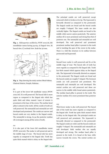
Fig. 1. <strong>Selenoportax</strong> <strong>vexillarius</strong>, PUPC 2010/26, left<br />
mandibular ramus having p4-m3, A) lingual view, B)<br />
buccal view, C) occlusal view. Scale bar 30 mm.<br />
The principal conids are well preserved except<br />
entoconid which is broken at its tip. The hypoconid is<br />
forwardly directed as compared to <strong>the</strong> protoconid.<br />
The lingual conids are broad and <strong>the</strong> buccal conids<br />
are roughly V-shaped. The inner conids are also<br />
vertically higher. The lingual conids are broad in <strong>the</strong><br />
middle while narrow antero-posteriorly. The anterior<br />
median rib is more prominent and strong than that of<br />
posterior one. The metastylid and entostylid are well<br />
developed. The well preserved and prominent<br />
prominent median basal pillar is present in <strong>the</strong> tooth<br />
and is touching <strong>the</strong> apex of <strong>the</strong> crown in <strong>the</strong> molar.<br />
There is a fold like structure in its outline between<br />
protoconid and hypoconid.<br />
Fig. 2. Map showing <strong>the</strong> study section <strong>Dhok</strong> <strong>Pathan</strong>,<br />
<strong>Chakwal</strong> <strong>District</strong>, <strong>Punjab</strong>, <strong>Pakistan</strong>.<br />
p4<br />
It is part of <strong>the</strong> lower left mndibular ramus (PUPC<br />
2010/26). It is well preserved. The buccal side is more<br />
rugose as compared to <strong>the</strong> lingual side which has<br />
quite thick and shiny enamel. Layer of cement is<br />
prominent at <strong>the</strong> base of <strong>the</strong> crown. The median basal<br />
pillar is absent in <strong>the</strong> tooth. All <strong>the</strong> conids of tooth are<br />
well preserved. The metastylid and entostylid are well<br />
developed and prominent. The conids are narrow and<br />
buccal conids are very similar to <strong>the</strong> lingual conids.<br />
The metastylid is strong. In p4 <strong>the</strong> posterior median<br />
rib is strongest among all <strong>the</strong> series of teeth.<br />
m1<br />
It is also part of <strong>the</strong> lower left mandibular ramus<br />
(PUPC 2010/26). The molar is well preserved and in<br />
<strong>the</strong> middle stage of wear. The buccal side has more<br />
rugosity as compared to <strong>the</strong> lingual side. Tooth has<br />
quite thick enamel which is shiny on <strong>the</strong> lingual side.<br />
m2<br />
Second lower molar is well preserved and lie at <strong>the</strong><br />
middle stage of wear. The buccal side of tooth has<br />
more rugosity as compared to <strong>the</strong> lingual side. Tooth<br />
has thick enamel which apperas shiny on <strong>the</strong> lingual<br />
side. The hypoconid is forwardly directed as compare<br />
to <strong>the</strong> protoconid. The lingual conids are broad and<br />
<strong>the</strong> buccal conids are roughly V-shaped. The foremost<br />
conids are well preserved and prominent. The<br />
metastylid and entostylid are well developed. The<br />
central cavities are well preserved and <strong>the</strong>se are<br />
narrow in <strong>the</strong> middle while broad antero-posteriorly.<br />
The median basal pillar is present in <strong>the</strong> tooth. The<br />
anterior median rib is more prominent than posterior<br />
one.<br />
m3<br />
Third lower molar is also well preserved. The buccal<br />
side of <strong>the</strong> tooth has more rugosity as compared to<br />
<strong>the</strong> lingual side. Tooth has quite thick enamel which<br />
is shiny on <strong>the</strong> lingual side. The principal conids are<br />
well preserved and prominent. The hypoconid is<br />
forwardly directed as compared to <strong>the</strong> protoconid.<br />
The lingual conids are wider and <strong>the</strong> buccal conids are<br />
roughly V-shaped. The metastylid and entostylid are<br />
well developed. Both median ribs are equally<br />
developed and strong. The median basal pillar is<br />
present in <strong>the</strong> tooth. It is broad at <strong>the</strong> base while<br />
narrow towards <strong>the</strong> crown. The central cavities are<br />
187 | Samiullah et al.


