Moving an incisor across the midline_ A treatment alternative in an adolescent patient

You also want an ePaper? Increase the reach of your titles
YUMPU automatically turns print PDFs into web optimized ePapers that Google loves.
536 Bosio, Bradley, <strong>an</strong>d Hefti<br />
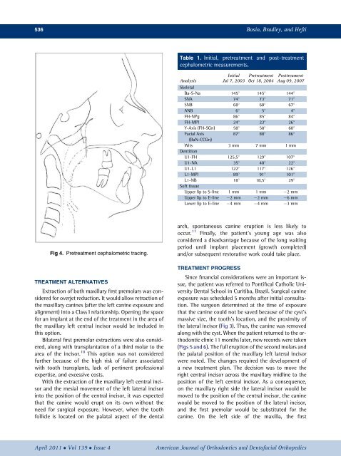
Table 1. Initial, pre<strong>treatment</strong> <strong>an</strong>d post-<strong>treatment</strong><br />
cephalometric measurements.<br />
Analysis<br />
Initial<br />
Jul 7, 2003<br />
Pre<strong>treatment</strong><br />
Oct 18, 2004<br />
Posttreament<br />
Aug 09, 2007<br />
Skeletal<br />
Ba-S-Na 145 145 144 <br />
SNA 74 73 71 <br />
SNB 68 68 67 <br />
ANB 6 5 4 <br />
FH-NPg 86 85 84 <br />
FH-MPl 24 23 26 <br />
Y-Axis (FH-SGn) 58 58 60 <br />
Facial Axis<br />
87 88 86 <br />
(BaN-CCGn)<br />
Wits 3 mm 7 mm 1 mm<br />
Dentition<br />
U1-FH 125,5 129 107 <br />
U1-NA 35 40 22 <br />
U1-L1 122 117 126 <br />
L1-MPl 89 91 101 <br />
L1-NB 18 18,5 29 <br />
Soft tissue<br />
Upper lip to S-l<strong>in</strong>e 1 mm 1 mm 2mm<br />
Upper lip to E-l<strong>in</strong>e 2mm 2mm 6mm<br />
Lower lip to E-l<strong>in</strong>e 4mm 4mm 3mm<br />
Fig 4. Pre<strong>treatment</strong> cephalometric trac<strong>in</strong>g.<br />
TREATMENT ALTERNATIVES<br />
Extraction of both maxillary first premolars was considered<br />
for overjet reduction. It would allow retraction of<br />
<strong>the</strong> maxillary c<strong>an</strong><strong>in</strong>es (after <strong>the</strong> left c<strong>an</strong><strong>in</strong>e exposure <strong>an</strong>d<br />
alignment) <strong>in</strong>to a Class I relationship. Open<strong>in</strong>g <strong>the</strong> space<br />
for <strong>an</strong> impl<strong>an</strong>t at <strong>the</strong> end of <strong>the</strong> <strong>treatment</strong> <strong>in</strong> <strong>the</strong> area of<br />
<strong>the</strong> maxillary left central <strong><strong>in</strong>cisor</strong> would be <strong>in</strong>cluded <strong>in</strong><br />
this option.<br />
Bilateral first premolar extractions were also considered,<br />
along with tr<strong>an</strong>spl<strong>an</strong>tation of a third molar to <strong>the</strong><br />
area of <strong>the</strong> <strong><strong>in</strong>cisor</strong>. 10 This option was not considered<br />
fur<strong>the</strong>r because of <strong>the</strong> high risk of failure associated<br />
with tooth tr<strong>an</strong>spl<strong>an</strong>ts, lack of pert<strong>in</strong>ent professional<br />
expertise, <strong>an</strong>d excessive costs.<br />
With <strong>the</strong> extraction of <strong>the</strong> maxillary left central <strong><strong>in</strong>cisor</strong><br />
<strong>an</strong>d <strong>the</strong> mesial movement of <strong>the</strong> left lateral <strong><strong>in</strong>cisor</strong><br />
<strong>in</strong>to <strong>the</strong> position of <strong>the</strong> central <strong><strong>in</strong>cisor</strong>, it was expected<br />
that <strong>the</strong> c<strong>an</strong><strong>in</strong>e would erupt on its own without <strong>the</strong><br />
need for surgical exposure. However, when <strong>the</strong> tooth<br />
follicle is located on <strong>the</strong> palatal aspect of <strong>the</strong> dental<br />
arch, spont<strong>an</strong>eous c<strong>an</strong><strong>in</strong>e eruption is less likely to<br />
occur. 11 F<strong>in</strong>ally, <strong>the</strong> <strong>patient</strong>’s young age was also<br />
considered a disadv<strong>an</strong>tage because of <strong>the</strong> long wait<strong>in</strong>g<br />
period until impl<strong>an</strong>t placement (growth completed)<br />
<strong>an</strong>d/or subsequent restorative work could take place.<br />
TREATMENT PROGRESS<br />
S<strong>in</strong>ce f<strong>in</strong><strong>an</strong>cial considerations were <strong>an</strong> import<strong>an</strong>t issue,<br />
<strong>the</strong> <strong>patient</strong> was referred to Pontifical Catholic University<br />
Dental School <strong>in</strong> Curitiba, Brazil. Surgical c<strong>an</strong><strong>in</strong>e<br />
exposure was scheduled 5 months after <strong>in</strong>itial consultation.<br />
The surgeon determ<strong>in</strong>ed at <strong>the</strong> time of exposure<br />
that <strong>the</strong> c<strong>an</strong><strong>in</strong>e could not be saved because of <strong>the</strong> cyst’s<br />
massive size, <strong>the</strong> tooth’s location, <strong>an</strong>d <strong>the</strong> proximity of<br />
<strong>the</strong> lateral <strong><strong>in</strong>cisor</strong> (Fig 3). Thus, <strong>the</strong> c<strong>an</strong><strong>in</strong>e was removed<br />
along with <strong>the</strong> cyst. When <strong>the</strong> <strong>patient</strong> returned to <strong>the</strong> orthodontic<br />
cl<strong>in</strong>ic 11 months later, new records were taken<br />
(Figs 5 <strong>an</strong>d 6). The full eruption of <strong>the</strong> second molars <strong>an</strong>d<br />
<strong>the</strong> palatal position of <strong>the</strong> maxillary left lateral <strong><strong>in</strong>cisor</strong><br />
were noted. The ch<strong>an</strong>ges required <strong>the</strong> development of<br />
a new <strong>treatment</strong> pl<strong>an</strong>. The decision was to move <strong>the</strong><br />
right central <strong><strong>in</strong>cisor</strong> <strong>across</strong> <strong>the</strong> maxillary <strong>midl<strong>in</strong>e</strong> to <strong>the</strong><br />
position of <strong>the</strong> left central <strong><strong>in</strong>cisor</strong>. As a consequence,<br />
on <strong>the</strong> maxillary right side <strong>the</strong> lateral <strong><strong>in</strong>cisor</strong> would be<br />
moved to <strong>the</strong> position of <strong>the</strong> central <strong><strong>in</strong>cisor</strong>, <strong>the</strong> c<strong>an</strong><strong>in</strong>e<br />
would be moved to <strong>the</strong> position of <strong>the</strong> lateral <strong><strong>in</strong>cisor</strong>,<br />
<strong>an</strong>d <strong>the</strong> first premolar would be substituted for <strong>the</strong><br />
c<strong>an</strong><strong>in</strong>e. On <strong>the</strong> left side of <strong>the</strong> maxilla, <strong>the</strong> first<br />
April 2011 Vol 139 Issue 4<br />
Americ<strong>an</strong> Journal of Orthodontics <strong>an</strong>d Dentofacial Orthopedics