3D Anatomy Series
Human Anatomy Reproductions with an extra dimension
Human Anatomy Reproductions with an extra dimension

You also want an ePaper? Increase the reach of your titles
YUMPU automatically turns print PDFs into web optimized ePapers that Google loves.
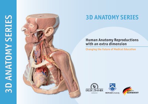
<strong>3D</strong> ANATOMY SERIES<br />
<strong>3D</strong> ANATOMY SERIES<br />
Human <strong>Anatomy</strong> Reproductions<br />
with an extra dimension<br />
Changing the Future of Medical Education
The ground-breaking Monash <strong>Anatomy</strong> <strong>Series</strong> represents a unique and unrivalled<br />
collection of colour-augmented human anatomy body replicas designed specifically<br />
for enhanced teaching and learning. This premium collection of highly accurate<br />
normal human anatomy has been generated directly from either radiographic data<br />
or actual cadaveric specimens using advanced imaging techniques. The Monash<br />
<strong>3D</strong> Human <strong>Anatomy</strong> <strong>Series</strong> provides a cost effective means to meet your specific<br />
educational and demonstration needs in a range of curricula from medicine, allied<br />
health sciences and biological sciences. A detailed description of the anatomy<br />
displayed in each <strong>3D</strong>printed body replica is provided<br />
What advantages does The Monash <strong>3D</strong> <strong>Anatomy</strong> <strong>Series</strong> offer over either plastic<br />
models or plastinated human specimens?<br />
Each body replica has been carefully created from selected radiographic patient<br />
data or high quality real human prosected cadaver specimens selected by a<br />
highly qualified team of anatomists at the Centre for Human <strong>Anatomy</strong> Education;<br />
Monash University to illustrate a range of clinically important areas of anatomy<br />
with a quality and fidelity that is not possible in conventional models – this is real<br />
anatomy and not stylised.<br />
Each body replica has been rigorously checked by a team of highly qualified<br />
anatomists at The Centre for Human <strong>Anatomy</strong> Education, Monash University, to<br />
ensure the anatomical accuracy of the final product<br />
The body replicas are not real human tissue and therefore not subject to any barriers<br />
of transportation, importation or use in educational facilities that do not possess<br />
an anatomy license. The Monash <strong>3D</strong> <strong>Anatomy</strong> <strong>Series</strong> avoids these and other ethical<br />
issues that are raised when dealing with plastinated human remains.<br />
Ms Michelle Quayle & Prof. Paul McMenamin<br />
Prof. Paul McMenamin
Conversion and comparison of cadaver specimen to human anatomy reproduction<br />
Cadaver / Specimen<br />
Human <strong>Anatomy</strong> Reproduction<br />
Human <strong>Anatomy</strong> Reproduction<br />
Cadaver / Specimen<br />
Human <strong>Anatomy</strong> Reproduction<br />
Cadaver / Specimen<br />
1
Advantages of our Human <strong>Anatomy</strong> Reproductions<br />
Anatomically accurate and identical to real specimen<br />
No ethical issues - not real human body parts<br />
Reasonably priced<br />
Available within a short lead time<br />
Reproducible, several identical prints can be used as a classroom set<br />
Can be produced in different sizes to cater for the needs of the teacher e.g.<br />
a larger teacher version can be created<br />
2
Current educational tools have a number of shortcomings<br />
Human Cadavers<br />
Plastinates<br />
Access to cadavers can be problematic. Many countries cannot access<br />
cadavers for cultural and religious reasons<br />
Cadavers cost a lot of money<br />
High cost of establishing your own plastination suite (OHS issues)<br />
Wet specimens cannot be used in uncertified labs –special labs are needed<br />
Costs –VERY HIGH<br />
Ethical issues<br />
Timeframe for plastination process<br />
Many countries do not allow their importation<br />
Dissection of cadavers is a lot of staff time and that is a cost<br />
Storage of cadaver material needs special refrigeration etc. which has a cost<br />
If you want another specimen you have to start all over again<br />
3
MP1250 Head Neck Shoulder with angiosomes<br />
This large, multipart <strong>3D</strong> printed specimen<br />
displays a great deal of anatomy spanning the<br />
head, neck, thorax, axillae and upper limbs.<br />
Head and neck:<br />
The head and neck of the specimen provides<br />
views of both superficial and deep structures in<br />
the region. The calotte has been removed ~2cm<br />
superior to the orbits to expose the brain in<br />
relation to the endocranial cavity. The transverse<br />
section through the cerebrum demonstrates the<br />
relation of the grey matter cortex to the white<br />
matter medulla, as well as the lateral ventricles<br />
with a small amount of choroid plexus visible in<br />
the base of both spaces. The skin and superficial<br />
fascia on the right side has been retained and<br />
false-coloured to display the angiosomes of<br />
the face and posterior neck. On the left side,<br />
the superficial tissues have been dissected to<br />
expose the muscles of facial expression, muscles<br />
of mastication, and deeper structures of the<br />
infratemporal fossa including the lingual nerve,<br />
terminal branches of the external carotid artery<br />
into the superficial temporal and maxillary<br />
arteries...*<br />
The root of the neck – axillary junction:<br />
The clavicle has been partially removed on the<br />
left side of the specimen (medial to the<br />
origin of the deltoid) to expose the first<br />
rib and the insertion of anterior scalene<br />
muscle. The roots of the brachial plexus<br />
(C5-T1) can be seen forming the trunks<br />
posterior to this muscle but anterior<br />
to middle and posterior scalene<br />
muscles they emerge from the interscalene plane.<br />
While the subclavian vein has been removed, the<br />
subclavian artery is also seen passing behind the<br />
scalenus anterior. The transition of the subclavian<br />
artery to the axillary artery is exposed, as is its<br />
position relative to the cords of the brachial<br />
plexus (medial, lateral and posterior)...*<br />
Thorax:<br />
The thorax has been opened via a ‘window’ on<br />
the left to display the internal thoracic wall and<br />
mediastinum. The left lung has been removed<br />
and the intercostal spaces are discernable deep<br />
to the parietal pleura although intercostal<br />
neurovascular bundles are only discernable<br />
posteriorly. The pericardium has been removed<br />
to expose the heart with its apex pointing<br />
inferiorly, anteriorly, and to the left. The left side<br />
of the heart is exposed as are the left pulmonary<br />
veins and arteries (above left main bronchus),<br />
ascending aorta, aortic arch and commencement<br />
of the descending thoracic aorta. The left vagus<br />
nerve and left recurrent laryngeal nerve are easily<br />
identified. The right half of the anterior and<br />
lateral thoracic wall are intact and display the<br />
muscles of the intercostal spaces and inserting<br />
hypaxial muscles from the right upper limb. If<br />
the specimen is viewed from below, the right lung<br />
and pleural spaces along with the diaphragmatic<br />
surface of the heart are all evident. While the<br />
skin and superficial fascia posterior thorax has<br />
been left intact, the distribution of cutaneous<br />
branches of dorsal rami have been illustrated<br />
along the left side of the specimen.<br />
4<br />
* Complete description can be found at: www.3danatomyseries.com
MP1300 Posterior Abdominal wall<br />
This large, multipart <strong>3D</strong> printed specimen<br />
displays the entire male posterior abdominal<br />
wall from the diaphragm to the pelvic brim,<br />
as well as pelvic anatomy and to the proximal<br />
thigh. This same individual specimen is<br />
also available as a pelvic and proximal thigh<br />
specimen (MP1770).<br />
The parietal peritoneum has been removed from<br />
the posterior abdominal wall to expose the<br />
muscular wall including the psoas, the quadratus<br />
lumborum, transversus abdominus, and the<br />
iliacus below the iliac crest. The muscular portions<br />
of the dome shaped diaphragm are clearly distinct<br />
from the central tendon. The fibres originate<br />
from the circumference of the internal walls of<br />
the bony thorax at its margin (sternal fibres,<br />
costal portion, lumbar portion). The origins of<br />
the diaphragm and the left and right crura are<br />
clearly identifiable originating from the vertebral<br />
bodies (L1-L3 on the right and L1-L2 on the left.<br />
The crura are connected by a tendinous band, the<br />
median arcuate ligament, which arches in front of<br />
the aorta; however in this specimen the aorta has<br />
been removed. The fibres of the diaphragm arising<br />
from the tendinous arches over psoas and the<br />
lateral arcuate ligaments are partly hidden by the<br />
kidneys. The oesophageal opening through the<br />
arching fibres of the right crus is present above<br />
(level of T10) and to the left of the aortic opening<br />
(level of T12). The opening in the central tendon<br />
that transmits the inferior vena cava (level of<br />
T8/9 intervertebral disc).<br />
The somatic nerves of the posterior abdominal<br />
wall are clearly identifiable and consist of<br />
from above downwards – the subcostal, the<br />
iliohypogastric and ilioinguinal nerves lie on<br />
the quadratus lumborum (in this individual they<br />
arise together and– this can often occur and<br />
they split later in abdominal muscle layers), the<br />
lateral cutaneous nerve of thigh, the femoral<br />
lying in the groove between psoas and iliacus),<br />
and the genitofemoral nerve lying superficially<br />
upon psoas. The sympathetic trunks can be<br />
seen descending lateral to the lumbar vertebral<br />
bodies.<br />
The aorta and inferior cava are<br />
transected around the level of<br />
L3 vertebral body. The aortic<br />
bifurcation into the right and<br />
left common iliac arteries is<br />
slightly higher than normal.<br />
Finally, the kidneys have<br />
dissected from the peri- and<br />
pararenal fat of the posterior<br />
abdominal wall. The renal<br />
vessels (arteries anteriorly, veins<br />
posteriorly) have been preserved but<br />
as the aorta and inferior cava have been<br />
removed this does display the origin and<br />
arrangements of these vessels fully.<br />
The more inferior position of the<br />
right kidney is clearly visible and the<br />
ureters can be seen emerging from<br />
the hilum and descending initially<br />
lateral to psoas, then anterior to<br />
this muscle before crossing pelvic<br />
brim anterior to the bifurcation of<br />
the common iliac arteries to reach the<br />
true pelvis.<br />
5
MP1400 Nervous System Dissection (posterior view)<br />
This <strong>3D</strong> printed specimen presents a unique<br />
view of axial anatomy, presenting a dorsal deep<br />
dissection of the head, neck, axillae, thorax,<br />
abdomen, and gluteal regions. The removal<br />
of the posterior portions of the cranium and<br />
laminectomy from the cervical region to the<br />
opening of the sacral canal affords a continuous<br />
view of the central nervous system structures<br />
and origin of the segmental nerves relative to<br />
other axillary and appendicular structures.<br />
In the cranium the two cerebral hemispheres<br />
are exposed in coronal section, separated by a<br />
falx cerebri that preserves the superior sagittal<br />
sinus, and supported by a partial tentorium<br />
cerebelli. The cerebellum has been removed and<br />
along the lateral margins, the sigmoid sinus has<br />
been opened. This exposes the fourth ventricle,<br />
pons and medulla oblongata, posterior inferior<br />
cerebellar arteries, and cranial nerves (CN VII –<br />
XII) arising from these brainstem structures.<br />
Inferior to the cranium, the posterior cervical<br />
portion of the spinal cord is exposed through<br />
deep dissection and laminectomies (with the<br />
exception of the posterior arch of the atlas). At<br />
this level of dissection, the vertebral arteries<br />
can be observed ascending through the vertebral<br />
foramina and curving anteriorly on the superior<br />
surface of the atlas towards the foramen magnum.<br />
The roots of the cervical and brachial plexus are<br />
exposed, resting on the scalene musculature,<br />
cervical vasculature (common carotid artery on<br />
the right, internal jugular and common carotid<br />
on the left) and sternocleidomastoid muscles<br />
and can be traced anteriorly towards the margins<br />
of the dissection. Removal of the scapulae (fully<br />
on the right, partially on the left) the more distal<br />
portions of the brachial plexus can be followed<br />
passing superior to the first ribs and into the<br />
axilla, with the cords, divisions and terminal<br />
branches surrounding the axillary arteries. On<br />
the left side, the musculature is largely removed<br />
(with parts of the deltoid, infraspinatus, and teres<br />
minor muscles preserved) and the long thoracic<br />
nerve and lateral thoracic artery descend near<br />
the serratus anterior. The subscapular artery is<br />
shown dividing into the circumflex scapular artery<br />
(passing to the triangular space) and the root of<br />
the thoracodorsal artery. On the right side, the<br />
more full removal of the scapula affords a view<br />
of the brachial plexus structures and the passage<br />
of the axillary nerve and posterior circumflex<br />
humeral artery laterally towards the surgical<br />
neck of the humerus.<br />
In the midline of the thorax the spinal cord<br />
is exposed through both laminectomy and<br />
dissection of the dura mater. The dorsal roots<br />
and rootlets of the mixed spinal nerves are<br />
exposed and pass laterally to the dorsal root<br />
ganglia (enclosed within dura). On the right<br />
side the thoracic mixed spinal nerves and the<br />
posterior thoracic musculoskeletal wall has<br />
been removed (from the 2nd rib to the level of<br />
the 11th and 12th ribs ) to expose the posterior<br />
surface of the lung and the posterior diaphragm.<br />
On the left side, most of the posterior thoracic<br />
wall has been removed, but the 3rd-5th ribs are<br />
retained to demonstrate the external intercostal<br />
musculature and the position of the 5th<br />
intercostal nerve within the space. In addition,<br />
the full sequence of intercostal nerves has been<br />
retained...*<br />
6<br />
* Complete description can be found at: www.3danatomyseries.com
MP1500 Upper Limb<br />
Ms. Michelle Quayle<br />
This <strong>3D</strong> print demonstrates the superficial<br />
anatomy of a left upper limb from the blade of<br />
the scapula to the hand. The skin and superficial<br />
and deep fascia has been removed from most of<br />
the limb except over the dorsum of the scapula,<br />
proximal arm, and over the hand. The superficial<br />
veins, including the median cubital vein, have<br />
been maintained; with the cephalic and basilic<br />
preserved from the wrist to the deltopectoral<br />
groove and termination in the brachial vein,<br />
respectively.<br />
In the axilla, cross-sections of the deltoid,<br />
supraspinatus, infraspinatus, teres minor, teres<br />
major, and subscapularis muscles are visible<br />
relative to the bony blade and spine of the scapula.<br />
The coracobrachialis and tendon of the latissimus<br />
dorsi are also preserved, as well as the tendon<br />
of the pectoralis major. The lateral portions of<br />
the axillary artery and vein are preserved, as<br />
well as the most lateral extend of the cords of<br />
the brachial plexus (medial, lateral, posterior).<br />
Terminal nerves of the brachial plexus visible in<br />
the axilla include: the upper subscapular, ulnar,<br />
median, musculocutaneous, axillary and radial.<br />
The course of the deep vessels and nerves of<br />
the upper limb is exposed through the arm from<br />
proximal to distal, as well as the muscles of the<br />
anterior and posterior compartments. In the<br />
cubital region, part of the bicipital aponeurosis<br />
is preserved. The superficial layer of anterior<br />
and posterior forearm muscles are exposed from<br />
their origin to their tendons distally, with a<br />
small portion of deep forearm fascia over<br />
the extensor compartment maintained<br />
for reference. At the most distal extent<br />
of the dissected forearm, the ulnar and<br />
radial arteries and median nerve are<br />
visible.<br />
7
MP1510 Upper Limb<br />
elbow, forearm and hand<br />
This is a large print which displays a great deal<br />
of upper limb anatomy.<br />
In the distal arm and elbow/cubital fossa region<br />
we can see the arrangement of the biceps tendon,<br />
brachial artery and median nerve arranged from<br />
lateral to medial. The bicipital aponeurosis has<br />
been divided to reveal the structures deep to it.<br />
The ulnar nerve can be seen passing behind the<br />
medial epicondyle with an ulnar collateral artery<br />
close by. The superficial branch of the radial<br />
nerve can just be seen in the space between<br />
brachioradialis and brachialis muscles since the<br />
belly of the latter muscle has been displaced<br />
slightly laterally.<br />
In the forearm, the superficial flexor muscles<br />
arising from the common flexor origin can be<br />
clearly seen (from lateral to medial– pronator<br />
teres, flexor carpi radialis (FCR), flexor digitorum<br />
superficialis (FDS) and flexor carpi ulnaris (FCU).<br />
There is not a palmaris longus muscle in this<br />
cadaver. The radial artery and superficial branch<br />
of the radial nerve (emerging half way down the<br />
forearm from behind the brachioradialis muscle<br />
and tendon) are clearly identifiable. The ulnar<br />
artery can be seen in the distal forearm emerging<br />
from beneath FCU muscle.<br />
On the posterior aspect of the forearm the<br />
extensor muscles arising from the common<br />
extensor origin are clearly identifiable. These<br />
include from medial to lateral the extensor<br />
carpi ulnaris (ECU), extensor digiti minimi,<br />
extensor digitorum and extensor carpi radialis<br />
brevis (ECRB). The extensor carpi radialis<br />
longus (ECRL) can be seen arising from the<br />
inferior aspect of the lateral supracondylar<br />
ridge. Further distally the abductor pollicis<br />
longus (APL) and extensor pollicis brevis (EPB)<br />
can be seen emerging from deep to superficial<br />
and ‘wrapping’ around the radius. They along<br />
with extensor pollicis longus (EPL) (partly<br />
hidden) travel distally to insert into the<br />
extensor or dorsal surface of the base of the<br />
1st metacarpal, proximal phalynx and distal<br />
phalynx of the the thumb respectively. The<br />
anatomical snuffbox is clearly evident with the<br />
radial artery in its floor (suurrounded by fat)<br />
and the cutaneous branch of the radial nerve<br />
in its roof. The extensor retinaculum is clearly<br />
visible on the dorsum of the wrist and distal to<br />
it the tendons of extensor indicis and ECRB and<br />
ECRL can be seen inserting into the 2nd and 3rd<br />
metacarpals.<br />
In the hand, the superficial dissection reveals<br />
muscles of the thenar and hypothenar eminences,<br />
the flexor retinaculum (roof of the carpal tunnel),<br />
the long tendons of the hand, the lumbricals, and<br />
the superficial palmar arch arising from the ulnar<br />
artery, which passes into the hand lateral to the<br />
pisiform bone above the retinaculum, along with<br />
the superfical branch of the ulnar nerve. The large<br />
median nerve can be seen passing beneath the<br />
carpal ligament or flexor retinaculum between<br />
the FCR and the FDS tendons. Digital arteries<br />
and nerves can be clearly seen further distally in<br />
the palm entering the digits. Note in particular<br />
the small recurrent branch of the median nerve<br />
crossing over the flexor pollicis brevis close to<br />
its origin from the retinaculum. The extensor<br />
expansion is dissected on the middle finger.<br />
8
MP1515 Upper Limb<br />
biceps, bones and ligaments<br />
This <strong>3D</strong> print shows the origin and insertion<br />
of biceps (most other arm and shoulder<br />
muscle bellies have been removed). The long<br />
head of biceps arises from the supraglenoid<br />
tubercle (hidden from view) and travels<br />
inferiorly in the bicipital groove, whereas the<br />
short head of biceps arises from the coracoid<br />
process. The bifid insertion of the muscle as<br />
the bicipital aponeurosis and the rounded<br />
tendon which can be seen winding around the<br />
radius to insert into the radial tuberosity are<br />
clearly discernable.<br />
At the shoulder region short stumps of some<br />
muscles (subclavius, subscapularis, pectoralis<br />
major, teres minor, infraspinatus, long head<br />
of triceps) and the tendinous insertion of<br />
latissimus dorsi can be identified close to<br />
the ‘floor’ of the medial lip of the bicipital<br />
groove. The tendon of teres major lies on the<br />
medial lip of the groove and the pectoralis<br />
major tendon inserts into the lateral lip of the<br />
groove. The tendon of pectoralis minor arises<br />
from the coracoid process medial to the origin<br />
of the short head of biceps. Ligaments of the<br />
shoulder region such as the coracoclavicular,<br />
coracoacromial, coracohumeral can be seen as<br />
can the capsule of the shoulder joint and that of<br />
the acromioclavicular joint. The supraspinatus<br />
muscle is the only rotator cuff muscle that has<br />
been completely preserved. The suprascapular<br />
ligament which bridges across the suprascapular<br />
notch is also evident on the superior border of<br />
the scapula.<br />
At the elbow the capsule of the joint can be<br />
seen as can the annular ligament of the radius.<br />
The radial collateral ligaments are also just<br />
discernable. The ulnar collateral ligament<br />
is not visible as the two heads of flexor carpi<br />
ulnaris have been retained.<br />
9
MP1520 Upper Limb Ligaments<br />
This is a large print which displays a great deal<br />
of upper limb anatomy.<br />
In the distal arm and elbow/cubital fossa region<br />
we can see the arrangement of the biceps tendon,<br />
brachial artery and median nerve arranged from<br />
lateral to medial. The bicipital aponeurosis has<br />
been divided to reveal the structures deep to it. The<br />
ulnar nerve can be seen passing behind the medial<br />
epicondyle with an ulnar collateral artery close<br />
by. The superficial branch of the radial nerve can<br />
just be seen in the space between brachioradialis<br />
and brachialis muscles since the belly of the latter<br />
muscle has been displaced slightly laterally.<br />
In the forearm, the superficial flexor muscles<br />
arising from the common flexor origin can be clearly<br />
seen (from lateral to medial– pronator teres, flexor<br />
carpi radialis (FCR), flexor digitorum superficialis<br />
(FDS) and flexor carpi ulnaris (FCU). There is not<br />
a palmaris longus muscle in this cadaver. The radial<br />
artery and superficial branch of the radial nerve<br />
(emerging half way down the forearm from behind<br />
the brachioradialis muscle and tendon) are clearly<br />
identifiable. The ulnar artery can be seen in the<br />
distal forearm emerging from beneath FCU muscle.<br />
On the posterior aspect of the forearm the extensor<br />
muscles arising from the common extensor origin<br />
are clearly identifiable. These include from medial<br />
to lateral the extensor carpi ulnaris (ECU), extensor<br />
digiti minimi, extensor digitorum and extensor carpi<br />
radialis brevis (ECRB). The extensor carpi radialis<br />
longus (ECRL) can be seen arising from the inferior<br />
aspect of the lateral supracondylar ridge. Further<br />
distally the abductor pollicis longus (APL) and<br />
extensor pollicis brevis (EPB) can be seen emerging<br />
from deep to superficial and ‘wrapping’ around the<br />
radius. They along with extensor pollicis longus<br />
(EPL) (partly hidden) travel distally to insert into<br />
the extensor or dorsal surface of the base of the 1st<br />
metacarpal, proximal phalynx and distal phalynx<br />
of the the thumb respectively. The anatomical<br />
snuffbox is clearly evident with the radial artery<br />
in its floor (suurrounded by fat) and the cutaneous<br />
branch of the radial nerve in its roof. The extensor<br />
retinaculum is clearly visible on the dorsum of the<br />
wrist and distal to it the tendons of extensor indicis<br />
and ECRB and ECRL can be seen inserting into the<br />
2nd and 3rd metacarpals.<br />
In the hand, the superficial dissection reveals<br />
muscles of the thenar and hypothenar eminences,<br />
the flexor retinaculum (roof of the carpal tunnel),<br />
the long tendons of the hand, the lumbricals, and<br />
the superficial palmar arch arising from the ulnar<br />
artery, which passes into the hand lateral to the<br />
pisiform bone above the retinaculum, along with<br />
the superfical branch of the ulnar nerve. The large<br />
median nerve can be seen passing beneath the<br />
carpal ligament or flexor retinaculum between<br />
the FCR and the FDS tendons. Digital arteries and<br />
nerves can be clearly seen further distally in the<br />
palm entering the digits. Note in particular the<br />
small recurrent branch of the median nerve crossing<br />
over the flexor pollicis brevis close to its origin<br />
from the retinaculum. The extensor expansion is<br />
dissected on the middle finger.<br />
Ms. Michelle Quayle<br />
10
MP1521 Right thoracic wall<br />
axilla, and the root of the neck<br />
This <strong>3D</strong> printed specimen preserves a dissection<br />
of the right thoracic wall, axilla, and the root of<br />
the neck. The specimen is cut just parasagittally<br />
and the visceral contents of the chest have been<br />
removed. Structures within the right chest wall<br />
are visible deep to the parietal pleura, including<br />
the ribs, muscles of the intercostal spaces and<br />
the origins of the neurovascular bundle in each<br />
intercostal space. The pectoralis major has been<br />
reflected medially towards the sectioned edge of<br />
the specimen to expose pectoralis minor which<br />
acts as a useful landmark as it divides the axillary<br />
artery into its three parts. The clavicle has had its<br />
middle 1/3 removed, but the subclavius muscle<br />
has been retained. The brachial plexus and many<br />
of its branches are seen almost in its entirety<br />
from the roots of C5-T1 to its termination as<br />
it exits the axilla to enter the arm. Of the<br />
structures preserved on the specimen:<br />
Nerves: The medial pectoral nerves can be seen<br />
penetrating pectoralis minor, while the lateral<br />
pectoral nerve can be identified on the medial<br />
upper border of the pectoralis minor (one branch<br />
of which is reflected with the transected humeral<br />
portion of the pectoralis major). The cords of the<br />
brachial plexus can be identified around the 2nd<br />
part of the axillary artery. The major terminal<br />
nerves of the plexus (musculocutaneous,<br />
median, ulnar, radial and axillary nerves ) are<br />
all identifiable. The long thoracic nerve is visible<br />
lying on the surface of serratus anterior, as is the<br />
thoracodorsal nerve alongside the thoracodorsal<br />
artery as they descend to enter the latissimus<br />
dorsi muscle. The dorsal scapular nerve and<br />
artery are visible above and below omohyoid.<br />
The axillary nerve accompanied by the posterior<br />
circumflex humeral artery can be seen passing<br />
posteriorly just below the neck of the humerus.<br />
In the root of the neck the phrenic nerve is just<br />
visible as it passes on the anterior surface of<br />
scalenus anterior muscle from its lateral border<br />
to its medial border and a thin accessory phrenic<br />
nerve is identifiable.<br />
Vessels: Some of the branches of the subclavian<br />
artery (e.g., the transverse cervical and<br />
suprascapular arteries passing transversely across<br />
the root of the neck) can be clearly seen, however<br />
the subclavian artery itself is partly hidden from<br />
view as it crosses the first rib behind the insertion of<br />
scalenus anterior muscle. Most of the deep veins have<br />
been removed to expose branches of the three parts<br />
of the axillary artery, including the thoracoacromial<br />
artery and its branches, the lateral thoracic artery,<br />
thoracodorsal artery and the anterior and posterior<br />
circumflex humeral arteries. While the deep veins<br />
have been removed the cephalic vein can be seen<br />
ascending superficially in the deltopectoral groove<br />
into the dectopectoral triangle where it passes<br />
through the clavipectoral fascia.<br />
Muscles: The digitations of the serratus anterior<br />
muscle are clearly visible on the lateral chest<br />
wall. As mentioned above, the pectoralis major<br />
muscle is reflected to reveal the pectoralis minor<br />
which together form the anterior wall of the axilla.<br />
Posteriorly the large fan shaped latissimus dorsi<br />
is the most obvious muscle along with the teres<br />
major. When viewed posteriorly a few vertical fibers<br />
of trapezius can be seen, as can some descending<br />
fibers of the lower part of rhomboid major<br />
attaching to the medial border of the scapula.<br />
Below this the triangle of auscultation is clearly<br />
visible. The infraspinatus and teres minor muscles<br />
are also visible arising from the infraspinous fossa<br />
and lateral border of the scapula respectively. The<br />
triceps brachii muscle can be seen in the extensor<br />
compartment of the arm.<br />
In the root of the neck the insertion of<br />
sternocleidomastoid is visible medially and the<br />
trapezius is visible posteriorly. In the floor of<br />
the posterior triangle the scalene muscles are<br />
visible as is the omohyoid as it lies obliquely in<br />
the triangle.<br />
11
MP1523 Shoulder (left)<br />
Superficial muscles and axillary/brachial artery<br />
This printed <strong>3D</strong> model of the left shoulder<br />
displays the superficial muscles around the<br />
shoulder joint, the rotator cuff muscles and<br />
the axillary artery as it progresses distally to<br />
become the brachial artery.<br />
The specimen consists of the scapula, humerus<br />
and and the clavicle which has been sectioned<br />
approximately at midpoint. Inferiorly the humerus<br />
has been cut transversely at approximately at the<br />
level of the mid shaft.<br />
The muscles attached to the clavicle have<br />
been preserved; with the subclavius muscle<br />
attachment to the inferior border of the<br />
clavicle, and the deltoid covering the lateral<br />
aspect of the proximal upper limb, concealing<br />
the origins of the long head of biceps brachii<br />
and the lateral head of triceps brachii. The<br />
clavicular head of the pectoralis major has been<br />
preserved. On the posterior aspect the superior<br />
fibers of trapezius can also be observed where<br />
they attach attached to the posterior border<br />
of the lateral third of the clavicle, and to the<br />
acromion process and the spine of the scapula.<br />
Other muscles attached to the scapula which<br />
have been preserved include the subscapularis<br />
and serratus anterior on the anterior or costal<br />
aspect. Inspection of the anterior aspect<br />
reveals that the pectoralis minor insertion<br />
onto the coracoid process of the scapula has<br />
been preserved. Posteriorly the teres major<br />
and teres minor muscles are clearly visible<br />
arising from the lateral border of the scapula.<br />
Supraspinatus is preserved but infraspinatus<br />
has partly been removed to show branches<br />
of the suprascapular artery passing from the<br />
supraspinous fossa around the base of the<br />
spine to enter the infraspinous fossa housing<br />
the infraspinatus muscle. A small stump of the<br />
attachment of omohyoid is also visible above<br />
the suprascapular ligament.<br />
The muscles of the proximal upper limb have all<br />
been preserved, and those of the superficial layer,<br />
i.e. long head of biceps brachii, and long and<br />
lateral heads of triceps brachii, can be observed<br />
to form a complete layer of musculature around<br />
the humerus.<br />
The axillary artery below the inferior border of the<br />
clavicle can be seen to give off the thoracoacromial<br />
branch anteriorly and just slightly more distally<br />
the suprascapular artery can be seen passing<br />
posteriorly. Coursing distally, it gives off<br />
posterior branches of the circumflex scapular and<br />
subscapular arteries. The anterior and posterior<br />
circumflex humeral arteries are hidden from<br />
view when viewed from in front, however the<br />
latter artery can just be made out deep to the<br />
posteior fibres of deltoid as it emerges though<br />
quadrangular space. Below the inferior border<br />
of teres major the axillary artery becomes the<br />
brachial artery. A radial collateral artery is visible<br />
arising from the brachial artery. The axillary<br />
artery becomes the brachial artery beyond the<br />
lower margin of the teres major muscle.<br />
A small remnant of the suprascapular nerve<br />
passing under the suprascapular ligament is<br />
visible.<br />
The cross section of the mid shaft of the humerus<br />
nicely displays the relations of the major<br />
neurovascular bundles and the muscles in the<br />
anterior and posterior compartments.<br />
12
MP1525 Shoulder<br />
deep dissection of the left shoulder joint, musculature, and associated nerves and vessels<br />
This <strong>3D</strong> printed specimen displays a deep<br />
dissection of the left shoulder joint, musculature,<br />
and associated nerves and vessels of the scapula<br />
and proximal humerus (to near midshaft).<br />
Anteriorly, the deltoid muscle has been detached<br />
from its origin to expose the underlying deeper<br />
structures of the shoulder joint and rotator cuff<br />
musculature. The suprascapular nerve and artery<br />
are visible passing deep to, and superficial<br />
to, the superior transverse scapular ligament<br />
respectively. The multipennate subscapularis<br />
muscle is fully exposed with its tendinous<br />
insertion visible deep to the short head of the<br />
biceps brachii muscle. The insertion of the<br />
deltoid is preserved just overlying the long head<br />
of the biceps brachii, which can ascends through<br />
the bicipital groove towards the glenohumeral<br />
joint capsule.<br />
Adjacent to the short head of the biceps brachii<br />
is the neurovascular bundle of the brachial<br />
artery, brachial vein, and terminal nerves of<br />
the brachial plexus (radial, ulnar, median,<br />
and the medial antebrachial cutaneous). The<br />
tendon of the latissimus dorsi, teres major,<br />
teres major and long head of the triceps<br />
brachii muscles have been cut enhance the<br />
visibility of the medial aspect of the humerus,<br />
including the passage of the axillary nerve<br />
into the quadrangular space, the origin of<br />
the profunda brachii artery accompanying<br />
the radial nerve, and the insertion of the<br />
short head of the triceps brachii muscle. On<br />
the posterior aspect, the infraspinatus and<br />
supraspinatus muscles are fully exposed from<br />
their origins to insertions on the proximal<br />
humerus. The glenohumeral joint capsule<br />
is intact, with the extracapsular ligaments<br />
(e.g., acromioclavicular, coracoacromial, and<br />
coracoclavicular [both conoid and trapezoid<br />
portions]) preserved.<br />
13
MP1527 Shoulder<br />
deep dissection of a right shoulder girdle, preserving a complete<br />
scapula, lateral clavicle, and proximal humerus<br />
This <strong>3D</strong> printed specimen preserves a<br />
deep dissection of a right shoulder girdle,<br />
preserving a complete scapula, lateral clavicle,<br />
and proximal humerus. In the anterior view,<br />
the subscapularis muscle is preserved but<br />
sectioned to highlight the cross-sectional<br />
thickness of the belly within the subscapular<br />
fossa.<br />
The coracoclavicular ligament and coracoacromial<br />
ligaments are preserved just medial to the<br />
insertions of the coracobrachialis and pectoralis<br />
minor muscles on the coracoid process of the<br />
scapula. The insertion of the tendon of the<br />
latissimus dorsi is covered by the tendon of the<br />
long head of the biceps brachii muscle passing in<br />
the bicipital groove towards the glenohumeral<br />
joint capsule. The capsule has been opened<br />
anteriorly to expose the passage of this tendon,<br />
as well as the suprapinatus muscle (covered by<br />
the collapsed subdeltoid bursa). In the posterior<br />
view the supraspinatus muscle is preserved,<br />
with the infraspinatus and teres muscle removed<br />
to expose the posterior glenohumeral joint<br />
capsule. The insertions of the long head of the<br />
triceps brachii, infraspinatus, and teres minor<br />
are all preserved.<br />
14
MP1530 Hand <strong>Anatomy</strong><br />
This <strong>3D</strong> print demonstrates a superficial<br />
dissection of the hand and wrist region.<br />
Anteriorly, the transverse carpal and palmar<br />
carpal ligaments have been removed to expose<br />
the tendons and nerves traversing the carpal<br />
tunnel and Canal of Guyon.<br />
The palmar aponeurosis has been removed to<br />
demonstrate the course of the tendons through<br />
the palm, the superficial muscles of the thenar<br />
and hypothenar eminences (abductors and<br />
flexors), and the lumbrical muscles arising from<br />
the flexor digitorum tendon. In the digits, the<br />
fibrous sheaths have been removed to expose<br />
the flexor pollicis longus tendon and the spatial<br />
relationships between the flexor digitorum<br />
superficialis and profundus tendons as they insert<br />
into the intermediate and terminal phalanges.<br />
Also visible in the midpalm is the superficial palmar<br />
arch with contributions from superficial branches<br />
of the ulnar and radial arteries. The superficial<br />
palmar arch branches (common palmar) and<br />
terminal arteries (proper palmar digital) are<br />
visible to the terminal phalanges. Accompanying<br />
these vessels are the corresponding common and<br />
proper palmar digital nerves from the median<br />
and ulnar nerves. Also visible in the wrist are the<br />
tendons of the flexor carpi radialis and flexor carpi<br />
ulnaris tendons, and the radial and ulnar arteries.<br />
Posteriorly, the radial artery can be seen traversing<br />
the floor of the anatomical snuffbox and giving rise<br />
to both the deep branch (piercing the first dorsal<br />
interosseous muscle) and the dorsal carpal branch.<br />
The superficial fascia and extensor retinaculum<br />
has been removed to display the course and<br />
insertions of the extensor muscle tendons, as well<br />
as the tendons of the extensor pollicis longus,<br />
brevis, and abductor pollicis longus muscles. Both<br />
intertendinous connections and the extensor<br />
expansions (with insertions from the first dorsal<br />
interosseous and lumbrical) visible.<br />
15
MP1600 Circle of Willis<br />
This model demonstrates the intracranial<br />
arteries that supply the brain. The model<br />
was created by careful segmentation of<br />
angiographic data. There would be no physical<br />
manner in which to dissect these arteries in<br />
situ as displayed. The model shows the paired<br />
vertebral arteries entering the cranial cavity<br />
through the foramen magnum and uniting to<br />
form the basilar artery. The basilar can be seen<br />
dividing into their terminal posterior cerebral<br />
arteries. The superior cerebellar arteries arise<br />
just proximal to this termination.<br />
The internal carotid arteries (ICAs) can be traced<br />
from the point where they enter the skull base<br />
at the carotid canal on the temporal bone and<br />
travel medially and anteriorly within the canal in<br />
the petrous part of the temporal bone to emerge<br />
through the upper opening of the foramen<br />
lacerum. It is here that each ICA lies within the<br />
cavernous sinus (not shown). The S-shaped<br />
carotid siphon on both left and right sides are<br />
most beautifully demonstrated lateral to the sella<br />
turcica . They then pass medial to the anterior<br />
clinoid processes. They then divide into anterior<br />
and middle cerebral arteries. The paired posterior<br />
communicating arteries are clearly visible<br />
connecting the posterior cerebral and middle<br />
cerebral arteries. The completion of the circle of<br />
Willis, made by the single anterior communicating<br />
artery between the anterior cerebrals arteries<br />
is difficult to discern as the anterior cerebral<br />
arteries lie so close together.<br />
16
MP1610 Dural Skull<br />
In this <strong>3D</strong> print of the skull and cranial cavity<br />
part and dural folds have been preserved. A thin<br />
strip of the calvaria have been retained in the mid<br />
sagittal plane in order to preserve the attachment<br />
of the falx cerebri. The tentorium cerebelli is also<br />
preserved and demonstrates the tentorial notch<br />
which normally houses the mid-brain.<br />
In the region of the sella turcica the point where<br />
internal carotid arteries enter the cranial cavity<br />
through the roof of the cavernous sinuses is<br />
coloured red. The main purpose of this model<br />
is to show the disposition of the dural folds and<br />
the position of the dural venous sinuses (in<br />
pale blue). The following sinuses are indicated:<br />
superior sagittal sinus, inferior sagittal sinus,<br />
straight sinus, transverse sinuses, superior<br />
and inferior petrosal sinuses, sphenoparietal<br />
sinuses and the cavernous sinuses.<br />
17
MP1620 Temporal Bone Model, Set of 3<br />
1<br />
This 3 part <strong>3D</strong> printed model derived from<br />
CT data highlights the complex anatomy of<br />
the temporal bone including bone ossicles,<br />
canals, chambers, foramina and air spaces. In<br />
addition, the inter-relations between temporal<br />
bone and other structures of otological<br />
importance, i.e. carotid artery, dural venous<br />
sinuses, related nerves and the dura mater are<br />
indicated. Internal casts (endocasts) of the<br />
bony chambers and canals have been created to<br />
aid visualisation of the internal anatomy of the<br />
temporal bone<br />
Part 1 Skull Preparation<br />
Specimen has been trimmed to reveal the posterior<br />
quadrant of the left side of the skull including<br />
the posterior (cerebellar part only) and middle<br />
cranial fossa. The model shows the location of the<br />
temporal bone and its inter-relationship with the<br />
adjoining sphenoid, parietal and occipital bones.<br />
The superior aspect of the petrous part of the<br />
temporal bone including the tegmen tympani<br />
has been removed to reveal its detailed internal<br />
architecture, and structure associated with the<br />
auditory and vestibular apparatus.<br />
The middle ear (coloured orange) is revealed<br />
to show the tympanum, along with the aditus,<br />
antrum (laterally), and the ‘bone’ part of the<br />
pharangotympanic tube and the bony canal of the<br />
tensor tympani muscle (medially). Collectively,<br />
these form a direct anterior-posterior passage<br />
between the nasopharynx and the mastoid air<br />
cells (coloured blue). The anatomical position of<br />
the incus relative to the tympanic membrane can<br />
be seen via the external auditory meatus.<br />
The bony labyrinth of the vestibular apparatus<br />
of the inner ear (green) is seen juxtapositioned<br />
against the middle ear. The orthogonal<br />
arrangement of the anterior, lateral, and posterior<br />
semicircular canals and the spiral organisation of<br />
the cochlea can be clearly identified.<br />
The passage of the facial nerve (CN VII) through<br />
the petrous part of the temporal bone, and its<br />
intimate spatial relationship with the auditory and<br />
vestibular apparatus is shown in yellow. Proximad,<br />
the nerve courses in an anterolateral direction<br />
before descending distally to emerge from the<br />
bone via the stylomastoid foramen located<br />
between the mastoid and styloid processes...*<br />
The model also shows the foramen magnum and first<br />
three cervical vertebrae cut in parasagittal section.<br />
Note the sphenoid sinus located axially within the<br />
base of the sphenoid bone (coloured blue)<br />
Part 2 The petrous part of the temporal bone<br />
This model has been enlarged (x3) to further<br />
illustrate the detailed internal architecture of the<br />
petrous part of the temporal bone, and structure<br />
associated with the auditory and vestibular<br />
apparatus. As in Part 1 internal casts of the bony<br />
labyrinth of the inner ear, mastoid air cells, and<br />
the bony canal of the internal carotid artery are<br />
used to aid comprehension of this complex and<br />
important bone.<br />
The bony ossicles of the middle ear (incus, malleus<br />
and stapes) are shown within the middle ear<br />
cavity, and the bony prominence of the lateral<br />
semicircular canal of the vestibular apparatus<br />
can be seen protuting into the the middle ear.<br />
The connection from the tympanum or middle ear<br />
to the mastoid aid cells (Blue) via the aditus and<br />
antrum is visible...*<br />
Part 3 The auditory and vestibular apparatus.<br />
This model has been enlarged (x3) to highlight<br />
the detailed internal architecture of the auditory<br />
and vestibular apparatus and its relationship to<br />
anatomical features of otological importance.<br />
The petrous part of the temporal bone and<br />
tegmen tympani have been removed to expose<br />
the tympanum, aditus and antrum of the tympanic<br />
cavity of the middle ear, The model shows the direct<br />
connection between nasopharanx and mastoid air<br />
cells, via the bony canal of the pharangotympanic<br />
tube. The ossicles of the middle ear are seen<br />
within the tympanum or middle ear.<br />
The model also shows the bone labyrinth of the<br />
vestibular apparatus of the inner ear, and the<br />
tympanic prominence of the lateral semicircular<br />
canal can be seen within the tympanum of the<br />
middle ear...*<br />
2 3<br />
18<br />
* Complete description can be found at: www.3danatomyseries.com
MP1630 Paranasal Sinus model<br />
This unique model has been created from data<br />
in the dural skull (MP1610). The data has been<br />
obtained by CT imaging and segmentation of<br />
the internal spaces. Part of the skull has been<br />
retained but sections or windows have been<br />
removed to expose the paranasal sinuses.<br />
The paired frontal sinuses, with the right being<br />
partially subdivided, are coloured blue. The left<br />
one has been partly left surrounded by frontal<br />
bone. The right is completely exposed and shows<br />
the frontonasal ostium which drain as a funnel<br />
shaped tube into the infundibuklum of the middle<br />
meatus of the nasal cavity. The ethmoid sinuses<br />
or air cells, coloured purple, are only shown on<br />
the left. The medial wall of the orbit composed of<br />
the orbital plate of the ethmoid bone is retained.<br />
The maxillary sinus (green) on the left has been<br />
partly exposed and partly left within the maxilla.<br />
It is clear to see why this sinus is an important<br />
inferior relation of the orbit. The opening of the<br />
maxillary sinus into the lateral wall of the nose is<br />
barely discernable as a small patch green in the<br />
middle meatus. The left sphenoid sinus (pink) lies<br />
within the sphenoid bone which has been partly<br />
digitally removed. However the impression for<br />
the carorotid is identifiable.<br />
19
MP1640 Arterial and Venous Circulation<br />
This <strong>3D</strong> print integrates segmented angiographic<br />
data of both the cranial arterial and venous<br />
circulation into a single model.<br />
Further description of the visible structures can be<br />
found under the ‘Circle of Willis’, ‘Cranial Arterial<br />
Circulation’ and ‘Cranial Venous Circulation’ prints.<br />
Article will be fixed in a transparent cube<br />
20
MP1645 Venous Circulation<br />
This <strong>3D</strong> print presents the same dataset<br />
that underlies our circle of Willis and cranial<br />
arterial circulation <strong>3D</strong> prints and is derived<br />
from careful segmentation of angiographic<br />
data.<br />
Here, the dural venous sinus network has been<br />
segmented based on structures visible from<br />
the circulation of contrast medium. As a result,<br />
while most of the sinuses are present, the lack of<br />
contrast in the anterior portions of the venous<br />
system means that some structures (cavernous<br />
sinus, petrosal sinuses) are not included in the<br />
model.<br />
The extensive network of dural veins and venous<br />
lacunae are visible, joined in the midline to the<br />
superior sagittal sinus. Deep to this network of sinus<br />
veins are the great cerebral vein, the inferior sagittal<br />
sinus and the straight sinus to its convergence with<br />
the superior sagittal at the confluence of sinuses.<br />
Several dural veins drain into the left and right<br />
transverse sinuses as they pass anterior towards the<br />
petrous portion of the temporal bone. The sigmoid<br />
sinuses can be seen in the posterior cranial fossa<br />
prior to exiting the skull at the jugular foramen and<br />
forming the internal jugular vein (visible on the<br />
inferior surface of the skull).<br />
21
MP1650 Arterial Circulation<br />
This <strong>3D</strong> print presents an expanded version<br />
of the same dataset that underlies our<br />
circle of Willis <strong>3D</strong> print derived from careful<br />
segmentation of angiographic data.<br />
Like our circle of Willis print, this model<br />
demonstrates the internal carotid and vertebral<br />
arteries entering the skull, branching into the<br />
intracranial arteries that supply the brain. This<br />
more expanded <strong>3D</strong> print of the internal carotid<br />
and vertebral artery anastomoses and branches,<br />
inclusive of the circle of Willis, displays the full<br />
branching pattern of the cerebral and cerebellar<br />
arteries. This includes the pericallosal arteries<br />
(from the anterior cerebrals) with its named<br />
branches, the superior and inferior divisions of<br />
the middle cerebral (including sulcal, temporal,<br />
and parietal arteries), and the posterior cerebral<br />
artery branches.<br />
Article will be fixed in a transparent cube<br />
22
MP1660 Head and Neck<br />
This <strong>3D</strong> print demonstrates a number of<br />
aspects of head and neck anatomy depending<br />
on how it is viewed.<br />
Lateral aspect of the face. A window has been<br />
created to expose the parotid region. The pinna<br />
of the ear has been left intact, however the<br />
mastoid process has been exposed by reflection<br />
of the sternocleidomastoid (SCM) muscle. The<br />
parotid gland has been carefully removed to<br />
display structures which are normally embedded<br />
or hidden by the gland. The attachment of the<br />
posterior belly of digastric arising from the<br />
digastric groove medial to the mastoid process<br />
can be clearly seen. The masseter muscle is<br />
identifiable as it inserts into the lateral surface<br />
of the ramus and angle of the mandible. The<br />
condylar of the mandible can be seen in the<br />
temporamandibular joint (TMJ) which has had<br />
the capsule removed. The articular disc of the<br />
TMJ is indicated by a blue/grey colouration. The<br />
external carotid artery (ECA) can be seen passing<br />
deep to the digastic muscle and tendon. The<br />
branches of the ECA including facial artery, the<br />
maxillary artery, occipital artery and posterior<br />
auricular artery are preserved. At the inferior<br />
aspect of the dissected window one can see the<br />
cut remains of the internal jugular vein (IJV)<br />
and the cut upper surface of the submandibular<br />
gland and the hypoglossal nerve winding around<br />
the ECA on its lateral surface. The vagus nerve<br />
is just visible between the ECA/common carotid<br />
and the IJV. Emerging posterior to digastric one<br />
can see the spinal part of the accessory nerve<br />
being stretched due to the manner in which the<br />
SCM has been reflected. It lies superficial to the<br />
levator scapulae muscle.<br />
The facial nerve can be seen emerging from the<br />
stylomastoid foramen immediately posterior to<br />
the styloid process and ramifying into temporal,<br />
zygomatic, buccal and marginal mandibular<br />
branches on the face.<br />
The branches of the trigeminal that supply<br />
the dermatomes of the face are illustrated<br />
diagramatically by painted nerves on the skin of<br />
the face.<br />
Brain and Cranial Cavity: The medial surface of<br />
the cerebrum with the corpus collosum, thalamus<br />
and sulci and gyri are demonstrated. The septum<br />
pelucidum has been removed. The left hemisphere<br />
of the cerebellum and cerebral hemispheres<br />
have been removed to expose the floor of the<br />
left anterior, middle and posterior cranial fossa<br />
and the 4th ventricle. The anatomy around the<br />
cavernous sinus and sella turcica is well displayed.<br />
The intracranial course of cranial nerves II, III, V,<br />
VII, VIII, IX, X and spinal part of XI are displayed.<br />
The facial canal has been opened by removal of<br />
part of the temporal bone to expose the facial<br />
nerve, the geniculate ganglion and its course in<br />
the middle ear (due to removal of the tegmen<br />
tympani). In addition the origins of these nerves<br />
from the brain stem is shown.<br />
Medial surface: The parasagittal cut surface<br />
shows the lateral ventricle, the right cerebral<br />
peduncle, posterior cerebral artery, and the cut<br />
edge of the tentorium cerebelli. In the region<br />
of the sphenoid the internal carotid artery and<br />
the carotid siphon are visible in the cavernous<br />
sinus and as it pierces the dural roof (pale green)<br />
to commence its intracranial course. Here it lies<br />
lateral to the right optic chiasm. The mouth,<br />
tongue, associated muscles, lateral aspect of the<br />
nasal cavity, nasopharynx, and cut muscles and<br />
vertebrae are also visible on the medial surface of<br />
this parasagittal section.<br />
23
MP1665 Deep Face<br />
In this specimen the ramus, coronoid process and<br />
head of the mandible have been removed to expose<br />
the deep part of the infratemporal fossa. The<br />
pterygoid muscles have been removed to expose<br />
the lateral pteygoid plate and posterior surface<br />
of the maxilla. The buccinator has been retianed<br />
and can be seen originating from the outer aspect<br />
of the maxilla, the pterygomandibular raphe<br />
and the outer aspect of the mandible (which<br />
edentolous in this specimen).The superior<br />
constrictor arises from the posterior aspect of the<br />
pterygomandibular raphe.<br />
The internal laryngeal nerve has been preserved.<br />
Muscles in the neck that are identifiable include<br />
mylohyoid, the strap muscles and the inferior<br />
constrictor.<br />
The styloid muscles can be seen descending from<br />
the process to their insertions (not shown). The<br />
internal carotid artery can be seen deep to the<br />
styloid process which gives origin to stylohyoid,<br />
styloglossus and stylopharyngeus.<br />
On the medial aspect of the sagittal surface the<br />
features of the lateral wal of the nasal cavity<br />
(superior, middle and inferior conchae and<br />
sphenoethmoidal recess, superior meatus, middle<br />
meatus and inferior meatus), the nasopharynx,<br />
the opening of the auditory tube, the hard palate,<br />
soft palate, oropharynx, laryngopharynx, hyoid<br />
bone (white) and laryngeal carticlages (blue/<br />
grey). The muscles of the tongue are discernible.<br />
The parts of the larynx and the pharynx are clerly<br />
seen. The verterbral bodies of C2-C5 as well as the<br />
arch of C1(atlas) and the dens of C2 or axis are<br />
clearly seen in the mid sagittal cut.<br />
24
MP1670 Head and visceral column of the neck<br />
This <strong>3D</strong> print focuses on the head and visceral<br />
column of the neck.<br />
The face: On the head the right side is dissected<br />
to reveal the facial nerve and all its branches<br />
(temporal, zygomatic, buccal, marginal<br />
mandibular and cervical) which have been<br />
exposed due to removal of the parotid gland. The<br />
relations of structures embedded in the gland from<br />
superficial to deep (facial nerve, retromandibular<br />
vein, external carotid artery) are evident.<br />
Muscles of the head shown include temporalis,<br />
masseter and posterior belly of digastric. Arteries<br />
include facial artery, transverse facial artery and<br />
superficial temporal artery. The facial vein and<br />
transverse facial vein are clearly visible uniting<br />
to form the common facial vein which is joined<br />
by the retromandibular vein to form the external<br />
jugular vein...*<br />
The neck: The musculoskeletal portion of the<br />
neck have been removed to display the pharynx<br />
from behind, the larynx anteriorly and the<br />
neurovascular bundles laterally. The suprahyoid<br />
and infrahyoid muscles can bee seen on the<br />
neck. Indeed the vocal folds can be seen by<br />
looking up the length of the trachea from below.<br />
The cricothyroid muscle is also visible. The<br />
hypoglossal nerve can be seen winding around the<br />
lateral surface of the ECA and the external branch<br />
of superior laryngeal nerve is seen descending in<br />
the neck. The internal jugular vein, the common<br />
carotid artery and its bifurcation into ECA and ICA<br />
are clearly seen on both left and right. The vagus<br />
nerve in the carotid sheath is also visible. The<br />
superior thyroid artery branching from the ECA is<br />
seen descending in the anterior neck...*<br />
Posterior view of the pharynx:The superior,<br />
middle and inferior constrictors are indicated<br />
on the pharynx wall. The oesophagus can be<br />
identified emerging from the lower end of the<br />
pharynx. The posterior horn of the hyoid bone<br />
acts as a useful landmark. The carotid sheath<br />
seen from behind clearly shows the vagus nerve<br />
and ist pharyngeal branches on the left. The<br />
recurrent laryngeal nerve is briefly visible on<br />
the left lying medial to the inferior thyroid<br />
artery. The occipital arteries are visible as they<br />
curve around the mastoid process. The vertebral<br />
arteries are seen either side of the brainstem as<br />
they enter the foramen magnum. The cerebellum<br />
has been removed to allow the fourth ventricle<br />
to be exposed. The cut surfaces of the cerebellar<br />
peduncles are clearly visible. A large portion of<br />
the posterior inferior cerebellar artery on the<br />
right is still visible as it winds around around the<br />
brainstem.<br />
Cranial Cavity: The left and right orbits have<br />
been opened to reveal the orbital nerves and<br />
vessels along with the eyes and optic nerves.<br />
The optic chiasm, optic tracts and the lateral<br />
geniculate bodies are retained thus showing a<br />
large part of the visual pathways. The brainstem<br />
is cut at the level of the superior colliculi on the<br />
left and slightly lower on the right. The olfactory<br />
tracts and bulbs are also demonstrated. The<br />
origins of many of the cranial nerves from the<br />
brainstem are clearly visible.<br />
* Complete description can be found at: www.3danatomyseries.com<br />
25
MP1675 Superior Orbit<br />
This <strong>3D</strong> printed model captures a dissection<br />
in which the calvaria and cerebrum have been<br />
removed to expose the floors of the anterior<br />
and middle cranial fossae. The mid brain has<br />
been sectioned at the level of the tentorium<br />
cerebelli and on the cross sectional surface<br />
one can identify the superior colliculi, cerebral<br />
peduncles and the substantia nigra.<br />
Anterior to the mid-brain the vertebral artery<br />
can be clearly identified rising from the posterior<br />
cranial fossa and dividing into the posterior cerebral<br />
arteries. Anterior to this in the region of the sella<br />
turcica one can identify the internal carotid arteries<br />
emerging from the roof of the cavernous sinus<br />
medial to the anterior clinoid processes and beneath<br />
and lateral to the optic nerves and chiasm. The<br />
oculomotor nerves are clearly visible penetrating<br />
the roof of the cavernous sinuses on the left and<br />
right posterior to the point where the internal<br />
carotid arteries emerge.<br />
Anteriorly in the midline of the anterior cranial<br />
fossa lies the crista galli with the olfactory bulbs still<br />
present above the cribriform plates on either side.<br />
On the right the orbital plate of the frontal bone<br />
(the roof of the orbit) has been removed to expose<br />
the frontal nerve splitting into the supraorbital and<br />
supratrochlear nerves lying superior to the levator<br />
palpebrae superioris. The trochlear nerve is clearly<br />
visible entering the superior aspect of the superior<br />
oblique muscle belly on the medial aspect of the<br />
orbit. Ethmoidal air cells have been exposed in<br />
the medial orbital wall by removal of the part of the<br />
lamina papyracea. On the left the levator palpebrae<br />
and superior rectus muscles have been divided along<br />
with the frontal nerve to expose the optic nerve,<br />
nasociliary nerve, ophthalmic artery and superior<br />
ophthalmic vein in the intraconal space.<br />
The face has been dissected to show facial muscles<br />
around the orbit on the right and the infraorbital<br />
nerve on the left. The infratrochlear nerve is also<br />
shown on the right and facial veins and arteries are<br />
also visible.<br />
26
MP1680 Lateral Orbit<br />
This <strong>3D</strong> print shows the orbit from the lateral<br />
perspective when the bony lateral wall and part<br />
of the calvaria of the skull have been removed.<br />
The frontal and temporal lobes of the brain are<br />
exposed. In the orbit the lateral rectus (LR)<br />
has been divided to demonstrate the intraconal<br />
space. The muscle near its insertion has been<br />
reflected anteriorly to reveal the insertion of<br />
inferior oblique muscle (IO). The portion near<br />
its origin from the annulus is reflected to reveal<br />
the abducens nerve (VI Nv) entering the bulbar<br />
aspect of the muscle belly. Other features shown<br />
include the tarsal plate (TP), lacrimal gland (LG),<br />
the lacrimal artery (LA) and lacrimal nerve (LNv)<br />
and numerous other nerves and vessels around<br />
the optic nerve.<br />
27
MP1685 Medial Orbit<br />
This <strong>3D</strong> print gives a unique perspective on<br />
the orbital contents and its close relations as<br />
viewed from the medial perspective when the<br />
majority of the lateral wall of the nasal cavity<br />
and the intervening ethmoidal sinuses are<br />
carefully removed.<br />
The posterior ethmoidal nerve (PEN) (a branch<br />
of the nasociliary nerve, V1) can be seen passing<br />
between the medial rectus (MR) inferiorly and<br />
the superior oblique muscle superiorly. A small<br />
piece of the orbital plate of the ethmoid bone<br />
(EB) has been retained to illustrate its path<br />
as it enters the posterior ethmoidal foramen.<br />
Other structures visible include the frontal nerve<br />
(FN), the sphenoid sinus (SS), the pituitary<br />
gland (PG) and the frontal sinus mucosal lining<br />
exposed after removal of the orbital plate of the<br />
frontal bone on the anterior roof of the orbit.<br />
The internal carotid and optic nerve are clearly<br />
visible also.<br />
28
MP1690 Bronchial Tree<br />
This <strong>3D</strong> printed specimen preserves a human<br />
trachea, carina, and complete right and left<br />
bronchial trees to the level of the tertiary<br />
lobar bronchi. Each set of lobar bronchi<br />
have been colour-coded to demonstrate the<br />
bronchopulmonary segments of the right and<br />
left lobes.<br />
From the right primary bronchus, the secondary<br />
bronchus to the upper lobe gives rise to tertiary<br />
bronchi to the apical (yellow), anterior (brown)<br />
and posterior (sienna) segments. The bronchus<br />
intermedius divides to supply the middle lobe<br />
(lateral [lilac] and medial [tan]) segments.<br />
The lower lobe bronchus then gives rise to the<br />
tertiary bronchi of the superior (yellow) and basal<br />
(anterior [purple], posterior [sienna], lateral<br />
[brown], medial [tan]) segments.<br />
From the left primary bronchus, the secondary<br />
bronchus to the upper lobe gives rise to the<br />
tertiary bronchi to the apical-posterior (sienna),<br />
anterior (brown), superior lingual (tan), and<br />
inferior lingual (lilac) segments. The lower lobe<br />
bronchus gives rise to the tertiary bronchi to<br />
the superior (yellow) and basal (anteromedial<br />
[brown], lateral [tan], posterior [sienna])<br />
segments.<br />
29
MP1700 Heart<br />
This <strong>3D</strong> print preserves the superficial<br />
anatomy of an isolated heart and bases of the<br />
great vessels. All four chambers (atria and<br />
ventricles) are preserved, with the pericardial<br />
reflections on the left atrium demarcating<br />
the position of the transverse and oblique<br />
pericardial sinuses.<br />
The right marginal branch of the right coronary<br />
artery is visible exiting from the fat-filled<br />
coronary sulcus, as well as the posterior<br />
interventricular (posterior descending) artery<br />
within its sulcus. The anterior interventricular (left<br />
anterior descending) and diagonal branches from<br />
the left coronary artery are also visible anteriorly,<br />
as well as the terminal portion of the circumflex<br />
branch deep to the left auricle and great cardiac<br />
vein. On the posterior aspect, the coronary sinus<br />
receives all the cardiac veins (great, middle,<br />
small) and a prominent posterior vein of the left<br />
ventricle. The aortic and pulmonary semilunar<br />
valves are visible at the bases of the ascending<br />
aorta and pulmonary trunk, respectively.<br />
30
MP1710 Heart and the distal trachea<br />
carina and primary bronchi<br />
This <strong>3D</strong> printed specimen demonstrates the<br />
external anatomy of the heart and the distal<br />
trachea , carina, and primary bronchi in the<br />
posterior mediastinum relative to the great<br />
vessels and left atrium (which demonstrates<br />
the pericardial reflections of the transverse and<br />
oblique pericardial sinuses.<br />
An anterior window has been dissected into the<br />
right atrium and base of the auricle, exposing<br />
the right atrioventricular (tricuspid) valve and<br />
passage into the right ventricle. Both the right<br />
and left coronary arteries and named branches<br />
are visible, with the posterior interventricular<br />
artery arising from the right coronary. The left<br />
auricle has been sectioned to demonstrate the<br />
course of the circumflex artery in the coronary<br />
groove. The cardiac veins have been removed,<br />
but the coronary sinus has been retained<br />
inferior to the left atrium. The pulmonary<br />
trunk has been removed to expose the (open)<br />
pulmonary semilunar valves, while the arch of<br />
the aorta is intact to display the origins of the<br />
brachiocephalic trunk, left common carotid,<br />
and left subclavian. Adjacent to the aorta, the<br />
termination of the left and right brachiocephalic<br />
veins and azygos vein into the superior vena<br />
cava is preserved.<br />
31
MP1715 Heart internal structures<br />
This <strong>3D</strong> printed heart has been dissected to<br />
display the internal structures of the chambers.<br />
At the base of the heart the termination of the<br />
superior vena cava is preserved entering the<br />
right atrium. Part of the inferior vena cava is<br />
also preserved on the inferior aspect of the right<br />
atrium; however, most of the vessel lumen and<br />
much of the anterior wall has been removed to<br />
expose the pectinate muscles of the right auricle<br />
and the fossa ovalis (which is nearly translucent<br />
in the <strong>3D</strong> print).<br />
The anterior wall of the right ventricle has also<br />
been removed to exposed the right atrioventricular<br />
valve and its three cusps (anterior, posterior,<br />
and septal), including the chordae tendineae<br />
connecting them to respective papillary muscles<br />
projecting from trabeculae carneae (including a<br />
septomarginal trabecula entering the anterior<br />
papillary muscle from the interventricular<br />
septum). The smooth wall of the conus arteriosus<br />
is also exposed leading to the pulmonary<br />
semilunar valve (left, right, and anterior cusps)<br />
at the base of the pulmonary trunk. Preserved and<br />
encircling the right atrioventricular valve is the<br />
right coronary artery, ultimately passing to the<br />
posterior aspect and the origin of the posterior<br />
interventricular artery and atrioventricular nodal<br />
artery.<br />
On the posterior side of heart the terminations<br />
of the pulmonary veins are visible entering<br />
the opened left atrium. Just anterior to the<br />
depression of the fossa ovalis in the interatrial<br />
septum the left atrioventricular valve with its<br />
two cusps (anterior and posterior) is preserved,<br />
along with the associated chordae tendineae and<br />
papillary muscles in the ventricle. The walls of the<br />
opened left ventricle preserve well-developed<br />
trabeculae carneae. At the apex of the ventricle<br />
the aortic semilunar valve (with left, right, and<br />
posterior cusps preserved) can be seen at the<br />
base of the sectioned aorta alongside the origin<br />
of both coronary arteries. The left coronary artery<br />
in this specimen is very short, giving rise almost<br />
immediately from its origin to the left anterior<br />
descending artery, the diagonal artery, the<br />
ramus intermedius, and the circumflex branch.<br />
The latter branch passes between the left atrium<br />
and ventricle adjacent to the opened coronary<br />
sinus leading to the right atrium. The left anterior<br />
descending branch penetrates the myocardium<br />
in this individual and travels through the tissue,<br />
only emerging superficially to become visible<br />
again near the apex.<br />
32
MP1725 Bowel - Portion of Ileum<br />
This small loop of ileum illustrates the nature<br />
of the the arterial arcades in the mesentery<br />
(many short vasa rectae and more numerous<br />
arcades than in jejenum).<br />
This is shown in one small patch of the<br />
mesentery in which the fat and visceral<br />
peritoneum has been dissected to show the<br />
vessels and one ot two large lymph nodes<br />
surrounding larger vessels near the root of<br />
the mesentery. The distinct feature of the fat<br />
in the mesentery extending upto and indeed<br />
beyond the mesenteric border of the bowel is<br />
clearly evident. A small segment of ileum has<br />
been opened to reveal the nature of the mucosal<br />
folding (fewer but larger folds than jejenum)<br />
lining the lumen.<br />
33
MP1730 Bowel - Portion of Jejunum<br />
This small loop of jejenum illustrates the nature<br />
of the the arterial arcades in the mesentery<br />
(many long straight vasa rectae and fewer<br />
vascular arcades than in ileum).<br />
This is shown in one small patch of the mesentery<br />
in which the fat and visceral peritoneum has been<br />
dissected or removed. Also note the presence<br />
of lymph nodes (grey-light green) which are a<br />
prominent feature of the mesentery, especially<br />
near its root close to larger vessels. Classically<br />
the fat in the mesentery in the jejenum does not<br />
extend to the mesenteric border of the jejenum<br />
and would normally allow the observer to view the<br />
long straight vessels (vasa recti). However in this<br />
example, which is from a patient with a reasonably<br />
large amount of abdominal fat, this ‘window’<br />
is not apparent as fat extends further towards<br />
the mesenteric border of the jejenoum A small<br />
segment has been opened to reveal the nature of<br />
the mucosal folding (more folds and smaller folds<br />
than the ileum) lining the lumen.<br />
34
MP1750 Cubital Fossa<br />
This <strong>3D</strong> printed cubital fossa displays a<br />
superficial dissection of the right distal arm<br />
and proximal forearm. The skin and superficial<br />
fascia has been removed anteriorly, medially<br />
and laterally to expose the superficial veins<br />
(basilic, cephalic, and median cubital) and<br />
cutaneous (medial, lateral and posterior<br />
antebrachial) nerves. The deep fascia underlying<br />
these superficial structures has been largely<br />
removed, although the antebrachial fascia has<br />
been retained medially to demonstrate the<br />
merging of connective tissue fibers with the<br />
tendon of the biceps brachii through the bicipital<br />
aponeurosis. Medially, the ulnar artery is visible<br />
entering the cubital tunnel proximal to the medial<br />
epicondyle of the humerus. Anteriorly, the median<br />
nerve, brachial artery and accompanying veins in<br />
parallel to the biceps brachii. On the lateral aspect,<br />
the cephalic vein rests on the brachioradialis<br />
muscle, and the posterior antebrachial cutaneous<br />
nerve rests on the common origin of the forearm<br />
extensor muscles (and just anterior to the<br />
exposed origin of the triceps brachii muscle).<br />
The proximal cross-section displays the anterior<br />
and posterior arm compartment muscles (biceps<br />
brachii, brachialis, triceps brachii), neurovascular<br />
bundles (median, ulnar, radial nerves; brachial<br />
artery and veins) and superficial veins (basilic,<br />
cephalic) visible at the midshaft of the humerus.<br />
The distal cross-section displays the anterior<br />
and posterior forearm compartment muscles<br />
separated by the interosseous membrane, as<br />
well as the superficial and deep neurovascular<br />
bundles (radial artery, vein and superficial<br />
branch of the radial nerve; ulnar artery, vein<br />
and nerve; median nerve; anterior and posterior<br />
interosseous arteries, veins and nerves) and the<br />
distal continuations of the superficial veins and<br />
cutaneous nerves.<br />
35
MP1755 Cubital Fossa<br />
muscles, large nerves and the brachial artery<br />
This <strong>3D</strong> print shows the distal arm, a patially<br />
flexed elbow region and the proximal forearm.<br />
Its main purpose is to display the arrangement<br />
of muscles, large nerves and the brachial artery<br />
at the cubital fossa.<br />
All fat and superficial cutaneous nerves and veins<br />
have been removed.<br />
Viewed from the the anterior aspect the most<br />
obvious feaure is the biceps muscle with its<br />
insertion in the form of the flattened bicipital<br />
aponeurosis passing medially over the muscles of<br />
the common flexor origin and the more rounded<br />
tendon passing deep to insert into the radial<br />
tuberosity. The brachialis muscle lies deep to<br />
biceps and is particularly obvious when looked at<br />
from the lateral aspect. In the proximal part of<br />
the forearm the brachioradialis muscle (slightly<br />
elevated and reflected laterally to reveal deeper<br />
structures) and extensor carpi radialis longus are<br />
identifiable. On the medial side one can see the<br />
classic arrangement of the biceps tendon, brachial<br />
artery and median nerve (TAN) from lateral to<br />
medial. They are partially covered by the bicipital<br />
aponeurosis as they course distally. The ulnar<br />
nerve can be seen changing position from the<br />
anterior compartment of the arm to the posterior<br />
compartment (the intermuscular septum has<br />
not been preserved but triceps muscle is clearly<br />
evident) to pass behind the medial epicondyle<br />
and enter the cubital tunnel. It travels distally<br />
between the two heads of flexor carpi ulnaris.<br />
Close inspection of the groove between brachilais<br />
and brachioradialis reveals the radial nerve which<br />
would not be visible if the brachioradialis muscle<br />
had not been partly reflected. It lies amongst<br />
some fat (yellow) but its superficial branch passes<br />
distally below brachioradialis.<br />
Viewed from behind the most obvious structure is<br />
the triceps tendon inserting into the olecrenon<br />
process of the ulna. The medial and lateral<br />
epicondyles are also clearly visible (grey/white in<br />
colouration). The medial one is clearly identifiable<br />
as it has the ulnar nerve passing posteriorly before<br />
penetrating the deep fascia covering the gap<br />
between the two heads of flexor carpi ulnaris.<br />
The cut surface of the arm reveals in transverse<br />
section the biceps muscle lying anteriorly with<br />
the neurovascular bundle on its medial side<br />
which contains the brachial artery together<br />
with median nerve and ulnar nerve (veins<br />
have been removed). The three heads<br />
of triceps (lateral, long and the<br />
deeper placed medial head) are<br />
clearly visible in the posterior<br />
compartment.<br />
On the cut surface of the forearm it is<br />
more difficult to discern each muscle<br />
but the cut surfaces of the radius and<br />
ulna are clearly visible as is the brachial<br />
artery lying medial to pronator teres<br />
muscle and the median nerve lying just deep<br />
to this muscle which is the most lateral of the<br />
muscles arising from the common flexor origin.<br />
36
MP1765 Male left pelvis and proximal thigh<br />
This <strong>3D</strong> printed male left pelvis and proximal<br />
thigh (sectioned through the midsagittal plane<br />
in the midline and transversely through the<br />
L3/4 intervertebral disc) shows superficial and<br />
deep structures of the true and false pelves,<br />
inguinal and femoral region. In the transverse<br />
section, the epaxial musculature, abdominal<br />
wall musculature (rectus abdominis, external<br />
and internal abdominal obliques, transversus<br />
abdominis), psoas major and quadratus<br />
lumborum are visible and separated from each<br />
other and the superficial fat by fascial layers<br />
such as the rectus sheath and the thoracolumbar<br />
fascia. The psoas major muscle lies lateral to<br />
the external iliac artery, with the left testicular<br />
artery and vein lying on its superficial surface.<br />
More laterally (and moving inferiorly), the<br />
ilioinguinal nerve, the lateral cutaneous<br />
nerve of the thigh and the femoral nerve are<br />
positioned over the superficial surface of the<br />
iliacus muscle.<br />
The left common iliac artery bifurcates at the<br />
level of the sacral promontory into the external<br />
and internal iliac arteries. This specimen does not<br />
possess a clearly defined anterior and posterior<br />
division of the internal iliac artery; instead, the<br />
terminal arteries sequentially radiate from the<br />
internal iliac. The lateral sacral, inferior rectal,<br />
inferior gluteal, internal pudendal, superior<br />
vesical, obturator and umbilical arteries (which<br />
terminates in the medial umbilical ligament) are<br />
visible adjacent to the sacral ventral rami. The<br />
inferior gluteal and internal pudendal arteries<br />
have not bifurcated in this view and track<br />
inferiorly over piriformis.<br />
The deep circumflex iliac artery and vein can<br />
be seen passing deep posterior to the inguinal<br />
ligament, while the branches from the inferior<br />
epigastric artery and veins can be seen perforating<br />
rectus abdominis and the overlying rectus sheath.<br />
The left common iliac vein lies deep to the left<br />
common iliac artery; the obturator branch and<br />
the external iliac vein have been preserved.<br />
In the midline the pubic symphysis and sagittal<br />
sections of the pelvic viscera are visible: from<br />
anterior to posterior, the bladder (receiving the<br />
left ureter, which passes over the iliac vessels<br />
at the level of the pelvic brim), the left seminal<br />
vesicles and vas deferens, and rectum (with<br />
surrounding external anal sphincter muscle). The<br />
pathway of the urethra is visible from the inferior<br />
pole of the bladder through the prostate gland,<br />
pelvic diaphragm and the corpus spongiosum of<br />
the penis. Inferior to the sectioned erectile bodies<br />
(corpus cavernosa and corpus spongiosum) lies<br />
the scrotum, where the skin has been removed to<br />
reveal the parietal tunica vaginalis.<br />
On the preserved proximal thigh the fascia lata<br />
has been removed to highlight the transition of<br />
the neurovasculature and musculature from the<br />
pelvic region. Superior to the inguinal ligament<br />
a window has been cut to reveal the underlying<br />
aponeurosis of the transversus abdominis<br />
muscle. From medial to lateral, the femoral vein<br />
and artery have been removed from the femoral<br />
sheath, and the termination of the femoral nerve<br />
lies superficial to the iliopsoas muscle.. The great<br />
saphenous vein can be seen coursing medially<br />
over the pectineus, adductor longus and<br />
gracilis muscles, while branches of the femoral<br />
nerve pass over the profunda femoris artery.<br />
The thigh musculature is visible, with the cut<br />
sartorius muscle overlying the iliacus muscles<br />
and the origins of anterior thigh muscles (rectus<br />
femoris, vastus lateralis, vastus intermedius,<br />
vastus medialis). The tensor fasciae latae can<br />
be seen inserting on the anterior border of the<br />
iliotibial tract, which extends over the lateral<br />
surface of the thigh. A window has been cut to<br />
expose the underlying gluteus medius muscle,<br />
which terminates at the lateral aspect of the<br />
greater trochanter.<br />
37
MP1770 Male Pelvis<br />
This multipart <strong>3D</strong> printed specimen represents<br />
the inferior portions of our larger posterior<br />
abdominal wall print (MP1300) that displays<br />
the inferior posterior abdominal wall, the pelvic<br />
cavity and the proximal thigh (including the<br />
gluteal regions and femoral triangles).<br />
Lower posterior abdominal wall and false pelvis:<br />
The specimen is transected at approximately the<br />
level of the L2/L3 intervertebral disc. The common<br />
iliac veins unite to form the inferior vena cava.<br />
The common iliac arteries are close to uniting at<br />
the top of the print. The iliacus and psoas muscles<br />
are easy to identify, the latter has a prominent<br />
psoas minor tendon. They can be seen to unite<br />
as they pass under the inguinal ligament. The<br />
nerves of the iliac fossa (from superior to inferior:<br />
ilioinguinal nerve, lateral cutaneous nerve of<br />
thigh, femoral nerve ) and their course is clearly<br />
visible, as is the genitofemoral nerves on the<br />
surface of psoas muscle. The ureters also descend<br />
on the superficial surface of the psoas and cross<br />
from its lateral to its medial border. They enter<br />
the pelvis at the bifurcation of the common iliac<br />
arteries into external and internal arteries. The<br />
external iliac arteries and veins running along<br />
the pelvic brim are clearly visible, as is the vas<br />
deferens crossing the brim from the deep inguinal<br />
ring to enter the pelvis.<br />
True pelvis: The pelvis is dominated by a dilated<br />
rectum, dissected to demonstrate a transverse<br />
fold. The bladder is seen anteriorly in the pelvis<br />
(with the obliterated urachus passing towards the<br />
anterior abdominal wall) and the ureters can be<br />
seen entering the bladder wall posteriorly. The<br />
branches of the internal iliac artery can be clearly<br />
seen, with the obturator exiting the pelvis through<br />
the obturator foramen with its accompanying<br />
artery and vein. There is an accessory obturator<br />
vein crossing the brim in addition to the usual<br />
branch which drains to the internal iliac vein. The<br />
obliterated umbilical arteries are seen exiting the<br />
pelvis anteriorly and ascending on the anterior<br />
abdominal wall (reflected anteriorly).<br />
The femoral triangle: On the right the muscles on<br />
the floor of the triangle are dissected. On the left<br />
the vein, artery and nerve have been retained as<br />
they pass deep to the inguinal ligament.<br />
Gluteal region: The right gluteal region is<br />
dissected down to the gluteus maximus and<br />
no further. The perforating cutaneous nerves<br />
(S2-S3)/cutaneous branches of the inferior<br />
gluteal nerve can be seen winding around the<br />
lower edge of the gluteus maximus muscle. The<br />
extensive origin of the gluteus maximus is readily<br />
seen and its course inferiorly to its insertion<br />
on the femur is visible (though not the actual<br />
insertion). The tensor fascia lata and iliotibial<br />
tract are evident on the lateral aspect. On the<br />
left a ‘window’ has been made in the gluteus<br />
maximus to reveal the deeper lying gluteus<br />
medius and piriformis. The sciatic nerve arises<br />
deep to piriformis, and passes superficially to the<br />
superior and inferior gemelli, obturator internus<br />
and the quadratus femoris muscles. Descending<br />
adjacent to the sciatic nerve is the inferior gluteal<br />
nerve with its accompanying artery. The inferior<br />
cluneal nerve and perineal branch of the posterior<br />
cutaneous nerve of thigh can be seen just briefly<br />
lying above semitendinosus. The superior gluteal<br />
artery can be seen just superior to the piriformis.<br />
38
MP1780 Female left pelvis and proximal thigh<br />
This <strong>3D</strong> printed female left pelvis and proximal<br />
thigh preserves both superficial and deep<br />
structures of the true and false pelves, inguinal<br />
region, femoral triangle, and gluteal region.<br />
The specimen has been sectioned transversely<br />
through the fourth lumbar vertebra, displaying<br />
the cross-section of the musculature (epaxial<br />
musculature, psoas and quadratus lumborum<br />
muscles) and cauda equina within the vertebral<br />
canal. The ventral and dorsal roots of the cauda<br />
equina are also visible exiting the intervertebral<br />
and sacral foramina in the sagittal section.<br />
The abdominal aorta is preserved from the fourth<br />
lumbar vertebra to the bifurcation into the<br />
common iliac arteries; the root of the inferior<br />
mesenteric artery, lumbar arteries, and median<br />
sacral artery are also preserved. At the level of<br />
the sacral promontory, the common iliac artery<br />
bifurcates into the external and internal iliac<br />
arteries. Superficial to the sacral ventral rami<br />
the major branches of the internal iliac artery are<br />
visible (iliolumbar, lateral sacral, superior gluteal,<br />
inferior gluteal, internal pudendal, obturator,<br />
superior vesicle, obturator, uterine). These latter<br />
arterial branches pass to the sectioned bladder<br />
(collapsed against the pubis and receiving the<br />
ureters), uterus and vagina. The pelvic viscera<br />
(bladder, uterus, vagina and rectum) are visible<br />
in the midsagittal section extending to the<br />
anterior and posterior triangles of the perineum;<br />
in the anterior triangle the sagittal section of the<br />
clitoral body and part of the corpus spongiosum<br />
are visible inferior to the pubic symphysis.<br />
The external iliac artery passes anteriorly<br />
along the pelvic brim, giving rise to the inferior<br />
epigrastric and deep circumflex iliac arteries<br />
before passing deep to the inguinal ligament. The<br />
psoas major and minor muscles pass lateral to the<br />
external iliac artery, with the femoral nerve and<br />
lateral cutaneous nerve of the thigh resting on<br />
the superficial surface of the iliacus muscle.<br />
The fascia lata inferior to the inguinal ligament<br />
has been removed to expose the muscular<br />
borders and contents of the femoral triangle<br />
(and surrounding anterior and medial thigh<br />
musculature). The great saphenous vein is visible<br />
joining the femoral vein adjacent to the femoral<br />
artery, with the branches of the femoral nerve<br />
just overlying the deep artery of the thigh. The<br />
distal cross-section through the proximal femur<br />
displays the anterior, medial and posterior<br />
compartment musculature, neurovascular<br />
bundles (femoral artery and vein, deep artery of<br />
the thigh, and the sciatic nerve), and tributaries<br />
of the great saphenous vein.<br />
Posteriorly the gluteal region has been dissected<br />
to demonstrate deep structures. The gluteus<br />
maximus and gluteus medius muscles have<br />
been removed exposing the piriformis muscle.<br />
Superior to piriformis the superior gluteal<br />
artery and nerve pass laterally towards the<br />
gluteus minimus. Inferior to piriformis the<br />
inferior gluteal artery and nerve are visible<br />
(and pinned towards the sectioned edge of<br />
the gluteus maximus. The sciatic nerve and<br />
posterior cutaneous nerve of the thigh are<br />
also visible exiting the greater sciatic foramen<br />
inferior to the piriformis, running superficial<br />
to the lateral rotators (superior and inferior<br />
gemelli, obturator internus, quadratus femoris)<br />
and common origin of posterior thigh muscles<br />
(semitendinosus, semimembranosus, long head<br />
of biceps femoris) from the ischial tuberosity. The<br />
sacrotuberous ligament has been sectioned to<br />
show the internal pudendal artery and pudendal<br />
nerve exiting the greater sciatic foramen to wrap<br />
around the sacrospinous ligament and coccygeus<br />
muscle to enter the lesser sciatic foramen. The<br />
fat of the ischioanal fossa has been removed to<br />
demonstrate the course of these vessels in the<br />
perineum just lateral to the levator ani and<br />
external anal sphincter muscles.<br />
39
MP1783 Female right pelvis<br />
superficial and deep structures<br />
This <strong>3D</strong> printed female right pelvis preserves<br />
both superficial and deep structures of the<br />
true and false pelves, as well as the inguinal<br />
ligament, the obturator membrane and<br />
canal, and both the greater and lesser sciatic<br />
foramina. Somewhat unique is the removal of<br />
portions of the peritoneum (a grayish colour)<br />
to create ‘windows’ displaying extraperitoneal<br />
structures.<br />
The specimen has been sectioned transversely<br />
through the L4 vertebra, displaying a cross<br />
section of the colon, the epaxial musculature<br />
(psoas and quadratus lumborum muscles), and<br />
the abdominal wall musculature. The common iliac<br />
artery has been preserved from the level of the L4<br />
vertebra, and its bifurcation into the external and<br />
internal iliac arteries can be observed at the level<br />
of the sacral promontory. Deep to the arteries the<br />
common iliac vein and the origin of the inferior<br />
vena cava are visible.<br />
The external iliac artery and vein passes<br />
anteroinferiorly along the pelvic brim, giving<br />
rise to the inferior epigastric and deep circumflex<br />
arteries and veins before passing deep to the<br />
inguinal ligament. The psoas major muscle lies<br />
lateral to the external iliac artery, with the<br />
femoral nerve evident on its lateral margin close<br />
to the inguinal ligament. The lateral cutaneous<br />
nerve of the thigh travels laterally on the<br />
superficial surface of the iliacus muscle to exit the<br />
‘false’ pelvis close to the anterior superior iliac<br />
spine.<br />
Following the course of the internal iliac artery<br />
deep to the undissected peritoneum, many of<br />
the major branches of its anterior and posterior<br />
divisions can be identified. The anterior division<br />
divides (deep to the peritoneum) into the superior<br />
vesical, obturator and obliterated umbilical<br />
artery. With a course parallel to the obturator<br />
artery, the obturator nerve can be seen running<br />
over obturator internus before entering the<br />
obturator canal together with the obturator vein<br />
(nerve, artery, vein in that order from superior to<br />
inferior).<br />
Branches of the posterior division of the internal<br />
iliac artery, iliolumbar, and several lateral sacral<br />
arteries, can be seen arising from the posterior<br />
aspect of the internal iliac just below the sacral<br />
promontory. Its terminal branch, the superior<br />
gluteal, usually passes posteriorly between<br />
the lumbosacral trunk and S1 nerve, but this is<br />
hidden from view. The internal iliac vein and its<br />
tributaries - the obturator veins, uterine vein,<br />
vesical veins, etc. can be seen lying internal to the<br />
nerves and muscles. The large S1 and S2 roots and<br />
the smaller S3 nerve root can be seen emerging<br />
from the sacral foramina to pass laterally where<br />
it is joined by the lumbosacral trunk (L4 and l5<br />
roots) which is not visible, to form the sciatic<br />
nerve which exits through the greater sciatic<br />
foramen to emerge on the posterior aspect in<br />
the gluteal region. In the pelvis as these roots<br />
pass laterally they are interdigitated between the<br />
fibres of piriformis muscle.<br />
The right ureter can be clearly seen as it passes<br />
inferiorly on the posterior abdominal wall<br />
superficial to psoas muscle. It passes over the<br />
pelvic brim at the bifurcation of the common iliac<br />
artery to descend on the lateral wall of the pelvis<br />
before passing medially in the base of the broad<br />
ligament (hidden from view as the peritoneal<br />
folds that ‘drape’ over the uterine [Fallopian]<br />
tubes are still intact) to reach the lateral angles<br />
of the bladder...*<br />
40<br />
* Complete description can be found at: www.3danatomyseries.com
MP1785 Female right pelvis<br />
This <strong>3D</strong> printed specimen represents a female<br />
right pelvis, sectioned along the midsagittal<br />
plane and transversely across the level of the L4<br />
vertebrae and the proximal thigh. The specimen<br />
has been dissected to demonstrate the deep<br />
structures of the true and false pelves, the inferior<br />
anterior abdominal wall and inguinal region,<br />
femoral triangle and gluteal region.<br />
Internal anatomy: The muscular boundaries<br />
of the inferior abdominal cavity are defined<br />
posterolaterally by the quadratus lumborum, iliacus<br />
and psoas muscles; anteriorly by the (varyingly<br />
exposed) external and internal abdominal oblique<br />
muscles, the transversus abdominis and rectus<br />
abdominis. Inferiorly in the pelvic cavity, the<br />
obturator internus is visible traversing through the<br />
lesser sciatic foramen inferior to the sacrospinous<br />
ligament. Fibres of coccygeus merge with those<br />
of the sacrospinous ligament. Piriformis has been<br />
sectioned, with both origin visible within the<br />
cavity (and part visible in the gluteal region). The<br />
common iliac artery arises from its cut edge at the<br />
level of L5, bifurcating at the level of the sacral<br />
promontory into the external and internal iliac<br />
arteries. The external iliac artery crosses the pelvic<br />
brim to give off the deep circumflex iliac artery and<br />
inferior epigastric artery before exiting the pelvis<br />
deep to the inguinal ligament. The internal iliac<br />
artery runs posterolaterally, giving the iliolumbar<br />
artery posteriorly and lateral sacral arteries which<br />
enter the anterior sacral foramina. A radicular<br />
artery entering the anterior foramina of the coccyx<br />
can also be seen. Inferiorly, the superior gluteal<br />
artery, inferior gluteal artery and internal pudendal<br />
artery exit the pelvic cavity through the greater<br />
sciatic foramen. A branch from the inferior gluteal<br />
artery, supplying psoas, travels anteriorly along<br />
the pectineal line. Anteriorly, the umbilical artery<br />
gives off the superior vesical artery (supplying the<br />
bladder) before terminating against the anterior<br />
abdominal wall as the medial umbilical ligament.<br />
Posteriorly, the inferior vesical artery arises from<br />
the obturator artery before exiting the pelvis<br />
through the obturator canal. The uterine artery<br />
crosses over the ureter to enter the remnants of the<br />
broad ligament. The major veins preserved are the<br />
inferior epigastric vein and deep circumflex iliac<br />
vein draining into the external iliac vein, and the<br />
iliolumbar vein and lateral sacral vein draining into<br />
the internal iliac vein. The external iliac vein and<br />
internal iliac vein unite to form the right common<br />
iliac vein which, at the level of L5, joins the (cut<br />
edge) of the left common iliac vein to become the<br />
inferior vena cava. Two veins pass horizontally<br />
across iliacus and quadratus lumborum.<br />
External anatomy: In the posterior view, most of<br />
the multifidus and origin of the gluteus maximus<br />
have been removed over the lumbr and sacral region,<br />
and the laminae of L4 and L5 and the dorsal sacrum<br />
have been sectioned to reveal the cauda equina<br />
in the vertebral and sacral canal. The dura mater<br />
has been partially sectioned to expose the roots<br />
traversing the region, including the passage of the<br />
sacral ventral rami through the ventral foramina.<br />
Laterally, a large window into the gluteal maximus<br />
has been opened to expose the deeper structures of<br />
the gluteal region. Part of the sectioned piriformis is<br />
visible in the greater sciatic foramen, with the sciatic<br />
nerve (preserving an early division of the common<br />
peroneal and tibial nerves within the gluteal region)<br />
surrounded by the superior and inferior gluteal<br />
arteries. The sectioned internal pudendal artery<br />
and pudendal nerve rest on the sacrotuberous<br />
ligament as they descend towards the lesser sciatic<br />
foramen. Inferior to the sacrotuberous ligament the<br />
obturator internus muscle (along with the superior<br />
and inferior gemelli muscles) passes laterally deep<br />
to the common peroneal and tibial nerves. Inferior<br />
to these lateral rotators, the quadratus femoris and<br />
common origin of the hamstring group are visible<br />
just proximal to the remaining portion of the gluteus<br />
maximus.<br />
41
MP1800 Flexed knee joint<br />
This <strong>3D</strong> printed specimen demonstrates the<br />
ligaments of the knee joint with the leg in<br />
flexion. In the anterior view, with the patella<br />
and part of the patellar ligament removed, the<br />
medial and lateral menisci and anterior and<br />
posterior cruciate ligaments are visible. Both<br />
tibial and fibular collateral ligaments are intact.<br />
Medially, the insertions of the adductor magnus<br />
and semimembranosus muscles are retained, with<br />
the oblique popliteal ligament reflected onto the<br />
posterior aspect of the joint capsule. Laterally,<br />
the insertion of the biceps femoris and origins<br />
of the popliteus and soleus muscles have been<br />
preserved.<br />
42
MP1805 Knee Joint extended<br />
This <strong>3D</strong> printed specimen demonstrates the<br />
ligaments of the knee joint with the leg in<br />
extension; it represents the same specimen as<br />
MP1800 knee joint imaged in a flexed position.<br />
In the anterior view, with the patella and part<br />
of the patellar ligament removed. Both tibial<br />
and fibular collateral ligaments are intact.<br />
Medially, the insertions of the adductor magnus<br />
and semimembranosus muscles are retained,<br />
with the oblique popliteal ligament reflected<br />
onto the posterior aspect of the joint capsule.<br />
Laterally, the insertion of the biceps femoris and<br />
origins of the popliteus (covered by the arcuate<br />
popliteal ligament) and soleus muscles have been<br />
preserved.<br />
43
MP1807 Flexed knee joint deep dissection<br />
This <strong>3D</strong> printed specimen displays a deep dissection<br />
of a left knee joint that displays the internal joint<br />
capsule structures relative to superficial tissues<br />
in a flexed position. The proximal cross-section<br />
through the distal thigh captures a small portion<br />
of the quadriceps femoris and sartorius anteriorly<br />
(with the thickened connective tissue of the<br />
iliotibial tract), the fat-filled popliteal fossa (with<br />
the popliteal vessels, tibial and common peroneal<br />
nerves), and the termination of the medial<br />
(adductor magnus tendon, gracilis) and posterior<br />
thigh muscles (biceps femoris, semitendinosus,<br />
semimembranosus) posteriorly. The distal<br />
cross-section through the leg preserves the most<br />
proximal portion of the muscles of the anterior,<br />
lateral and posterior compartments. Also visible<br />
in the section are the associated neurovascular<br />
structures: the anterior tibial artery, vein and<br />
deep peroneal nerve; the posterior tibial artery,<br />
vein and tibial nerve; and the fibular artery and<br />
vein.<br />
Anteriorly, the skin, subcutaneous tissue and<br />
patella have been removed, with only remnant<br />
portions of the tendon of the quadriceps femoris<br />
and patellar ligament retained. With the joint<br />
capsule opened, the anterior and posterior<br />
cruciate ligaments and the menisci are visible<br />
positioned between the femoral condyles and<br />
tibial plateau. On the medial aspect, the tibial<br />
(medial) collateral ligament passes just anterior<br />
to the insertion of the semitendinosus of the pes<br />
anserinus (the sartorius and gracilis tendons are<br />
sectioned), which in turn is just anterior to the<br />
posterior compartment musculature (covered by<br />
crural fascia). On the lateral aspect, the fibular<br />
(lateral) collateral ligament is preserved, and the<br />
anterior crural musculature is exposed. Passing<br />
from the thigh are the inserting tendon of the<br />
biceps femoris onto the head of the fibula, as well<br />
as the common peroneal nerve.<br />
44
MP1809 Lower limb – deep dissection<br />
This <strong>3D</strong> printed specimen consists of a right<br />
partial lower limb sectioned just proximal to<br />
the knee joint and complete through a partially<br />
dissected foot exposing the structures on<br />
the dorsum. In the proximal cross section,<br />
the patella articulates with the distal femur<br />
anteriorly, while the posterior portion of<br />
the specimen preserves structures within<br />
the superior portion of the popliteal fossa<br />
(including the popliteal artery, vein, and<br />
terminal portion of the sciatic nerve).<br />
On the posterior aspect of the specimen distal<br />
to the knee joint, most of the musculature has<br />
been removed to demonstrate the passage of the<br />
neurovascular structures (common peroneal nerve,<br />
tibial nerve, posterior tibial artery, anterior tibial<br />
artery) relative to the deep musculature (e.g.,<br />
popliteus muscle) and the interosseous membrane<br />
between the exposed posterior surfaces of the tibia<br />
and fibula. Medially the pes anserinus is visible<br />
inserting onto the medial aspect of the proximal<br />
tibia, while laterally the biceps femoris is seen<br />
inserting into the head of the fibula adjacent to the<br />
common peroneal nerve.<br />
The bulk of the posterior compartment musculature<br />
(excepting the proximal deep posterior muscular<br />
origins and insertions and the distal tendons of<br />
the tibialis posterior, flexor digitorum longus and<br />
flexor hallucis longus) and the lateral compartment<br />
musculature (excepting the proximal portion of<br />
the fibularis longus muscle) have been removed<br />
to the ankle joint, while the anterior compartment<br />
musculature has been maintained and exposed deep<br />
to the crural fascia. Deep to the exposed posterior<br />
surface of the interosseous membrane the anterior<br />
tibial artery and vein can be seen passing distally<br />
through the anterior compartment. On the anterior<br />
and distal aspect of the specimen the tendons of<br />
the anterior musculature pass deep to the extensor<br />
and peroneal retinaculae and are visible passing to<br />
their respective insertions. The dorsalis pedis and<br />
the terminal portion of the deep peroneal nerve<br />
is visible lateral to the extensor hallucis longus<br />
tendon and medial to the extensor hallucis brevis<br />
tendons, and a well-developed extensor digitorum<br />
brevis is visible deep to the extensor digitorum<br />
longus and peroneus tertius tendons.<br />
45
MP1810 Lower limb musculature<br />
This <strong>3D</strong> printed specimen preserves a superficial<br />
dissection of the lower limb musculature from<br />
the mid-thigh to mid-leg, as well as nerves and<br />
vessels of the popliteal fossa. The insertions of<br />
the muscles of the anterior, middle and posterior<br />
compartments of the thigh are visible, including<br />
the pes anserinus medially and the iliotibial tract<br />
laterally. The capsule of the knee joint has been<br />
opened anterior to demonstrate the menisci and<br />
the tibial and fibular collateral ligaments. The<br />
superficial muscles of the leg are visible, with<br />
the anterior and lateral compartment muscles<br />
highlighted deep to the crural fascia. Within the<br />
popliteal fossa, both the popliteal artery and<br />
vein are visible descending from the adductor<br />
hiatus. The sciatic nerve is seen bifurcating in<br />
the fossa, with the common fibular, tibial, and<br />
sural nerves preserved.<br />
The proximal cross-section provides a view of<br />
the distal thigh musculature of the anterior,<br />
medial and posterior compartments. The femoral<br />
artery and vein and saphenous nerve are visible<br />
within the adductor canal. The sciatic nerve and<br />
perforating branches of the profunda femoris<br />
artery (and accompanying veins) are visible in the<br />
posterior compartment. In the distal cross-section,<br />
muscles of the anterior, lateral and posterior<br />
compartment are visible. The superficial and<br />
deep fibular nerves are visible in the anterior and<br />
lateral compartments, respectively. In the anterior<br />
compartment, the deep fibular nerve is adjacent to<br />
the anterior tibial artery and veins. In the posterior<br />
compartment, deep to soleus, both the posterior<br />
tibial and fibular arteries and veins are highlighted<br />
near the tibial nerve.<br />
46
MP1813 Lower Limb<br />
deep dissection of a left pelvis and thigh<br />
This <strong>3D</strong> printed specimen presents a deep<br />
dissection of a left pelvis and thigh to show the<br />
course of the femoral artery and sciatic nerve<br />
from their proximal origins to the midshaft<br />
of the femur. Proximally, the pelvis has been<br />
sectioned along the mid-sagittal plane and<br />
the pelvic viscera has been removed. In the<br />
pelvis the coccygeus muscle spans between the<br />
sacrum and iliac spine and the obturator artery<br />
and nerve entering the obturator canal superior<br />
to the obturator membrane. The lumbosacral<br />
trunk is visible descending to join the S1-S3<br />
ventral rami to form the sciatic nerve. The nerve<br />
exits the pelvis via the greater sciatic foramen<br />
(defined by the preserved sacrotuberous and<br />
sacrospinous ligaments) and passes superficial<br />
to the preserved gluteus minimus, piriformis,<br />
obturator internus, superior and inferior<br />
gemellus, and quadratus femoris muscles.<br />
The posterior compartment muscles of the<br />
thigh have been dissected to demonstrate the<br />
course of the sciatic (and constituent tibial and<br />
common peroneal components) as it descends<br />
towards the popliteal fossa.<br />
Just lateral to the lumbosacral trunk in the pelvis<br />
are the iliacus and (partial) psoas muscles, as<br />
well as the proximal portion of the rectus femoris.<br />
The femoral artery is preserved as it crosses the<br />
superior pubic ramus, giving rise to the superficial<br />
circumflex iliac and superficial epigastric arteries<br />
as they enter the proximal thigh. As the femoral<br />
artery crosses through the femoral triangle, the<br />
medial and lateral circumflex femoral arteries<br />
arise and are distributed through the medial<br />
and anterior compartment; including several<br />
branches entering the preserved vastus lateralis<br />
muscle. The profunda femoris (deep artery of<br />
the thigh) also arises proximally and descends<br />
giving off perforating branches to the posterior<br />
thigh muscles. The removal of the anterior and<br />
posterior thigh muscles provides a view of the<br />
femoral artery passing across the superficial<br />
surface of the adductor muscles and exiting<br />
the femoral triangle. It also affords a view of<br />
the obturator externus muscle passing from the<br />
anterior surface of the obturator membrane<br />
towards the trochanteric fossa of the greater<br />
trochanter.<br />
47
MP1815 Lower Limb superficial veins<br />
This <strong>3D</strong> printed specimen provides a superficial<br />
dissection of a left lower limb, from just<br />
proximal to the knee joint to a complete foot.<br />
The skin and superficial fascia have been removed<br />
to display the superficial venous structures of<br />
the leg including the dorsal venous plexus,<br />
great saphenous vein (including numerous<br />
tributaries), and the small saphenous vein<br />
(including numerous tributaries) on the crural<br />
fascia. Accompanying these venous structures<br />
are several cutaneous nerves, including the sural<br />
nerve posteriorly, the saphenous nerve medially,<br />
and the superficial peroneal nerve anteriorly. On<br />
the dorsum of the foot, and lateral to the tendon<br />
of the extensor hallucis longus muscle passing<br />
over the first metatarsal towards the hallux, the<br />
dorsal digital branch of the deep peroneal nerve<br />
is visible emerging to supply the skin between the<br />
first two pedal digits.<br />
48
MP1816 Lower limb – superficial dissection<br />
This <strong>3D</strong> printed specimen represents the<br />
remainder of the lower limb portions of our male<br />
abdominopelvic and proximal thigh specimen<br />
(MP1765), sectioned proximally near midthigh<br />
and continuous to the partially dissected<br />
foot. The transverse section through the thigh<br />
exposes the neurovascular structures of the<br />
anterior, medial and posterior compartments.<br />
This includes the great saphenous vein<br />
superficial to the terminal branches of the<br />
femoral nerve, femoral artery and vein in<br />
the anterior compartment, and perforating<br />
branches of the deep femoral artery in the<br />
medial and posterior compartments.<br />
The remainder of the thigh, leg and dorsum of<br />
the foot have been dissected to demonstrate<br />
superficial structures and compartmental<br />
musculature, except the posterior aspect of the<br />
specimen which has been left undissected. The<br />
course of the great saphenous vein is displayed<br />
from the medial aspect of the thigh to the<br />
medial malleolus and the medial aspect of the<br />
dorsal venous plexus. The origin of the small<br />
saphenous vein from lateral branches of the<br />
dorsal venous plexus is also visible to the margin<br />
of the dissected superficial fascia near the lateral<br />
malleolus. The deeper femoral artery, vein and<br />
nerve branches are visible deep to the anterior<br />
compartment musculature (and a sectioned<br />
sartorius muscle) entering the adductor canal.<br />
Near the medial aspect of the knee joint the<br />
saphenous nerve is visible passing superficially<br />
near the great saphenous vein on the surface of<br />
the posterior crural fascia and terminating as<br />
the medial cutaneous nerve of the leg branches.<br />
On the lateral aspect of the leg the medial and<br />
intermediate dorsal cutaneous branches from the<br />
superficial fibular nerve are preserved passing<br />
onto the dorsum of the foot adjacent to the dorsal<br />
venous plexus tributaries.<br />
49
MP1818 Lower limb<br />
superficial dissection with male left pelvis<br />
This <strong>3D</strong> printed specimen combines the Lower<br />
limb – superficial dissection (MP1816) with the<br />
male left pelvis (MP1765)<br />
Lower Limb:<br />
sectioned proximally near midthigh and<br />
continuous to the partially dissected foot. The<br />
transverse section through the thigh exposes<br />
the neurovascular structures of the anterior,<br />
medial and posterior compartments. This<br />
includes the great saphenous vein superficial<br />
to the terminal branches of the femoral<br />
nerve, femoral artery and vein in the anterior<br />
compartment, and perforating branches of the<br />
deep femoral artery in the medial and posterior<br />
compartments.<br />
The remainder of the thigh, leg and dorsum of<br />
the foot have been dissected to demonstrate<br />
superficial structures and compartmental<br />
musculature, except the posterior aspect of the<br />
specimen which has been left undissected. The<br />
course of the great saphenous vein is displayed<br />
from the medial aspect of the thigh to the<br />
medial malleolus and the medial aspect of the<br />
dorsal venous plexus. The origin of the small<br />
saphenous vein from lateral branches of the<br />
dorsal venous plexus is also visible to the margin<br />
of the dissected superficial fascia near the lateral<br />
malleolus. The deeper femoral artery, vein and<br />
nerve branches are visible deep to the anterior<br />
compartment musculature (and a sectioned<br />
sartorius muscle) entering the adductor canal.<br />
Near the medial aspect of the knee joint the<br />
saphenous nerve is visible passing superficially<br />
near the great saphenous vein on the surface of<br />
the posterior crural fascia and terminating as<br />
the medial cutaneous nerve of the leg branches.<br />
On the lateral aspect of the leg the medial and<br />
intermediate dorsal cutaneous branches from<br />
the superficial fibular nerve are preserved<br />
passing onto the dorsum of the foot adjacent to<br />
the dorsal venous plexus tributaries.<br />
Male left pelvis:<br />
shows superficial and deep structures of the true<br />
and false pelves, inguinal and femoral region. In<br />
the transverse section, the epaxial musculature,<br />
abdominal wall musculature (rectus abdominis,<br />
external and internal abdominal obliques,<br />
transversus abdominis), psoas major and<br />
quadratus lumborum are visible and separated<br />
from each other and the superficial fat by<br />
fascial layers such as the rectus sheath and the<br />
thoracolumbar fascia. The psoas major muscle<br />
lies lateral to the external iliac artery, with<br />
the left testicular artery and vein lying on its<br />
superficial surface. More laterally (and moving<br />
inferiorly), the ilioinguinal nerve, the lateral<br />
cutaneous nerve of the thigh and the femoral<br />
nerve are positioned over the superficial surface<br />
of the iliacus muscle.<br />
The left common iliac artery bifurcates at the<br />
level of the sacral promontory into the external<br />
and internal iliac arteries. This specimen does not<br />
possess a clearly defined anterior and posterior<br />
division of the internal iliac artery; instead, the<br />
terminal arteries sequentially radiate from the<br />
internal iliac. The lateral sacral, inferior rectal,<br />
inferior gluteal, internal pudendal, superior<br />
vesical, obturator and umbilical arteries (which<br />
terminates in the medial umbilical ligament) are<br />
visible adjacent to the sacral ventral rami. The<br />
inferior gluteal and internal pudendal arteries<br />
have not bifurcated in this view and track<br />
inferiorly over piriformis.<br />
The deep circumflex iliac artery and vein can<br />
be seen passing deep posterior to the inguinal<br />
ligament, while the branches from the inferior<br />
epigastric artery and veins can be seen perforating<br />
rectus abdominis and the overlying rectus sheath.<br />
The left common iliac vein lies deep to the left<br />
common iliac artery; the obturator branch and<br />
the external iliac vein have been preserved...*<br />
50<br />
* Complete description can be found at: www.3danatomyseries.com
MP1820 Popliteal Fossa distal thigh and proximal leg<br />
This <strong>3D</strong> printed specimen preserves the distal<br />
thigh and proximal leg, dissected posteriorly<br />
to demonstrate the contents of the popliteal<br />
fossa and surrounding region. The proximal<br />
cross-section demonstrates the anterior,<br />
posterior and medial compartment muscles,<br />
with the origin of the popliteal artery and vein<br />
just as they have entered the popliteal fossa<br />
via the adductor hiatus. The sciatic nerve and<br />
great saphenous vein are also visible. The skin,<br />
superficial fascia, fascia lata and crural fascia<br />
has been removed posteriorly to demonstrate<br />
the course of the popliteal vessels, tibial<br />
nerve and common peroneal nerve. Medially,<br />
the semitendinosus and semimembranosus<br />
muscles have been sectioned to demonstrate<br />
the superior medial genicular artery and the<br />
medial head of the gastrocnemius. Distally,<br />
the medial gastrocnemius itself has been<br />
sectioned to expose the popliteus muscle and<br />
the tendon of the plantaris muscle.<br />
The course of the popliteal artery and vein can<br />
be traced through the fossa to the passage of the<br />
vessels deep to soleus. They are accompanied<br />
by the tibial nerve; with the lateral head of<br />
the gastrocnemius removed several muscular<br />
branches of the tibial nerve are visible in the<br />
fossa, as is the medial sural cutaneous nerve<br />
(and the distalmost part of the lateral sural<br />
cutaneous nerve). Running in parallel, the<br />
common peroneal descends and passes laterally<br />
over the exposed soleus muscle to the neck<br />
of the fibula just distal to the attachment of<br />
the biceps femoris muscle. Deep to the biceps<br />
femoris, the superior lateral genicular branch<br />
can be observed passing towards the anterior<br />
compartment.<br />
The distal cross-section demonstrates the<br />
continuation of popliteal contents and branches.<br />
The great saphenous vein, small saphenous vein<br />
and sural nerves are visible within the superficial<br />
fascia. Between the muscles of the posterior,<br />
lateral, and anterior compartments are the<br />
neurovascular bundles of the leg (posterior<br />
tibial artery, veins and tibial nerve; peroneal<br />
artery and veins; anterior tibial artery, veins and<br />
deep peroneal nerve).<br />
51
MP1830 Popliteal Fossa<br />
This <strong>3D</strong> printed specimen preserves the<br />
distal thigh and proximal leg, dissected<br />
posteriorly to demonstrate the contents of<br />
the popliteal fossa and surrounding region.<br />
The proximal cross-section demonstrates the<br />
anterior, posterior and medial compartment<br />
muscles, with the femoral artery and vein<br />
visible within the adductor canal. The sciatic<br />
nerve and great saphenous vein are also<br />
visible.<br />
The skin, superficial fascia, and deep fascia<br />
have been removed over the popliteal fossa to<br />
expose the contents of the space. The muscular<br />
borders of the space are intact except for a<br />
window cut into the semimembranosus muscle<br />
to allow a view of the popliteal artery and<br />
vein near the adductor magnus. On the medial<br />
aspect of the window the great saphenous<br />
vein descends on the surface of the sartorius<br />
muscle. Distally the sartorius is visible joining<br />
the semitendinosus and semimembranosus<br />
muscles to form the pes anserinus. All major<br />
deep and superficial nerves and vessels of the<br />
space are visible, including the superior lateral<br />
genicular artery passing towards the anterior<br />
compartment of the thigh. Along the lateral<br />
margin the posterior aspect of iliotibial tract<br />
is visible descending to the lateral epicondyle<br />
of the tibia.<br />
The distal cross-section demonstrates the<br />
continuation of popliteal contents and<br />
branches. The great saphenous and small<br />
saphenous veins are visible within the<br />
superficial fascia, as are the medial and<br />
lateral sural cutaneous nerves. Between the<br />
muscles of the posterior, lateral, and anterior<br />
compartments are the neurovascular bundles<br />
of the leg (posterior tibial artery, veins<br />
and tibial nerve; peroneal artery and veins;<br />
anterior tibial artery, veins and deep peroneal<br />
nerve; superficial peroneal nerve).<br />
52
MP1850 Foot<br />
Parasagittal cross-section<br />
This <strong>3D</strong> printed specimen provides a<br />
parasagittal cross-section through the medial<br />
aspect of the right distal tibia and foot,<br />
displaying the skeletal structures of the medial<br />
longitudinal arch of the foot and surrounding<br />
soft-tissue structures.<br />
Proximally, the tendocalcaneus is visible<br />
superficial to the deep posterior compartment<br />
muscles and can be seen inserting into the<br />
posterior calcaneus. On the plantar surface of<br />
the medial arch, the plantar aponeurosis extends<br />
from the calcaneus towards the digits (were a<br />
sectioned lateral sesamoid is positioned at the<br />
head of the hallux). Part of the lateral head of<br />
the flexor hallucis brevis and muscular fibres<br />
at the origin of the flexor digitorum brevis and<br />
quadratus plantae are preserved (with the lateral<br />
plantar neurovascular bundle sectioned). Deep to<br />
these muscular portions is the flexor digitorum<br />
longus tendon (passing obliquely) near the<br />
calcaneus and neck of the talus, and the terminal<br />
insertion of the tibialis posterior tendon is visible<br />
at the articulation of the navicular and medial<br />
cuneiform.<br />
53
MP1900 Foot<br />
Structures of the plantar surface<br />
This <strong>3D</strong> print records the anatomy of a right<br />
distal leg and foot, including the deep<br />
structures of the plantar surface of the foot.<br />
Proximally, the tibia, fibula, interosseous<br />
membrane, and leg muscles are discernable in<br />
cross-section. At the level of the ankle joint<br />
medially, the long tendons of the dorsi- and<br />
plantar-flexors are visible superficial to the<br />
capsular and extra capsular ligaments. The<br />
posterior tibial artery, veins, and tibial nerve<br />
are exposed through their course from the<br />
posterior aspect of the leg to the plantar surface<br />
of the foot. Laterally, the course and insertion<br />
of the fibularis muscles (longus, brevis and<br />
tertius) are visible. On the dorsum of the foot,<br />
the anterior tibial artery and deep fibular nerve<br />
emerge from deep to the extensor hallucis<br />
longus just superficial to the extensor hallucis<br />
brevis and extensor digitorum brevis muscles.<br />
On the plantar surface of the foot, the plantar<br />
aponeurosis and portions of the superficial<br />
and deep musculature (flexor digitorum brevis,<br />
abductor hallucis, abductor digiti minimi,<br />
quadratus plantae) has been removed between<br />
the calcaneous and bases of the metatarsals<br />
to display the course of the tibialis posterior,<br />
flexor digitorum longus, flexor hallucis longus,<br />
and fibularis longus tendons. The origins of<br />
both the flexor hallucis brevis and flexor digiti<br />
minimi brevis are visible, as are lumbribals<br />
arising from the flexor digitorum longus<br />
tendons.<br />
54
MP1910 Foot<br />
Plantar surface & superficial dissection on the dorsum<br />
This <strong>3D</strong> printed specimen is dissected to<br />
expose the plantar surface of the foot, with<br />
some superficial dissection on the dorsum.<br />
The anterior portions of the plantar<br />
aponeurosis have been largely removed the<br />
plantar surface of the foot has been dissected<br />
to the first layer of muscles. A small portion of<br />
the lateral band of the plantar aponeurosis is<br />
preserved with the attachment to the fourth<br />
metatarsal visible. The flexor digitorum<br />
brevis muscle and tendons overlie the flexor<br />
digitorum longus tendon, although both the<br />
divisions of the tendon and the lumbricals<br />
muscles are visible approaching the flexor<br />
sheaths. The superficial branches of the<br />
medial and lateral plantar nerves radiate from<br />
the margins of the flexor digitorum brevis<br />
muscle, and can be seen dividing into the<br />
common and proper plantar digital branches.<br />
At the margins of the plantar dissection,<br />
both the abductors and flexors of the hallux<br />
and fifth digit are exposed, with both medial<br />
and lateral heads of the flexor hallucis brevis<br />
inserting into prominent sesamoids on either<br />
side of the flexor hallucis longus tendon.<br />
On the dorsum, a window of skin has been<br />
removed to expose the dorsal fascia of the<br />
foot and underlying tendons from the anterior<br />
compartment of the leg. The dorsal fascia has<br />
been removed over the lateral metatarsals to<br />
expose the extensor hallucis brevis, the tendons<br />
of the extensor digitorum longus and brevis, and<br />
the dorsal interosseous muscles.<br />
At the proximal end of the specimen, the<br />
distal tibia and fibula are visible joined by<br />
the interosseous membrane. The tendons and<br />
muscles of the leg compartment muscles are<br />
visible, including the tendocalcaneous. Both<br />
the anterior and posterior tibial arteries (with<br />
accompanying veins) are visible in cross section,<br />
as are the superficial fibular nerve and tibial<br />
nerve.<br />
55
MP1920 Foot<br />
Superficial and deep dissection of the distal leg and foot<br />
This <strong>3D</strong> printed specimens preserves a mixed<br />
superficial and deep dissection of the distal<br />
leg and foot. Posteriorly, the compartment<br />
muscles and neurovascular structures have<br />
been removed to isolate the tendocalcaneous<br />
and expose the body of the calcaneous.<br />
Medially, the tibialis posterior and flexor<br />
digitorum longus tendons are visible deep to the<br />
crural fascia, joined by the tendon of the flexor<br />
hallucis longus as the tendons passing deep to the<br />
flexor retinaculum (opened to demonstrate the<br />
tendon passage) to the medial foot. The adductor<br />
hallucis, medial head of the flexor hallucis brevis,<br />
and flexor digitorum brevis muscles are all<br />
exposed on the medial aspect of the foot.<br />
On the dorsum of the foot, both superior and<br />
inferior extensor retinacula are preserved, with<br />
the muscles of the anterior compartment of<br />
the leg extending to their distal attachments<br />
(including the fibularis tertius). The anterior<br />
tibial artery is exposed through to the dorsalis<br />
pedis artery. Deep to these long tendons are the<br />
extensor hallucis brevis and extensor digitorum<br />
brevis muscles, as well as the dorsal interosseous<br />
muscles. On the lateral aspect, both fibularis<br />
longus and brevis are visible deep to the crural<br />
fascia, with their tendons passing deep to both<br />
superior and inferior fibular retinacula. On the<br />
lateral margin of the foot the abductor digit<br />
minimi muscle is exposed.<br />
56
MP1930 Foot<br />
Superficial and deep structures of the leg and foot<br />
This <strong>3D</strong> printed specimen presents both superficial<br />
and deep structures of the distal leg and foot.<br />
Proximally, the posterior compartment of the leg<br />
has been dissected to remove the triceps surae<br />
muscles and tendocalcaneous to demonstrate<br />
the deep muscles of the compartment (tibialis<br />
posterior, flexor digitorum longus, flexor hallucis<br />
longus). Adjacent to these muscles the course<br />
of the tibial nerve and posterior tibial artery<br />
can be followed to the origin of the medial and<br />
lateral plantar branches at the level of the flexor<br />
retinaculum. The origin of the abductor hallucis<br />
brevis muscle has been removed to expose more<br />
of the artery and nerve branches.<br />
The origin of the great saphenous vein from<br />
the medial aspect of the dorsal venous arch is<br />
preserved, with the vessel ascending to the cut<br />
edge of the specimen. Although the anterior<br />
compartment muscles have been removed to<br />
demonstrate the interosseous membrane, the<br />
course of the anterior tibial artery, and the<br />
deep fibular nerve to the dorsum of the foot;<br />
the tendinous insertions of the tibialis anterior,<br />
extensor hallucis longus, and the hallucal<br />
tendon of the extensor digitorum longus have<br />
been retained passing deep to the inferior<br />
extensor retinaculum. The anterior tibial artery<br />
is continuous through dorsalis pedis to the<br />
arcuate artery and the dorsal metatarsal arteries.<br />
The removal of the dorsal interosseous muscles<br />
demonstrate the approach of these terminal<br />
branches to the plantar interosseous muscles.<br />
On the lateral aspect of the specimen, the fibularis<br />
longus and fibularis brevis muscles and tendons<br />
are visible, with tendons passing deep to the<br />
cut edge of the superior fibular retinaculum and<br />
complete inferior fibular retinaculum. Adjacent to<br />
the inserton of the fibularis brevis is the preserved<br />
tendon of the extensor digitorum longus to the<br />
fifth digit and the termination of the superficial<br />
fibular nerve; adjacent to the fibularis longus<br />
tendon entering the plantar surface of the foot is<br />
the origin of the abductor digiti minimi muscle.<br />
Deep to these more superficial structures are several<br />
of the distal leg and foot ligaments, including<br />
the anterior and posterior tibiofibular ligaments,<br />
calcaneofibular ligament, dorsal and posterior<br />
talonavicular ligaments, and the deltoid ligament.<br />
57
MP1940 Foot<br />
Deep plantar structures<br />
This <strong>3D</strong> printed specimen provides a view of<br />
deep plantar structures of the foot. Medially, the<br />
cut edge of the great saphenous vein is visible<br />
within the superficial fascia, just anterior to<br />
the cut edges of the medial and lateral plantar<br />
arteries and nerves overlying the insertion of<br />
the tibialis posterior muscle.<br />
The superficial fascias, the plantar aponeurosis,<br />
and superficial musculature have been removed<br />
to expose the ‘third layer’ of musculature. The cut<br />
edges of the first, second and third layer muscles<br />
are preserved on the calcaneous for orientation,<br />
as is the cut tendon of the flexor digitorum<br />
longus muscle descending into the foot and the<br />
exposed distal tendons of the flexor digitorum<br />
longus and brevis muscles. The transverse and<br />
oblique heads of the adductor hallucis are visible<br />
deep to the tendon of the flexor hallucis longus<br />
muscle (surrounded by a complete lateral head<br />
and partial medial head of the flexor hallucis<br />
brevis muscle). The plantar interosseous muscles<br />
are visible deep to the adductor hallucis. Deep<br />
to the musculature the ligaments of the tarsal<br />
and metatarsal joint capsules are exposed, as<br />
well as the long and short plantar ligaments and<br />
the plantar calcaneonavicular ligament. On the<br />
lateral aspect, the abductor digiti minimi muscle<br />
has been sectioned to expose the insertions of the<br />
peroneus longus and brevis tendons are exposed.<br />
58
Coming soon...<br />
Further human body replicas in progress:<br />
MP1410 Posterior Body Wall<br />
MP1512 Forearm and Hand – superficial dissection<br />
MP1514 Forearm and Hand – deep dissection<br />
59
Table of Contents<br />
Conversion and comparison of cadaver specimen to<br />
human anatomy reproduction.................................................1<br />
Advantages of our Human <strong>Anatomy</strong> Reproductions......................2<br />
Current educational tools have a number of shortcomings............3<br />
MP1250 Head Neck Shoulder with angiosomes...........................4<br />
MP1300 Posterior Abdominal wall............................................5<br />
MP1400 Nervous System Dissection (posterior view)...................6<br />
MP1500 Upper Limb..............................................................7<br />
MP1510 Upper Limb - elbow, forearm and hand...........................8<br />
MP1515 Upper Limb - biceps, bones and ligaments......................9<br />
MP1520 Upper Limb Ligaments...............................................10<br />
MP1521 Right thoracic wall - axilla, and the root of the neck.........11<br />
MP1523 Shoulder (left) - Superficial muscles and<br />
axillary/brachial artery.........................................................12<br />
MP1525 Shoulder - deep dissection of the left shoulder joint,<br />
musculature, and associated nerves and vessels.........................13<br />
MP1527 Shoulder - deep dissection of a right shoulder<br />
girdle, preserving a complete scapula, lateral clavicle,<br />
and proximal humerus...........................................................14<br />
MP1530 Hand <strong>Anatomy</strong>..........................................................15<br />
MP1600 Circle of Willis...........................................................16<br />
MP1610 Dural Skull...............................................................17<br />
MP1620 Temporal Bone Model, Set of 3.....................................18<br />
MP1630 Paranasal Sinus model...............................................19<br />
MP1640 Arterial and Venous Circulation....................................20<br />
MP1645 Venous Circulation.....................................................21<br />
MP1650 Arterial Circulation....................................................22<br />
MP1660 Head and Neck..........................................................23<br />
MP1665 Deep Face................................................................24<br />
MP1670 Head and visceral column of the neck............................25<br />
MP1675 Superior Orbit...........................................................26<br />
MP1680 Lateral Orbit.............................................................27<br />
MP1685 Medial Orbit.............................................................28<br />
MP1690 Bronchial Tree..........................................................29<br />
MP1700 Heart......................................................................30<br />
MP1710 Heart and the distal trachea - carina and<br />
primary bronchi...................................................................31<br />
MP1715 Heart internal structures............................................32<br />
MP1725 Bowel - Portion of Ileum.............................................33<br />
MP1730 Bowel - Portion of Jejunum.........................................34<br />
MMP1750 Cubital Fossa..........................................................35<br />
MP1755 Cubital Fossa - muscles, large nerves and the<br />
brachial artery.....................................................................36<br />
MP1765 Male left pelvis and proximal thigh...............................37<br />
MP1770 Male Pelvis...............................................................38<br />
MP1780 Female left pelvis and proximal thigh............................39<br />
MP1783 Female right pelvis - superficial and deep structures.........40<br />
MP1785 Female right pelvis....................................................41<br />
MP1800 Flexed knee joint......................................................42<br />
MP1805 Knee Joint extended..................................................43<br />
MP1807 Flexed knee joint deep dissection.................................44<br />
MP1809 Lower limb – deep dissection.......................................45<br />
MP1810 Lower limb musculature..............................................46<br />
MP1813 Lower Limb - deep dissection of a left pelvis and thigh......47<br />
MP1815 Lower Limb superficial veins........................................48<br />
MP1816 Lower limb – superficial dissection................................49<br />
MP1818 Lower limb - superficial dissection with male left pelvis.....50<br />
MP1820 Popliteal Fossa distal thigh and proximal leg..................51<br />
MP1830 Popliteal Fossa.........................................................52<br />
MP1850 Foot - Parasagittal cross-section..................................53<br />
MP1900 Foot - Structures of the plantar surface.........................54<br />
MP1910 Foot - Plantar surface & superficial<br />
dissection on the dorsum.......................................................55<br />
MP1920 Foot - Superficial and deep dissection of the<br />
distal leg and foot................................................................56<br />
MP1930 Foot - Superficial and deep structures of<br />
the leg and foot...................................................................57<br />
MP1940 Foot - Deep plantar structures.....................................58<br />
Coming soon - Further human body replicas in progress...............59<br />
Table of Contents..................................................................60<br />
60
Photo by Gerard Hynes| (standing) Dr. Justin Adams, Prof. Paul McMenamin, Ms Michelle Quayle
Human <strong>Anatomy</strong> Reproductions<br />
with an extra dimension<br />
Changing the Future of Medical Education<br />
Erler-Zimmer GmbH & Co.KG<br />
Hauptstrasse 27<br />
77886 Lauf / Germany<br />
Email: mprobst@erler-zimmer.de<br />
Tel.: (+49) 7841 / 60030<br />
Fax: (+49) 7841 / 600320<br />
... be a part of it.