Looking into the future... Today

Create successful ePaper yourself
Turn your PDF publications into a flip-book with our unique Google optimized e-Paper software.
LOOKING INTO<br />
THE FUTURE …TODAY<br />
Is <strong>the</strong> entrance to your operating room a portal to <strong>the</strong> past?<br />
When it comes to visualizing your surgical field, are you taking<br />
a step back in time? This is entirely likely, according to several<br />
retina specialists who have seen <strong>the</strong> <strong>future</strong>.<br />
“<strong>Today</strong>, we have amazing imaging modalities in our clinics—<br />
OCTs, fluorescein angiograms, ultrasounds—but as soon as we<br />
enter our operating rooms, we leave it all behind, as we position<br />
ourselves at a microscope that uses circa-1960 technology,” says<br />
Thomas M. Aaberg Jr., MD, founder and president of Retina<br />
Specialists of Michigan in Grand Rapids. “O<strong>the</strong>r than <strong>the</strong> sleekness<br />
of <strong>the</strong> design and <strong>the</strong> addition of some peripheral features,<br />
<strong>the</strong> surgical microscope has changed very little.”<br />
What distinguishes old technology from advanced technology<br />
in imaging and visualization? In a word, digitization, says<br />
Christopher D. Riemann, MD, who is in private practice at<br />
Cincinnati Eye Institute and vitreoretinal fellowship director at<br />
<strong>the</strong> University of Cincinnati.<br />
“From an optical perspective, <strong>the</strong> microscopes of today are<br />
only incrementally better than <strong>the</strong>y were a half century ago,<br />
while everything else in <strong>the</strong> OR, from our vitrectomy machines<br />
to <strong>the</strong> music we listen to, has been digitized,” Dr. Riemann<br />
says. “The microscope has lagged because digitizing an image<br />
for microsurgery requires <strong>the</strong> highest quality video at dynamic<br />
ranges and resolutions that are comparable to or better than<br />
what we see through <strong>the</strong> microscope. That technology has only<br />
recently become available.”<br />
Dr. Riemann and Dr. Aaberg, who each hold degrees in<br />
biomedical engineering, are early adopters of digitally assisted<br />
vitreoretinal surgery, using a new 3D, high-definition, surgical<br />
visualization platform called Ngenuity (Alcon), which converts<br />
an optical microscope <strong>into</strong> a digital imaging system. Both<br />
surgeons have provided input <strong>into</strong> <strong>the</strong> design and functionality<br />
of <strong>the</strong> system for retina surgery. In fact, Dr. Riemann has<br />
been advising TrueVision 3D Surgical, <strong>the</strong> original creators of<br />
this technology, for nearly a decade. By 2008, he was already<br />
performing this type of vitreoretinal surgery on select cases<br />
using an early version of this system, and in 2010, he presented<br />
<strong>the</strong> first retina cases using this technology at <strong>the</strong> American<br />
Academy of Ophthalmology Annual Meeting. 1<br />
“Back <strong>the</strong>n, it was a labor of love, but <strong>the</strong>re have been huge<br />
improvements in <strong>the</strong> past 7 years,” Dr. Riemann says. “There is no<br />
surgery that I know how to do that I would not be comfortable<br />
doing with this system. In fact, I would prefer doing all of my surgeries<br />
with this new digital microscopy system. It has <strong>the</strong> very best<br />
high dynamic range, high-resolution 3D camera on <strong>the</strong> market. It<br />
is an exquisitely powerful camera with fantastic latency.”<br />
Dr. Riemann notes <strong>the</strong> lure of new technology is strong among<br />
retina specialists, but to replace an iconic piece of equipment, such<br />
as <strong>the</strong> surgical microscope, surgeons require unequivocal, tangible<br />
benefits. “Those benefits exist, right now, today,” he says.<br />
Perhaps <strong>the</strong> most obvious is <strong>the</strong> improved ergonomics with<br />
digital vitreoretinal surgery.<br />
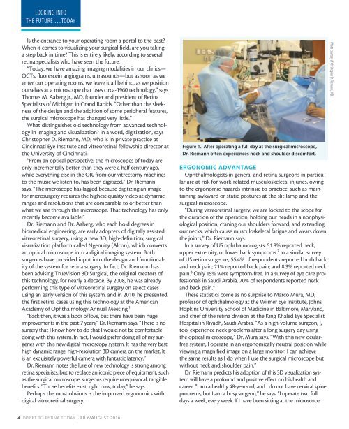
Figure 1. After operating a full day at <strong>the</strong> surgical microscope,<br />
Dr. Riemann often experiences neck and shoulder discomfort.<br />
ERGONOMIC ADVANTAGE<br />
Ophthalmologists in general and retina surgeons in particular<br />
are at risk for work-related musculoskeletal injuries, owing<br />
to <strong>the</strong> ergonomic hazards intrinsic to practice, such as maintaining<br />
awkward or static postures at <strong>the</strong> slit lamp and <strong>the</strong><br />
surgical microscope.<br />
“During vitreoretinal surgery, we are locked to <strong>the</strong> scope for<br />
<strong>the</strong> duration of <strong>the</strong> operation, holding our heads in a nonphysiological<br />
position, craning our shoulders forward, and extending<br />
our necks, which cause musculoskeletal fatigue and wears down<br />
<strong>the</strong> joints,” Dr. Riemann says.<br />
In a survey of US ophthalmologists, 51.8% reported neck,<br />
upper extremity, or lower back symptoms. 2 In a similar survey<br />
of US retina surgeons, 55.4% of respondents reported both back<br />
and neck pain; 21% reported back pain; and 8.3% reported neck<br />
pain. 3 Only 15% were symptom-free. In a survey of eye care professionals<br />
in Saudi Arabia, 70% of respondents reported neck<br />
and back pain. 4<br />
These statistics come as no surprise to Marco Mura, MD,<br />
professor of ophthalmology at <strong>the</strong> Wilmer Eye Institute, Johns<br />
Hopkins University School of Medicine in Baltimore, Maryland,<br />
and chief of <strong>the</strong> retina division at <strong>the</strong> King Khaled Eye Specialist<br />
Hospital in Riyadh, Saudi Arabia. “As a high-volume surgeon, I,<br />
too, experience neck problems after a long surgery day using<br />
<strong>the</strong> optical microscope,” Dr. Mura says. “With this new ocularfree<br />
system, I operate in an ergonomically neutral position while<br />
viewing a magnified image on a large monitor. I can achieve<br />
<strong>the</strong> same results as I do when I use <strong>the</strong> surgical microscope but<br />
without neck and shoulder pain.”<br />
Dr. Riemann predicts his adoption of this 3D visualization system<br />
will have a profound and positive effect on his health and<br />
career. “I am a healthy 48-year-old, and I do not have cervical spine<br />
problems, but I am a busy surgeon,” he says. “I operate two full<br />
days a week, every week. If I have been sitting at <strong>the</strong> microscope<br />
Photo courtesy of Christopher D. Riemann, MD.<br />
4 INSERT TO RETINA TODAY | JULY/AUGUST 2016