Drug resistance of Staphylococcus aureus in sinusitis patients
Abstract In this study on Sinusitis patients, we obtained 45 strains of Staphylococcus aureus. Antibiotic pattern of Staphyloccus aureus showed that resistance to Quinolones was 21% and 33% towards ciprofloxacin and oflaxacin respectively. Resistance to cephalosporins was 50% to cefuroxime, 41% and 50% to cefaperazone and cefotaxime respectively. Least resistance was noticed against aminoglycosides viz. Amikacin 47% and Gentamicin 21%. Resistance to Ampicillin and amoxicillin was 60% and 64% respectively. Oxacillin resistance was seen in 26% of the strains. Of the 45 isolates, 6 were found to be resistant for oxacillin . All these six isolates were subjected to Polymerase Chain Reaction (PCR) and they possessed the mecA gene. Correlation existed between the presence of mecA gene and oxacillin resistance in Staphylococcus aureus and these strains can be considered as MRSA and the patients can be advised for vancomycin therapy. Oxacillin resistance determination by phenotypic methods takes 24 hours to infer whereas PCR for mecA gene took only 6 hours. So the PCR techniques for the detection of mecA gene can be considered as gold standard (Rapid, Quick and accurate diagnosis) method for the detection of MRSA in spite of the cost involved.
Abstract
In this study on Sinusitis patients, we obtained 45 strains of Staphylococcus aureus. Antibiotic pattern of Staphyloccus aureus showed that resistance to Quinolones was 21% and 33% towards ciprofloxacin and oflaxacin respectively. Resistance to cephalosporins was 50% to cefuroxime, 41% and 50% to cefaperazone and cefotaxime respectively. Least resistance was noticed against aminoglycosides viz. Amikacin 47% and Gentamicin 21%. Resistance to Ampicillin and amoxicillin was 60% and 64% respectively. Oxacillin resistance was seen in 26% of the strains. Of the 45 isolates, 6 were found to be resistant for oxacillin . All these six isolates were subjected to Polymerase Chain Reaction (PCR) and they possessed the mecA gene. Correlation existed between the presence of mecA gene and oxacillin resistance in Staphylococcus aureus and these strains can be considered as MRSA and the patients can be advised for vancomycin therapy. Oxacillin resistance determination by phenotypic methods takes 24 hours to infer whereas PCR for mecA gene took only 6 hours. So the PCR techniques for the detection of mecA gene can be considered as gold standard (Rapid, Quick and accurate diagnosis) method for the detection of MRSA in spite of the cost involved.
Create successful ePaper yourself
Turn your PDF publications into a flip-book with our unique Google optimized e-Paper software.
acid precipitates unhydrolysed DNA. DNase<br />
produc<strong>in</strong>g colonies are therefore surrounded by clear<br />
areas due to DNA hydrolysis.<br />
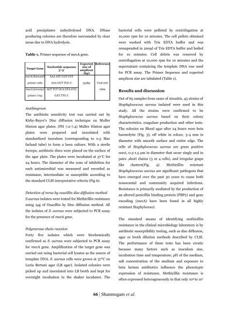
Table 1. Primer sequence <strong>of</strong> mecA gene.<br />
Target Gene<br />
mecA (forward)<br />
primer 1282<br />
mecA (reverse)<br />
primer 1793<br />
Antibiogram<br />
Nucleotide sequence<br />
5’-3’<br />
AAA ATC GAT CGT<br />
The antibiotic sensitivity test was carried out by<br />
Kirby-Buyer’s Disc diffusion technique on Muller<br />
H<strong>in</strong>ton agar plates. (PH 7.2-7.4) Muller H<strong>in</strong>ton agar<br />
plates were prepared and <strong>in</strong>oculated with<br />
standardized <strong>in</strong>oculum (correspond<strong>in</strong>g to 0.5 Mac<br />
farland tube) to form a lawn culture. With a sterile<br />
forceps, antibiotic discs were placed on the surface <strong>of</strong><br />
the agar plate. The plates were <strong>in</strong>cubated at 37 ° C for<br />
24 hours. The diameter <strong>of</strong> the zone <strong>of</strong> <strong>in</strong>hibition for<br />
each antimicrobial was measured and recorded as<br />
<strong>resistance</strong>, <strong>in</strong>termediate or susceptible accord<strong>in</strong>g to<br />
the standard CLSI <strong>in</strong>terpretative criteria (Fig 6).<br />
Detection <strong>of</strong> mrsa by oxacill<strong>in</strong> disc diffusion method<br />
S.<strong>aureus</strong> isolates were tested for Methicill<strong>in</strong> <strong>resistance</strong><br />
us<strong>in</strong>g 1μg <strong>of</strong> Oxacill<strong>in</strong> by Disc diffusion method. All<br />
the isolates <strong>of</strong> S. <strong>aureus</strong> were subjected to PCR assay<br />
for the presence <strong>of</strong> mecA gene.<br />
Polymerase cha<strong>in</strong> reaction<br />
Expected<br />
size <strong>of</strong><br />
amplicon<br />
(bp)<br />
Reference<br />
AAA GGT TGG C 533bp Unal etal<br />
AGT TCT GCA GTA CCG<br />
GAT TTG C<br />
1994<br />
Forty five isolates which were biochemically<br />
confirmed as S. <strong>aureus</strong> were subjected to PCR assay<br />
for mecA gene. Amplification <strong>of</strong> the target gene was<br />
carried out us<strong>in</strong>g bacterial cell lysates as the source <strong>of</strong><br />
template DNA. S. <strong>aureus</strong> cells were grown at 37°C on<br />
Luria Bertani agar (LB agar). Isolated colonies were<br />
picked up and <strong>in</strong>oculated <strong>in</strong>to LB broth and kept for<br />
overnight <strong>in</strong>cubation <strong>in</strong> the shaker <strong>in</strong>cubator. The<br />
bacterial cells were pelleted by centrifugation at<br />
10,000 rpm for 10 m<strong>in</strong>utes. The cell pellets obta<strong>in</strong>ed<br />
were washed with Tris EDTA buffer and was<br />
resuspended <strong>in</strong> 200μl <strong>of</strong> Tris EDTA buffer and boiled<br />
for 10 m<strong>in</strong>utes. Cell debris was removed by<br />
centrifugation at 10,000 rpm for 10 m<strong>in</strong>utes and the<br />
supernatant conta<strong>in</strong><strong>in</strong>g the template DNA was used<br />
for PCR assay. The Primer Sequence and expected<br />
amplicon size are tabulated (Table 1).<br />
Results and discussion<br />
Out <strong>of</strong> 65 samples from cases <strong>of</strong> s<strong>in</strong>usitis, 45 stra<strong>in</strong>s <strong>of</strong><br />
<strong>Staphylococcus</strong> <strong>aureus</strong> isolated were used <strong>in</strong> this<br />
study. All the stra<strong>in</strong>s were confirmed to be<br />
<strong>Staphylococcus</strong> <strong>aureus</strong> based on their colony<br />
characteristics, coagulase production and other tests.<br />
The colonies on Blood agar after 24 hours were beta<br />
haemolytic (Fig. 3), <strong>of</strong>f white <strong>in</strong> colour, 3-4 mm <strong>in</strong><br />
diameter with smooth surface and entire edge. The<br />
cells <strong>of</strong> <strong>Staphylococcus</strong> <strong>aureus</strong> are gram positive<br />
cocci, 0.5-1.5 µm <strong>in</strong> diameter that occur s<strong>in</strong>gly and <strong>in</strong><br />
pairs ,short cha<strong>in</strong>s (3 or 4 cells), and irregular grape<br />
like clusters(Fig. 4). Methicill<strong>in</strong> resistant<br />
<strong>Staphylococcus</strong> <strong>aureus</strong> are significant pathogens that<br />
have emerged over the past 30 years to cause both<br />
nosocomial and community acquired <strong>in</strong>fections.<br />
Resistance is primarily mediated by the production <strong>of</strong><br />
an altered penicill<strong>in</strong> b<strong>in</strong>d<strong>in</strong>g prote<strong>in</strong> (PBP2) and gene<br />
encod<strong>in</strong>g (mecA) have been found <strong>in</strong> all highly<br />
resistant Staphylococci.<br />
The standard means <strong>of</strong> identify<strong>in</strong>g methicill<strong>in</strong><br />
<strong>resistance</strong> <strong>in</strong> the cl<strong>in</strong>ical microbiology laboratory is by<br />
antibiotic susceptibility test<strong>in</strong>g, such as disc diffusion,<br />
agar or broth dilution methods described by CLSI.<br />
The performance <strong>of</strong> these tests has been erratic<br />
because many factors such as <strong>in</strong>oculum size,<br />
<strong>in</strong>cubation time and temperature, pH <strong>of</strong> the medium,<br />
salt concentration <strong>of</strong> the medium and exposure to<br />
beta lactam antibiotics <strong>in</strong>fluence the phenotypic<br />
expression <strong>of</strong> <strong>resistance</strong>. Methicill<strong>in</strong> <strong>resistance</strong> is<br />
<strong>of</strong>ten expressed heterogeneously <strong>in</strong> that only 10 4 to 10 7<br />
66 | Shanmugam et al.




![Review on: impact of seed rates and method of sowing on yield and yield related traits of Teff [Eragrostis teff (Zucc.) Trotter] | IJAAR @yumpu](https://documents.yumpu.com/000/066/025/853/c0a2f1eefa2ed71422e741fbc2b37a5fd6200cb1/6b7767675149533469736965546e4c6a4e57325054773d3d/4f6e6531383245617a537a49397878747846574858513d3d.jpg?AWSAccessKeyId=AKIAICNEWSPSEKTJ5M3Q&Expires=1714978800&Signature=xoFUXxPHfHo%2F2KL8Z5ixsGiy%2Fmw%3D)


