

In this issue
ACNR-ASO16-full-pdf-2
ACNR-ASO16-full-pdf-2

Create successful ePaper yourself
Turn your PDF publications into a flip-book with our unique Google optimized e-Paper software.
e v i e w a r t i c l e<br />
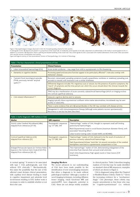
Figure 1: Neuropathological changes observed in CAA: the Vonsattel grading scheme for CAA severity.<br />
<strong>In</strong> mild (grade 1) CAA, Aβ deposits are present in a proportion of the vessel wall. <strong>In</strong> moderately severe (grade 2) CAA Aβ is deposited circumferentially in the media. <strong>In</strong> severe (grade 3) CAA, in<br />
addition to concentric Aβ deposition, there is splitting and double-barrelling of the vessel wall. Very severe (grade 4) CAA is associated with marked obliteration of the lumen often associated<br />
with vascular necrosis, recanalisation and scarring.<br />
Figure and legend courtesy of Zane Jaunmuktane, Division of Neuropathology, UCL <strong>In</strong>stitute of Neurology.<br />
Table 1: The four characteristic clinical presentations of CAA<br />
Presentation<br />
Clinical Features<br />
• Lobar intracerebral haemorrhage (ICH) Acute stroke syndrome – may range from mild or asymptomatic to life-threatening.<br />
• Dementia or cognitive decline Processing speed and executive function appear to be particularly affected 50 ; note also overlap with<br />
Alzheimer’s disease.<br />
• Transient focal neurological episodes<br />
(TFNE: previously termed “amyloid<br />
spells”) 51,52<br />
Recurrent, stereotyped, spreading symptoms (usually paraesthesia, numbness or weakness); spreading over<br />
seconds to minutes with resolution over a similar timeframe.<br />
Differential diagnosis includes TIA, migrainous aura or seizure, but the presence of positive symptoms, the<br />
“march” of symptoms and the time period over which <strong>this</strong> occurs should direct the clinician towards the<br />
correct diagnosis.<br />
TFNE may be a manifestation of acute convexity subarachnoid haemorrhage (which on imaging evolves<br />
into cortical superficial siderosis).<br />
• CAA-related inflammation 53-56 Subacute cognitive decline and/or seizures.<br />
Imaging typically shows asymmetrical confluent white matter abnormalities; microbleeds may be seen<br />
acutely or subacutely.<br />
There is some evidence that anti-Aβ autoantibodies in the CSF may correlate with disease activity.<br />
Management is with immunosuppressive therapy (although some patients recover spontaneously).<br />
Recurrence is rare but has been described.<br />
Table 2: Useful diagnostic MRI markers in CAA<br />
Marker MRI sequence Description<br />
Strictly Lobar Cerebral Microbleeds (MBs)<br />
Adapted from references [57-59]<br />
Cortical Superficial Siderosis (cSS)<br />
Adapted from reference [51]<br />
Enlarged Perivascular Spaces (or Virchow-Robin<br />
Spaces) in the Centrum Semi Ovale (CSO-PVS)<br />
Adapted from references [60, 61]<br />
Paramagnetic sequences<br />
e.g. T2*-GRE, SWI<br />
Paramagnetic sequences<br />
e.g. T2*-GRE, SWI<br />
T2<br />
“Haemorrhagic” marker of CAA, thought to represent small self-limiting<br />
parenchymal haemorrhages.<br />
Black (hypointense) round or ovoid lesions (maximum diameter 10mm), with<br />
associated “blooming effect”.<br />
Lobar location (rating scales include MARS and BOMBS).<br />
“Haemorrhagic” marker of CAA, believed to be the result of evolution of<br />
previous convexity subarachnoid haemorrhage.<br />
Dark (hypointense) bilinear ‘track-like’ rim around convexities of the cerebral<br />
hemisphere; restricted to supratentorial compartment in CAA.<br />
“Non-haemorrhagic” marker of CAA, demonstrating enlargement of the<br />
interstitial fluid channels that surround small arterioles<br />
Small white (hyperintense/high signal) round or linear lesions (CSF<br />
isointense).<br />
in normal ageing. 17 It seems to be associated<br />
with type 1 CAA pathologically, and CAA<br />
without ICH clinically. 5,15 Mechanistically,<br />
<strong>this</strong> raises the possibility that the size of the<br />
affected vessel dictates clinical presentation,<br />
with capillary level disease tending to result<br />
in cognitive impairment and arteriolar level<br />
involvement resulting in ICH; further work is<br />
necessary in order to establish whether or not<br />
<strong>this</strong> is the case.<br />
Imaging Markers<br />
The recent advances in our understanding<br />
of CAA have been made possible by the<br />
identification of new neuroimaging measures<br />
that allow a diagnosis to be made without<br />
pathological material. 2 Although a number of<br />
novel imaging techniques, including diffusion<br />
tensor imaging, visual functional MRI and<br />
amyloid-PET, have diagnostic potential in<br />
CAA, 2 these are not always widely available<br />
in clinical practice. Table 2 describes imaging<br />
markers of CAA that may be easily identified<br />
on standard clinical MR sequences, examples<br />
of which are shown in Figure 2.<br />
CAA is diagnosed using either the Classical<br />
or Modified Boston Criteria (Table 3). 3,4 Given<br />
the increasing evidence for a “non-haemorrhagic”<br />
CAA phenotype, these criteria may<br />
require amendments so that those who may<br />
be “cognitive-predominant” (i.e. without<br />
ACNR > VOLUME 16 NUMBER 2 > AUG-OCT 2016 > 9