- Page 2 and 3:
U.S. Fire Administration Mission St
- Page 4 and 5:
The IAFF would also like to thank t
- Page 6 and 7:
David Prezant, MD Professor of Medi
- Page 8 and 9:
This page was intentionally left bl
- Page 10 and 11:
Pneumonias.........................
- Page 12 and 13:
Chapter 2-7 • Pulmonary Fibrosis
- Page 14 and 15:
Clinical Manifestations............
- Page 16 and 17:
Reactive (Lower) Airways Dysfunctio
- Page 18 and 19:
Diagnostic Approach................
- Page 20 and 21:
This page was intentionally left bl
- Page 22 and 23:
phase (fire is extinguished, clean-
- Page 24 and 25:
In recognition of the causal relati
- Page 26 and 27:
and adjudicatory purposes and are n
- Page 28 and 29:
escue workers responded on the morn
- Page 30 and 31:
This page was intentionally left bl
- Page 32 and 33:
The wall of the alveoli is primaril
- Page 34 and 35:
Figure 1.1.1 Figure 1-1.1 Lung Volu
- Page 36 and 37:
This page was intentionally left bl
- Page 38 and 39:
helium, nitrogen and nitrogen oxide
- Page 40 and 41:
years. At the initial evaluation th
- Page 42 and 43:
and indirect (mannitol) airway chal
- Page 44 and 45:
Tornling et al 1994: Observed less
- Page 46 and 47:
27. Feuer E, Rosenman K. Mortality
- Page 48 and 49:
Oral Cavity, Pharynx, and Larynx Fo
- Page 50 and 51:
where the offending agents can be d
- Page 52 and 53:
The typical evaluation of chronic l
- Page 54 and 55:
This page was intentionally left bl
- Page 56 and 57:
fall and early spring. Bacteria are
- Page 58 and 59:
mucus plugging can lead to progress
- Page 60 and 61:
In adults, this is often aspirated
- Page 62 and 63:
Antibiotic treatment is sometimes g
- Page 64 and 65:
Pneumonia can also occur as patchy
- Page 66 and 67:
inhalation of an infected aerosol g
- Page 68 and 69:
ill or “toxic". The chest radiogr
- Page 70 and 71:
Currently, effective treatment is w
- Page 72 and 73:
20. Kaplan V, Angus DC, Griffin MF,
- Page 74 and 75:
A Person with Active TB Disease Has
- Page 76 and 77:
Figure 2-3.2: TB is spread from per
- Page 78 and 79:
Clinical Aspects of TB Disease Prog
- Page 80 and 81:
Drug-Resistant TB Drug-resistant TB
- Page 82 and 83:
Patients who have a positive TST re
- Page 84 and 85:
ecommended that close contacts with
- Page 86 and 87:
Individuals with immunosuppressive
- Page 88 and 89:
Vaccination with Live Attenuated Va
- Page 90 and 91:
than 18 years should be counseled a
- Page 92 and 93:
Treatment for Latent TB Infection D
- Page 94 and 95:
Directly Observed Therapy (DOT) for
- Page 96 and 97:
Monthly liver function tests (LFTs)
- Page 98 and 99:
19. Nolan CM, Goldberg SV, Buskin S
- Page 100 and 101:
to such allergens as house dust mit
- Page 102 and 103:
CLINICAL MANIFESTATION Patients usu
- Page 104 and 105:
who has persistent asthma. Patients
- Page 106 and 107:
Long-Term Control Medications Inhal
- Page 108 and 109:
Table 2-4.2: Stepwise approach to t
- Page 110 and 111:
Treatment should be started as soon
- Page 112 and 113:
21. Nathan RA, Sorkness CA, Kosinsk
- Page 114 and 115:
Pathology COPD affects the three ma
- Page 116 and 117:
CLASSIFICATION AND DIAGNOSIS OF COP
- Page 118 and 119:
different survey methods and variab
- Page 120 and 121:
A few other risk factors deserve me
- Page 122 and 123:
Suggested Questions for Follow-Up V
- Page 124 and 125:
counseling, social support and medi
- Page 126 and 127:
SUMMARY In summary, COPD is a progr
- Page 128 and 129:
or fungal), autoimmune diseases or
- Page 130 and 131:
Figure 2-6.2: Chest radiograph of S
- Page 132 and 133:
Other skin findings may occur and w
- Page 134 and 135:
prior to the start of our prospecti
- Page 136 and 137:
WTC and therefore had follow-up for
- Page 138 and 139:
18. Baughman RP, Teirstein AS, Juds
- Page 140 and 141: products like cotton. The key chara
- Page 142 and 143: signs are specific for interstitial
- Page 144 and 145: Certain features of the high-resolu
- Page 146 and 147: lung diseases without a known cause
- Page 148 and 149: An APR or PAPR will not protect aga
- Page 150 and 151: This page was intentionally left bl
- Page 152 and 153: heart. Pulmonary hypertension is th
- Page 154 and 155: suppressant drug, amironex fumarate
- Page 156 and 157: MEDICAL MANAGEMENT General Measures
- Page 158 and 159: Clinical Risk Factors Clinical symp
- Page 160 and 161: with renal failure. In many patient
- Page 162 and 163: Pulmonary Edema Associated with Inh
- Page 164 and 165: This page was intentionally left bl
- Page 166 and 167: Tobacco Use in the US, 1900-2000 50
- Page 168 and 169: Environmental Causes of Lung Cancer
- Page 170 and 171: Figure 2-9.3 depicts the frequency
- Page 172 and 173: are allowing even small lung nodule
- Page 174 and 175: Figure 2-9.6: Chest X-ray and CT Sc
- Page 176 and 177: Figure 2-9.8: Chest CT Scan Showing
- Page 178 and 179: Certain types of SCLC can secrete b
- Page 180 and 181: Physicians Guidelines for the Diagn
- Page 182 and 183: 17. Alberg AJ and Samet JM. Epidemi
- Page 184 and 185: This page was intentionally left bl
- Page 186 and 187: use. Cases of mesothelioma, a type
- Page 188 and 189: The mechanism by which asbestos cau
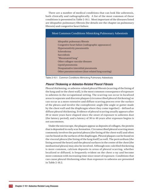
- Page 192 and 193: diagnosis is made by excluding othe
- Page 194 and 195: The role of lung scarring in the de
- Page 196 and 197: screening through the use of the IA
- Page 198 and 199: Normal nocturnal sleep is divided i
- Page 200 and 201: However, during sleep there is gene
- Page 202 and 203: from sleep, eye movements, breathin
- Page 204 and 205: Figure 2-11.3: Effects of nasal CPA
- Page 206 and 207: People with OSAS undergoing surgery
- Page 208 and 209: with or without cataplexy. The cond
- Page 210 and 211: 26. Appel D, Schmidt-Nowarra WW, Po
- Page 212 and 213: ACUTE COUGH As mentioned above, a c
- Page 214 and 215: cough occurred in adults and adoles
- Page 216 and 217: Gastroesophageal Reflux Disease (GE
- Page 218 and 219: WORLD TRADE CENTER COUGH Immediatel
- Page 220 and 221: This page was intentionally left bl
- Page 222 and 223: materials may produce smoke that is
- Page 224 and 225: The effects of frequent smoke expos
- Page 226 and 227: the most intense exposure to air po
- Page 228 and 229: seizures, coma and death 64 . Addit
- Page 230 and 231: highly water-soluble and, thus, wil
- Page 232 and 233: Diagnostic Testing Initial pulmonar
- Page 234 and 235: symptoms in an inhalation lung inju
- Page 236 and 237: 17. Musk AW, Peters JM, Wegman DW.
- Page 238 and 239: 42. Herbert R, Moline J, Skloot G,
- Page 240 and 241:
71. Kales S, Christiani D. Acute ch
- Page 242 and 243:
DECONTAMINATION OF RADIOLOGICAL CAS
- Page 244 and 245:
Procedure for Determining Approxima
- Page 246 and 247:
Following a three Gy exposure, lymp
- Page 248 and 249:
decontamination of the patient and
- Page 250 and 251:
treated as per standard burn protoc
- Page 252 and 253:
time course for this phenomenon, re
- Page 254 and 255:
This page was intentionally left bl
- Page 256 and 257:
groups have been identified to be a
- Page 258 and 259:
fever accompanied by mouth sores an
- Page 260 and 261:
and Aspergillus species which are u
- Page 262 and 263:
enal failure. Encephalitis and bact
- Page 264 and 265:
progress to severe, fulminant illne
- Page 266 and 267:
may be present. A systemic disease
- Page 268 and 269:
one percent are primary pneumonic p
- Page 270 and 271:
gentamicin is recommended. In a mas
- Page 272 and 273:
9. Kilbourne E. Influenza. 1st ed.
- Page 274 and 275:
44. Marik PE, Bowles SA. Medical as
- Page 276 and 277:
74. Advisory Committee on Immunizat
- Page 278 and 279:
the Mt. St. Helens eruption in 1980
- Page 280 and 281:
UPPER RESPIRATORY DISEASE Reactive
- Page 282 and 283:
Pulmonary function declines or abno
- Page 284 and 285:
Currently, for asthma in general an
- Page 286 and 287:
Thirteen were identified during the
- Page 288 and 289:
(FDNY, NY/NJ consortium coordinated
- Page 290 and 291:
For example at the WTC site, FDNY h
- Page 292 and 293:
21. Jalloul AS and Banks DE. The he
- Page 294 and 295:
49. Banauch GI, Hall C, Weiden M, e
- Page 296 and 297:
This page was intentionally left bl
- Page 298 and 299:
PEAK FLOW/SPIROMETRY/BRONCHODILATOR
- Page 300 and 301:
Spirometry results can usually diff
- Page 302 and 303:
of these conditions on spirometry i
- Page 304 and 305:
At beginning of gas dilution test A
- Page 306 and 307:
calculate a single-breath estimate
- Page 308 and 309:
is often referred to as cardiopulmo
- Page 310 and 311:
Classification of Respiratory Impai
- Page 312 and 313:
This page was intentionally left bl
- Page 314 and 315:
taken with the plate pressed agains
- Page 316 and 317:
• Granulomas: Small nodules, ofte
- Page 318 and 319:
clarity can be obtained in a shorte
- Page 320 and 321:
CT findings may be non-specific and
- Page 322 and 323:
Sarcoidosis is a common chronic gra
- Page 324 and 325:
Risks Risks from the procedure incl
- Page 326 and 327:
while certain slow growing tumors (
- Page 328 and 329:
the insertion of needle after preli
- Page 330 and 331:
CT Pulmonary Angiography (CTPA) CTP
- Page 332 and 333:
Risks • Although the total amount
- Page 334 and 335:
INCIDENCE AND PREVALENCE The freque
- Page 336 and 337:
Hamartomas are the most common beni
- Page 338 and 339:
Figure 4-3.1: Spiculated lesion on
- Page 340 and 341:
Assessment of Nodule Growth Rate an
- Page 342 and 343:
Estimating Probability of Malignanc
- Page 344 and 345:
advanced emphysema); those with bul
- Page 346 and 347:
c. If old chest images are availabl
- Page 348 and 349:
4. Henschke CI, Yankelevitz DF, Nai
- Page 350 and 351:
While most people are aware that sm
- Page 352 and 353:
Modified Fagerström Test for Smoke
- Page 354 and 355:
nicotine gum, inhalers, or nicotine
- Page 356 and 357:
Common Reasons Fire Fighters Expres
- Page 358 and 359:
and while anyone can temporarily ex
- Page 360 and 361:
heart and lungs, increasing the ris
- Page 362 and 363:
your doctor, healthcare professiona
- Page 364 and 365:
protection from tobacco cravings, w
- Page 366 and 367:
cessation. The entire program is ac
- Page 368 and 369:
drive, the neural control of breath
- Page 370 and 371:
Figure 4-5.2: The balance between i
- Page 372 and 373:
TREATMENT OF RESPIRATORY FAILURE Tr
- Page 374 and 375:
Mechanical Ventilatory Support Fact
- Page 376 and 377:
WEANING OR REMOVING A PATIENT FROM
- Page 378 and 379:
7. Esteban A, Frutos F, Tobin MJ, A
- Page 380:
International Association of Fire F