Impact of chronic exercise training on pro-inflammatory cytokine interleukine-6 in adult men with asthma
Abstract Evidence supports an important contribution of low-grade systemic inflammation in asthma or chronic obstructive pulmonary disease. The objective of present study was to determine whether aerobic training program affect serum IL-6 in males with asthma. For this purpose, fasting blood samples were collected before and at the end of aerobic training (3 months/3 days weekly) in order to measuring serum interleukin-6 in twenty two adult men with chronic asthma that randomly divided into exercise or control groups. Anthropometrical markers were also measured before and after exercise program in two groups. Independent student T test was used for between group’s comparison at baseline and paired T test used for determine significant changes in variables by exercise intervention. Exercise program decreases body weight, body mass index and body fat percentage compared to baseline. There were no significant differences for serum IL-6 [pre, 5.33(3.6); post, 5.65(2.71) pg/ml (p = 0.76)]. These data suggest that long term aerobic training is not associated with an anti-inflammatory property in asthma patients. Further studies are necessary to elucidate the significance of this exercise program on other inflammatory cytokines.
Abstract
Evidence supports an important contribution of low-grade systemic inflammation in asthma or chronic obstructive pulmonary disease. The objective of present study was to determine whether aerobic training program affect serum IL-6 in males with asthma. For this purpose, fasting blood samples were collected before and at the end of aerobic training (3 months/3 days weekly) in order to measuring serum interleukin-6 in twenty two adult men with chronic asthma that randomly divided into exercise or control groups. Anthropometrical markers were also measured before and after exercise program in two groups. Independent student T test was used for between group’s comparison at baseline and paired T test used for determine significant changes in variables by exercise intervention. Exercise program decreases body weight, body mass index and body fat percentage compared to baseline. There were no significant differences for serum IL-6 [pre, 5.33(3.6); post, 5.65(2.71) pg/ml (p = 0.76)]. These data suggest that long term aerobic training is not associated with an anti-inflammatory property in asthma patients. Further studies are necessary to elucidate the significance of this exercise program on other inflammatory cytokines.
Create successful ePaper yourself
Turn your PDF publications into a flip-book with our unique Google optimized e-Paper software.
J. Bio. & Env. Sci. 2014<br />
patients. However, our f<strong>in</strong>d<strong>in</strong>gs are somewhat<br />
c<strong>on</strong>troversial. The f<strong>in</strong>d<strong>in</strong>gs <str<strong>on</strong>g>of</str<strong>on</strong>g> this study are observed<br />
while most studies report higher levels <str<strong>on</strong>g>of</str<strong>on</strong>g> IL-6 <strong>in</strong><br />
<strong>asthma</strong>tic patients than <strong>in</strong> healthy subjects. However,<br />
it is recognized that <strong>in</strong>creased levels <str<strong>on</strong>g>of</str<strong>on</strong>g> IL-6 or some<br />
other <strong><strong>in</strong>flammatory</strong> <strong>cytok<strong>in</strong>e</strong>s such as CRP and TNF-α<br />
are associated <strong>with</strong> impaired lung functi<strong>on</strong> (Wu et al.,<br />
2005). IL-6 plays an important role <strong>in</strong> the<br />
pathophysiology <str<strong>on</strong>g>of</str<strong>on</strong>g> <strong>asthma</strong> and its level drastically<br />
<strong>in</strong>creases <strong>in</strong> these patients especially dur<strong>in</strong>g<br />
<strong>asthma</strong>tic attacks (Yokoyama et al., 1995). In additi<strong>on</strong><br />
to adipose tissue, alveolar macrophages, br<strong>on</strong>chial<br />
epithelial cells and mast cells are also <strong>in</strong>volved <strong>in</strong><br />
secreti<strong>on</strong> <str<strong>on</strong>g>of</str<strong>on</strong>g> iL6 <strong>in</strong> <strong>asthma</strong>tic patients (Gosset et al.,<br />
1991; Mar<strong>in</strong>i et al., 1992).<br />
or narrow<strong>in</strong>g <str<strong>on</strong>g>of</str<strong>on</strong>g> the respiratory tract. Increased<br />
mucus <strong>pro</strong>ducti<strong>on</strong> by lung epithelium caused by IL-6<br />
dur<strong>in</strong>g <strong>in</strong>flammati<strong>on</strong> <str<strong>on</strong>g>of</str<strong>on</strong>g> respiratory pathways <strong>in</strong><br />
<strong><strong>in</strong>flammatory</strong> diseases such as <strong>asthma</strong> can physically<br />
block the respiratory pathways, which are associated<br />
<strong>with</strong> <strong>in</strong>creased resistance <str<strong>on</strong>g>of</str<strong>on</strong>g> respiratory pathways and<br />
ultimately leads to lung dysfuncti<strong>on</strong> (Rogers, 2004;<br />
Agrawal et al., 2007). Increased accumulati<strong>on</strong> <str<strong>on</strong>g>of</str<strong>on</strong>g><br />
eos<strong>in</strong>ophils <strong>in</strong> the lungs <strong>in</strong> resp<strong>on</strong>se to <strong>in</strong>creased<br />
levels <str<strong>on</strong>g>of</str<strong>on</strong>g> IL-6 has been observed previously (Wang et<br />
al., 2000; Qiu et al., 2004). Furthermore, <strong>in</strong>hibiti<strong>on</strong><br />
<str<strong>on</strong>g>of</str<strong>on</strong>g> IL-6 functi<strong>on</strong> by neutraliz<strong>in</strong>g or <strong>in</strong>hibit<strong>in</strong>g its<br />
receptor <strong>in</strong> <strong>asthma</strong>tic rats reduces the accumulati<strong>on</strong><br />
<str<strong>on</strong>g>of</str<strong>on</strong>g> eos<strong>in</strong>ophils <strong>in</strong> the lungs (Doganci et al., 2005).<br />
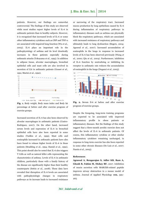
Fig. 1. Body weight, Body mass <strong>in</strong>dex and Body fat<br />
percentage at before and after <str<strong>on</strong>g>exercise</str<strong>on</strong>g> <strong>pro</strong>gram <str<strong>on</strong>g>of</str<strong>on</strong>g><br />
<str<strong>on</strong>g>exercise</str<strong>on</strong>g> groups.<br />
Increased secreti<strong>on</strong> <str<strong>on</strong>g>of</str<strong>on</strong>g> IL-6 has also been observed by<br />
alveolar macrophages <strong>in</strong> <strong>asthma</strong>tic patients (Castro-<br />
Rodríguez, 2007). On the other hand, <strong>in</strong>creased<br />
serum levels and expressi<strong>on</strong> <str<strong>on</strong>g>of</str<strong>on</strong>g> IL-6 <strong>in</strong> br<strong>on</strong>chial<br />
epithelial cells have also been reported <strong>in</strong> some<br />
studies (Yudk<strong>in</strong> et al., 1999). Mast cells and<br />
eos<strong>in</strong>ophils <strong>in</strong>creased <strong>in</strong> <strong>asthma</strong>tic patients have also<br />
been found to release higher levels <str<strong>on</strong>g>of</str<strong>on</strong>g> IL-6 <strong>in</strong> these<br />
patients (Bradd<strong>in</strong>g et al., 1994; Hamid et al., 1992).<br />
This po<strong>in</strong>t should also be noted that IL-6 also triggers<br />
T Cells as well as natural killer cells represent<strong>in</strong>g the<br />
characteristics <str<strong>on</strong>g>of</str<strong>on</strong>g> <strong>asthma</strong>. Levels <str<strong>on</strong>g>of</str<strong>on</strong>g> IL-6 <strong>in</strong> <strong>asthma</strong>tic<br />
children, particularly those <strong>with</strong> a family history <str<strong>on</strong>g>of</str<strong>on</strong>g><br />
the disease are significantly higher than their healthy<br />
counterparts (Sett<strong>in</strong> et al., 2008). These data have<br />
revealed that disrupti<strong>on</strong> <str<strong>on</strong>g>of</str<strong>on</strong>g> IL-6 levels are associated<br />
<strong>with</strong> pathophysiologic changes <strong>in</strong> respiratory<br />
pathways as its <strong>in</strong>crease leads to <strong>in</strong>creased resistance<br />
Fig. 2. Serum Il-6 at before and after <str<strong>on</strong>g>exercise</str<strong>on</strong>g><br />
<strong>pro</strong>gram <str<strong>on</strong>g>of</str<strong>on</strong>g> <str<strong>on</strong>g>exercise</str<strong>on</strong>g> groups.<br />
Despite the forego<strong>in</strong>g, l<strong>on</strong>g-term <str<strong>on</strong>g>tra<strong>in</strong><strong>in</strong>g</str<strong>on</strong>g> <strong>pro</strong>grams<br />
are expected to be associated <strong>with</strong> im<strong>pro</strong>ved<br />
<strong><strong>in</strong>flammatory</strong> pr<str<strong>on</strong>g>of</str<strong>on</strong>g>ile <strong>in</strong> obese patients or<br />
<strong><strong>in</strong>flammatory</strong> diseases. But the f<strong>in</strong>d<strong>in</strong>gs <str<strong>on</strong>g>of</str<strong>on</strong>g> this study<br />
suggest that a three-m<strong>on</strong>th aerobic <str<strong>on</strong>g>exercise</str<strong>on</strong>g> does not<br />
affect the levels <str<strong>on</strong>g>of</str<strong>on</strong>g> IL-6 <strong>in</strong> <strong>asthma</strong>tic patients. Of<br />
course, this <strong><strong>in</strong>flammatory</strong> <strong>cytok<strong>in</strong>e</strong> or other similar<br />
<strong><strong>in</strong>flammatory</strong> <strong>cytok<strong>in</strong>e</strong>s rema<strong>in</strong><strong>in</strong>g unchanged, <strong>in</strong><br />
resp<strong>on</strong>se to l<strong>on</strong>g-term <str<strong>on</strong>g>exercise</str<strong>on</strong>g> has also been reported<br />
<strong>in</strong> some other <str<strong>on</strong>g>chr<strong>on</strong>ic</str<strong>on</strong>g> diseases (De Luis et al., 2007;<br />
Nassis et al., 2005).<br />
References<br />
Agrawal A, Rengarajan S, Adler KB, Ram A,<br />
Ghosh B, Fahim M, Dickey BF. 2007. Inhibiti<strong>on</strong><br />
<str<strong>on</strong>g>of</str<strong>on</strong>g> muc<strong>in</strong> secreti<strong>on</strong> <strong>with</strong> MARCKS-related peptide<br />
im<strong>pro</strong>ves airway obstructi<strong>on</strong> <strong>in</strong> a mouse model <str<strong>on</strong>g>of</str<strong>on</strong>g><br />
<strong>asthma</strong>. Journal <str<strong>on</strong>g>of</str<strong>on</strong>g> Applied Physiology 102, 399–<br />
405.<br />
361 | Jalalvand et al




![Review on: impact of seed rates and method of sowing on yield and yield related traits of Teff [Eragrostis teff (Zucc.) Trotter] | IJAAR @yumpu](https://documents.yumpu.com/000/066/025/853/c0a2f1eefa2ed71422e741fbc2b37a5fd6200cb1/6b7767675149533469736965546e4c6a4e57325054773d3d/4f6e6531383245617a537a49397878747846574858513d3d.jpg?AWSAccessKeyId=AKIAICNEWSPSEKTJ5M3Q&Expires=1716706800&Signature=jodl1CmWoQwthINChYQDJrcxxlA%3D)


