Vicon Standard 2012

Create successful ePaper yourself
Turn your PDF publications into a flip-book with our unique Google optimized e-Paper software.
The <strong>Standard</strong><br />
vicon.com/standard<br />
Several markers on the stage<br />
could be seen by a different<br />
number of cameras. This allowed<br />
the simulation of a real test<br />
condition, where not all markers<br />
are simultaneously seen by all<br />
cameras. The accuracy of the<br />
setup (six MX13+ and four T40<br />
cameras) was dependant on the<br />
number of cameras tracking a<br />
marker: with six or more cameras,<br />
the accuracy was ±0.022mm; with<br />
two cameras, it was ±0.036mm.<br />
The limiting factor of the system<br />
was the noise level of the cameras.<br />
During static data collection, it<br />
was possible to detect movements<br />
as small as 0.01mm; however, due<br />
to the noise level of the cameras<br />
this wasn’t expected for dynamic<br />
measurements. Even taking into<br />
account the noise level, the<br />
accuracy of the <strong>Vicon</strong> system<br />
was still approximately 20 times<br />
better than the resolution of a<br />
medical CT-scanner.<br />
The measurement of 3D-deformation<br />
of a human pelvic bone was done<br />
in a two-leg standing position.<br />
First the soft tissue was removed<br />
from the fresh frozen and thawed<br />
specimen, while leaving the joint<br />
capsule and the ligaments intact.<br />
Then an adjustable fixture was<br />
fixed at the sacrum, allowing<br />
unconstrained rotational and<br />
transversal motion [Widmer<br />
1997]. After adjusting the pelvis<br />
on the testing machine, about 80<br />
reflective markers were fixed at<br />
anatomically defined positions<br />
on the bone with cyanoacrylate.<br />
Finally, the dynamic loading of the<br />
specimen could be performed. It<br />
consisted of 100 sinusoidal cycles<br />
at 1 Hz with amplitudes between<br />
100 N and either 0.5; 1; or 1.5×<br />
body weight respectively. The<br />
movement was tracked at a frame<br />
rate of 60 Hz.<br />
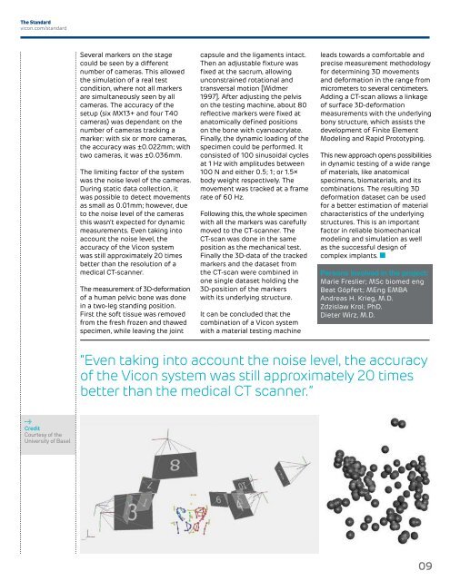
Following this, the whole specimen<br />
with all the markers was carefully<br />
moved to the CT-scanner. The<br />
CT-scan was done in the same<br />
position as the mechanical test.<br />
Finally the 3D-data of the tracked<br />
markers and the dataset from<br />
the CT-scan were combined in<br />
one single dataset holding the<br />
3D-position of the markers<br />
with its underlying structure.<br />
It can be concluded that the<br />
combination of a <strong>Vicon</strong> system<br />
with a material testing machine<br />
leads towards a comfortable and<br />
precise measurement methodology<br />
for determining 3D movements<br />
and deformation in the range from<br />
micrometers to several centimeters.<br />
Adding a CT-scan allows a linkage<br />
of surface 3D-deformation<br />
measurements with the underlying<br />
bony structure, which assists the<br />
development of Finite Element<br />
Modeling and Rapid Prototyping.<br />
This new approach opens possibilities<br />
in dynamic testing of a wide range<br />
of materials, like anatomical<br />
specimens, biomaterials, and its<br />
combinations. The resulting 3D<br />
deformation dataset can be used<br />
for a better estimation of material<br />
characteristics of the underlying<br />
structures. This is an important<br />
factor in reliable biomechanical<br />
modeling and simulation as well<br />
as the successful design of<br />
complex implants.<br />
Persons involved in the project:<br />
Marie Freslier; MSc biomed eng<br />
Beat Göpfert; MEng EMBA<br />
Andreas H. Krieg, M.D.<br />
Zdzislaw Krol; PhD.<br />
Dieter Wirz, M.D.<br />
“Even taking into account the noise level, the accuracy<br />
of the <strong>Vicon</strong> system was still approximately 20 times<br />
better than the medical CT scanner.”<br />
Credit<br />
Courtesy of the<br />
University of Basel<br />
09