

ABC of Burns

You also want an ePaper? Increase the reach of your titles
YUMPU automatically turns print PDFs into web optimized ePapers that Google loves.
Mechanical restriction <strong>of</strong> breathing—Deep dermal or full<br />
thickness circumferential burns <strong>of</strong> the chest can limit chest<br />
excursion and prevent adequate ventilation. This may require<br />
escharotomies (see next article).<br />
Blast injury—If there has been an explosion, blast lung can<br />
complicate ventilation. Penetrating injuries can cause tension<br />
pneumothoraces, and the blast itself can cause lung contusions<br />
and alveolar trauma and lead to adult respiratory distress<br />
syndrome.<br />
Smoke inhalation—The products <strong>of</strong> combustion, though<br />
cooled by the time they reach the lungs, act as direct irritants to<br />
the lungs, leading to bronchospasm, inflammation, and<br />
bronchorrhoea. The ciliary action <strong>of</strong> pneumocytes is impaired,<br />
exacerbating the situation. The inflammatory exudate created is<br />
not cleared, and atelectasis or pneumonia follows. The situation<br />
can be particularly severe in asthmatic patients. Non-invasive<br />
management can be attempted, with nebulisers and positive<br />
pressure ventilation with some positive end-expiratory pressure.<br />
However, patients may need a period <strong>of</strong> ventilation, as this<br />
allows adequate oxygenation and permits regular lung toileting.<br />
Carboxyhaemoglobin—Carbon monoxide binds to<br />
deoxyhaemoglobin with 40 times the affinity <strong>of</strong> oxygen. It also<br />
binds to intracellular proteins, particularly the cytochrome<br />
oxidase pathway. These two effects lead to intracellular and<br />
extracellular hypoxia. Pulse oximetry cannot differentiate<br />
between oxyhaemoglobin and carboxyhaemoglobin, and may<br />
therefore give normal results. However, blood gas analysis will<br />
reveal metabolic acidosis and raised carboxyhaemoglobin levels<br />
but may not show hypoxia. Treatment is with 100% oxygen,<br />
which displaces carbon monoxide from bound proteins six<br />
times faster than does atmospheric oxygen. Patients with<br />
carboxyhaemoglobin levels greater than 25-30% should be<br />
ventilated. Hyperbaric therapy is rarely practical and has not<br />
been proved to be advantageous. It takes longer to shift the<br />
carbon monoxide from the cytochrome oxidase pathway than<br />
from haemoglobin, so oxygen therapy should be continued<br />
until the metabolic acidosis has cleared.<br />
C—Circulation<br />
Intravenous access should be established with two large bore<br />
cannulas preferably placed through unburnt tissue. This is an<br />
opportunity to take blood for checking full blood count, urea<br />
and electrolytes, blood group, and clotting screen. Peripheral<br />
circulation must be checked. Any deep or full thickness<br />
circumferential extremity burn can act as a tourniquet,<br />
especially once oedema develops after fluid resuscitation. This<br />
may not occur until some hours after the burn. If there is any<br />
suspicion <strong>of</strong> decreased perfusion due to circumferential burn,<br />
the tissue must be released with escharotomies (see next article).<br />
Pr<strong>of</strong>ound hypovolaemia is not the normal initial response<br />
to a burn. If a patient is hypotensive then it is may be due to<br />
delayed presentation, cardiogenic dysfunction, or an occult<br />
source <strong>of</strong> blood loss (chest, abdomen, or pelvis).<br />
D—Neurological disability<br />
All patients should be assessed for responsiveness with the<br />
Glasgow coma scale; they may be confused because <strong>of</strong> hypoxia<br />
or hypovolaemia.<br />
E—Exposure with environment control<br />
The whole <strong>of</strong> a patient should be examined (including the back)<br />
to get an accurate estimate <strong>of</strong> the burn area (see later) and to<br />
check for any concomitant injuries. Burn patients, especially<br />
children, easily become hypothermic. This will lead to<br />
hypoperfusion and deepening <strong>of</strong> burn wounds. Patients should<br />
be covered and warmed as soon as possible.<br />
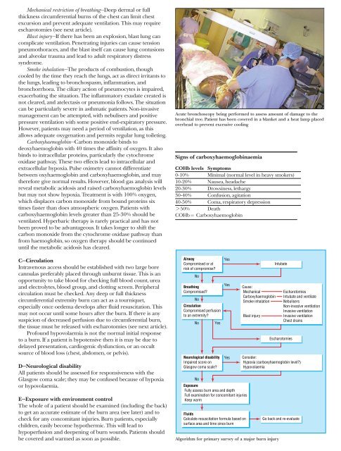
Acute bronchoscopy being performed to assess amount <strong>of</strong> damage to the<br />
bronchial tree. Patient has been covered in a blanket and a heat lamp placed<br />
overhead to prevent excessive cooling<br />
Signs <strong>of</strong> carboxyhaemoglobinaemia<br />
COHb levels Symptoms<br />
0-10% Minimal (normal level in heavy smokers)<br />
10-20% Nausea, headache<br />
20-30% Drowsiness, lethargy<br />
30-40% Confusion, agitation<br />
40-50% Coma, respiratory depression<br />
> 50% Death<br />
COHb = Carboxyhaemoglobin<br />
Airway<br />
Compromised or at<br />
risk <strong>of</strong> compromise?<br />
No<br />
Breathing<br />
Compromised?<br />
No<br />
Circulation<br />
Compromised perfusion<br />
to an extremity?<br />
No<br />
Yes<br />
Neurological disability<br />
Impaired score on<br />
Glasgow coma scale?<br />
No<br />
Yes<br />
Yes<br />
Yes<br />
Exposure<br />
Fully assess burn area and depth<br />
Full examination for concomitant injuries<br />
Keep warm<br />
Fluids<br />
Calculate resuscitation formula based on<br />
surface area and time since burn<br />
Cause:<br />
Mechanical<br />
Carboxyhaemoglobin<br />
Smoke inhalation<br />
Blast injury<br />
Intubate<br />
Escharotomies<br />
Escharotomies<br />
Intubate and ventilate<br />
Nebulisers<br />
Non-invasive ventilation<br />
Invasive ventilation<br />
Invasive ventilation<br />
Chest drains<br />
Consider:<br />
Hypoxia (carboxyhaemoglobin level?)<br />
Hypovolaemia<br />
Algorithm for primary survey <strong>of</strong> a major burn injury<br />
Go back and re-evaluate