

Preparation of RC System---9780723610892

Create successful ePaper yourself
Turn your PDF publications into a flip-book with our unique Google optimized e-Paper software.
PREPARATION OF THE ROOT CANAL SYSTEM 6<br />
preparation is used to dissolve tissue and aid in<br />
debridement <strong>of</strong> the chamber. This will also reduce<br />
the opportunity for the inadvertent inoculation <strong>of</strong><br />
microorganisms from the pulp chamber into the<br />
root canal system. Ultrasonically energized tips<br />
(Fig. 6.11) are useful adjuncts during debridement<br />
<strong>of</strong> the pulp chamber to break up calcific masses<br />
with relative safety and to facilitate removal <strong>of</strong><br />
debris. These instruments are contra-angled to<br />
enhance access.<br />
ROOT CANAL ORIFICES<br />
(a)<br />
(b)<br />
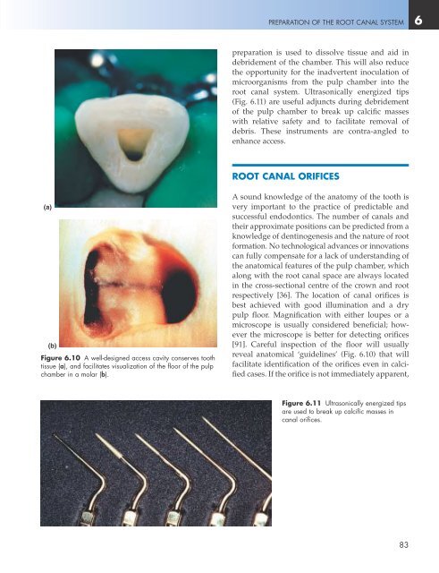
Figure 6.10 A well-designed access cavity conserves tooth<br />
tissue (a), and facilitates visualization <strong>of</strong> the floor <strong>of</strong> the pulp<br />
chamber in a molar (b).<br />
Asound knowledge <strong>of</strong> the anatomy <strong>of</strong> the tooth is<br />
very important to the practice <strong>of</strong> predictable and<br />
successful endodontics. The number <strong>of</strong> canals and<br />
their approximate positions can be predicted from a<br />
knowledge <strong>of</strong> dentinogenesis and the nature <strong>of</strong> root<br />
formation. No technological advances or innovations<br />
can fully compensate for a lack <strong>of</strong> understanding <strong>of</strong><br />
the anatomical features <strong>of</strong> the pulp chamber, which<br />
along with the root canal space are always located<br />
in the cross-sectional centre <strong>of</strong> the crown and root<br />
respectively [36]. The location <strong>of</strong> canal orifices is<br />
best achieved with good illumination and a dry<br />
pulp floor. Magnification with either loupes or a<br />
microscope is usually considered beneficial; however<br />
the microscope is better for detecting orifices<br />
[91]. Careful inspection <strong>of</strong> the floor will usually<br />
reveal anatomical ‘guidelines’ (Fig. 6.10) that will<br />
facilitate identification <strong>of</strong> the orifices even in calcified<br />
cases. If the orifice is not immediately apparent,<br />
Figure 6.11 Ultrasonically energized tips<br />
are used to break up calcific masses in<br />
canal orifices.<br />
83