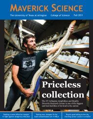
Maverick Science mag 2013-14

Create successful ePaper yourself
Turn your PDF publications into a flip-book with our unique Google optimized e-Paper software.
B<br />
reakthroughs in technology<br />
are expanding the<br />
limits of what’s possible<br />
in biophysics research.<br />
Samarendra Mohanty is<br />
helping to push those<br />
boundaries by developing<br />
new tools to discover<br />
safer, more efficient ways<br />
to treat disease and study the human body.<br />
Mohanty, a UT Arlington assistant professor of<br />
physics, is using the principles of biophysics –<br />
studying biological processes and materials by<br />
means of the theories as well as the tools of physics<br />
– to find new and innovative ways to alter biological<br />
systems down to the molecular level. He and<br />
his students are improving cutting-edge tools such<br />
as optical tweezers, specialized optical microscopes<br />
and optogenetics (the science of controlling brain<br />
activity with light) to understand and influence<br />
processes in cells and cellular networks.<br />
“We have taken an integrated approach of cellular<br />
manipulation, activation and control by optical<br />
as well as hybrid approaches, combined with<br />
a variety of i<strong>mag</strong>ing methods to visualize and<br />
quantify responses in in-vitro and in-vivo models,”<br />
Mohanty said. “In order to evaluate miniscule<br />
changes to cell membranes during optical manipulation,<br />
we developed a unique multimodal i<strong>mag</strong>ing<br />
platform integrated with laser scissors,<br />
tweezers, spanners, transporters and stimulators<br />
here at UT Arlington.”<br />
At the moment, Mohanty and the Biophysics<br />
and Physiology Laboratory group he leads are<br />
working on projects in five major research areas,<br />
along with a number of smaller projects. He keeps<br />
up a relentless pace in preparing and submitting<br />
grant proposals which have brought millions of<br />
dollars in research support from the National Institutes<br />
of Health, National <strong>Science</strong> Foundation<br />
and other sources. He authors and co-authors<br />
manuscripts which are regularly published in top<br />
journals.<br />
“His projects are highly i<strong>mag</strong>inative. He’s doing<br />
an intriguing combination of physics, biology,<br />
chemistry and biomechanics,” said Alex Weiss,<br />
professor and chair of the Department of Physics.<br />
“He’s doing very interesting research with nerve<br />
cells and optical stimulation of the brain, among<br />
other things. The work his group is doing has a lot<br />
of potential to improve medical research and treatment<br />
of disease down the road.”<br />
A<br />
s excited as he is by his<br />
group’s current work,<br />
Mohanty is even more<br />
thrilled by the research<br />
he envisions happening<br />
in his lab in the years<br />
ahead. He wants to shift<br />
his main focus from different<br />
aspects of neuronal<br />
manipulation, i<strong>mag</strong>ing and control, to the<br />
Brain Research through Advancing Innovative<br />
Neurotechnologies (BRAIN) initiative, an effort<br />
unveiled by President Barack Obama in April <strong>2013</strong><br />
which is intended to revolutionize understanding<br />
of the human brain through groundbreaking research.<br />
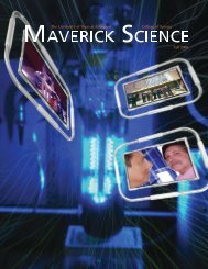
This i<strong>mag</strong>e shows a highly-controlled laser transfection of<br />
ChR2-YFP gene into a targeted area of retina (green is<br />
ChR2-YFP and blue is nuclei) by a near-infrared femtosecond<br />
laser microbeam. I<strong>mag</strong>e courtesy of Samarendra Mohanty.<br />
This illustration<br />
demonstrates the<br />
non-invasiveness of<br />
two-photon optogenetic<br />
stimulation. It<br />
shows brain tissue<br />
da<strong>mag</strong>e by an invasive<br />
fiber delivering<br />
one-photon stimulation<br />
(shown in<br />
blue) vs. non-invasive,<br />
two-photon<br />
stimulation (shown<br />
in red). Illustration<br />
courtesy of<br />
Samarendra<br />
Mohanty.<br />
One of Mohanty’s current projects which could<br />
be useful in the BRAIN initiative is the development<br />
of a tiny tool which could help scientists map<br />
and track interactions between neurons inside different<br />
areas of the brain. The fiber-optic, two-photon,<br />
optogenetic stimulator builds on a previous<br />
Mohanty discovery that near-infrared (NIR) light<br />
can be used to stimulate a light-sensitive protein<br />
introduced into living cells and neurons in the<br />
brain. This new method could show how different<br />
parts of the brain react when a linked area is stimulated.<br />
“Scientists have spent a lot of time looking at<br />
the physical connections between different regions<br />
of the brain. But that information is not sufficient<br />
unless we examine how those connections function,”<br />
Mohanty said. “That's where two-photon optogenetics<br />
comes into play. This is a tool not only<br />
to control the neuronal activity but to understand<br />
how the brain works.”<br />
The two-photon optogenetic stimulation involves<br />
introducing the gene for ChR2, a protein<br />
that responds to light, into a sample of excitable<br />
tissue cells. A fiber-optic infrared beam of NIR<br />
light can then be used to precisely excite the neurons<br />
in a tissue circuit. In the brain, researchers<br />
could then observe responses in the excited area<br />
as well as other parts of the neural circuit. In living<br />
subjects, scientists could also observe the behavioral<br />
outcome, Mohanty said.<br />
Optogenetic stimulation avoids da<strong>mag</strong>e to living<br />
tissue by stimulating neurons with light instead<br />
of electric pulses used in past research. Mohanty’s<br />
method of using low-energy NIR<br />
light also enables more precision and a<br />
deeper focus than the blue or green light<br />
beams often used in optogenetic stimulation.<br />
Kamal Dhakal, a third-year doctoral<br />
student in Mohanty’s lab, works in optogenetics<br />
and optical manipulation of cells. He<br />
says the multidisciplinary approach of Mohanty’s<br />
research is preparing him well for<br />
a career in optics and biophotonics. Dhakal<br />
was lead author of a paper on the brain<br />
mapping research that was published in<br />
the June 1, <strong>2013</strong> edition of the journal Optics<br />
Letters; Mohanty, doctoral student<br />
Bryan Black and postdoctoral researcher<br />
Ling Gu were co-authors.<br />
“Dr. Mohanty is very good researcher<br />
and has fancy ideas and visions,” Dhakal<br />
said. “He is very frank, like a friend, and<br />
helpful. Before joining his lab, I did not<br />
have any technical knowledge such as<br />
using computer software for data analysis,<br />
interfacing instruments with computers,<br />
i<strong>mag</strong>ing, programming, even how to make<br />
a good graph. But today, I know all of them.<br />
In addition, my projects require a wide variety<br />
of knowledge, from genetics to optics,<br />
mammalian cells to bacterial cells, electrophysiology<br />
to digital holography. These<br />
<strong>Maverick</strong> <strong>Science</strong> <strong>2013</strong>-<strong>14</strong><br />
33