

Mediworld Final Draft for Print

Create successful ePaper yourself
Turn your PDF publications into a flip-book with our unique Google optimized e-Paper software.
News & Updates<br />
Innovative microscope poised to propel<br />
optogenetics studies<br />
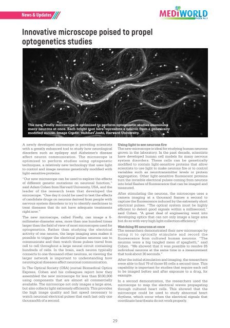
The new Firefly microscope is optimized to per<strong>for</strong>m optogenetic studies examining<br />
many neurons at once. Each bright spot here represents a neuron from a genetically<br />
modified mouse. Image Credit: Vaibhav Joshi, Harvard University.<br />
A newly developed microscope is providing scientists<br />
with a greatly enhanced tool to study how neurological<br />
disorders such as epilepsy and Alzheimer's disease<br />
affect neuron communication. The microscope is<br />
optimized to per<strong>for</strong>m studies using optogenetic<br />
techniques, a relatively new technology that uses light<br />
to control and image neurons genetically modified with<br />
light-sensitive proteins.<br />
“Our new microscope can be used to explore the effects<br />
of different genetic mutations on neuronal function,”<br />
said Adam Cohen from Harvard University, USA, and the<br />
leader of the research team that developed the<br />
microscope. “One day it could be used to test the effects<br />
of candidate drugs on neurons derived from people with<br />
nervous system disorders to try to identify medicines to<br />
treat diseases that do not have adequate treatments<br />
right now.”<br />
The new microscope, called Firefly, can image a 6-<br />
millimeter-diameter area, more than one hundred times<br />
larger than the field of view of most microscopes used <strong>for</strong><br />
optogenetics. Rather than studying the electrical<br />
activity of one neuron, the large imaging area makes it<br />
possible to trigger the electrical pulses neurons use to<br />
communicate and then watch those pulses travel from<br />
cell to cell throughout a large neural circuit containing<br />
hundreds of cells. In the brain, each neuron typically<br />
connects to one thousand other neurons, so viewing the<br />
larger network is important to understanding how<br />
neurological diseases affect neuronal communication.<br />
In The Optical Society (OSA) journal Biomedical Optics<br />
Express, Cohen and his colleagues report how they<br />
assembled the new microscope <strong>for</strong> less than $100,000<br />
using components that are almost all commercially<br />
available. The microscope not only images a large area,<br />
but also collects light extremely efficiently. This provides<br />
the high image quality and fast speed necessary to<br />
watch neuronal electrical pulses that each last only one<br />
thousandth of a second.<br />
Using light to see neurons fire<br />
The new microscope is ideal <strong>for</strong> studying human neurons<br />
grown in the laboratory. In the past decade, scientists<br />
have developed human cell models <strong>for</strong> many nervous<br />
system disorders. These cells can be genetically<br />
modified to contain light-sensitive proteins that allow<br />
scientists to use light to make neurons fire or to control<br />
variables such as neurotransmitter levels or protein<br />
aggregation. Other light-sensitive fluorescent proteins<br />
turn the invisible electrical pulses coming from neurons<br />
into brief flashes of fluorescence that can be imaged and<br />
measured.<br />
After stimulating the neurons, the microscope uses a<br />
camera imaging at a thousand frames a second to<br />
capture the fluorescence induced by the extremely short<br />
electrical pulses. “The optical system must be highly<br />
efficient to detect good signals within a millisecond,”<br />
said Cohen. “A great deal of engineering went into<br />
developing optics that can not only image a large area<br />
but do so with very high light collection efficiency.”<br />
Watching 85 neurons at once<br />
The researchers demonstrated their new microscope by<br />
using it to optically stimulate and record the<br />
fluorescence from cultured human neurons. “The<br />
neurons were a big tangled mess of spaghetti,” said<br />
Cohen. “We showed that it was possible to resolve 85<br />
individual neurons at the same time in a measurement<br />
that took about 30 seconds.”<br />
After the initial stimulation and imaging, the researchers<br />
were able to find 79 of those 85 cells a second time. This<br />
capability is important <strong>for</strong> studies that require each cell<br />
to be imaged be<strong>for</strong>e and after exposure to a drug, <strong>for</strong><br />
example.<br />
In a second demonstration, the researchers used the<br />
microscope to map the electrical waves propagating<br />
through cultured heart cells. This showed that the<br />
microscope could be used to study abnormal heart<br />
rhythms, which occur when the electrical signals that<br />
coordinate heartbeats do not work properly.<br />
29


