A study of histopathological effects and functional tests in liver and kidney of laboratory male mice treated with ammonium chloride
Abstract The present study was aimed to investigate the effects of ammonium chloride on mice. The animals were divided into three groups: groups 2 and 3were treated orally with ammonium chloride at doses of 2 and 4 mg/body weight, respectively for a period of three weeks, while group 1 served as a control group. The tissue sections which were made from mice organs (liver and kidney) which treated with 4mg/ body weight of ammonium chloride proved that this dose has toxic effect only on the liver and kidneys. In the liver, the histopathological effects are represented by hypertrophy and irregular shape of nucleus, degeneration of cytoplasm, congestion of sinusoid, bleeding and infiltration of inflammatory cells. In kidneys, the effects focus on renal tubules only which are represented by the degenerative changes, necrosis and infiltration of inflammatory cells. This study was also carried out to investigate the influence of ammonium chloride on levels of urea, creatinine, total protein, lipid (triglyceride and cholesterol), and liver marker enzymes such as AST, ALT and ALP. Oral administration of ammonium chloride (4mg/body weight) caused a significant increase in the levels of AST and a significant decrease in the level of ALP and total protein in mice. Treated mice with ammonium chloride at a dose of 2 and 4mg/ body weight for 21 days showed a significant decrease in levels of creatinine, triglyceride and cholesterol, while ALT and urea had no affect at two doses of ammonium chloride. In conclusion, ammonium chloride causes direct hepatotoxicity and nephrotoxicity.
Abstract
The present study was aimed to investigate the effects of ammonium chloride on mice. The animals were divided into three groups: groups 2 and 3were treated orally with ammonium chloride at doses of 2 and 4 mg/body weight, respectively for a period of three weeks, while group 1 served as a control group. The tissue sections which were made from mice organs (liver and kidney) which treated with 4mg/ body weight of ammonium chloride proved that this dose has toxic effect only on the liver and kidneys. In the liver, the histopathological effects are represented by hypertrophy and irregular shape of nucleus, degeneration of cytoplasm, congestion of sinusoid, bleeding and infiltration of inflammatory cells. In kidneys, the effects focus on renal tubules only which
are represented by the degenerative changes, necrosis and infiltration of inflammatory cells. This study was also carried out to investigate the influence of ammonium chloride on levels of urea, creatinine, total protein, lipid (triglyceride and cholesterol), and liver marker enzymes such as AST, ALT and ALP. Oral administration of ammonium chloride (4mg/body weight) caused a significant increase in the levels of AST and a significant decrease in the level of ALP and total protein in mice. Treated mice with ammonium chloride at a dose of 2 and
4mg/ body weight for 21 days showed a significant decrease in levels of creatinine, triglyceride and cholesterol, while ALT and urea had no affect at two doses of ammonium chloride. In conclusion, ammonium chloride causes direct hepatotoxicity and nephrotoxicity.

Create successful ePaper yourself
Turn your PDF publications into a flip-book with our unique Google optimized e-Paper software.
Int. J. Biosci. 2016<br />
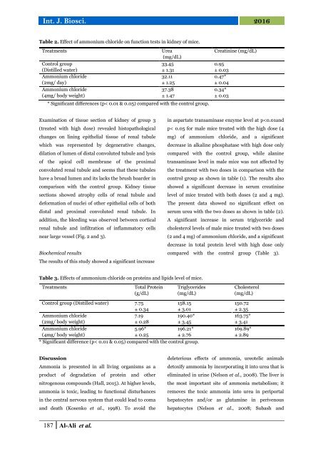
Table 2. Effect <strong>of</strong> <strong>ammonium</strong> <strong>chloride</strong> on function <strong>tests</strong> <strong>in</strong> <strong>kidney</strong> <strong>of</strong> <strong>mice</strong>.<br />
Treatments<br />
Control group<br />
(Distilled water)<br />
Ammonium <strong>chloride</strong><br />
(2mg/ day)<br />
Ammonium <strong>chloride</strong><br />
(4mg/ body weight)<br />
Urea<br />
(mg/dL)<br />
33.45<br />
± 1.31<br />
32.11<br />
± 1.25<br />
37.38<br />
± 1.47<br />
* Significant differences (p< 0.01 & 0.05) compared <strong>with</strong> the control group.<br />
Creat<strong>in</strong><strong>in</strong>e (mg/dL)<br />
0.95<br />
± 0.03<br />
0.47*<br />
± 0.04<br />
0.34*<br />
± 0.03<br />
Exam<strong>in</strong>ation <strong>of</strong> tissue section <strong>of</strong> <strong>kidney</strong> <strong>of</strong> group 3<br />
(<strong>treated</strong> <strong>with</strong> high dose) revealed <strong>histopathological</strong><br />
changes on l<strong>in</strong><strong>in</strong>g epithelial tissue <strong>of</strong> renal tubule<br />
which was represented by degenerative changes,<br />
dilation <strong>of</strong> lumen <strong>of</strong> distal convoluted tubule <strong>and</strong> lysis<br />
<strong>of</strong> the apical cell membrane <strong>of</strong> the proximal<br />
convoluted renal tubule <strong>and</strong> seems that these tubules<br />
have a broad lumen <strong>and</strong> its lacks the brush boarder <strong>in</strong><br />
comparison <strong>with</strong> the control group. Kidney tissue<br />
sections showed atrophy cells <strong>of</strong> renal tubule <strong>and</strong><br />
deformation <strong>of</strong> nuclei <strong>of</strong> other epithelial cells <strong>of</strong> both<br />
distal <strong>and</strong> proximal convoluted renal tubule. In<br />
addition, the bleed<strong>in</strong>g was observed between cortical<br />
renal tubule <strong>and</strong> <strong>in</strong>filtration <strong>of</strong> <strong>in</strong>flammatory cells<br />
near large vessel (Fig. 2 <strong>and</strong> 3).<br />
Biochemical results<br />
The results <strong>of</strong> this <strong>study</strong> showed a significant <strong>in</strong>crease<br />
<strong>in</strong> aspartate transam<strong>in</strong>ase enzyme level at p

![Review on: impact of seed rates and method of sowing on yield and yield related traits of Teff [Eragrostis teff (Zucc.) Trotter] | IJAAR @yumpu](https://documents.yumpu.com/000/066/025/853/c0a2f1eefa2ed71422e741fbc2b37a5fd6200cb1/6b7767675149533469736965546e4c6a4e57325054773d3d/4f6e6531383245617a537a49397878747846574858513d3d.jpg?AWSAccessKeyId=AKIAICNEWSPSEKTJ5M3Q&Expires=1714957200&Signature=C6JpywBV3rXeiqdofowD7yrWR1w%3D)