

7 - E-Lib FK UWKS
7 - E-Lib FK UWKS
7 - E-Lib FK UWKS

Create successful ePaper yourself
Turn your PDF publications into a flip-book with our unique Google optimized e-Paper software.
Modular Training in EERP<br />
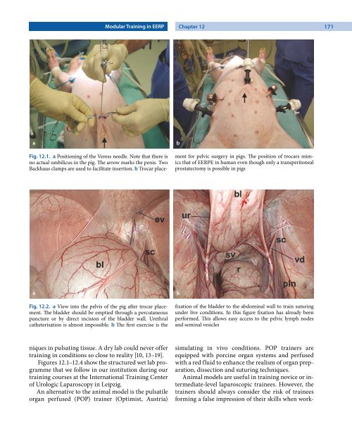
Fig. 12.1. a Positioning of the Veress needle. Note that there is<br />
no actual umbilicus in the pig. The arrow marks the penis. Two<br />
Backhaus clamps are used to facilitate insertion. b Trocar place-<br />
Fig. 12.2. a View into the pelvis of the pig after trocar placement.<br />
The bladder should be emptied through a percutaneous<br />
puncture or by direct incision of the bladder wall. Urethral<br />
catheterisation is almost impossible. b The first exercise is the<br />
niques in pulsating tissue. A dry lab could never offer<br />
training in conditions so close to reality [10, 13–19].<br />
Figures 12.1–12.4 show the structured wet lab programme<br />
that we follow in our institution during our<br />
training courses at the International Training Center<br />
of Urologic Laparoscopy in Leipzig.<br />
An alternative to the animal model is the pulsatile<br />
organ perfused (POP) trainer (Optimist, Austria)<br />
Chapter 12 171<br />
ment for pelvic surgery in pigs. The position of trocars mimics<br />
that of EERPE in human even though only a transperitoneal<br />
prostatectomy is possible in pigs<br />
fixation of the bladder to the abdominal wall to train suturing<br />
under live conditions. In this figure fixation has already been<br />
performed. This allows easy access to the pelvic lymph nodes<br />
and seminal vesicles<br />
simulating in vivo conditions. POP trainers are<br />
equipped with porcine organ systems and perfused<br />
with a red fluid to enhance the realism of organ preparation,<br />
dissection and suturing techniques.<br />
Animal models are useful in training novice or intermediate-level<br />
laparoscopic trainees. However, the<br />
trainers should always consider the risk of trainees<br />
forming a false impression of their skills when work-


![SISTEM SENSORY [Compatibility Mode].pdf](https://img.yumpu.com/20667975/1/190x245/sistem-sensory-compatibility-modepdf.jpg?quality=85)


