Ellegaard_Minipigs_Newsletter_51

You also want an ePaper? Increase the reach of your titles
YUMPU automatically turns print PDFs into web optimized ePapers that Google loves.
later saline and PFA are collected directly in containers through<br />
the venous cannula.<br />
After perfusion, both inflow and outflow tubes are clamped,<br />
and the cadaver left untouched for 1 hour to allow further<br />
fixation. Then the head is removed from the body and maxilla<br />
and mandibula are dissected and the bone is cut in between<br />
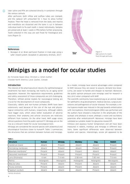
individual teeth to fix each tooth (+ bone) individually. Samples<br />
are preserved at least 1 week in PFA before further processing.<br />
Teeth collected in this way are well fixed for histological analysis<br />
(Figure 3).<br />
References<br />
1 Musigazi et al. Brain perfusion fixation in male pigs using a<br />
safer closed system. Accepted in Laboratory Animals, 2017.<br />
Figure 3:<br />
H&E section of a tooth<br />
fixed through perfusion.<br />
<strong>Minipigs</strong> as a model for ocular studies<br />
By Fernando Negro Silva, Christian Li, Simon Authier<br />
Citoxlab North America, Laval, Quebec, Canada<br />
Introduction<br />
The interest of the pharmaceutical industry for ophthalmological<br />
treatments has been increasing, led mainly by an aging world<br />
population. However, the registration requirements, guidelines<br />
and safety assessment of those compounds are still challenging.<br />
Therefore, refined animal models for toxicological testing are<br />
crucial for the development of novel compounds.<br />
Classically, rabbits and non-human primates (NHP) have been<br />
extensively used because of the size of the eye and physiological<br />
similarities with humans, respectively. Although rabbits<br />
have bigger eyes, which allow better evaluation of adverse<br />
reactions, their anatomy and cellular structures are relatively<br />
different from humans. On the other hand, NHP usage raises<br />
ethical questions from the public opinion (1,2) . <strong>Minipigs</strong> are on the<br />
rise as a relevant model for ocular toxicological tests.<br />
<strong>Minipigs</strong> are considered the second species with anatomical and<br />
physiological functions closer to human (3) . Table 1 summarizes<br />
the structures that are common between humans and minipigs.<br />
As a model, minipigs have several advantages when compared<br />
to NHP, because they are easier to acquire, demand less biosecurity,<br />
are easier to handle and cheaper to maintain. Moreover,<br />
the public opinion pressure over minipigs used for research is<br />
less strict when compared with NHP.<br />
The scientific community have explored pig as an animal model<br />
for ophthalmic drug development, medical devices, surgical procedures<br />
and pathogenesis of ocular diseases. For example, a retina<br />
hypoxia model was induced in the pig towards embolization<br />
with microspheres. Electroretinogram (ERG) was performed and<br />
the authors have showed a reduction in the amplitudes of the<br />
scotopic and photopic b-wave, photopic a-wave and oscillatory<br />
potentials after embolization (4) . Moreover, minipigs have been<br />
validated as suitable model for glaucoma research (5) .<br />
Vitreous and aqueous humor temperature and pH have been<br />
measured in rabbits, monkeys and minipigs in different locations.<br />
Some significant differences were observed between<br />
location and species. Interestingly, ocular pH appeared to be<br />
Table 1<br />
Structure Human Minipig Monkey Dog Rabbit<br />
Harderian Gland Absent Present Absent Absent Present<br />
Nictitating membrane Absent Present Absent Present Present<br />
Bowman membrane* Present Absent Present Absent Absent<br />
Descemet membrane* Present Present Present Present Present<br />
Lens (thickness) 4 mm 7.4 mm 2.98 mm 7.85 mm 7.9 mm<br />
Retina Holangiotic Holangiotic Holangiotic Holangiotic Merangiotic<br />
Macula lutea Present Absent Present Absent Absent<br />
Fovea centralis Present Absent Present Absent Absent<br />
Tapetum lucidum Absent Absent Absent Present Absent<br />
*cornea structures<br />
4