Entrez Search Services Enhanced NCBI Home Page Redesigned
Entrez Search Services Enhanced NCBI Home Page Redesigned
Entrez Search Services Enhanced NCBI Home Page Redesigned

Create successful ePaper yourself
Turn your PDF publications into a flip-book with our unique Google optimized e-Paper software.
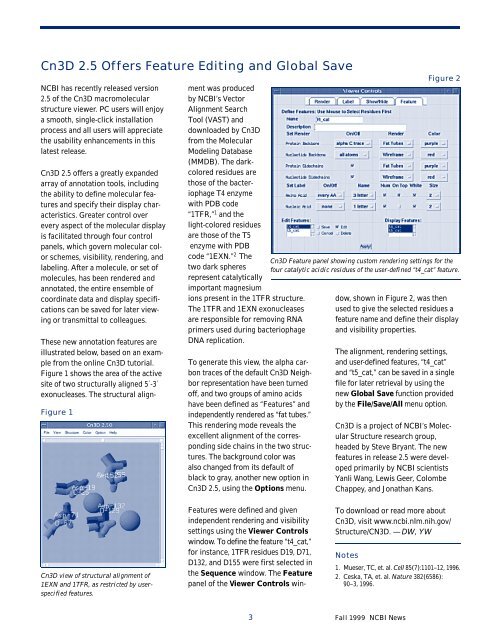
Cn3D 2.5 Offers Feature Editing and Global Save<br />
<strong>NCBI</strong> has recently released version<br />
2.5 of the Cn3D macromolecular<br />
structure viewer. PC users will enjoy<br />
a smooth, single-click installation<br />
process and all users will appreciate<br />
the usability enhancements in this<br />
latest release.<br />
Cn3D 2.5 offers a greatly expanded<br />
array of annotation tools, including<br />
the ability to define molecular features<br />
and specify their display characteristics.<br />
Greater control over<br />
every aspect of the molecular display<br />
is facilitated through four control<br />
panels, which govern molecular color<br />
schemes, visibility, rendering, and<br />
labeling. After a molecule, or set of<br />
molecules, has been rendered and<br />
annotated, the entire ensemble of<br />
coordinate data and display specifications<br />
can be saved for later viewing<br />
or transmittal to colleagues.<br />
These new annotation features are<br />
illustrated below, based on an example<br />
from the online Cn3D tutorial.<br />
Figure 1 shows the area of the active<br />
site of two structurally aligned 5´-3´<br />
exonucleases. The structural align-<br />
Figure 1<br />
Cn3D view of structural alignment of<br />
1EXN and 1TFR, as restricted by userspecified<br />
features.<br />
ment was produced<br />
by <strong>NCBI</strong>’s Vector<br />
Alignment <strong>Search</strong><br />
Tool (VAST) and<br />
downloaded by Cn3D<br />
from the Molecular<br />
Modeling Database<br />
(MMDB). The darkcolored<br />
residues are<br />
those of the bacteriophage<br />
T4 enzyme<br />
with PDB code<br />
“1TFR,” 1 and the<br />
light-colored residues<br />
are those of the T5<br />
enzyme with PDB<br />
code “1EXN.” 2 The<br />
two dark spheres<br />
represent catalytically<br />
important magnesium<br />
ions present in the 1TFR structure.<br />
The 1TFR and 1EXN exonucleases<br />
are responsible for removing RNA<br />
primers used during bacteriophage<br />
DNA replication.<br />
To generate this view, the alpha carbon<br />
traces of the default Cn3D Neighbor<br />
representation have been turned<br />
off, and two groups of aminoacids<br />
have been defined as “Features”and<br />
independently rendered as “fat tubes.”<br />
This rendering mode reveals the<br />
excellent alignment of the corresponding<br />
side chains in the two structures.<br />
The background color was<br />
also changed from its default of<br />
black to gray, another new option in<br />
Cn3D 2.5, using the Options menu.<br />
Features were defined and given<br />
independent rendering and visibility<br />
settings using the Viewer Controls<br />
window. To define the feature “t4_cat,”<br />
for instance, 1TFR residuesD19, D71,<br />
D132, and D155 were first selected in<br />
the Sequence window. The Feature<br />
panel of the Viewer Controls win-<br />
Cn3D Feature panel showing custom rendering settings for the<br />
four catalytic acidic residues of the user-defined “t4_cat” feature.<br />
dow, shown in Figure 2, was then<br />
used to give the selected residues a<br />
feature name and define their display<br />
and visibility properties.<br />
The alignment, rendering settings,<br />
and user-defined features, “t4_cat”<br />
and “t5_cat,” can be saved in a single<br />
filefor later retrieval by usingthe<br />
new Global Savefunction provided<br />
by the File/Save/Allmenu option.<br />
Cn3D is a project of <strong>NCBI</strong>’s Molecular<br />
Structure research group,<br />
headed by Steve Bryant. The new<br />
features in release 2.5 were developed<br />
primarily by <strong>NCBI</strong> scientists<br />
Yanli Wang, Lewis Geer, Colombe<br />
Chappey, and Jonathan Kans.<br />
To download or read more about<br />
Cn3D, visit www.ncbi.nlm.nih.gov/<br />
Structure/CN3D. — DW, YW<br />
Notes<br />
1. Mueser, TC, et. al. Cell85(7):1101–12, 1996.<br />
2. Ceska, TA, et. al. Nature382(6586):<br />
90–3, 1996.<br />
3 Fall1999 <strong>NCBI</strong> News<br />
Figure 2