Guidelines and Standards for Performance of a Pediatric ...
Guidelines and Standards for Performance of a Pediatric ...
Guidelines and Standards for Performance of a Pediatric ...

You also want an ePaper? Increase the reach of your titles
YUMPU automatically turns print PDFs into web optimized ePapers that Google loves.
Journal <strong>of</strong> the American Society <strong>of</strong> Echocardiography<br />
1420 Lai et al December 2006<br />
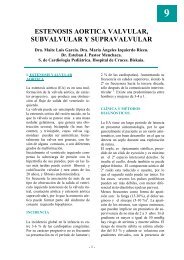
Figure 2 Subxiphoid short-axis views. 1, At rightward aspect <strong>of</strong> heart, superior vena cava (SVC) <strong>and</strong><br />
inferior vena cava enter right atrium (RA). Right pulmonary artery (RPA) is seen in cross section behind<br />
SVC <strong>and</strong> above left atrium (LA). 2, With leftward angulation, base <strong>of</strong> left ventricle (LV) <strong>and</strong> right ventricle<br />
(RV) <strong>and</strong> atrioventricular valves are seen. Aortic (Ao) valve is seen in cross section at this level. 3, Further<br />
leftward angulation reveals cross-sectional view <strong>of</strong> LV <strong>and</strong> mitral valve (MV), <strong>and</strong> RV outflow tract <strong>and</strong><br />
pulmonary valve (PV). 4, Sweep then passes through midmuscular septum <strong>and</strong> LV papillary muscles<br />
toward apical portions <strong>of</strong> both ventricles. (Reproduced with permission from: Geva T. Echocardiography<br />
<strong>and</strong> Doppler ultrasound. In: Garson A, Bricker JT, Fisher DJ, Neish SR, editors. The science <strong>and</strong> practice<br />
<strong>of</strong> pediatric cardiology. Baltimore: Williams <strong>and</strong> Wilkins; 1997, p. 789-843).<br />
5) is positioned over the LVOT, allowing visualization<br />
<strong>of</strong> the aortic <strong>and</strong> mitral valves. The long-axis<br />
sweep includes the RV inflow <strong>and</strong> outflow. The<br />
long-axis or modified parasagittal view <strong>of</strong> the aorta<br />
best demonstrates the position <strong>of</strong> the right coronary<br />
artery ostium relative to the sinotubular junction.<br />
The parasternal short-axis view (Figure 6) at the base<br />
<strong>of</strong> the heart can provide detailed imaging <strong>of</strong> aortic<br />
valve morphology <strong>and</strong> views <strong>of</strong> the RV infundibulum,<br />
pulmonary valve, <strong>and</strong> main/proximal branch<br />
pulmonary arteries. The short-axis view also allows<br />
<strong>for</strong> visualization <strong>of</strong> the coronary ostia, <strong>and</strong> imaging<br />
<strong>and</strong> color flow mapping <strong>of</strong> the proximal coronary<br />
arteries. Clockwise rotation <strong>of</strong> the transducer into a<br />
transverse view elongates the left coronary artery<br />
<strong>and</strong> <strong>of</strong>ten demonstrates its bifurcation. The morphology<br />
<strong>of</strong> the mitral leaflets <strong>and</strong> valve apparatus is<br />
<strong>of</strong>ten best seen on a parasternal short-axis view. In<br />
larger hearts, color flow mapping <strong>of</strong> the ventricular<br />
septum may require separate short-axis sweeps <strong>of</strong><br />
the anterior <strong>and</strong> posterior portions <strong>of</strong> the septum.<br />
Examination <strong>of</strong> the apical muscular septum may<br />
require repositioning <strong>of</strong> the transducer toward the<br />
apex during the short-axis sweep.<br />
A ductal view is obtained from imaging in a<br />
parasagittal plane from a high left parasternal window.<br />
This window lines up the ultrasound beam<br />
with the main pulmonary artery-ductus continuum<br />
<strong>and</strong> allows <strong>for</strong> exclusion <strong>of</strong> a normally located small<br />
patent ductus arteriosus. The distal aortic arch <strong>and</strong><br />
superior thoracic aorta may also be visualized<br />
through the heart <strong>and</strong> the main pulmonary artery,<br />
<strong>and</strong>, in larger individuals, the aortic isthmus is<br />
sometimes best seen from this position. Clockwise<br />
rotation <strong>of</strong> the transducer to a high left parasternal<br />
transverse plane may better demonstrate the left<br />
pulmonary veins in some larger individuals.<br />
Suprasternal notch views. Imaging from the suprasternal<br />
notch 38-40 includes positioning <strong>of</strong> the<br />
transducer in the right supraclavicular region <strong>and</strong><br />
occasionally in the left supraclavicular region. In<br />
the coronal, or short-axis, plane (Figure 7), the