Materials - Sumisan SA
Materials - Sumisan SA
Materials - Sumisan SA

You also want an ePaper? Increase the reach of your titles
YUMPU automatically turns print PDFs into web optimized ePapers that Google loves.
308 A. WEILER ET AL.<br />
TABLE 1. Phases of Degradation of Amorphous Biodegradable Implants and Tissue Reactions According to Pistner et al. 47<br />
Phase Tissue Reaction<br />
1. Healing phase Unchanged implant, development of a fibrous capsule with a high amount of fibroblasts<br />
2. Latency phase Unchanged implant, fibrous capsule gets thinner with less cells and more fibers or direct implant contact to bone<br />
3. Protracted resorptive Mainly central degradation of the implant, development of cracks, mild to moderate cellular response with inva-<br />
phase<br />
sion of macrophages and foreign-body giant cells<br />
4. Progressive resorptive<br />
phase<br />
Progressive disintegration of the implant with a severe tissue response (macrophages, foreign-body giant cells)<br />
5. Recovery phase No polymer remnants detectable, development of scar tissue or osseous replacement of the former implant site<br />
OSSEOUS REPLACEMENT<br />
A major intent of biodegradable implants is complete<br />
tissue replacement at the former implant site.<br />
Although an early replacement with fibrous granulation<br />
tissue takes place during degradation, 46,48-53 less is<br />
known about the long-term fate of the former implant<br />
site and its osseous replacement. Although a complete<br />
osseous replacement has been anticipated for all<br />
biodegradable implants, it has not yet been shown<br />
either experimentally or clinically in most cases. To<br />
facilitate uncompromised revision surgery, a complete<br />
osseous replacement should occur within a 2- to 3-year<br />
time frame to allow for a second interference fit or tack<br />
fixation as, for example, in cruciate ligament and<br />
shoulder revision surgery.<br />
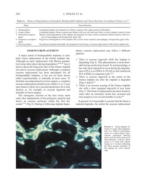
The osteogenic reaction of the host tissue starts<br />
early after implantation of the polymeric material and<br />
shows an osseous enclosure within the first few<br />
weeks 51,53 (Fig 3). During or following implant degra-<br />
dation, osseous replacement may follow 3 different<br />
patterns:<br />
1. There is osseous ingrowth while the implant is<br />
degrading (Fig 4). This phenomenon is most desirable<br />
but has rarely been found. To our knowledge, it<br />
has only been reported to occur during the degradation<br />
of PLLA-co-PDLLA (70:30) or self-reinforced<br />
PLLA/PDLLA composite rods. 50,51<br />
2. There is osseous ingrowth in the center of the<br />
former implant site after the implant is degraded<br />
(Figs 5 and 6). 46<br />
3. There is an osseous scaring of the former implant<br />
site with a slow marginal ingrowth of new bone<br />
(Fig 7). This kind of replacement has been found in<br />
cases after an osteolytic lesion has occurred and<br />
may progress over several months or years. 46<br />
In general, it is reasonable to assume that the faster a<br />
material degrades, the earlier the osseous replacement<br />
FIGURE 3. Tissue-implant interface<br />
6 weeks after implantation<br />
of a PDLLA interference<br />
screw in a sheep femur. Polychrome<br />
sequential labeling<br />
shows activity of the early given<br />
fluorochromes (calcein green<br />
given at 4 weeks and xylenol<br />
orange at 6 weeks) indicating<br />
the early osseous enclosure of<br />
the implant (S, screw threading).