This thesis is dedicated to my grandparents
This thesis is dedicated to my grandparents
This thesis is dedicated to my grandparents

Create successful ePaper yourself
Turn your PDF publications into a flip-book with our unique Google optimized e-Paper software.
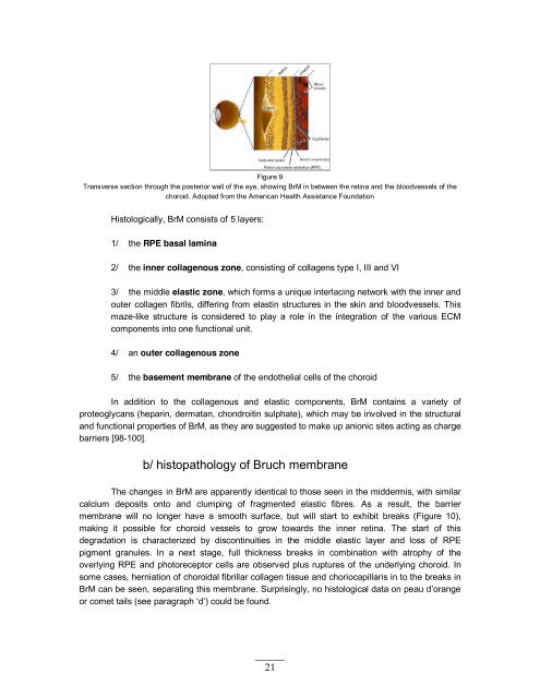
Figure 9<br />
Transverse section through the posterior wall of the eye, showing BrM in between the retina and the bloodvessels of the<br />
choroid. Adopted from the American Health Ass<strong>is</strong>tance Foundation<br />
H<strong>is</strong><strong>to</strong>logically, BrM cons<strong>is</strong>ts of 5 layers:<br />
1/ the RPE basal lamina<br />
2/ the inner collagenous zone, cons<strong>is</strong>ting of collagens type I, III and VI<br />
3/ the middle elastic zone, which forms a unique interlacing network with the inner and<br />
outer collagen fibrils, differing from elastin structures in the skin and bloodvessels. <strong>Th<strong>is</strong></strong><br />
maze-like structure <strong>is</strong> considered <strong>to</strong> play a role in the integration of the various ECM<br />
components in<strong>to</strong> one functional unit.<br />
4/ an outer collagenous zone<br />
5/ the basement membrane of the endothelial cells of the choroid<br />
In addition <strong>to</strong> the collagenous and elastic components, BrM contains a variety of<br />
proteoglycans (heparin, dermatan, chondroitin sulphate), which may be involved in the structural<br />
and functional properties of BrM, as they are suggested <strong>to</strong> make up anionic sites acting as charge<br />
barriers [98-100].<br />
b/ h<strong>is</strong><strong>to</strong>pathology of Bruch membrane<br />
The changes in BrM are apparently identical <strong>to</strong> those seen in the midderm<strong>is</strong>, with similar<br />
calcium deposits on<strong>to</strong> and clumping of fragmented elastic fibres. As a result, the barrier<br />
membrane will no longer have a smooth surface, but will start <strong>to</strong> exhibit breaks (Figure 10),<br />
making it possible for choroid vessels <strong>to</strong> grow <strong>to</strong>wards the inner retina. The start of th<strong>is</strong><br />
degradation <strong>is</strong> characterized by d<strong>is</strong>continuities in the middle elastic layer and loss of RPE<br />
pigment granules. In a next stage, full thickness breaks in combination with atrophy of the<br />
overlying RPE and pho<strong>to</strong>recep<strong>to</strong>r cells are observed plus ruptures of the underlying choroid. In<br />
some cases, herniation of choroidal fibrillar collagen t<strong>is</strong>sue and choriocapillar<strong>is</strong> in <strong>to</strong> the breaks in<br />
BrM can be seen, separating th<strong>is</strong> membrane. Surpr<strong>is</strong>ingly, no h<strong>is</strong><strong>to</strong>logical data on peau d’orange<br />
or comet tails (see paragraph ‘d’) could be found.<br />
21