Neutron Scattering - JUWEL - Forschungszentrum Jülich
Neutron Scattering - JUWEL - Forschungszentrum Jülich
Neutron Scattering - JUWEL - Forschungszentrum Jülich

Create successful ePaper yourself
Turn your PDF publications into a flip-book with our unique Google optimized e-Paper software.
18 M. Meven<br />
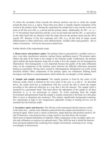
To direct the secondary beam towards the detector position one has to orient the sample<br />
around the three axes �, � and �. These three axes allow a virtually random orientation of the<br />
crystal in the primary beam. During the experiment the sample has to stay exactly in the cross<br />
point of all four axes (2�, ���� and �) and the primary beam. Additionally, for 2� = ����� =<br />
� = 0° the primary beam direction and the � axis on one hand side and the 2�-, �- and �-axes<br />
on the other hand side are identical while the angle between the primary beam and the 2�-is<br />
exactly 90°. Because of the four rotational axes (2�, ����, �) this kind of single crystal<br />
diffractometer is often called four circle diffractometer. Another often used geometry - the so<br />
called �-Geometrie - will not be discussed in detail here.<br />
Further details of the experimental setup:<br />
1. Beam source and primary optics: The primary beam is generated by a suitable source (xrays:<br />
x-ray tube, synchrotron; neutrons: nuclear fission, spallation source). The primary optics<br />
defines the path of the beam to the sample in the Eulerian cradle. Furthermore, the primary<br />
optics defines the beam diameter using slits to make it fit to the sample size for homogeneous<br />
illumination. This homogeneity is very important because the quality of the data refinement<br />
relies on the comparison of the intensity ratios between the different reflections measured<br />
during an experiment. Wrong ratios caused by inhomogeneous illumination can yield wrong<br />
structural details! Other components of the primary optics are collimators defining beam<br />
divergence and filters or monochromators which define the wavelength � of the radiation.<br />
2. Sample and sample environment: The sample position is fixed by the centre of the<br />
Eulerian cradle which is defined by the cross point of the axes ���� and �. As described<br />
above, the cradle itself has in combination with the �-circle the task to orient the sample<br />
according to the observed reflection in a way that it hits the detector. The sample itself is<br />
mounted on a goniometer head. This head allows the adjustment of the sample in all three<br />
directions x; y; z, via microscope or camera. To avoid scattering from the sample<br />
environment and goniometer head the sample is usually connected to the head via a thin glass<br />
fibre (x-rays) or aluminum pin (neutrons). This reduces significantly background scattering.<br />
For experiments at high or low temperatures adjustable cooling or heating devices can be<br />
mounted into the Eulerian cradle.<br />
3. Secondary optics and detector: The 2� arm of the instrument hold the detector which –<br />
in the ideal case – catches only radiation scattered from the sample and transforms it to an<br />
electrical signal. There exists a variety of detectors, single detectors and position sensitive 1D<br />
and 2D detectors. Area detectors have a large sensitive area that allows the accurate<br />
observation of spatial distribution of radiation. Other components of the secondary optics are<br />
slits and collimators or analyser (as optional units). They fulfil the task to shield the detector<br />
from unwanted radiation like scattering from sample environment, scattering in air, wrong<br />
wavelengths or flourescence