Editorial Cover Story ESOP/NZW 2011 Congress Report Oncology ...
Editorial Cover Story ESOP/NZW 2011 Congress Report Oncology ...
Editorial Cover Story ESOP/NZW 2011 Congress Report Oncology ...
You also want an ePaper? Increase the reach of your titles
YUMPU automatically turns print PDFs into web optimized ePapers that Google loves.
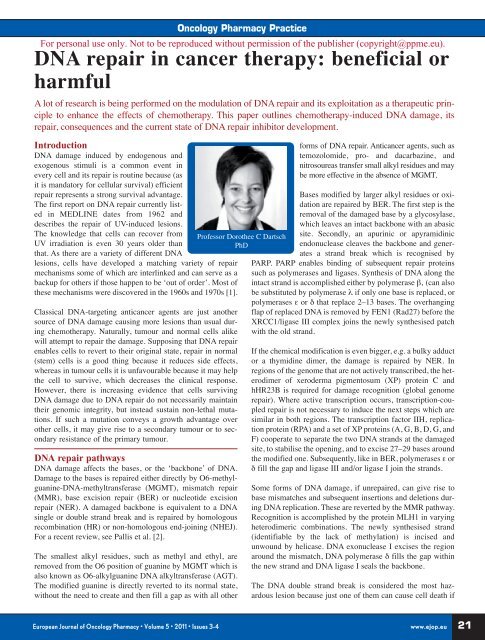
Introduction<br />
DNA damage induced by endogenous and<br />
exogenous stimuli is a common event in<br />
every cell and its repair is routine because (as<br />
it is mandatory for cellular survival) efficient<br />
repair represents a strong survival advantage.<br />
The first report on DNA repair currently listed<br />
in MEDLINE dates from 1962 and<br />
describes the repair of UV-induced lesions.<br />
The knowledge that cells can recover from<br />
UV irradiation is even 30 years older than<br />
that. As there are a variety of different DNA<br />
lesions, cells have developed a matching variety of repair<br />
mechanisms some of which are interlinked and can serve as a<br />
backup for others if those happen to be ‘out of order’. Most of<br />
these mechanisms were discovered in the 1960s and 1970s [1].<br />
Classical DNA-targeting anticancer agents are just another<br />
source of DNA damage causing more lesions than usual during<br />
chemotherapy. Naturally, tumour and normal cells alike<br />
will attempt to repair the damage. Supposing that DNA repair<br />
enables cells to revert to their original state, repair in normal<br />
(stem) cells is a good thing because it reduces side effects,<br />
whereas in tumour cells it is unfavourable because it may help<br />
the cell to survive, which decreases the clinical response.<br />
However, there is increasing evidence that cells surviving<br />
DNA damage due to DNA repair do not necessarily maintain<br />
their genomic integrity, but instead sustain non-lethal mutations.<br />
If such a mutation conveys a growth advantage over<br />
other cells, it may give rise to a secondary tumour or to secondary<br />
resistance of the primary tumour.<br />
DNA repair pathways<br />
DNA damage affects the bases, or the ‘backbone’ of DNA.<br />
Damage to the bases is repaired either directly by O6-methylguanine-DNA-methyltransferase<br />
(MGMT), mismatch repair<br />
(MMR), base excision repair (BER) or nucleotide excision<br />
repair (NER). A damaged backbone is equivalent to a DNA<br />
single or double strand break and is repaired by homologous<br />
recombination (HR) or non-homologous end-joining (NHEJ).<br />
For a recent review, see Pallis et al. [2].<br />
The smallest alkyl residues, such as methyl and ethyl, are<br />
removed from the O6 position of guanine by MGMT which is<br />
also known as O6-alkylguanine DNA alkyltransferase (AGT).<br />
The modified guanine is directly reverted to its normal state,<br />
without the need to create and then fill a gap as with all other<br />
<strong>Oncology</strong> Pharmacy Practice<br />
For personal use only. Not to be reproduced without permission of the publisher (copyright@ppme.eu).<br />
DNA repair in cancer therapy: beneficial or<br />
harmful<br />
A lot of research is being performed on the modulation of DNA repair and its exploitation as a therapeutic principle<br />
to enhance the effects of chemotherapy. This paper outlines chemotherapy-induced DNA damage, its<br />
repair, consequences and the current state of DNA repair inhibitor development.<br />
Professor Dorothee C Dartsch<br />
PhD<br />
forms of DNA repair. Anticancer agents, such as<br />
temozolomide, pro- and dacarbazine, and<br />
nitrosoureas transfer small alkyl residues and may<br />
be more effective in the absence of MGMT.<br />
Bases modified by larger alkyl residues or oxidation<br />
are repaired by BER. The first step is the<br />
removal of the damaged base by a glycosylase,<br />
which leaves an intact backbone with an abasic<br />
site. Secondly, an apurinic or apyramidinic<br />
endonuclease cleaves the backbone and generates<br />
a strand break which is recognised by<br />
PARP. PARP enables binding of subsequent repair proteins<br />
such as polymerases and ligases. Synthesis of DNA along the<br />
intact strand is accomplished either by polymerase β‚ (can also<br />
be substituted by polymerase λ if only one base is replaced, or<br />
polymerases ε or δ that replace 2–13 bases. The overhanging<br />
flap of replaced DNA is removed by FEN1 (Rad27) before the<br />
XRCC1/ligase III complex joins the newly synthesised patch<br />
with the old strand.<br />
If the chemical modification is even bigger, e.g. a bulky adduct<br />
or a thymidine dimer, the damage is repaired by NER. In<br />
regions of the genome that are not actively transcribed, the heterodimer<br />
of xeroderma pigmentosum (XP) protein C and<br />
hHR23B is required for damage recognition (global genome<br />
repair). Where active transcription occurs, transcription-coupled<br />
repair is not necessary to induce the next steps which are<br />
similar in both regions. The transcription factor IIH, replication<br />
protein (RPA) and a set of XP proteins (A, G, B, D, G, and<br />
F) cooperate to separate the two DNA strands at the damaged<br />
site, to stabilise the opening, and to excise 27–29 bases around<br />
the modified one. Subsequently, like in BER, polymerases ε or<br />
δ fill the gap and ligase III and/or ligase I join the strands.<br />
Some forms of DNA damage, if unrepaired, can give rise to<br />
base mismatches and subsequent insertions and deletions during<br />
DNA replication. These are reverted by the MMR pathway.<br />
Recognition is accomplished by the protein MLH1 in varying<br />
heterodimeric combinations. The newly synthesised strand<br />
(identifiable by the lack of methylation) is incised and<br />
unwound by helicase. DNA exonuclease I excises the region<br />
around the mismatch, DNA polymerase δ fills the gap within<br />
the new strand and DNA ligase I seals the backbone.<br />
The DNA double strand break is considered the most hazardous<br />
lesion because just one of them can cause cell death if<br />
European Journal of <strong>Oncology</strong> Pharmacy • Volume 5 • <strong>2011</strong> • Issues 3-4 www.ejop.eu<br />
21


