Urinary Bladder
Urinary Bladder
Urinary Bladder

Create successful ePaper yourself
Turn your PDF publications into a flip-book with our unique Google optimized e-Paper software.
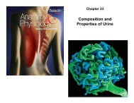
Chapter 23<br />
The Ureters and <strong>Urinary</strong><br />
<strong>Bladder</strong>
Urine Storage and Elimination<br />
• urine is produced continually<br />
• does not drain continually from the body<br />
• urination is episodic – occurring when we allow it<br />
• made possible by storage apparatus<br />
• and neural controls of this timely release<br />
23-2
The Ureter<br />
• ureters – retroperitoneal, muscular tube that extends from<br />
the kidney to the urinary bladder<br />
– about 25 cm long<br />
– passes posterior to bladder and enters it from below<br />
– flap of mucosa acts as a valve into bladder<br />
• keeps urine from backing up in the ureter when bladder contracts<br />
– 3 layers of ureter<br />
• adventitia – connective tissue layer that connects ureter to<br />
surrounding structures<br />
• muscularis - 2 layers of smooth muscle with 3 rd layer in lower<br />
ureter<br />
– urine enters, it stretches and contracts in peristaltic wave<br />
• mucosa - transitional epithelium<br />
– begins at minor calyces and extends through the bladder<br />
– lumen very narrow, easily obstructed kidney stones<br />
23-3
<strong>Urinary</strong> <strong>Bladder</strong><br />
• urinary bladder - muscular sac located on floor of pelvic<br />
cavity<br />
– inferior to peritoneum and posterior to pubic symphysis<br />
• 3 layers<br />
– parietal peritoneum, superiorly, fibrous adventitia other areas<br />
– muscularis - detrusor muscle - 3 layers of smooth muscle<br />
– mucosa - transitional epithelium<br />
• rugae - conspicuous wrinkles in relaxed bladder<br />
• trigone – smooth-surfaced triangular area marked with<br />
openings of ureters and urethra<br />
• capacity - mod. full is 500 ml, max. is 700 - 800 ml<br />
– highly distensible<br />
– as it fills, it expands superiorly<br />
– rugae flatten<br />
– epithelium thins from five or six layers to two or three<br />
23-4
Ureter<br />
Detrusor<br />
muscle<br />
Ureteral<br />
openings<br />
Trigone<br />
Urethra<br />
Urogenital<br />
diaphragm<br />
External urethral<br />
orifice<br />
<strong>Urinary</strong> <strong>Bladder</strong><br />
Copyright © The McGraw-Hill Companies, Inc. Permission required for reproduction or display.<br />
(a) Female<br />
Internal urethral<br />
sphincter<br />
External urethral<br />
sphincter<br />
Figure 23.23a<br />
23-5
Kidney Stones<br />
• renal calculus (kidney stone) - hard granule of calcium phosphate,<br />
calcium oxalate, uric acid, or a magnesium salt called struvite<br />
• form in the renal pelvis<br />
• usually small enough to pass unnoticed in the urine flow<br />
– large stones might block renal pelvis or ureter and can cause pressure<br />
build up in kidney which destroys nephrons<br />
• passage of large jagged stones is excruciatingly painful and may damage<br />
ureter causing hematuria<br />
• causes include hypercalcemia, dehydration, pH imbalances, frequent<br />
urinary tract infections, or enlarged prostate gland causing urine<br />
retention<br />
• treatment includes stone dissolving drugs, often surgery, or<br />
lithotripsy –nonsurgical technique that pulverizes stones with<br />
ultrasound<br />
23-6
Copyright © The McGraw-Hill Companies, Inc. Permission required for reproduction or display.<br />
Ureter<br />
Detrusor<br />
muscle<br />
Ureteral<br />
openings<br />
Trigone<br />
Urethra<br />
Urogenital<br />
diaphragm<br />
External urethral<br />
orifice<br />
(a) Female<br />
Figure 23.23a<br />
Female Urethra<br />
Internal urethral<br />
sphincter<br />
External urethral<br />
sphincter<br />
• 3 to 4 cm long<br />
• bound to anterior wall of vagina<br />
• external urethral orifice<br />
– between vaginal orifice and<br />
clitoris<br />
• internal urethral sphincter<br />
– detrusor muscle thickening<br />
– smooth muscle under<br />
involuntary control<br />
• external urethral sphincter<br />
– where the urethra passes<br />
through the pelvic floor<br />
– skeletal muscle under<br />
voluntary control<br />
23-7
Copyright © The McGraw-Hill Companies, Inc. Permission required for reproduction or display.<br />
Ureter<br />
Rugae<br />
Ureteral<br />
openings<br />
Trigone<br />
Prostate gland<br />
Prostatic urethra<br />
Membranous<br />
urethra<br />
Bulbourethral<br />
gland<br />
Spongy (penile)<br />
urethra<br />
Penis<br />
External urethral orifice<br />
(b) Male<br />
Male Urethra<br />
Detrusor<br />
muscle<br />
Internal urethral<br />
sphincter<br />
Urogenital<br />
diaphragm<br />
External urethral<br />
sphincter<br />
Figure 23.23b<br />
• 18 cm long<br />
• 3 regions of male urethra<br />
– prostatic urethra (2.5 cm)<br />
• passes through prostate gland<br />
– membranous urethra (.5 cm)<br />
• passes through muscular floor of<br />
pelvic cavity<br />
– spongy (penile) urethra (15 cm)<br />
• passes through penis in corpus<br />
spongiosum<br />
• internal urethral sphincter<br />
– detrusor muscle thickening<br />
• external urethral sphincter<br />
– part of skeletal muscle of pelvic<br />
floor<br />
23-8
<strong>Urinary</strong> Tract Infection (UTI)<br />
• cystitis – infection of the urinary bladder<br />
– especially common in females due to short<br />
urethra<br />
– frequently triggered by sexual intercourse<br />
– can spread up the ureter causing pyelitis<br />
• pyelitis – infection of the renal pelvis<br />
• pyelonephritis – infection that reaches the<br />
cortex and the nephrons<br />
– can result from blood-borne bacteria<br />
23-9
Voiding Urine<br />
• between acts of urination, the bladder is filling<br />
– detrusor muscle relaxes<br />
– urethral sphincters are tightly closed<br />
• accomplished by sympathetic pathway from upper lumbar spinal<br />
cord<br />
• postganglionic fibers travel through the hypogastric nerve to the<br />
detrusor muscle (relax) and internal urethral sphincter (excite)<br />
– somatic motor fibers from upper sacral spinal cord through<br />
pudendal nerve to supply the external sphincter give us<br />
voluntary control<br />
• micturition – the act of urinating<br />
• micturition reflex - spinal reflex that partly controls<br />
urination<br />
23-10
Voiding Urine – Micturition Reflex<br />
• involuntary control (steps 1 – 4)<br />
– filling of the bladder to about 200 mL excites stretch receptors<br />
in the bladder wall<br />
– send sensory signals through fibers in pelvic nerve to sacral<br />
spinal cord (S2 or S3)<br />
– motor signals travel back from the spinal cord to the bladder<br />
by way of motor fibers in pelvic nerve and parasympathetic<br />
ganglion in bladder wall<br />
– excites detrusor muscle and relaxes internal urethral sphincter<br />
– results in emptying bladder<br />
– if there was no voluntary control over urination, this reflex<br />
would be the only means of control<br />
23-11
Voiding Urine – Micturition Reflex<br />
• voluntary control (steps 5 – 8)<br />
– micturition center - nucleus in the pons that receives some input from<br />
bladder stretch receptors that ascends the spinal cord<br />
– nucleus integrates information about bladder tension with information from<br />
other brain centers<br />
• urination can be prompted by fear<br />
• inhibited by knowledge that the circumstances are inappropriate for urination<br />
– fibers from micturition center descend the spinal cord<br />
• through reticulospinal tracts<br />
– some fibers inhibit sympathetic fibers than normally keep internal urethral<br />
sphincter contracted<br />
– others descend farther to sacral spinal cord<br />
• excite parasympathetic neurons that stimulate the detrusor to contract and relax the internal<br />
urethral sphincter<br />
– initial detrusor contraction raises pressure in bladder, stimulate stretch<br />
receptors, bringing about more forceful contraction<br />
– external urethral sphincter receives nerve fibers from cerebral cortex by<br />
way of corticospinal tract<br />
• inhibit somatic motor neurons that normally keep that sphincter constricted<br />
23-12
Voiding Urine – Micturition Reflex<br />
• urge to urinate usually arises at an inconvenient time<br />
– one must suppress it<br />
– stretch receptors fatigue and stop firing<br />
• as bladder tension increases<br />
– signals return with increasing frequency and persistence<br />
• there are times when the bladder is not full enough to trigger<br />
the micturition reflex but one wishes to ‘go’ anyway<br />
– Valsalva maneuver used to compress bladder<br />
– excites stretch receptors early getting the reflex started<br />
23-13
Stretch receptors<br />
Motor fibers to<br />
detrusor muscle<br />
Neural Control of Micturition<br />
Full<br />
urinary bladder<br />
Internal urethral<br />
sphincter (involuntary)<br />
External urethral<br />
sphincter (voluntary)<br />
Copyright © The McGraw-Hill Companies, Inc. Permission required for reproduction or display.<br />
Sensory<br />
fiber<br />
4<br />
Urethra<br />
8<br />
Pelvic nerve<br />
1<br />
3<br />
Motor<br />
fiber<br />
To pons<br />
2<br />
Parasympathetic<br />
ganglion in<br />
bladder wall<br />
Somatic motor fiber<br />
of pudendal nerve<br />
5 6 7<br />
Figure 23.24<br />
From pons<br />
Sacral segments<br />
of spinal cord<br />
S2<br />
S3<br />
S4<br />
Involuntary micturition reflex<br />
1<br />
2<br />
3<br />
4<br />
5<br />
6<br />
7<br />
8<br />
Stretch receptors detect filling<br />
of bladder, transmit afferent<br />
signals to spinal cord.<br />
Signals return to bladder from<br />
spinal cord segments S2 and S3<br />
via parasympathetic fibers in<br />
pelvic nerve.<br />
Efferent signals excite<br />
detrusor muscle.<br />
Efferent signals relax internal<br />
urethral sphincter. Urine is<br />
involuntarily voided if not<br />
inhibited by brain.<br />
Voluntary control<br />
For voluntary control, micturition<br />
center in pons receives signals<br />
from stretch receptors.<br />
If it is timely to urinate,<br />
pons returns signals to<br />
spinal interneurons that<br />
excite detrusor and relax<br />
internal urethral sphincter.<br />
Urine is voided.<br />
If it is untimely to urinate,<br />
signals from pons excite<br />
spinal interneurons that<br />
keep external urethral<br />
sphincter contracted. Urine<br />
is retained in bladder.<br />
If it is timely to urinate, signals<br />
from pons cease and external<br />
urethral sphincter relaxes. Urine<br />
is voided.<br />
23-14
Artery<br />
Vein<br />
Shunt<br />
Hemodialysis<br />
Copyright © The McGraw-Hill Companies, Inc. Permission required for reproduction or display.<br />
Figure 23.25<br />
Hank Morgan/Photo Researchers, Inc.<br />
Blood<br />
pump<br />
Bubble<br />
trap<br />
Thermometer<br />
Dialysis<br />
tubing<br />
Cutaway view<br />
of dialysis<br />
chamber<br />
To<br />
drain<br />
Dialysis<br />
fluid<br />
Flow<br />
meter<br />
23-15
Renal Insufficiency & Hemodialysis<br />
• renal insufficiency – a state in which the kidneys cannot maintain<br />
homeostasis due to extensive destruction of their nephrons<br />
• causes of nephron destruction<br />
– hypertension, chronic kidney infections, trauma, prolonged ischemia and<br />
hypoxia, poisoning by heavy metals or solvents, blockage of renal tubules in<br />
transfusion reaction, atherosclerosis, or glomerulonephritis<br />
• nephrons can regenerate and restore kidney function after short-term<br />
injuries<br />
– others nephrons hypertrophy to compensate for lost kidney function<br />
• can survive with one-third of one kidney<br />
• when 75% of nephrons are lost and urine output of 30 mL/hr is insufficient<br />
(normal 50 -60 mL/hr) to maintain homeostasis<br />
– causes azotemia, acidosis, and uremia develops, also anemia<br />
• hemodialysis – procedure for artificially clearing wastes from the blood<br />
– wastes leave bloodstream and enter the dialysis fluid as blood flows through a<br />
semipermeable cellophane tube; also removes excess body water<br />
23-16