Recommendations from Gynaecological (GYN) GEC-ESTRO ...
Recommendations from Gynaecological (GYN) GEC-ESTRO ...
Recommendations from Gynaecological (GYN) GEC-ESTRO ...

Create successful ePaper yourself
Turn your PDF publications into a flip-book with our unique Google optimized e-Paper software.
242<br />
spread as it presents in these examinations forms the frame<br />
for the delineation process.<br />
Gross tumour volume (diagnosis) (GTVD) includes<br />
macroscopic tumour extension at diagnosis as detected by<br />
clinical examination (visualisation and palpation) and as<br />
visualised on MRI: high signal intensity mass(es) at fast spin<br />
echo sequences (FSE) T2 in cervix/corpus, parametria,<br />
vagina, bladder and rectum (Fig. 4a,b).<br />
Gross tumour volume (BT) (GTV B1, GTV B2, GTV B3,.)<br />
for BT includes macroscopic tumour extension at time of<br />
BT as detected by clinical examination and as visualised on<br />
MRI: High signal intensity mass(es) (FSE, T2) in cervix/corpus,<br />
parametria, vagina, bladder and rectum. In patients<br />
treated with upfront BT or with BT alone, GTV B is identical<br />
with GTVD.<br />
High risk CTV for BT (HR CTVB1, HR CTVB2,.)<br />
carrying a high tumour load, includes GTV B1, B2,., always<br />
the whole cervix and the presumed extracervical tumour<br />
extension at time of BT. In limited disease GTVB is<br />
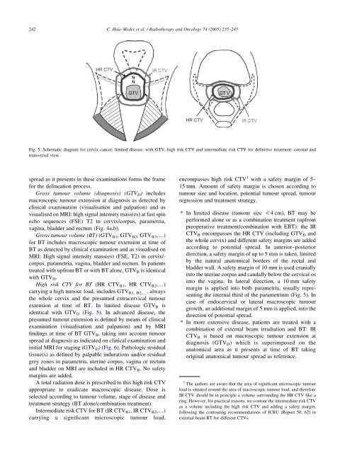
identical with GTVD (Fig. 5). In advanced disease, the<br />
presumed tumour extension is defined by means of clinical<br />
examination (visualisation and palpation) and by MRI<br />
findings at time of BT GTVB, taking into account tumour<br />
spread at diagnosis as indicated on clinical examination and<br />
initial MRI for staging (GTV D)(Fig. 6). Pathologic residual<br />
tissue(s) as defined by palpable indurations and/or residual<br />
grey zones in parametria, uterine corpus, vagina or rectum<br />
and bladder on MRI are included in HR CTVB. No safety<br />
margins are added.<br />
A total radiation dose is prescribed to this high risk CTV<br />
appropriate to eradicate macroscopic disease. Dose is<br />
selected according to tumour volume, stage of disease and<br />
treatment strategy (BT alone/combination treatment).<br />
Intermediate risk CTV for BT (IR CTV B1, IR CTV B2,.)<br />
carrying a significant microscopic tumour load,<br />
C. Haie-Meder et al. / Radiotherapy and Oncology 74 (2005) 235–245<br />
Fig. 5. Schematic diagram for cervix cancer, limited disease, with GTV, high risk CTV and intermediate risk CTV for definitive treatment: coronal and<br />
transversal view.<br />
encompasses high risk CTV 1 with a safety margin of 5–<br />
15 mm. Amount of safety margin is chosen according to<br />
tumour size and location, potential tumour spread, tumour<br />
regression and treatment strategy.<br />
* In limited disease (tumour size !4 cm), BT may be<br />
performed alone or as a combination treatment (upfront<br />
preoperative treatment/combination with EBT): the IR<br />
CTVB encompasses the HR CTV (including GTVD and<br />
the whole cervix) and different safety margins are added<br />
according to potential spread. In anterior–posterior<br />
direction, a safety margin of up to 5 mm is taken, limited<br />
by the natural anatomical borders of the rectal and<br />
bladder wall. A safety margin of 10 mm is used cranially<br />
into the uterine corpus and caudally below the cervical os<br />
into the vagina. In lateral direction, a 10 mm safety<br />
margin is applied into both parametria, usually representing<br />
the internal third of the parametrium (Fig. 5). In<br />
case of endocervical or lateral macroscopic tumour<br />
growth, an additional margin of 5 mm is applied, into the<br />
direction of potential spread.<br />
* In more extensive disease, patients are treated with a<br />
combination of external beam irradiation and BT: IR<br />
CTVB is based on macroscopic tumour extension at<br />
diagnosis (GTV D) which is superimposed on the<br />
anatomical area as it presents at time of BT taking<br />
original anatomical tumour spread as reference.<br />
1 The authors are aware that the area of significant microscopic tumour<br />
load is situated around the area of macroscopic tumour load, and therefore<br />
IR CTV should be in principle a volume surrounding the HR CTV like a<br />
ring. However, for practical reasons, we contour the intermediate risk CTV<br />
as a volume including the high risk CTV and adding a safety margin,<br />
following the contouring recommendations of ICRU (Report 50, 62) in<br />
external beam RT for different CTVs.