Aphanocladium macrosporum sp. nov. from Taiwan - Academia Sinica
Aphanocladium macrosporum sp. nov. from Taiwan - Academia Sinica
Aphanocladium macrosporum sp. nov. from Taiwan - Academia Sinica

Create successful ePaper yourself
Turn your PDF publications into a flip-book with our unique Google optimized e-Paper software.
334 Botanical Bulletin of <strong>Academia</strong> <strong>Sinica</strong>, Vol. 40, 1999<br />
characteristics of this isolate fit the generic concept of<br />
<strong>Aphanocladium</strong>. Phialide, conidial morphology and dimensions<br />
easily distinguish this isolate <strong>from</strong> other known<br />
<strong>sp</strong>ecies in the genus. Thus, a new <strong>sp</strong>ecies,<br />
<strong>Aphanocladium</strong> <strong>macro<strong>sp</strong>orum</strong> J.L. Chen, W.S. Lin and<br />
S.S. Tzean is proposed.<br />
Materials and Methods<br />
Samples were collected <strong>from</strong> rotten bark in Kuohsing,<br />
Nantou County during October, 1996 and incubated in<br />
moist chambers (plastic boxes, 30 × 20 × 12 cm, with<br />
three layers of moistened papers) to encourage fungal<br />
<strong>sp</strong>orulation. Pure culture was established by isolating a<br />
single <strong>sp</strong>ore or <strong>sp</strong>ores with a sterile glass microneedle<br />
on 3% agar. A piece of agar containing isolated <strong>sp</strong>ores<br />
was cut out and transferred to oat meal agar (OMA) slants<br />
or plates under a stereomicroscope. Details of fungal<br />
morphology and conidiogenesis were studied and<br />
recorded. The fungus was illustrated using a drawing tube<br />
and photographed using an Olympus light microscope<br />
(BX50). The taxonomic systems of Barron (1968),<br />
Hughes (1953), Tubaki (1963), Ellis (1971) and Saccardo<br />
(1882-1931) were used for identification. Both live cultures<br />
and dried <strong>sp</strong>ecimens were deposited in the Herbarium<br />
of the Chen-Fungi-Collection (Herb. CFC).<br />
Species Descriptions<br />
<strong>Aphanocladium</strong> <strong>macro<strong>sp</strong>orum</strong> J.L. Chen, W.S. Lin, and<br />
S.S. Tzean <strong>sp</strong>. <strong>nov</strong>. (Figures 1-2)<br />
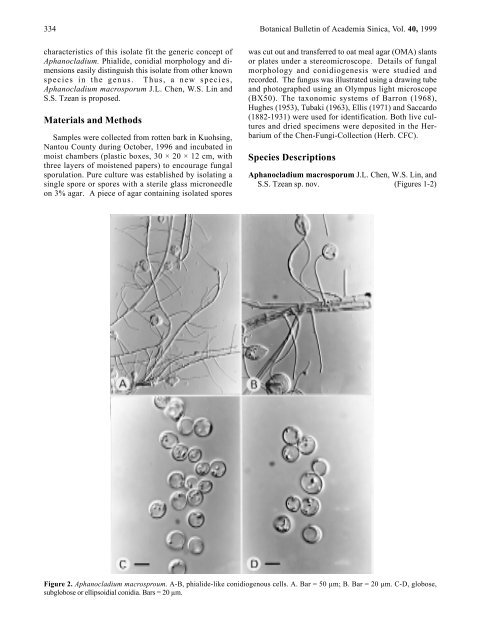
Figure 2. <strong>Aphanocladium</strong> macro<strong>sp</strong>roum. A-B, phialide-like conidiogenous cells. A. Bar = 50 µm; B. Bar = 20 µm. C-D, globose,<br />
subglobose or ellipsoidial conidia. Bars = 20 µm.