Spring 2010 Gustavus Quarterly
Spring 2010 Gustavus Quarterly
Spring 2010 Gustavus Quarterly

You also want an ePaper? Increase the reach of your titles
YUMPU automatically turns print PDFs into web optimized ePapers that Google loves.
Alex Messenger ’10<br />
22<br />
THE GUSTAVUS QUARTERLY<br />
continued from previous page<br />
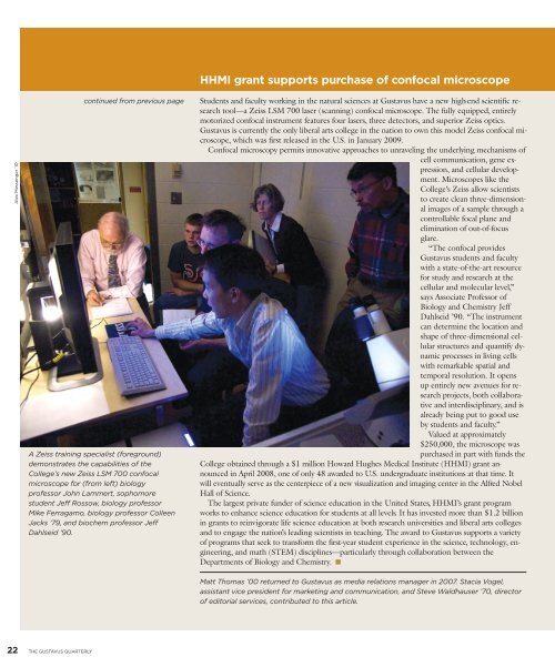
A Zeiss training specialist (foreground)<br />
demonstrates the capabilities of the<br />
College’s new Zeiss LSM 700 confocal<br />
microscope for (from left) biology<br />
professor John Lammert, sophomore<br />
student Jeff Rossow, biology professor<br />
Mike Ferragamo, biology professor Colleen<br />
Jacks ’79, and biochem professor Jeff<br />
Dahlseid ’90.<br />
HHMI grant supports purchase of confocal microscope<br />
Students and faculty working in the natural sciences at <strong>Gustavus</strong> have a new high-end scientific research<br />
tool—a Zeiss LSM 700 laser (scanning) confocal microscope. The fully equipped, entirely<br />
motorized confocal instrument features four lasers, three detectors, and superior Zeiss optics.<br />
<strong>Gustavus</strong> is currently the only liberal arts college in the nation to own this model Zeiss confocal microscope,<br />
which was first released in the U.S. in January 2009.<br />
Confocal microscopy permits innovative approaches to unraveling the underlying mechanisms of<br />
cell communication, gene expression,<br />
and cellular development.<br />
Microscopes like the<br />
College’s Zeiss allow scientists<br />
to create clean three-dimensional<br />
images of a sample through a<br />
controllable focal plane and<br />
elimination of out-of-focus<br />
glare.<br />
“The confocal provides<br />
<strong>Gustavus</strong> students and faculty<br />
with a state-of-the-art resource<br />
for study and research at the<br />
cellular and molecular level,”<br />
says Associate Professor of<br />
Biology and Chemistry Jeff<br />
Dahlseid ’90. “The instrument<br />
can determine the location and<br />
shape of three-dimensional cellular<br />
structures and quantify dynamic<br />
processes in living cells<br />
with remarkable spatial and<br />
temporal resolution. It opens<br />
up entirely new avenues for research<br />
projects, both collaborative<br />
and interdisciplinary, and is<br />
already being put to good use<br />
by students and faculty.”<br />
Valued at approximately<br />
$250,000, the microscope was<br />
purchased in part with funds the<br />
College obtained through a $1 million Howard Hughes Medical Institute (HHMI) grant announced<br />
in April 2008, one of only 48 awarded to U.S. undergraduate institutions at that time. It<br />
will eventually serve as the centerpiece of a new visualization and imaging center in the Alfred Nobel<br />
Hall of Science.<br />
The largest private funder of science education in the United States, HHMI’s grant program<br />
works to enhance science education for students at all levels. It has invested more than $1.2 billion<br />
in grants to reinvigorate life science education at both research universities and liberal arts colleges<br />
and to engage the nation’s leading scientists in teaching. The award to <strong>Gustavus</strong> supports a variety<br />
of programs that seek to transform the first-year student experience in the science, technology, engineering,<br />
and math (STEM) disciplines—particularly through collaboration between the<br />
Departments of Biology and Chemistry. ■<br />
Matt Thomas ’00 returned to <strong>Gustavus</strong> as media relations manager in 2007. Stacia Vogel,<br />
assistant vice president for marketing and communication, and Steve Waldhauser ’70, director<br />
of editorial services, contributed to this article.