Busse Beate Fisslthaler, Rüdiger Popp, U. Ruth ... - Hypertension
Busse Beate Fisslthaler, Rüdiger Popp, U. Ruth ... - Hypertension
Busse Beate Fisslthaler, Rüdiger Popp, U. Ruth ... - Hypertension

Create successful ePaper yourself
Turn your PDF publications into a flip-book with our unique Google optimized e-Paper software.
1430 <strong>Hypertension</strong> December 2001<br />
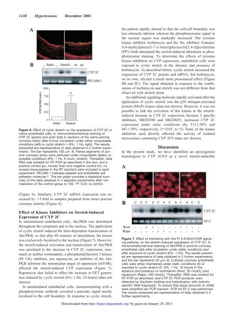
Figure 6. Effect of cyclic stretch on the expression of CYP 2C in<br />
native endothelial cells. A, Immunohistochemical staining of<br />
CYP 2C (green) and actin (red) in sections of the same porcine<br />
coronary artery after 6-hour incubation under either nonpulsatile<br />
conditions (left) or cyclic stretch (�8%, 1 Hz; right). The results<br />
presented are representative of data obtained in 2 further experiments.<br />
The bar represents 100 �m. B, Paired segments of porcine<br />
coronary artery were perfused under nonpulsatile (static) or<br />
pulsatile conditions (8%, 1 Hz, 6 hours, stretch). Thereafter, total<br />
RNA was isolated for RT-PCR as described in the text, and a<br />
positive control (pc, human liver) and negative control (nc, no<br />
reverse transcriptase in the RT reaction) were included in each<br />
experiment. PECAM-1 indicates platelet and endothelial cell<br />
adhesion molecule-1. The bar graph provides a statistical summary<br />
of the data obtained in 4 separate experiments after normalization<br />
of the control group to 100. *P�0.05 vs control.<br />
(Figure 6). Similarly, CYP 2C mRNA expression was increased<br />
by �5-fold in samples prepared from intact porcine<br />
coronary arteries (Figure 6).<br />
Effect of Kinase Inhibitors on Stretch-Induced<br />
Expression of CYP 2C<br />
In unstimulated endothelial cells, Akt/PKB was distributed<br />
throughout the cytoplasm and in the nucleus. The application<br />
of cyclic stretch induced the time-dependent translocation of<br />
Akt/PKB, so that after 60 minutes of stimulation, the kinase<br />
was exclusively localized to the nucleus (Figure 7). However,<br />
the stretch-induced activation and translocation of Akt/PKB<br />
was unrelated to the increase in CYP 2C expression, inasmuch<br />
as neither wortmannin, a phosphatidylinositol 3-kinase<br />
(PI 3-K) inhibitor, nor rapamycin, an inhibitor of the Akt/<br />
PKB substrate the mammalian target of rapamycin (mTOR),<br />
affected the stretch-induced CYP expression (Figure 7).<br />
Rapamycin also failed to affect the increase in EET generation<br />
induced by cyclic stretch (6%, 1 Hz, 18 hours) (data not<br />
shown).<br />
In unstimulated endothelial cells, immunostaining with a<br />
phosphotyrosine antibody revealed a punctate signal mostly<br />
localized to the cell boundary. In response to cyclic stretch,<br />
the pattern rapidly altered so that the cell-cell boundary was<br />
less intensely labeled, whereas the phosphotyrosine signal in<br />
the nuclear region was markedly increased. The tyrosine<br />
kinase inhibitor herbimycin and the Src inhibitor 4-amino-<br />
5(4-methylphenyl)-7-(t-butyl)phyrazolo[3,4-d]pyrimidine<br />
(PP1) both attenuated the stretch-induced alterations in phosphotyrosine<br />
staining. To determine the effects of tyrosine<br />
kinase inhibition on CYP expression, endothelial cells were<br />
exposed to cyclic stretch in the absence and presence of<br />
herbimycin. As described before, cyclic stretch increased the<br />
expression of CYP 2C protein and mRNA, but herbimycin,<br />
on its own, elicited a much more pronounced effect (Figure<br />
8B and 8C). The signal obtained in response to the combination<br />
of herbimycin and stretch was not different from that<br />
observed with stretch alone.<br />
An additional signaling molecule rapidly activated after the<br />
application of cyclic stretch was the p38 mitogen-activated<br />
protein (MAP) kinase (data not shown). However, it was not<br />
possible to link the activation of this kinase to the stretchinduced<br />
increase in CYP 2C expression, because 2 specific<br />
inhibitors, SB203580 and SB220025, increased CYP 2C<br />
expression under static conditions (by 513�20% and<br />
461�30%, respectively; P�0.01; n�5). None of the kinase<br />
inhibitors used directly affected the activity of isolated<br />
microsomes containing CYP 2C9 (data not shown).<br />
Discussion<br />
In the present study, we have identified an epoxygenase<br />
homologous to CYP 2C8/9 as a novel stretch-inducible<br />
Figure 7. Effect of interfering with the PI 3-K/Akt/mTOR signaling<br />
pathway on the stretch-induced expression of CYP 2C. A,<br />
Immunohistochemical staining of Akt/PKB in porcine coronary<br />
endothelial cells after incubation under static conditions and<br />
after exposure to cyclic stretch (6%, 1 Hz). The results presented<br />
are representative of data obtained in 2 further experiments,<br />
and the bar represents 20 �m. B, Cultured coronary endothelial<br />
cells were either maintained under static conditions (S) or<br />
exposed to cyclic stretch (C; 6%, 1 Hz, 18 hours) in the<br />
absence and presence of wortmannin (Wort, 20 nmol/L) and<br />
rapamycin (Rapa, 100 nmol/L). Thereafter, RNA was isolated for<br />
RT-PCR as described, and CYP 2C PCR products were<br />
detected by Southern blotting and hybridization with isoformspecific<br />
DNA fragments. To ensure that equal amounts of cDNA<br />
were amplified per PCR reaction, PCR for EF-2 was performed.<br />
The results presented are representative of data obtained in 2<br />
further experiments.<br />
Downloaded from<br />
http://hyper.ahajournals.org/ by guest on January 29, 2013