Gold Nanoparticles - Department of Chemistry
Gold Nanoparticles - Department of Chemistry
Gold Nanoparticles - Department of Chemistry

Create successful ePaper yourself
Turn your PDF publications into a flip-book with our unique Google optimized e-Paper software.
Nano Letters PERSPECTIVE<br />
<strong>Gold</strong>(III) is rarely used as a primary therapeutic agent as it is a<br />
strong oxidizing agent and thus very reactive. However, gold(I)<br />
can transform into gold(III) within phagolysosomes, which may<br />
account, in part, for some <strong>of</strong> its toxic effects. In brief, gold(I) is<br />
oxidized to gold(III) via a redox system involving myeloperoxidase<br />
and other lysosomal enzymes within phagolysosomes<br />
containing gold (i.e., aurosomes). <strong>Gold</strong>(III) then diffuses away<br />
from its site <strong>of</strong> generation where it can interact with and denature<br />
“self-proteins” surrounding proteins, thereby possibly explaining<br />
why autoimmunity occurs during a few cases <strong>of</strong> gold therapy.<br />
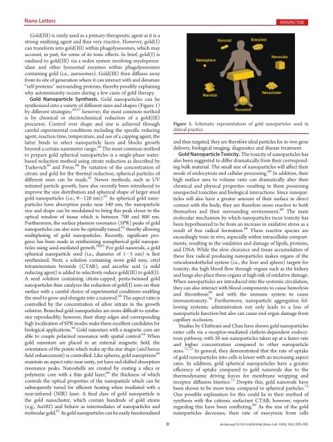
<strong>Gold</strong> Nanoparticle Synthesis. <strong>Gold</strong> nanoparticles can be<br />
synthesized into a variety <strong>of</strong> different sizes and shapes (Figure 1)<br />
by different strategies; 56,57 however, the most common method<br />
is by chemical or electrochemical reduction <strong>of</strong> a gold(III)<br />
precursor. Control over shape and size is achieved through<br />
careful experimental conditions including the specific reducing<br />
agent, reaction time, temperature, and use <strong>of</strong> a capping agent, the<br />
latter binds to select nanoparticle faces and blocks growth<br />
beyond a certain nanometer range. 58 The most common method<br />
to prepare gold spherical nanoparticles is a single-phase waterbased<br />
reduction method using citrate reduction as described by<br />
Turkevich 59 and Frens. 60 By variation <strong>of</strong> the concentration <strong>of</strong><br />
citrate and gold for the thermal reduction, spherical particles <strong>of</strong><br />
different sizes can be made. 61 Newer methods, such as UV<br />
initiated particle growth, have also recently been introduced to<br />
improve the size distribution and spherical shape <strong>of</strong> larger sized<br />
gold nanoparticles (i.e., 9 120 nm). 61 As spherical gold nanoparticles<br />
have absorption peaks near 540 nm, the nanoparticle<br />
size and shape can be modulated to bring this peak closer to the<br />
optical window <strong>of</strong> tissue which is between 700 and 800 nm.<br />
Furthermore, the surface plasmon resonance (SPR) peaks <strong>of</strong> gold<br />
nanoparticles can also now be optimally tuned, 62 thereby allowing<br />
multiplexing <strong>of</strong> gold nanoparticles. Recently, significant progress<br />
has been made in synthesizing nonspherical gold nanoparticles<br />
using seed-mediated growth. 56,63 For gold nanorods, a gold<br />
spherical nanoparticle seed (i.e., diameter <strong>of</strong> 1 5 nm) is first<br />
synthesized. Next, a solution containing more gold ions, cetyl<br />
tetrammonium bromide (CTAB), and ascorbic acid (a mild<br />
reducing agent) is added to selectively reduce gold(III) to gold(I).<br />
A seed solution containing citrate-capped, penta-twinned gold<br />
nanoparticles then catalyzes the reduction <strong>of</strong> gold(I) ions on their<br />
surface with a careful choice <strong>of</strong> experimental conditions enabling<br />
the seed to grow and elongate into a nanorod. 56 The aspect ratio is<br />
controlled by the concentration <strong>of</strong> silver nitrate in the growth<br />
solution. Branched gold nanoparticles are more difficult to synthesize<br />
reproducibly; however, their sharp edges and corresponding<br />
high localization <strong>of</strong> SPR modes make them excellent candidates for<br />
biological applications. 56 <strong>Gold</strong> nanostars with a magnetic core are<br />
able to couple polarized resonance with spatial control. 64 When<br />
gold nanostars are placed in an external magnetic field, the<br />
orientation <strong>of</strong> the points which make up the star shape (and hence<br />
field enhancement) is controlled. Like spheres, gold nanoprisms 65<br />
maintain an aspect ratio near unity, yet have red-shifted absorption<br />
resonance peaks. Nanoshells are created by coating a silica or<br />
polymeric core with a thin gold layer, 66 the thickness <strong>of</strong> which<br />
controls the optical properties <strong>of</strong> the nanoparticle which can be<br />
subsequently tuned for efficient heating when irradiated with a<br />
near-infrared (NIR) laser. A final class <strong>of</strong> gold nanoparticle is<br />
the gold nanocluster, which contain hundreds <strong>of</strong> gold atoms<br />
(e.g., Au102) and behave as intermediates <strong>of</strong> nanoparticles and<br />
molecular gold. 67 As gold nanoparticles can be easily functionalized<br />
Figure 1. Schematic representations <strong>of</strong> gold nanoparticles used in<br />
clinical practice.<br />
and thus targeted, they are therefore ideal particles for in vivo gene<br />
delivery, biological imaging, diagnostics and disease treatment.<br />
<strong>Gold</strong> Nanoparticle Toxicity. The toxicity <strong>of</strong> nanoparticles has<br />
also been suggested to differ dramatically from their corresponding<br />
bulk material. The small size <strong>of</strong> nanoparticles will affect their<br />
mode <strong>of</strong> endocytosis and cellular processing. 68 In addition, their<br />
high surface area to volume ratio can dramatically alter their<br />
chemical and physical properties resulting in them possessing<br />
unexpected toxicities and biological interactions. Since nanoparticles<br />
will also have a greater amount <strong>of</strong> their surface in direct<br />
contact with the body, they are therefore more reactive to both<br />
themselves and their surrounding environment. 69 The main<br />
molecular mechanism by which nanoparticles incur toxicity has<br />
been hypothesized to be from an increase in oxidative stress as a<br />
result <strong>of</strong> free radical formation. 68 These reactive species are<br />
exceedingly toxic in vivo, especially within intracellular compartments,<br />
resulting in the oxidation and damage <strong>of</strong> lipids, proteins,<br />
and DNA. While the slow clearance and tissue accumulation <strong>of</strong><br />
these free radical producing nanoparticles makes organs <strong>of</strong> the<br />
reticuloendothelial system (i.e., the liver and spleen) targets for<br />
toxicity, the high blood flow through organs such as the kidney<br />
and lungs also place these organs at high risk <strong>of</strong> oxidative damage.<br />
When nanoparticles are introduced into the systemic circulation,<br />
they can also interact with blood components to cause hemolysis<br />
and thrombosis 69 and with the immune system to cause<br />
immunotoxicity. 70 Furthermore, nanoparticle aggregation following<br />
systemic administration not only leads to a loss <strong>of</strong><br />
nanoparticle function but also can cause end organ damage from<br />
capillary occlusion.<br />
Studies by Chithrani and Chan have shown gold nanoparticles<br />
enter cells via a receptor-mediated clathrin-dependent endocytosis<br />
pathway, with 50 nm nanoparticles taken up at a faster rate<br />
and higher concentration compared to other nanoparticle<br />
sizes. 71,72 In general, they demonstrated that the rate <strong>of</strong> uptake<br />
<strong>of</strong> gold nanoparticles into cells is lower with an increasing aspect<br />
ratio. In addition, gold spherical nanoparticles have a greater<br />
efficiency <strong>of</strong> uptake compared to gold nanorods due to the<br />
thermodynamic driving forces for membrane wrapping and<br />
receptor diffusion kinetics. 71 Despite this, gold nanorods have<br />
been shown to be more toxic compared to spherical particles. 73<br />
One possible explanation for this could lie in their method <strong>of</strong><br />
synthesis with the cationic surfactant CTAB; however, reports<br />
regarding this have been conflicting. 69 As the size <strong>of</strong> the gold<br />
nanoparticles decreases, their rate <strong>of</strong> exocytosis from cells<br />
D dx.doi.org/10.1021/nl202559p |Nano Lett. XXXX, XXX, 000–000

![Radical salts of TTF derivatives with the metal–metal bonded [Re2Cl8]](https://img.yumpu.com/10115211/1/190x253/radical-salts-of-ttf-derivatives-with-the-metal-metal-bonded-re2cl8.jpg?quality=85)