Puneet Khandelwal, Soman N. Abraham and Gerard Apodaca
Puneet Khandelwal, Soman N. Abraham and Gerard Apodaca
Puneet Khandelwal, Soman N. Abraham and Gerard Apodaca

You also want an ePaper? Increase the reach of your titles
YUMPU automatically turns print PDFs into web optimized ePapers that Google loves.
layer of basal cells (Fig. 1). The intermediate cells are often<br />
pear-shaped (pyriform), have a single nucleus, are �10–15<br />
�m diameter, <strong>and</strong> are connected to one another <strong>and</strong> the overlying<br />
umbrella cell layer by desmosomes (102) <strong>and</strong>, possibly,<br />
by gap junctions (78). The number of intermediate cell strata is<br />
species dependent: in rodents, the intermediate cells are one to<br />
two layers thick (Fig. 1); in humans, up to five strata are<br />
observed (102). The apparent thickness of the overall intermediate<br />
cell layer varies with the state of bladder filling. The<br />
strata of intermediate cells appear fewer in number in the<br />
distended than in the voided bladder (85). How this phenomenon<br />
is accomplished is unknown but could result from adjacent<br />
strata of intermediate cells sliding past one another during<br />
THE UROEPITHELIUM<br />
AJP-Renal Physiol • VOL 297 • DECEMBER 2009 • www.ajprenal.org<br />
Review<br />
F1479<br />
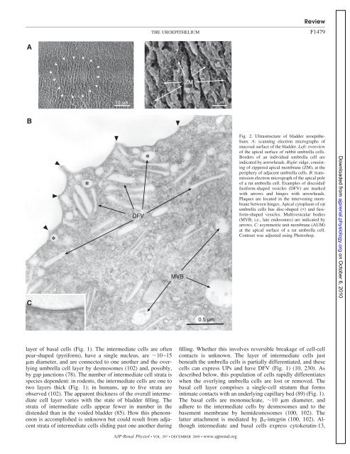
Fig. 2. Ultrastructure of bladder uroepithelium.<br />
A: scanning electron micrographs of<br />
mucosal surface of the bladder. Left: overview<br />
of the apical surface of rabbit umbrella cells.<br />
Borders of an individual umbrella cell are<br />
indicated by arrowheads. Right: ridge, consisting<br />
of zippered apical membrane (ZM), at the<br />
periphery of adjacent umbrella cells. B: transmission<br />
electron micrograph of the apical pole<br />
of a rat umbrella cell. Examples of discoidal/<br />
fusiform-shaped vesicles (DFV) are marked<br />
with arrows <strong>and</strong> hinges with arrowheads.<br />
Plaques are located in the intervening membrane<br />
between hinges. Apical cytoplasm of rat<br />
umbrella cells has disc-shaped (�) <strong>and</strong> fusiform-shaped<br />
vesicles. Multivesicular bodies<br />
(MVB; i.e., late endosomes) are indicated by<br />
arrows. C: asymmetric unit membrane (AUM)<br />
at the apical surface of a rat umbrella cell.<br />
Contrast was adjusted using Photoshop.<br />
filling. Whether this involves reversible breakage of cell-cell<br />
contacts is unknown. The layer of intermediate cells just<br />
beneath the umbrella cells is partially differentiated, <strong>and</strong> these<br />
cells can express UPs <strong>and</strong> have DFV (Fig. 1) (10, 230). As<br />
described below, this population of cells rapidly differentiates<br />
when the overlying umbrella cells are lost or removed. The<br />
basal cell layer comprises a single-cell stratum that forms<br />
intimate contacts with an underlying capillary bed (89) (Fig. 1).<br />
The basal cells are mononucleate, �10 �m diameter, <strong>and</strong><br />
adhere to the intermediate cells by desmosomes <strong>and</strong> to the<br />
basement membrane by hemidesmosomes (100, 102). The<br />
latter attachment is mediated by �4-integrin (100, 102). Although<br />
intermediate <strong>and</strong> basal cells express cytokeratin-13,<br />
Downloaded from<br />
ajprenal.physiology.org<br />
on October 6, 2010