Tissue viability by contrast echocardiography - EHJ Cardiovascular ...
Tissue viability by contrast echocardiography - EHJ Cardiovascular ...
Tissue viability by contrast echocardiography - EHJ Cardiovascular ...

Create successful ePaper yourself
Turn your PDF publications into a flip-book with our unique Google optimized e-Paper software.
<strong>Tissue</strong> <strong>viability</strong> <strong>by</strong> <strong>contrast</strong> <strong>echocardiography</strong> S27<br />
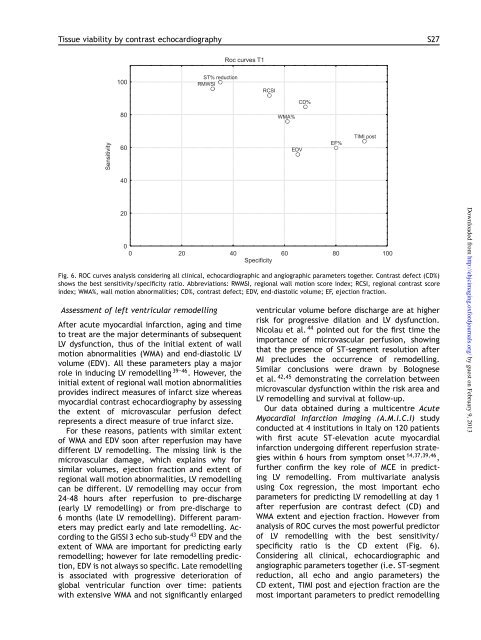
Fig. 6. ROC curves analysis considering all clinical, echocardiographic and angiographic parameters together. Contrast defect (CD%)<br />
shows the best sensitivity/specificity ratio. Abbreviations: RWMSI, regional wall motion score index; RCSI, regional <strong>contrast</strong> score<br />
index; WMA%, wall motion abnormalities; CD%, <strong>contrast</strong> defect; EDV, end-diastolic volume; EF, ejection fraction.<br />
Assessment of left ventricular remodelling<br />
After acute myocardial infarction, aging and time<br />
to treat are the major determinants of subsequent<br />
LV dysfunction, thus of the initial extent of wall<br />
motion abnormalities (WMA) and end-diastolic LV<br />
volume (EDV). All these parameters play a major<br />
role in inducing LV remodelling 39–46 . However, the<br />
initial extent of regional wall motion abnormalities<br />
provides indirect measures of infarct size whereas<br />
myocardial <strong>contrast</strong> <strong>echocardiography</strong> <strong>by</strong> assessing<br />
the extent of microvascular perfusion defect<br />
represents a direct measure of true infarct size.<br />
For these reasons, patients with similar extent<br />
of WMA and EDV soon after reperfusion may have<br />
different LV remodelling. The missing link is the<br />
microvascular damage, which explains why for<br />
similar volumes, ejection fraction and extent of<br />
regional wall motion abnormalities, LV remodelling<br />
can be different. LV remodelling may occur from<br />
24–48 hours after reperfusion to pre-discharge<br />
(early LV remodelling) or from pre-discharge to<br />
6 months (late LV remodelling). Different parameters<br />
may predict early and late remodelling. According<br />
to the GISSI 3 echo sub-study 43 EDV and the<br />
extent of WMA are important for predicting early<br />
remodelling; however for late remodelling prediction,<br />
EDV is not always so specific. Late remodelling<br />
is associated with progressive deterioration of<br />
global ventricular function over time: patients<br />
with extensive WMA and not significantly enlarged<br />
ventricular volume before discharge are at higher<br />
risk for progressive dilation and LV dysfunction.<br />
Nicolau et al. 44 pointed out for the first time the<br />
importance of microvascular perfusion, showing<br />
that the presence of ST-segment resolution after<br />
MI precludes the occurrence of remodelling.<br />
Similar conclusions were drawn <strong>by</strong> Bolognese<br />
et al. 42,45 demonstrating the correlation between<br />
microvascular dysfunction within the risk area and<br />
LV remodelling and survival at follow-up.<br />
Our data obtained during a multicentre Acute<br />
Myocardial Infarction Imaging (A.M.I.C.I) study<br />
conducted at 4 institutions in Italy on 120 patients<br />
with first acute ST-elevation acute myocardial<br />
infarction undergoing different reperfusion strategies<br />
within 6 hours from symptom onset 14,37,39,46 ,<br />
further confirm the key role of MCE in predicting<br />
LV remodelling. From multivariate analysis<br />
using Cox regression, the most important echo<br />
parameters for predicting LV remodelling at day 1<br />
after reperfusion are <strong>contrast</strong> defect (CD) and<br />
WMA extent and ejection fraction. However from<br />
analysis of ROC curves the most powerful predictor<br />
of LV remodelling with the best sensitivity/<br />
specificity ratio is the CD extent (Fig. 6).<br />
Considering all clinical, echocardiographic and<br />
angiographic parameters together (i.e. ST-segment<br />
reduction, all echo and angio parameters) the<br />
CD extent, TIMI post and ejection fraction are the<br />
most important parameters to predict remodelling<br />
Downloaded from<br />
http://ehjcimaging.oxfordjournals.org/ <strong>by</strong> guest on February 9, 2013