¿Cuál sería su diagnóstico y abordaje quirúrgico? - SciELO España
¿Cuál sería su diagnóstico y abordaje quirúrgico? - SciELO España
¿Cuál sería su diagnóstico y abordaje quirúrgico? - SciELO España

You also want an ePaper? Increase the reach of your titles
YUMPU automatically turns print PDFs into web optimized ePapers that Google loves.
Página del Residente<br />
Mujer de 51 años, ex-fumadora de un paquete de cigarrillos/día<br />
hace 25 años, con antecedente de fibroadenoma de mama, acude<br />
a nuestra con<strong>su</strong>lta presentando afonía y disfagia leve de 5 meses<br />
de evolución. Ante la exploración física intraoral normal, se decide<br />
realizar exploración con fibroscopio, observándose una masa<br />
exofítica de 4x3 cm en base derecha de lengua, que llega a la pared<br />
lateral de la faringe, pliegue gloso-amigdalino derecho y vallécula<br />
epiglótica.<br />
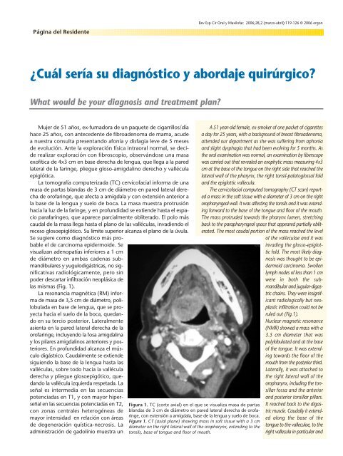
La tomografía computerizada (TC) cervicofacial informa de una<br />
masa de partas blandas de 3 cm de diámetro en pared lateral derecha<br />
de orofaringe, que afecta a amígdala y con extensión anterior a<br />
la base de la lengua y <strong>su</strong>elo de boca. La masa muestra protrusión<br />
hacia la luz de la faringe, y en profundidad se extiende hasta el espacio<br />
parafaríngeo, que aparece parcialmente obliterado. El polo más<br />
caudal de la masa llega hasta el plano de las valléculas, invadiendo el<br />
receso glosoepiglótico. Su límite <strong>su</strong>perior alcanza el plano de la úvula.<br />
Se <strong>su</strong>giere como <strong>diagnóstico</strong> más probable<br />
el de carcinoma epidermoide. Se<br />
vi<strong>su</strong>alizan adenopatías inferiores a 1 cm<br />
de diámetro en ambas cadenas <strong>su</strong>bmandibulares<br />
y yugulodigástricas, no significativas<br />
radiológicamente, pero sin<br />
poder descartar infiltración neoplásica de<br />
las mismas (Fig. 1).<br />
La resonancia magnética (RM) informa<br />
de masa de 3,5 cm de diámetro, polilobulada<br />
en base de lengua, que se proyecta<br />
hacia el <strong>su</strong>elo de la boca, quedando<br />
en <strong>su</strong> tercio posterior. Lateralmente<br />
asienta en la pared lateral derecha de la<br />
orofaringe, incluyendo la fosa amigdalina<br />
y los pilares amigdalinos anteriores y posteriores.<br />
En profundidad alcanza el músculo<br />
digástrico. Caudalmente se extiende<br />
siguiendo la base de la lengua hasta las<br />
valléculas, sobre todo hacia la vallécula<br />
derecha y pliegue glosoepigótico, quedando<br />
la vallécula izquierda respetada. La<br />
señal es intermedia en las secuencias<br />
potenciadas en T1, y con mayor hiper-<br />
señal en las secuencias potenciadas en T2,<br />
con zonas centrales heterogéneas de<br />
mayor intensidad en relación con áreas<br />
de degeneración quística-necrosis. La<br />
administración de gadolínio muestra un<br />
Rev Esp Cir Oral y Maxilofac 2006;28,2 (marzo-abril):119-126 © 2006 ergon<br />
<strong>¿Cuál</strong> <strong>sería</strong> <strong>su</strong> <strong>diagnóstico</strong> y <strong>abordaje</strong> <strong>quirúrgico</strong>?<br />
What would be your diagnosis and treatment plan?<br />
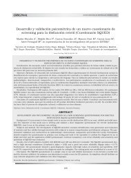
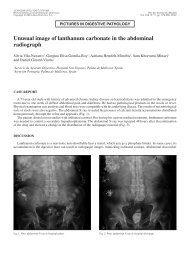
Figura 1. TC (corte axial) en el que se vi<strong>su</strong>aliza masa de partas<br />
blandas de 3 cm de diámetro en pared lateral derecha de orofaringe,<br />
con extensión a amígdala, base de la lengua y <strong>su</strong>elo de boca.<br />
Figure 1. CT (axial plane) showing mass in soft tis<strong>su</strong>e with a 3 cm<br />
diameter on the right lateral wall of the oropharynx, extending to the<br />
tonsils, base of tongue and floor of mouth.<br />
A 51 year-old female, ex-smoker of one packet of cigarettes<br />
a day for 25 years, with a background of breast fibroadenoma,<br />
attended our department as she was <strong>su</strong>ffering from aphonia<br />
and slight dysphagia that had been evolving for 5 months. As<br />
the oral examination was normal, an examination by fiberscope<br />
was carried out that revealed an exophytic mass mea<strong>su</strong>ring 4x3<br />
cm at the base of the tongue on the right side that reached the<br />
lateral wall of the pharynx, the right tonsil-palatoglossal fold<br />
and the epiglottic vallecula.<br />
The cervicofacial computed tomography (CT scan) reported<br />
a mass in the soft tis<strong>su</strong>e with a diameter of 3 cm on the right<br />
oropharyngeal wall. It was affecting the tonsils and it was extending<br />
forward to the base of the tongue and floor of the mouth.<br />
The mass protruded towards the pharynx lumen, stretching<br />
back to the parapharyngeal space that appeared partially obliterated.<br />
The most caudal portion of the mass reached the level<br />
of the valleculae and it was<br />
invading the glosso-epiglottic<br />
fold. The most likely diagnosis<br />
was thought to be epidermoid<br />
carcinoma. Swollen<br />
lymph nodes of less than 1 cm<br />
were in both the <strong>su</strong>bmandibular<br />
and jugular-digastric<br />
chains. They were insignificant<br />
radiologically but neoplastic<br />
infiltration could not be<br />
ruled out (Fig.1).<br />
Nuclear magnetic resonance<br />
(NMR) showed a mass with a<br />
3.5 cm diameter that was<br />
polylobulated and at the base<br />
of the tongue. It was extending<br />
towards the floor of the<br />
mouth from the posterior third.<br />
Laterally, it was attached to<br />
the right lateral wall of the<br />
oropharynx, including the tonsillar<br />
fossa and the anterior<br />
and posterior tonsillar pillars.<br />
It reached back to the digastric<br />
muscle. Caudally it extended<br />
along the base of the<br />
tongue to the valleculae, to the<br />
right vallecula in particular and
120 Rev Esp Cir Oral y Maxilofac 2006;28,2 (marzo-abril):119-126 © 2006 ergon<br />
realce intenso y uniforme. El <strong>diagnóstico</strong> de pre<strong>su</strong>nción es el de carcinoma<br />
epidermoide.<br />
La realización de gammagrafía con I 131 descarta la presencia<br />
de tejido tiroideo lingual. Se realiza biopsia de la lesión, que es informada<br />
como epitelio escamoso no queratinizado con focos de tejido<br />
linfoide a nivel del corion, e infiltrados de células atípicas, de<br />
citoplasma amplio y núcleos con marcadas variaciones de forma y<br />
tamaño, con nucleolos prominentes. Con la técnica de PAS se observa<br />
una ligera positividad celular. El estudio inmunohistoquímico<br />
reveló positividad para queratinas AE-1 y AE-3, y negatividad para<br />
músculo liso, S-100 y GFAP.<br />
Carcinoma mucoepidermoide de base de lungua. Abordaje <strong>quirúrgico</strong>...<br />
the glosso-epiglottic fold, leaving the left vallecula unaffected.<br />
The signal was intermediate on the T1-weighted sequences,<br />
and there was a larger hypersignal on the T2-weighted sequences<br />
with heterogeneous areas in the center of greater intensity when<br />
compared to areas of cystic-necrotic degeneration. The administration<br />
of gadolinium showed an intense and uniform contrast<br />
enhancement. The pre<strong>su</strong>med diagnosis was of epidermoid<br />
carcinoma.<br />
The gammagraphy with 131I ruled out the existence of lingual<br />
thyroid. The biopsy of the lesion revealed a non-keratinized<br />
squamous epithelium with foci of lymphoid tis<strong>su</strong>e by the chorion,<br />
and atypical cell infiltrate, with abundant cytoplasm and<br />
nuclei of various shapes and sizes, and prominent nucleoli. The<br />
PAS technique showed some positive cells. The immunohistochemical<br />
analysis was positive for keratins AE-1 and AE-3 and<br />
negative for S-100 and GFAP.
Página del Residente<br />
Carcinoma mucoepidermoide de base de<br />
lengua. Abordaje <strong>quirúrgico</strong> mediante<br />
labiomandibulotomía media<br />
Mucoepidermoid carcinoma of the base of the tongue. A <strong>su</strong>rgical approach by<br />
means of the median labiomandibulotomy<br />
R. González-García 1 , J. Sastre Pérez 2 , V. Escorial Hernández 1 , P.L. Martos 1 , M. Mancha de la Plata 1 ,<br />
F.J. Rodríguez Campo 2 , L. Naval Gías 2 , S. Nieto Llanos 3 , F.J. Díaz González 4<br />
Con el <strong>diagnóstico</strong> histológico inicial de adenocarcinoma, y estadio<br />
tumoral III (T3N0Mx), y ante la posibilidad de inclusión de la<br />
epiglostis y la totalidad de la laringe en una resección quirúrgica,<br />
se decide en Comité de Tumores de Cabeza y Cuello la administración<br />
de tratamiento radio-quimioterápico inicial. Se inicia quimioterapia<br />
de tipo Al-Sarraf y posteriormente CDDP concomitante<br />
a dosis de 100 mg/m 2 . El tratamiento radioterápico consistió<br />
en la administración fraccionada de 50 Gy. Durante el tratamiento<br />
combinado radio-quimioterápico presentó empeoramiento del estado<br />
general, náuseas y vómitos, que requirieron ingreso por Urgencias.<br />
La analítica general mostró un nivel de creatinina de 2, y fue<br />
diagnosticado de nefrotoxicidad secundaria a cisplatino y toxicidad<br />
gastrointestinal secundaria al tratamiento combinado radio-quimioterápico.<br />
En cabeza y cuello se objetivó mucositis de grado III.<br />
Dos meses después de iniciado el tratamiento, y ante la persistencia<br />
de la tumoración sin mejoría clínica ni radiológica, se decide la<br />
<strong>su</strong>spensión de las citadas modalidades terapéuticas y se opta por la<br />
opción quirúrgica.<br />
Bajo anestesia general e intubación nasotraqueal, y previa realización<br />
de disección cervical funcional derecha, se realizó la exposición<br />
de la sínfisis mandibular mediante una incisión cutánea vertical<br />
a nivel del mentón, continuando la incisión cervical hasta la<br />
línea media labial, y completando posteriormente la labiotomía<br />
media. Se realizó una incisión en el periostio, con despegamiento<br />
<strong>su</strong>ficiente para permitir la colocación del material de osteosíntesis.<br />
1 Médico Residente<br />
2 Médico Adjunto<br />
4 Jefe de Servicio<br />
Servicio de Cirugía Oral y Maxilofacial<br />
3 Médico Adjunto de Anatomía Patológica<br />
Servicio de Anatomía Patológica<br />
Hospital Universitario La Princesa, Madrid. <strong>España</strong>.<br />
Correspondencia:<br />
Raúl González García<br />
C/ Los Yébenes 35, 8ºC<br />
28047 Madrid, <strong>España</strong>.<br />
raugg@mixmail.com<br />
Rev Esp Cir Oral y Maxilofac 2006;28,2 (marzo-abril):119-126 © 2006 ergon<br />
Following the initial histological diagnosis of adenocarcinoma<br />
and of stage III tumor (T3NoMx), and as there was<br />
a possibility of having to included the epiglottis and all of<br />
the larynx in the <strong>su</strong>rgical resection, the Head and Neck Tumor<br />
Committee decided on first administering radiochemotherapy.<br />
Al-Sarraf type chemotherapy was started followed by<br />
a concomitant dose of CDDP of 100 mg/m 2 . The radiotherapy<br />
treatment consisted in the administration of 50 Gy in<br />
fractions. During the combined radiochemotherapy her general<br />
health deteriorated. She was hospitalized by the emergency<br />
services with nausea and vomiting. A general blood<br />
test showed a level of creatine of 2, and nephrotoxicity was<br />
diagnosed secondary to cisplatin together with gastrointestinal<br />
toxicity secondary to the combined radiochemotherapy.<br />
Grade III mucositis was observed in the head and neck.<br />
As there was no improvement in the tumor either clinically<br />
or radiologically two weeks after starting the treatment, the<br />
therapy was <strong>su</strong>spended and the <strong>su</strong>rgical option was decided<br />
upon.<br />
Under general anesthesia with nasotracheal intubation,<br />
and following functional neck dissection on the right side,<br />
the mandibular symphysis was exposed by means of a vertical<br />
cutaneous incision. The neck incision was continued to<br />
the lip midline and a midline lip splitting incision was then<br />
performed. An incision was made into the periosteum, and<br />
it was detached just enough to allow placing the osteosynthesis<br />
material. Three four hole non-bridging miniplates were<br />
then adapted, and the ones for the ends were placed below<br />
the apex and at the base of the mandible. Fixation was carried<br />
out with 7 mm screws. The miniplates were then fixed,<br />
the fixtures were released at one end and the mandibular<br />
osteotomy was carried out with a reciprocating saw under<br />
continuous saline irrigation. A median glossotomy was then<br />
carried out, and the mucosa and muscles of the floor of<br />
the mouth were sectioned. The tumor mass was resected<br />
with macroscopically healthy margins. It appeared to have<br />
a pedicle, as it was attached by a narrow base, and the
122 Rev Esp Cir Oral y Maxilofac 2006;28,2 (marzo-abril):119-126 © 2006 ergon<br />
Se procedió a la adaptación de tres miniplacas de 4 agujeros sin<br />
puente, colocando las de los extremos a nivel <strong>su</strong>bapical y de la basilar<br />
mandibular. Se utilizaron tornillos de 7 mm para <strong>su</strong> fijación.<br />
Seguido de la fijación de las miniplacas, se soltaron las fijaciones en<br />
uno de <strong>su</strong>s extremos y se realizó la osteotomía mandibular mediante<br />
una sierra reciprocante con irrigación continua con <strong>su</strong>ero salino.<br />
Posteriormente, se realizó una glosotomía media y sección de la<br />
mucosa y musculatura del <strong>su</strong>elo de la boca. Se realizó resección de<br />
la masa tumoral con márgenes macroscópicos, que presentaba una<br />
base de implantación estrecha, adoptando una conformación pediculada,<br />
respetando la epiglotis y la laringe. El estudio anatomopatológico<br />
intraoperatorio fue informado como masa tumoral con<br />
márgenes libres de infiltración. El defecto se reparó mediante cierre<br />
directo, sin necesidad de colgajos regional o libre microvascularizado.<br />
Tras el cierre de la glosotomía media y de la sección del<br />
<strong>su</strong>elo de la boca por planos con <strong>su</strong>tura reabsorbible, se procedió<br />
a la reaproximación de los segmentos mandibulares y fijación con<br />
las miniplacas y tornillos previamente retirados. La piel se <strong>su</strong>turó<br />
con monofilamento de 4-0.<br />
El examen macroscópico mostró una tumoración exofítica de<br />
3,3 cm de diámetro de coloración blanquecina, bien delimitada<br />
microscópicamente, sin alcanzar los bordes <strong>quirúrgico</strong>s de resección.<br />
La tumoración descrita se encontraba constituida en <strong>su</strong> mayor<br />
parte por una celularidad de tamaño intermedio de núcleos ligeramente<br />
irregulares con nucleolo evidente y citoplasmas claros; en<br />
otras zonas la celularidad tumoral presentaba citoplasmas más escasos<br />
y una disposición escamoide y con áreas de estructuras glandulares<br />
muy aisladas en las que se identificaron células mucinosas.<br />
La celularidad tumoral se disponía en forma de nidos irregulares<br />
o cordones gruesos en un estroma con abundantes fibroblastos<br />
y áreas de aspecto mixoide, objetivándose en algunos puntos la<br />
existencia de estructuras quísticas que ocupaban menos del 20%<br />
del tumor, sin que se observara necrosis ni anaplasia nuclear. Focalmente<br />
se objetivaron imágenes aisladas de invasión perineural. El<br />
índice mitósico fue menor de 4 mitosis por 10 campos de gran<br />
aumento. La tumoración asentaba en el corion de la lengua, sin<br />
encontrarse en ningún punto en continuidad con el epitelio de<br />
revestimiento lingual. El <strong>diagnóstico</strong> anatomopatológico definitivo<br />
fue carcinoma mucoepidermoide de bajo grado de glándula salivar<br />
menor de localización en base de lengua (Fig. 3). Los ganglios<br />
cervicales fueron negativos para infiltración metastática tumoral. Se<br />
administró tratamiento radioterápico posterior.<br />
Carcinoma mucoepidermoide de base de lungua. Abordaje <strong>quirúrgico</strong>...<br />
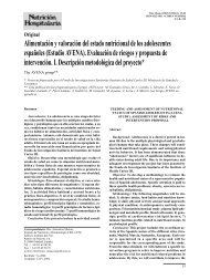
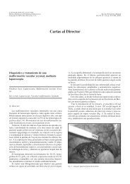
Figura 2. A) Vista preoperatoria de la lesión. B) Labiomandibulotomía media, marcado de la incisión de la glosotomía media. C) Glosotomía completada.<br />
D) Masa pediculada en base de lengua-orofarínge en proximidad a la epiglotis.<br />
Figure 2. A) Preoperative view of the lesion. B) Median labiomandibulotomy showing incision line of the median glossotomy. C) Completed glossotomy.<br />
D) Pedicled mass by the base of the tongue-oropharynx close to the epiglottis.<br />
epiglottis and larynx were preserved. The intraoperative<br />
pathologic examination reported a tumor mass with no infiltration<br />
of the margins. The defect was repaired by means of<br />
direct clo<strong>su</strong>re, and neither regional nor microvascularized<br />
free flaps were needed. Following clo<strong>su</strong>re of the median glossotomy<br />
and of the floor of the mouth layers following the<br />
incision with resorbable <strong>su</strong>tures, the mandibular segments<br />
were joined and fixed with the miniplates, and the screws<br />
that had previously been removed were inserted. The skin<br />
was <strong>su</strong>tured with 4-0 monofilament material.<br />
The macroscopic examination revealed a whitish exophytic<br />
tumor mea<strong>su</strong>ring 3.3 cm in diameter, that was well-circumscribed<br />
microscopically and that did not extend to the resection<br />
margins. It was made up largely of intermediate sized<br />
cells that had slightly irregular nuclei, prominent nucleoli and<br />
clear cytoplasm. Other areas of the tumor cellularity had scant<br />
cytoplasm and a squamous pattern, and there were areas of<br />
very isolated glandular structures in which mucinous cells could<br />
be identified. The tumoral cells were arranged in irregular nest<br />
shapes or thick cords in stroma with abundant fibroblasts and<br />
areas with a myxoid appearance, and in some areas there<br />
were cystic structures that made up less than 20% of the tumor,<br />
but neither necrosis nor nuclear anaplasia were observed. Isolated<br />
perineural invasion was observed focally. The mitotic<br />
index was less than 4 mitoses per 10 high power fields. The<br />
tumor was situated on the chorion of the tongue, but at no<br />
point did it continue to the epithelium or lining of the tongue.<br />
The definitive pathologic diagnosis was of low-grade mucoepidermoid<br />
carcinoma of the minor salivary glands at the base<br />
of the tongue (Fig. 3). The neck nodes were negative for<br />
metastatic infiltration. Radiation therapy treatment was <strong>su</strong>bsequently<br />
administered.<br />
Discussion<br />
Although a rare entity, the mucoepidermoid carcinoma<br />
(MEC) together with the cystic adenoid carcinoma, represents<br />
the most common malignant tumor of the salivary<br />
glands. It affects mainly adults during the fourth and sixth<br />
decades in life. MEC affects the major and minor salivary<br />
glands equally, although low-grade MEC is more frequent-
R. González y cols.<br />
Rev Esp Cir Oral y Maxilofac 2006;28,2 (marzo-abril):119-126 © 2006 ergon 123<br />
ly found in the minor salivary<br />
glands. Although more common<br />
in the minor salivary<br />
glands of the mouth, it has<br />
been reported that in 10% of<br />
patients it can appear in the<br />
maxillary sinuses, nasopharynx,<br />
nasal cavity, oropharynx,<br />
vocal cords, larynx and<br />
trachea. 1,2 With regard to this<br />
case, and given the localization<br />
in the oropharynx and<br />
base of the tongue, the differential<br />
diagnosis should be<br />
carried out with other entities<br />
<strong>su</strong>ch as the thyroglossal duct<br />
cyst, lingual thyroid, hyperplastic<br />
lymphoid tis<strong>su</strong>e, lymphangioma,<br />
fibroma, lipoma,<br />
dermoid cyst, squamous cell carcinoma, lymphoma and other<br />
minor salivary gland tumors. 3<br />
With regard to prognosis, Plambeck et al. 4 described<br />
global <strong>su</strong>rvival rates of 91,9 and 89,5% at 5 and 10 years.<br />
This rate fell to 63,5 and 52,0% at 5 and 10 years in patients<br />
with stage III and IV. The pre<strong>su</strong>med diagnosis, based on the<br />
initial biopsy was of adenocarcinoma of minor salivary gland<br />
origin. Both imaging studies showed nodal enlargement of<br />
not more than 1 cm in the <strong>su</strong>bmandibular and jugular-digastric<br />
chains. Sheahan et al. 5 reported a percentage of occult<br />
neck metastasis (N0 necks) of 40%, and that CT and NMR<br />
were not useful for detecting occult neck disease. They concluded<br />
that neck dissection is indicated for patients with highgrade<br />
salivary gland adenocarcinoma of the head and neck.<br />
As a re<strong>su</strong>lt of this information, homolateral functional neck<br />
dissection was carried out. This proved to be the right decision<br />
following the definitive histological diagnosis of MEC.<br />
With regard to tumor size, neck dissection for T2 MEC and<br />
above has quite rightly been indicated as a necessary requirement<br />
given the potential risk of regional metastasis. 6 Clinical<br />
stage and tumor grade have been reported as the most<br />
important prognostic factors in MEC. Other elements have<br />
been demonstrated as being independent prognostic factors:<br />
being above the age of 40, fixed tumors, T stage, N<br />
stage and histological grade. 7 The existence of disease-free<br />
<strong>su</strong>rgical margins represents in itself a prognostic factor for<br />
these tumors, and obtaining wide <strong>su</strong>rgical expo<strong>su</strong>re is essential<br />
for obtaining these margins. Given the oropharyngeal<br />
localization, this particular case required a <strong>su</strong>rgical approach<br />
using a median labiomandibulotomy.<br />
The labiomandibulotomy was first described in 1836 by<br />
Roux8 and it was re-introduced in 1929 by Trotter9 Discusión<br />
El carcinoma mucoepidermoide<br />
(CME) constituye, dentro de <strong>su</strong> rareza<br />
y, junto al carcinoma adenoide quístico,<br />
el tumor maligno más frecuente de<br />
las glándulas salivares. Tiene predilección<br />
por adultos en torno a las décadas<br />
cuarta y sexta. El CME se encuentra en<br />
el mismo porcentaje en glándulas salivares<br />
mayores y menores, aunque los<br />
CME de bajo grado asientan más frecuentemente<br />
en estas últimas. Aunque<br />
<strong>su</strong> localización más frecuente dentro de<br />
las glándulas salivares menores son las<br />
de localización intraoral, se han descrito<br />
otras en un 10% de los pacientes:<br />
seno maxilar, nasofaringe, cavidad nasal,<br />
orofaringe, cuerdas vocales, laringe y<br />
tráquea.<br />
who extended<br />
the approach of his predecessor by means of a median<br />
translingual pharyngotomy. Since it was first described, the<br />
median mandibulotomy has been modified several times, especially<br />
the osteotomy technique and posterior fixation. In 1961<br />
1,2 En el presente caso, y dada <strong>su</strong> localización en orofarínge<br />
y base de lengua, es preciso realizar el <strong>diagnóstico</strong> diferencial<br />
con otras entidades como el quiste del conducto tirogloso, tiroides<br />
lingual, tejido linfoide hiperplásico, linfangioma, fibroma, lipoma,<br />
quiste dermoide, carcinoma de células escamosas, linfoma y otro<br />
tumor de glándulas salivares menores. 3<br />
En relación a <strong>su</strong> pronóstico, Plambeck et al. 4 describieron tasas<br />
de <strong>su</strong>pervivencia global de 91,9 y 89,5% a los 5 y 10 años. Esta tasa<br />
desciende a 63,5 y 52,0% a los 5 y 10 años en pacientes con estadios<br />
III y IV. El <strong>diagnóstico</strong> de pre<strong>su</strong>nción, basado en la biopsia<br />
inicial, fue el de adenocarcinoma de glándula salivar menor. Ambas<br />
pruebas de imagen mostraron adenopatías de tamaño no <strong>su</strong>periores<br />
a 1 cm en cadenas <strong>su</strong>bmandibulares y yugulodigástricas. Sheahan<br />
y cols. 5 refieren un porcentaje de metástasis cervicales ocultas<br />
(cuellos N0) del 40%, y que la TC y RM no fueron útiles en la detección<br />
de enfermedad cervical oculta. Concluyen que la disección cervical<br />
está indicada en pacientes con adenocarcinoma de alto grado<br />
de glándula salivar de cabeza y cuello. En función de estos datos se<br />
decidió realizar disección cervical funcional homolateral. Esta actitud<br />
se mostró igualmente adecuada una vez obtenido el <strong>diagnóstico</strong><br />
histológico definitivo de CME. No en vano y, considerando el<br />
tamaño tumoral, se ha indicado la necesidad de disección cervical<br />
a partir de los CME T2, debido al riesgo potencial de metástasis<br />
regional. 6 El estadio clínico y el grado tumoral han sido referidos<br />
como los factores pronósticos más importantes en el CME. Otros<br />
han sido demostrados como factores pronósticos independientes:<br />
edad mayor de 40 años, tumores adheridos, estadio T y N y grado<br />
histológico. 7 La presencia de márgenes <strong>quirúrgico</strong>s libres constituye<br />
per se un factor pronóstico en estos tumores, y la obtención<br />
de un campo <strong>quirúrgico</strong> amplio es fundamental para obtener tales<br />
márgenes. Debido a <strong>su</strong> localización tumoral en orofaringe, el presente<br />
caso requirió un <strong>abordaje</strong> <strong>quirúrgico</strong> mediante labiomandibulotomía<br />
media.<br />
Inicialmente descrita por Roux en 1836, 8 la labiomandibulotomía<br />
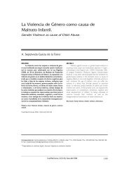
fue retomada por Trotter en 1929, 9 Figura 3. Imagen histológica correspondiente a CME de bajo grado.<br />
Figure 3. Histologic image corresponding to low-grade MEC.<br />
que amplió el <strong>abordaje</strong> de<br />
<strong>su</strong> precedente a través de una faringotomía media translingual.
124 Rev Esp Cir Oral y Maxilofac 2006;28,2 (marzo-abril):119-126 © 2006 ergon<br />
Desde <strong>su</strong> descripción inicial, la mandibulotomía media ha experimentado<br />
<strong>su</strong>cesivas modificaciones, sobre todo en relación con la<br />
técnica de osteotomía y <strong>su</strong> fijación posterior. Hayes Martin, en<br />
1961, 10 popularizó la labiomandibulotomía media y Cohen y cols. 11<br />
describió la modificación del trazado de la osteotomía mandibular<br />
mediante la creación de un escalón en la línea media y la exodoncia<br />
de un incisivo central.<br />
La labiomandibulotomía media ha sido referida en el tratamiento<br />
de tumores de la cavidad oral, orofaringe y espacio parafaríneo, ya<br />
que permite la obtención de un amplio campo <strong>quirúrgico</strong>. En un<br />
estudio previo por nuestro grupo, 12 se analizó esta técnica en 21<br />
pacientes que presentaron carcinoma epidermoide (18%) a nivel<br />
de lengua libre (4,7%), base de lengua (38,1%), <strong>su</strong>elo de boca<br />
(9,5%), orofaringe (33,3%) y clivus (14,2%). En esta serie previa,<br />
no se diagnosticó carcinoma mucoepidermoide en ningún caso. En<br />
todos los casos se obtuvo una exposición adecuada del tumor, 2<br />
pacientes presentaron dehiscencia de la <strong>su</strong>tura con comunicación<br />
orocervical, 1 presentó osteorradionecrosis de la línea de fractura,<br />
3 <strong>su</strong>frieron infección del lecho de la osteotomía y 2 presentaron<br />
alteraciones oclusales. En relación con esta serie, pueden considerarse<br />
otras opciones quirúrgicas, como la glosectomía pull through,<br />
en el <strong>abordaje</strong> de tumores de localización lingual y <strong>su</strong>elo de boca.<br />
Esta técnica evita la realización de la osteotomía y labiotomía, con<br />
un adecuado control tumoral. 13,14 En nuestra opinión, los tumores<br />
de localización en base de lengua y orofaringe presentan un acceso<br />
<strong>quirúrgico</strong> difícil, y el factor limitante más importante es la madíbula.<br />
La falta de exposición adecuada puede limitar asimismo la<br />
obtención de márgenes <strong>quirúrgico</strong>s amplios. En estos casos el <strong>abordaje</strong><br />
transmandibular re<strong>su</strong>lta adecuado. En el presente caso, la localización<br />
posterior del tumor hizo recomendable la utilización de esta<br />
técnica. La naturaleza pediculada del tumor no pudo ser precisada<br />
con seguridad en los estudios de imagen previos, y ante la posibilidad<br />
de una masa que infiltrara ampliamente la base de la lengua<br />
y orofaringe, con extensión al lado contralateral y con posible afectación<br />
de estructuras laríngeas, se optó por un <strong>abordaje</strong> que permitiera<br />
una exposición amplia.<br />
Otras técnicas empleadas para el <strong>abordaje</strong> de tumores de localización<br />
en cavidad oral y orofaringe son la faringotomía transhioidea,<br />
la faringotomía lateral y varias formas de mandibulotomía.<br />
La mandibulotomía puede realizarse posterior al orificio mentoniano,<br />
mandibulotomía lateral, o anterior al mismo, mandibulotomía<br />
media. Esta última puede <strong>su</strong>bdividirse en mandibulotomía de la<br />
línea media o sinfisaria, con la línea de osteotomía entre los dos incisivos<br />
centrales, y mandibulotomía parasinfisaria, que discurre entre<br />
el incisivo lateral y el canino. En un estudio comparativo entre estos<br />
dos tipos de mandibulotomía media, Dai y cols. 15 no encontraron<br />
diferencias estadísticamente significativas aunque, de un modo teórico,<br />
abogan por la mandibulotomía media parasinfisaria por la preservación<br />
de las inserciones de los músculos genihioideos y geniogloso,<br />
con mejores re<strong>su</strong>ltados en relación a la <strong>su</strong>cción y función<br />
deglutoria postquirúrgicas.<br />
La tasa global de complicaciones referidas en la literatura oscila<br />
en torno al 20%, generalmente consistentes en alteraciones<br />
oclusales, trastornos de la sensibilidad del territorio inervado por<br />
el nervio dentario inferior, dolor en la articulación temporoman-<br />
Carcinoma mucoepidermoide de base de lungua. Abordaje <strong>quirúrgico</strong>...<br />
the median labiomandibular glossotomy was popularized<br />
by Hayes Martin, 10 and Cohen et al 11 described the modification<br />
of the mandibular osteotomy line by means of creating<br />
a midline step and by extracting a central incisor.<br />
The median labiomandibulotomy has been reported in<br />
the treatment for tumors of the oral cavity, oropharynx, parapharyngeal<br />
space, as it permits obtaining wide-field expo<strong>su</strong>re.<br />
In a previous study by our group 12 this technique was<br />
analyzed in 21 patients that had epidermoid carcinoma (18)<br />
of the mobile tongue (4.7%), base of the tongue (38.1%),<br />
floor of the mouth (9.5%), oropharynx (33.3%) and clivus<br />
(14.2%). In this previous series, mucoepidermoid carcinoma<br />
was not diagnosed in any of the cases. In all the cases there<br />
was adequate expo<strong>su</strong>re of the tumor, 2 patients <strong>su</strong>ffered<br />
<strong>su</strong>ture dehiscence with orocervical communication, 1 <strong>su</strong>ffered<br />
osteoradionecrosis of the fracture line, 3 <strong>su</strong>ffered infection<br />
of the osteotomy bed, and 2 had occlusal alterations.<br />
With regard to this series, other <strong>su</strong>rgical options can be considered,<br />
<strong>su</strong>ch as a pull-through glossectomy for approaching<br />
tumors with a lingual or floor of the mouth localization.<br />
This would avoid osteotomies and lip-splitting techniques<br />
while en<strong>su</strong>ring proper tumor control. 13,14 It is our opinion<br />
that the <strong>su</strong>rgical access for tumors situated at the base of<br />
the tongue and oropharynx is difficult, and the most important<br />
limiting factor is the mandible. The lack of adequate<br />
expo<strong>su</strong>re can condition obtaining wide <strong>su</strong>rgical margins.<br />
In these cases the transmandibular approach is <strong>su</strong>itable.<br />
With regard to our case, this technique was advisable given<br />
the posterior location of the tumor. The pedicled nature of<br />
the tumor could not be confirmed by the previous imaging<br />
studies, and as there was a possibility that there was considerable<br />
infiltration of the base of the tongue and oropharynx<br />
by the mass, with extension to the contralateral side and<br />
possible damage to laryngeal structures, an approach that<br />
allowed wide expo<strong>su</strong>re was chosen.<br />
Other techniques used for approaching tumors located<br />
in the oral cavity and oropharynx are the transhyoid pharyngotomy,<br />
lateral pharyngotomy and various forms of mandibulotomies.<br />
Mandibulotomies may be posterior to the mental<br />
foramen, lateral, anterior or midline. The latter can be <strong>su</strong>bdivided<br />
into mandibulotomies of the midline or symphyseal,<br />
with the osteotomy line running between the two central<br />
incisors, and parasymphyseal with the line running between<br />
the lateral incisor and the canine tooth. In a comparative<br />
study between these two types of midline mandibulotomies,<br />
Dai et al 15 did not find any differences that were statistically<br />
significant although, from a theoretical point of view,<br />
they advocated the midline parasymphyseal mandibulotomy<br />
as the insertions of the geniohyoid and genioglossal muscles<br />
were preserved and there were better re<strong>su</strong>lts with regard<br />
to post-<strong>su</strong>rgical <strong>su</strong>cking and swallowing function.<br />
The global complication rate reported in the literature is<br />
around 20%, and this generally consists in occlusion disturbances,<br />
disorders relating to sensitivity in the area innervated<br />
by the lower dental nerve, temporomandibular joint pain,
R. González y cols.<br />
dibular, alteraciones periodontales, infección de la herida y alteraciones<br />
estéticas en relación a la cicatriz de la labiotomía. En un<br />
intento por disminuir la tasa de complicaciones en relación a la<br />
enfermedad periodontal, Bertrand y cols., 16 proponen la realización<br />
de la incisión mucosa a una distancia 2-3 dientes más allá de<br />
la zona de la osteotomía y el cierre mucoso lo más hermético posible<br />
mediante <strong>su</strong>turas transpapilares. Estos mismos autores refieren<br />
re<strong>su</strong>ltados estéticos desfavorables en un 10% de los casos,<br />
tasa <strong>su</strong>perponible a los re<strong>su</strong>ltados obtenidos por nuestro grupo. 12,15<br />
La exodoncia previa de un incisivo central ha sido propuesta con<br />
el objeto de evitar el daño de las dos raíces adyacentes a la osteotomía<br />
y disminuir el riesgo de alteraciones periodontales. Sin<br />
embargo, esta maniobra puede generar problemas oclusales posteriores<br />
y <strong>su</strong>pone un detrimento estético para el paciente. Preferimos<br />
no realizar exodoncia en ningún caso, a menos que exista<br />
enfermedad periodontal previa importante. La utilización de hojas<br />
de sierra finas y la utilización de escoplos finos a nivel del espacio<br />
interdental en la cresta alveolar permite obtener un corte limpio,<br />
con mínima pérdida de hueso entre las piezas dentarias y<br />
menor riesgo de alteraciones periodontales.<br />
En relación a la osteosíntesis mandibular, los procedimientos de<br />
fijación con alambre han sido progresivamente <strong>su</strong>stituidos por la<br />
fijación rígida con placas de compresión mandibular o semirrígida<br />
con miniplacas, con el objeto de disminuir la tasa de complicaciones<br />
oclusales y las derivadas de la inestabilidad del foco de fractura,<br />
como son la infección y la radionecrosis. Todos los casos de nuestra<br />
serie fueron tratados mediante fijación con miniplacas, sin evidenciarse<br />
pseudoartrosis ni retardo en la consolidación. El caso presentado<br />
no experimentó complicaciones en relación a la osteotomía<br />
en la evaluación a los 6 meses de la cirugía, a pesar del tratamiento<br />
radio-quimioterápico previo y radioterápico adyuvante posterior.<br />
En este sentido diversos autores, 6,17,18 no han encontrado una<br />
asociación estadísticamente significativa entre radioterapia y complicaciones<br />
en el foco de fractura. Más aún, Eisen y cols. 19 compararon<br />
las complicaciones aparecidas en pacientes en los que la radiación<br />
postoperatoria se administró en el campo de la osteotomía,<br />
con respecto a aquellos en los que la radioterapia no lo alcanzó. No<br />
encontraron diferencias estadísticamente significativas utilizando<br />
dosis de radioterapia moderadas.<br />
Tras la cirugía es fundamental el control de la vía aérea, precisamente<br />
por la localización de estos tumores. En este sentido obviamos<br />
la realización de traqueostomía porque se mantuvo al paciente<br />
bajo inducción anestésica intubado por vía nasotraqueal y conectado<br />
a respirador durante 72 horas. La realización de un <strong>abordaje</strong><br />
en el fondo del <strong>su</strong>elo de boca ipsilateral a la lesión podría haber<br />
<strong>su</strong>puesto un menor riesgo de compromiso post<strong>quirúrgico</strong> de la vía<br />
aérea. En nuestra experiencia, la tasa de complicaciones por fístula<br />
orocervical mediante esta técnica es mayor que con la glosotomía<br />
media.<br />
En conclusión, la labiomandibulotomía media constituye un<br />
<strong>abordaje</strong> óptimo en el tratamiento de tumores del área orofaríngea<br />
y espacio parafaríngeo, como el CME, aportando un campo <strong>quirúrgico</strong><br />
adecuado para la excisión tumoral con márgenes amplios.<br />
La utilización de miniplacas permite una fijación estable de la osteotomía<br />
mandibular, con escaso número de complicaciones.<br />
Rev Esp Cir Oral y Maxilofac 2006;28,2 (marzo-abril):119-126 © 2006 ergon 125<br />
periodontal problems, wound infection and aesthetic changes<br />
due to the lip-splitting procedure. In an attempt at reducing<br />
the rate of complications with regard to periodontal disease,<br />
Bertrand et al 16 proposed making a mucosal incision that was<br />
two to three teeth away from the area of the osteotomy, and<br />
that the mucosal clo<strong>su</strong>re should be carried out with transpapillary<br />
<strong>su</strong>tures and as hermetically as possible. These same<br />
authors reported unfavorable aesthetic re<strong>su</strong>lts in 10% of cases,<br />
a rate that was higher than those of our group. 12,15 The removal<br />
of a central incisor has been proposed so as not to damage<br />
both adjacent roots as a re<strong>su</strong>lt of the osteotomy, and to reduce<br />
the risk of periodontal disturbances. However, this procedure<br />
could lead to occlusal problems later on, and aesthetically this<br />
would be worse for the patient. We prefer not to perform any<br />
extractions at all, unless these is considerable periodontal disease.<br />
The use of fine saw blades and a fine chisel by the interdental<br />
space of the alveolar crest permits obtaining a clean<br />
cut, with a minimal amount of bone loss between the teeth,<br />
and the risk of periodontal disturbances is reduced.<br />
With regard to mandibular osteosynthesis, the fixation<br />
procedures using wires have been replaced progressively with<br />
rigid fixation and mandibular compression plates, or semirigid<br />
fixation with miniplates, in order to reduce the rate of<br />
occlusion complications and those derived from the instability<br />
of the fracture site, <strong>su</strong>ch as infection and radionecrosis.<br />
All the cases in our series were treated by means of fixation<br />
with miniplates, with no evidence of pseudoarthrosis<br />
or consolidation delays. No complications were observed<br />
in the case presented six months after the <strong>su</strong>rgery with regard<br />
to the osteotomy, in spite of the pre-operative radiochemotherapy<br />
and the post-operative adjuvant radiotherapy.<br />
In this sense, various authors 6,17,18 have not found a statistically<br />
significant correlation between radiotherapy and<br />
complications at the fracture site. Moreover Eisen et al 19 compared<br />
the complications that appeared in patients who<br />
received postoperative radiation therapy to the osteotomy<br />
site, with the fracture sites of other patients that were shielded.<br />
Statistically significant differences were not found with<br />
moderate doses of radiation therapy.<br />
Following the <strong>su</strong>rgery, airway control is fundamental,<br />
precisely because of the location of these tumors. Carrying<br />
out a tracheostomy was obviated as the patient was kept<br />
under anesthesia and with nasotracheal intubation and connected<br />
to a respirator for 72 hours. Had the approach been<br />
carried out at the back of the floor of the mouth, ipsilateral<br />
to the lesion, the post-<strong>su</strong>rgical risk of airway compromise<br />
would have been reduced. In our experience, the complication<br />
rate from orocervical fistulae as a re<strong>su</strong>lt of this technique<br />
is greater than with a median glossotomy.<br />
To conclude, the median labiomandibulotomy is an optimal<br />
approach for treating tumors of the oropharyngeal area<br />
and the parapharyngeal space, <strong>su</strong>ch as MEC, that provides<br />
an adequate <strong>su</strong>rgical field enabling tumor excision with wide<br />
margins. The use of miniplates allows stable fixation of the<br />
mandibular osteotomy, with very few complications.
126 Rev Esp Cir Oral y Maxilofac 2006;28,2 (marzo-abril):119-126 © 2006 ergon<br />
Bibliografía<br />
1. Spiro RH, Huvos AG, Berk R, Strong EW. Mucoepidermoid carcinoma<br />
of salivary gland origin: a clinicopathologic study of 367 cases. Am J Surg<br />
1978;136:461-8.<br />
2. Brandwein MS, Ivanov K, Wallace DI, et al. Mucoepidermoid carcinoma:<br />
a clinicopathologic study of 80 patients with special reference<br />
to histological grading. Am J Surg Pathol 2001;25:835-45.<br />
3. Perez JS, Muñoz M, Naval L, Blasco A, Díaz FJ. Papillary carcinoma arising<br />
in lingual thyroid. J Oral Craniomaxillofac Surg 2003;31:179-82.<br />
4. Koemueller H, Brueggemann N, Swennen G, Eckardt A. Mucoepidermoid<br />
carcinoma of the salivary gland-clinical review of 42 cases. Oral<br />
Oncol 2005;41:3-10.<br />
5. Sheahan P, Byrne M, Hafidh M, Toner M, Simon C. Neck dissection<br />
in primary head and neck high-grade adenocarcinoma. J Laryngol Otol<br />
2004;118:532-6.<br />
6. Pires FR, Alves FA, de Almeida OP, Lopes MA, Kowalski LP. Synchronous<br />
mucoepidermoid carcinoma of tongue and pleomorphic adenoma<br />
of <strong>su</strong>bmandibular gland. Oral Surg Oral Med Oral Pathol Oral<br />
Radiol Endod 2003;95:328-31.<br />
7. Pires FR, de Almeida OP, de Araujo VC, Kowalski LP. Prognostic factors<br />
in head and neck mucoepidermoid carcinoma. Arch Otolaryngol<br />
Head Neck Surg 2004;130:174-81.<br />
8. Butlin HT. Disease of the tongue. En: Cassells (ed). Clinical Mannuals<br />
for Practitioners and Students of Medicine. London 1885;331.<br />
9. Shah JP, Kumaraswamy SV, Kularni V. Comparative evaluation of fixation<br />
methods after mandiulotomy for oropharyneal tumors. Am J Surg<br />
1993;166:431-4.<br />
Carcinoma mucoepidermoide de base de lungua. Abordaje <strong>quirúrgico</strong>...<br />
10. Martin H, Tollefsen R, Gerald FP. Median labiomandibular glossotomy.<br />
Am J Surg 1961;102:755-9.<br />
11. Cohen JI, Marentette LJ, Maisel RH. The mandibular swing stabilization<br />
of the midline mandibular osteotomy. Laryngoscope 1988;98:1139-<br />
42.<br />
12. Sastre Pérez J, Naval Gías L, Rodríguez Campo FJ, Muñoz Guerra MF,<br />
Díaz González FJ. Labiomandibulotomía media como <strong>abordaje</strong> de neoplasias<br />
de orofaringe y ase de cráneo. Rev Esp Cir Oral Maxilofac<br />
2001;23:224-9.<br />
13. Burgueño García M. Labiomandibulotomía media como <strong>abordaje</strong> de<br />
neoplasias de orofaringe y ase de cráneo [discusión]. Rev Esp Cir Oral<br />
Maxilofac 2001;23:330.<br />
14. Zubillaga Rodríguez I, Sánchez Aniceto G, García Recuero I, Montalvo<br />
Moreno JJ. Tiroides lingual: un nuevo <strong>abordaje</strong> <strong>quirúrgico</strong>. Rev Esp<br />
Cir Oral Maxilofac 2004;26:316-24.<br />
15. Dai TS, Hao SP, Chang KP, Pan WL, Yeh HC, Tsang NM. Complications<br />
of mandibulotomy: midline ver<strong>su</strong>s paramidline. Otolaryngol Head<br />
Neck Surg 2003;128:137-41.<br />
16. Bertrand J, Luc B, Philippe M. Philippe P. Anterior mandibular osteotomy<br />
for tumor extirpation: a critical evaluation. Head Neck 2000;22:<br />
323-7.<br />
17. Dubner S, Spiro RH. Median mandibulotomy: a critical assessment.<br />
Head Neck 1991;13:389-93.<br />
18. Smeele LE, Slotman BJ, Mens JW, Tiwari R. Local radiation dose, fixation,<br />
and non-union of mandibulotomies. Head Neck 1999;21:315-8.<br />
19. Eisen MD, Weinstein GS, Chalian A, Machtay M, Kent K, Coia LR,<br />
Weber RS. Moridity after midline mandiulotomy and radiation therapy.<br />
Am J Otolaryngol 2000;21:312-7.