Estudio de la respuesta inmune humoral y celular ... - edigraphic.com
Estudio de la respuesta inmune humoral y celular ... - edigraphic.com
Estudio de la respuesta inmune humoral y celular ... - edigraphic.com

Create successful ePaper yourself
Turn your PDF publications into a flip-book with our unique Google optimized e-Paper software.
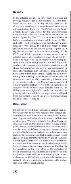
Results<br />
In the infected group, the PPE reached a maximum<br />
average of 5.2% at day 31 posinfection (pi) that diminished<br />
to less than 1% at day 46, and <strong>la</strong>ter on the<br />
rickettsia was no longer <strong>de</strong>tected in blood smears even<br />
after reinfection (Figure 1a). The PCV diminished until<br />
it reached an average of 6% at day 46 pi and was within<br />
normal limits from reinfection up to the end of the<br />
study (Figure 1b). The CD2+ values were simi<strong>la</strong>r in<br />
both groups during the study; while those of CD4+<br />
and CD8+ in the infected group significantly diminished<br />
(P < 0.05) at day 44 pi and <strong>la</strong>ter reached values<br />
simi<strong>la</strong>r to those of the control group (Figure 2). A<br />
significant increase of fluorescence intensity (FI) in<br />
CD2+ and CD8+ lymphocytes from animals in the<br />
infected group after reinfection (days 62 and 83) (P <<br />
0.05) with respect to the FI observed in the lymphocytes<br />
from the control group was noticed (Figure 3).<br />
Antibody titers (Abs) in the infected cattle increased<br />
above the basal values at day 51 pi and increased even<br />
more after reinfection. In the Control group, antibody<br />
titers were within basal values (Figure 4a). The interferon<br />
gamma (IFN-γ ) levels in the sera from infected<br />
animals increased steadily, particu<strong>la</strong>rly following day<br />
51 pi, while levels in the Control group remained<br />
within basal limits (Figure 4b). In the supernatants of<br />
<strong>com</strong>plete blood cultures from infected animals, the<br />
IFN-γ levels were higher after reinfection than after the<br />
primary infection; while in the supernatants of blood<br />
from the control group those levels remained within<br />
the basal limits (Figure 5).<br />
Discussion<br />
It has been stressed that vaccination against anap<strong>la</strong>smosis<br />
has been an effective measure for outbreak prevention;<br />
however the existent vaccines, live or attenuated,<br />
<strong>de</strong>pend on bovine blood as a source of infection<br />
or of antigen. 33 Vaccines obtained from blood have the<br />
inconvenience of transmitting other bovine pathogens,<br />
that are imperceptible at the time of blood collection;<br />
besi<strong>de</strong>s, the different Anap<strong>la</strong>sma marginale geographical<br />
iso<strong>la</strong>tes frequently do not induce cross protection.<br />
33 In this context, and taking into account recent<br />
studies on the different genotypes, 34,35 and on A. marginale<br />
antigenic variation36 it is mandatory to better un<strong>de</strong>rstand<br />
the host immune mechanisms responsible<br />
for protection in bovine anap<strong>la</strong>smosis, and to i<strong>de</strong>ntify<br />
the rickettsial epitopes involved in the induction of<br />
both <strong>humoral</strong> and cellu<strong>la</strong>r immune responses 33 so as to<br />
<strong>de</strong>sign or establish effective immunoprophy<strong>la</strong>ctic control<br />
measures. With this in mind, the information generated<br />
by the present study contributes to the un<strong>de</strong>rstanding<br />
of these mechanisms.<br />
252<br />
252<br />
do ese tiempo se colectaron los sobrenadantes y se<br />
examinaron por medio <strong>de</strong>l paquete <strong>com</strong>ercial <strong>de</strong> ELI-<br />
SA, siguiendo <strong>la</strong>s indicaciones <strong>de</strong>l fabricante. Las lecturas<br />
se llevaron a cabo en un lector <strong>de</strong> ELISA ‡ a una<br />
<strong>de</strong>nsidad óptica (DO) <strong>de</strong> 650 nm.<br />
Análisis estadístico<br />
La <strong>com</strong>paración <strong>de</strong> datos entre grupos se llevó a cabo<br />
por medio <strong>de</strong> un análisis <strong>de</strong> varianza. 32 Valores <strong>de</strong> P <<br />
0.05 fueron consi<strong>de</strong>rados <strong>com</strong>o significativos.<br />
Resultados<br />
En el grupo infectado el PEP alcanzó un promedio<br />
máximo <strong>de</strong> 5.2% el día 31 posinfección (pi) para bajar<br />
a menos <strong>de</strong>l 1% el día 46, y posteriormente ya no se<br />
<strong>de</strong>tectó <strong>la</strong> rickettsia en los frotis sanguíneos aun <strong>de</strong>spués<br />
<strong>de</strong> <strong>la</strong> reinfección (Figura 1a). El VCA disminuyó<br />
hasta alcanzar un promedio <strong>de</strong> 6% el día 46 pi y se<br />
mantuvo <strong>de</strong>ntro <strong>de</strong> los límites normales en <strong>la</strong> reinfección<br />
hasta el final <strong>de</strong>l estudio (Figura 1b). Los valores<br />
<strong>de</strong> CD2+ fueron simi<strong>la</strong>res en los dos grupos a lo <strong>la</strong>rgo<br />
<strong>de</strong>l estudio; en cambio los <strong>de</strong> CD4+ y CD8+ en el<br />
grupo infectado disminuyeron significativamente (P<br />
< 0.05) el día 44 pi y <strong>de</strong>spués alcanzaron valores<br />
simi<strong>la</strong>res a los <strong>de</strong>l grupo testigo (Figura 2). Al examinar<br />
<strong>la</strong> intensidad <strong>de</strong> fluorescencia (IF) se apreció un<br />
aumento significativo (P < 0.05) <strong>de</strong> ésta en los linfocitos<br />
CD2+ y CD8+ <strong>de</strong> los animales <strong>de</strong>l grupo infectado<br />
<strong>de</strong>spués <strong>de</strong> <strong>la</strong> ri (días 62 y 83) con respecto a <strong>la</strong> IF,<br />
<strong>de</strong>terminada en los bovinos <strong>de</strong>l grupo testigo (Figura<br />
3). Se observó que los títulos <strong>de</strong> anticuerpos (Acs) <strong>de</strong><br />
los bovinos infectados se incrementaron por arriba <strong>de</strong><br />
los valores basales el día 51 pi y aumentaron más<br />
<strong>de</strong>spués <strong>de</strong> <strong>la</strong> ri. En el grupo <strong>de</strong> los animales testigo,<br />
dichos títulos se mantuvieron <strong>de</strong>ntro <strong>de</strong> los límites<br />
basales (Figura 4a). Los niveles <strong>de</strong> interferon gamma<br />
(IFN-γ ) en el suero aumentaron continuamente en el<br />
grupo <strong>de</strong> animales infectados, particu<strong>la</strong>rmente <strong>de</strong>s<strong>de</strong><br />
el día 51 pi; mientras que dichos niveles se mantuvieron<br />
<strong>de</strong>ntro <strong>de</strong> los límites basales en el grupo <strong>de</strong> los<br />
bovinos testigo (Figura 4b). En los animales infectados,<br />
los niveles <strong>de</strong> IFN-γ en el sobrenadante <strong>de</strong> cultivos<br />
<strong>de</strong> sangre <strong>com</strong>pleta fueron más altos <strong>de</strong>spués <strong>de</strong><br />
<strong>la</strong> ri, que <strong>de</strong>spués <strong>de</strong> <strong>la</strong> primoinfección; mientras que<br />
en el grupo <strong>de</strong> los bovinos testigo, los niveles se<br />
mantuvieron <strong>de</strong>ntro <strong>de</strong> los límites basales (Figura 5)<br />
<strong>edigraphic</strong>.<strong>com</strong><br />
Discusión<br />
Se ha indicado que <strong>la</strong> vacunación contra <strong>la</strong> anap<strong>la</strong>smosis<br />
bovina ha sido una medida efectiva para <strong>la</strong> prevención<br />
<strong>de</strong> brotes; sin embargo, <strong>la</strong>s vacunas existentes,<br />
vivas o inactivadas, <strong>de</strong>pen<strong>de</strong>n <strong>de</strong> <strong>la</strong> sangre <strong>de</strong> bovino