Estudio de la respuesta inmune humoral y celular ... - edigraphic.com
Estudio de la respuesta inmune humoral y celular ... - edigraphic.com
Estudio de la respuesta inmune humoral y celular ... - edigraphic.com
Create successful ePaper yourself
Turn your PDF publications into a flip-book with our unique Google optimized e-Paper software.
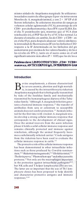
mismo ais<strong>la</strong>do <strong>de</strong> Anap<strong>la</strong>sma marginale. Se utilizaron siete bovinos <strong>de</strong> 12 a 14 meses <strong>de</strong> edad. El día 0 fueron<br />
inocu<strong>la</strong>dos cuatro <strong>de</strong> ellos (grupo I) por vía intravenosa con 1 × 10 8 eritrocitos parasitados (EP), con el ais<strong>la</strong>do<br />
Morelos <strong>de</strong> A. marginale/animal, y con 2 × 10 8 EP el día 60; mientras que los otros tres bovinos (grupo T) no<br />
fueron infectados. Se colectaron muestras <strong>de</strong> sangre periférica y suero <strong>de</strong> todos los animales, para evaluar el<br />
volumen celu<strong>la</strong>r aglomerado (VCA), porcentaje <strong>de</strong> eritrocitos parasitados (PEP), linfocitos T CD2+, CD4+ y<br />
CD8+ por citofluorometría y Acs e IFN-γ por ELISA. En el grupo I, el PEP alcanzó un promedio máximo <strong>de</strong> 5.2%<br />
el día 31 posinfección (pi), mientras que el VCA disminuyó hasta un promedio <strong>de</strong> 6% el día 46 pi. En <strong>la</strong><br />
reinfección (ri), el PEP fue <strong>de</strong> 0 y el VCA fue normal. Los valores <strong>de</strong> CD2+ fueron simi<strong>la</strong>res en los dos grupos<br />
durante el estudio; en cambio, los <strong>de</strong> CD4+ y CD8+ en el grupo I disminuyeron significativamente (P < 0.05)<br />
en el día 45 pi, para <strong>de</strong>spués alcanzar valores simi<strong>la</strong>res a los <strong>de</strong>l grupo T. Se observó un aumento (P < 0.05) <strong>de</strong><br />
<strong>la</strong> intensidad <strong>de</strong> fluorescencia (IF) en los linfocitos CD2+ y CD8+ <strong>de</strong>l grupo I <strong>de</strong>spués <strong>de</strong> <strong>la</strong> ri (días 62 y 83) con<br />
respecto a <strong>la</strong> IF <strong>de</strong>terminada en los linfocitos <strong>de</strong>l grupo T. Los niveles <strong>de</strong> Acs <strong>de</strong> los bovinos infectados<br />
aumentaron por encima <strong>de</strong> los valores basales y <strong>de</strong> los <strong>de</strong> los animales testigo, a partir <strong>de</strong>l día 51 pi. Asimismo,<br />
los niveles <strong>de</strong> IFN-γ , tanto en el suero <strong>com</strong>o en el sobrenadante <strong>de</strong> CSC <strong>de</strong> los bovinos infectados, fueron<br />
significativamente más altos (P < 0.05) que los <strong>de</strong> los animales no infectados, particu<strong>la</strong>rmente en <strong>la</strong> ri.<br />
Pa<strong>la</strong>bras Pa<strong>la</strong>bras c<strong>la</strong>ve: c<strong>la</strong>ve: LINFOCITOS LINFOCITOS T T CD2+, CD2+, CD2+, CD4+ CD4+ Y Y CD8+ CD8+ CD8+ DE DE SANGRE SANGRE PERIFÉRICA PERIFÉRICA DE DE DE BOVINO, BOVINO, AAAAANAPLASMA<br />
NAPLASMA<br />
NAPLASMA<br />
NAPLASMA<br />
NAPLASMA<br />
MARGINALE, MARGINALE<br />
MARGINALE<br />
MARGINALE<br />
MARGINALE , ANTICUERPOS ANTICUERPOS IgG, IgG, INTERFERON INTERFERON GAMMA.<br />
GAMMA.<br />
Introduction<br />
ovine anap<strong>la</strong>smosis, a disease characterized<br />
by severe anemia, abortion and high mortality,<br />
is caused by the intraerithrocytic rickettsia<br />
Anap<strong>la</strong>sma marginale that is biologically transmitted<br />
by ticks of the Ixodidae family and mechanically<br />
transmitted by hematophagous diptera of the Tabanidae<br />
family. 1 Although A. marginale infection generates<br />
a <strong>humoral</strong> immune response, 2,3 the transfer of<br />
antibodies from sera or colostrum to susceptible<br />
animals does not confer protection. 3-5 Animals infected<br />
for the first time with virulent A. marginale strains<br />
<strong>de</strong>velop a strong cellu<strong>la</strong>r immune response that<br />
corresponds to the <strong>de</strong>velopment of clinical signs.<br />
Once the animal recovers from the acute infection<br />
phase it holds a solid cellu<strong>la</strong>r immune response and<br />
remains clinically protected and immune against<br />
reinfection, although the animal frequently be<strong>com</strong>es<br />
a subclinically infected carrier. 6 Cru<strong>de</strong> antigens<br />
from the rickettsia have been used to evaluate the<br />
cellu<strong>la</strong>r immune response in cattle. 7-9<br />
The protective role of the cellu<strong>la</strong>r immune response<br />
has been <strong>de</strong>monstrated in other intracellu<strong>la</strong>r infections<br />
such as those produced by Cowdria ruminantium,<br />
10-12 Rickettsia tsutsugamushi, 13,14 Ehrlichia risticii, 15,16<br />
Theileria parva, 17 P<strong>la</strong>smodium spp18 and other parasitic<br />
protozoa. 19 Not only are the macrophages important<br />
in the protection against intracellu<strong>la</strong>r pathogens 20-22<br />
but NK cells and T helper lymphocytes (Th) are too.<br />
The <strong>la</strong>tter are so important that the use of Th1 lymphocyte<br />
clones has been proposed to help i<strong>de</strong>ntify<br />
and characterize protective antigens and immune<br />
responses. 23,24<br />
248<br />
248<br />
Introducción<br />
a anap<strong>la</strong>mosis bovina, enfermedad caracterizada<br />
por anemia severa, abortos y alta mortalidad, es<br />
causada por <strong>la</strong> rickettsia intraeritrocítica Anap<strong>la</strong>sma<br />
marginale que es transmitida biológicamente por garrapatas<br />
<strong>de</strong> <strong>la</strong> familia Ixodidae y mecánicamente por dípteros<br />
hematófagos <strong>de</strong> <strong>la</strong> familia Tabanidae. 1 Aunque <strong>la</strong> infección<br />
por A. marginale genera una <strong>respuesta</strong> <strong>inmune</strong> <strong>humoral</strong>, 2,3<br />
<strong>la</strong> transferencia <strong>de</strong> anticuerpos <strong>de</strong>l suero o <strong>de</strong>l calostro a<br />
animales susceptibles no confiere protección. 3-5 Los animales<br />
que se infectan por primera vez con cepas virulentas <strong>de</strong><br />
A. marginale <strong>de</strong>sarrol<strong>la</strong>n una <strong>respuesta</strong> <strong>inmune</strong> celu<strong>la</strong>r<br />
sólida que correspon<strong>de</strong> al <strong>de</strong>sarrollo <strong>de</strong> signos clínicos; una<br />
vez que el animal se recupera <strong>de</strong> <strong>la</strong> fase aguda <strong>de</strong> <strong>la</strong><br />
infección mantiene una inmunidad celu<strong>la</strong>r vigorosa y<br />
permanece clínicamente protegido e <strong>inmune</strong> contra <strong>la</strong><br />
reinfección, aunque con frecuencia el animal se transforma<br />
en un portador subclínicamente infectado. 6 Para evaluar <strong>la</strong><br />
<strong>respuesta</strong> <strong>inmune</strong> celu<strong>la</strong>r en anap<strong>la</strong>smosis bovina se han<br />
utilizado antígenos crudos <strong>de</strong> <strong>la</strong> rickettsia. 7-9<br />
Se ha <strong>de</strong>mostrado el papel protector <strong>de</strong> <strong>la</strong> inmunidad<br />
celu<strong>la</strong>r en otras infecciones intracelu<strong>la</strong>res, <strong>com</strong>o <strong>la</strong>s inducidas<br />
por Cowdria ruminantium, 10-12 Rickettsia tsutsugamushi,<br />
13,14 Ehrlichia risticii, 15,16 Theileria parva, 17 P<strong>la</strong>smodium spp18 y otros protozoarios parásitos. 19 No so<strong>la</strong>mente los macrófagos<br />
<strong>de</strong>sempeñan un papel importante en <strong>la</strong> protección<br />
contra patógenos intracelu<strong>la</strong>res20-22 sino también <strong>la</strong>s célu-<br />
<strong>edigraphic</strong>.<strong>com</strong><br />
<strong>la</strong>s NK y los linfocitos T cooperadores (Th). Tan importan-<br />
tes son los últimos que se ha propuesto utilizar clones <strong>de</strong><br />
linfocitos Th1 <strong>com</strong>o sondas para i<strong>de</strong>ntificar y caracterizar<br />
antígenos protectores y <strong>respuesta</strong>s <strong>inmune</strong>s. 23,24<br />
Con base en lo anterior, es necesario conocer los<br />
mecanismos inmunitarios protectores que operan en


