Parestesia del nervio dentario inferior provocada ... - Medicina Oral
Parestesia del nervio dentario inferior provocada ... - Medicina Oral
Parestesia del nervio dentario inferior provocada ... - Medicina Oral

You also want an ePaper? Increase the reach of your titles
YUMPU automatically turns print PDFs into web optimized ePapers that Google loves.
Med <strong>Oral</strong> 2003;8:299-303. <strong>Parestesia</strong> por tratamiento endodóncico / <strong>Parestesia</strong> by endodontic treatment<br />
<strong>Parestesia</strong> <strong>del</strong> <strong>nervio</strong> <strong>dentario</strong> <strong>inferior</strong> <strong>provocada</strong> por un<br />
tratamiento endodóncico<br />
Mª Mercedes Gallas Torreira (1), Mª Dolores Reboiras López (2), Abel García García (3),<br />
José Gándara Rey (4)<br />
(1) Profesora Ayudante Odontología Integrada Adultos. Facultad de <strong>Medicina</strong> y Odontología. Universidad de Santiago de<br />
Compostela<br />
(2) Odontóloga. Máster <strong>Medicina</strong> <strong>Oral</strong>, Cirugía <strong>Oral</strong> e Implantología <strong>Oral</strong>. Universidad de Santiago de Compostela<br />
(3) Profesor Titular de Cirugía <strong>Oral</strong> y Maxilofacial. Facultad de <strong>Medicina</strong> y Odontología. Universidad de Santiago de Compostela<br />
(4) Catedrático de <strong>Medicina</strong> <strong>Oral</strong> y Maxilofacial. Facultad de <strong>Medicina</strong> y Odontología. Universidad de Santiago de Compostela.<br />
España<br />
Correspondencia:<br />
Mercedes Gallas Torreira<br />
Facultad de <strong>Medicina</strong> y Odontología. Dpto. Estomatología<br />
Rúa Entrerríos, S/N<br />
15782 SANTIAGO DE COMPOSTELA (A CORUÑA)<br />
Tel: 981 562026/ 981 563100 ext. 12415<br />
Fax: 981 562226<br />
E-mail: mmgallas@usc.es<br />
Recibido: 23/11/2002 Aceptado: 2/3/2003<br />
Gallas-Torreira MM, Reboiras-López MD, García-García A, Gánda<br />
ra-Rey J. <strong>Parestesia</strong> <strong>del</strong> <strong>nervio</strong> <strong>dentario</strong> <strong>inferior</strong> <strong>provocada</strong> por un tratamiento<br />
endodóncico. Med <strong>Oral</strong> 2003;8:299-303.<br />
© <strong>Medicina</strong> <strong>Oral</strong> S. L. C.I.F. B 96689336 - ISSN 1137 - 2834<br />
RESUMEN<br />
Las parestesias <strong>del</strong> <strong>nervio</strong> <strong>dentario</strong> <strong>inferior</strong> constituyen una complicación<br />
que puede ocurrir tras la realización de varios procedimientos<br />
odontológicos como son: las cistectomías, la extracción<br />
de dientes retenidos, las apicectomías, los tratamientos<br />
endodónticos, la colocación de anestesia local, o la cirugía<br />
implantológica o preprotésica.<br />
Los posibles mecanismos de la lesión <strong>nervio</strong>sa son mecánicos,<br />
químicos y térmicos. El daño mecánico incluye compresión,<br />
estiramiento, resección parcial o total y laceración. La lesión<br />
puede ocasionar una discontinuidad <strong>del</strong> <strong>nervio</strong> con degeneración<br />
walleriana de las fibras distales e integridad de la cubierta<br />
(axonotmesis) o puede causar la total sección <strong>del</strong> <strong>nervio</strong><br />
(neurotmesis). El trauma químico puede deberse a determinados<br />
componentes tóxicos de los materiales de relleno<br />
endodóncicos (paraformaldehido, corticoides o eugenol) e<br />
irrigantes (hipoclorito sódico) o anestésicos locales. El daño<br />
térmico es consecuencia <strong>del</strong> sobrecalentamiento óseo durante<br />
la realización de técnicas quirúrgicas.<br />
Presentamos el caso clínico de parestesia <strong>del</strong> <strong>nervio</strong> <strong>dentario</strong><br />
<strong>inferior</strong> tras la introducción de una punta de gutapercha en el<br />
canal mandibular durante la realización de la endodoncia <strong>del</strong><br />
primer molar <strong>inferior</strong>. Se describe la etiología y el tratamiento<br />
de esta complicación endodóncica.<br />
Palabras clave : <strong>Parestesia</strong>s <strong>del</strong> <strong>nervio</strong> <strong>dentario</strong> <strong>inferior</strong>, complicaciones<br />
endodóncicas.<br />
299<br />
INTRODUCCION<br />
Las parestesias <strong>del</strong> <strong>nervio</strong> <strong>dentario</strong> <strong>inferior</strong> pueden ser el resultado<br />
de traumatismos, tumores, enfermedades <strong>del</strong> tejido<br />
conectivo, enfermedades infecciosas, enfermedades<br />
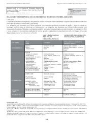
desmineralizantes o idiopáticas (Tabla 1). La causa más frecuente<br />
de neuropatía trigeminal es la traumática, siendo la más<br />
habitual de las neuropatías traumáticas en odontoestomatología<br />
la neuropatía <strong>del</strong> <strong>nervio</strong> alveolar <strong>inferior</strong> (1). Se trata de una<br />
neuropatía con afectación sensitiva deficitaria <strong>del</strong> territorio de<br />
inervación <strong>del</strong> mentoniano debida a la exodoncia <strong>del</strong> tercer molar<br />
<strong>inferior</strong> retenido (2,3). También pueden ser debidas a patología<br />
periodontal o iatrogenia endodóncica periapical (4,5) como<br />
consecuencia de la extrusión de material de relleno en el canal<br />
mandibular o sobreinstrumentación (6).<br />
La lesión <strong>del</strong> <strong>nervio</strong> <strong>dentario</strong> <strong>inferior</strong> después de tratamientos<br />
endodóncicos constituye una rarísima complicación en la terapia<br />
endodóncica.<br />
El primer síntoma de sobreobturación en el canal mandibular<br />
es un dolor repentino referido por el paciente durante la<br />
obturación <strong>del</strong> conducto y que persiste después de cesar los<br />
efectos de la anestesia local (7). El dolor puede acompañarse<br />
de signos inflamatorios locales, con percusión dolorosa <strong>del</strong> diente<br />
endodonciado, dolor a la palpación <strong>del</strong> proceso alveolar<br />
vestibular o la combinación de signos de lesión mecánica e<br />
inflamatoria <strong>del</strong> <strong>nervio</strong> <strong>dentario</strong> <strong>inferior</strong> con dolor o adormecimiento<br />
<strong>del</strong> labio <strong>inferior</strong> u otalgia (8). Algunos pacientes refie-
Med <strong>Oral</strong> 2003;8:299-303. <strong>Parestesia</strong> por tratamiento endodóncico / <strong>Parestesia</strong> by endodontic treatment<br />
ren la persistencia de la anestesia local (9).<br />
Para el tratamiento, actualmente se aconseja la inmediata remoción<br />
quirúrgica de la causa que comprima o dañe las fibras<br />
<strong>nervio</strong>sas para evitar agravar el cuadro (7,10,11).También se<br />
recomienda la prevención de esta complicación mediante la correcta<br />
evaluación radiográfica previa de la proximidad de los<br />
ápices <strong>dentario</strong>s de los molares <strong>inferior</strong>es al conducto <strong>dentario</strong><br />
<strong>inferior</strong> y el control radiológico durante la realización de la<br />
endodoncia (12).<br />
CASO CLINICO<br />
Paciente mujer de 45 años de edad, sin antecedentes médicos<br />
de interés que acude a consulta en el Servicio de Cirugía <strong>Oral</strong> y<br />
Maxilofacial <strong>del</strong> Complejo Hospitalario Universitario de Santiago<br />
de Compostela remitida por su odontoestomatólogo al<br />
presentar dolor en la hemimandíbula izquierda y adormecimiento<br />
<strong>del</strong> hemilabio izquierdo de 15 días de evolución. La paciente<br />
nos refiere la realización de un tratamiento endodóncico previo<br />
en el primer molar <strong>inferior</strong> izquierdo y posteriormente, al<br />
cabo de un mes al persistir el dolor y las molestias en el molar<br />
<strong>inferior</strong> su odontoestomatólogo comprueba que en el tratamiento<br />
endodóntico <strong>del</strong> conducto distal una punta de gutapercha había<br />
sobrepasado el ápice radicular hallándose en el conducto <strong>dentario</strong><br />
<strong>inferior</strong>, indicando entonces la exodoncia <strong>del</strong> molar. Apesar<br />
de la exodoncia, el dolor en la hemimandíbula izquierda y el<br />
adormecimiento <strong>del</strong> labio no ceden, por lo que nos remite a la<br />
paciente para valoración y tratamiento.<br />
A la exploración clínica intraoral observamos el tramo edéntulo<br />
correspondiente al primer molar <strong>inferior</strong> izquierdo en fase de<br />
cicatrización normal. La exploración neurológica <strong>del</strong> tercer par<br />
craneal evidenció la existencia de una hiperestesia dolorosa en<br />
la hemimandíbula izquierda y parestesia en el hemilabio izquierdo.<br />
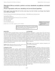
Se realiza un estudio radiológico mediante ortopantomografía<br />
y comprobamos la presencia de una punta de gutapercha retenida<br />
en el fondo <strong>del</strong> alvéolo de la raíz distal <strong>del</strong> primer molar<br />
<strong>inferior</strong> izquierdo (Fig.1). Se procede a la extracción de la punta<br />
de gutapercha con anestesia local en régimen ambulatorio.<br />
Para ello se levanta un colgajo mucoperióstico de espesor total<br />
que permite practicar una ostectomía a nivel de la cavidad ósea<br />
<strong>del</strong> alvéolo <strong>del</strong> primer molar <strong>inferior</strong> izquierdo, extraer la punta<br />
de gutapercha y curetear cuidadosamente la cavidad ósea.<br />
Pasados quince días, había desaparecido el dolor aunque persistía<br />
la parestesia de labio <strong>inferior</strong>. Esta se recuperó posteriormente,<br />
aproximadamente al mes de la extracción de la punta de<br />
gutapercha.<br />
DISCUSION<br />
La lesión de <strong>nervio</strong> <strong>dentario</strong> <strong>inferior</strong> tras un tratamiento<br />
endodóntico puede ser el resultado de un trauma físico, de un<br />
trauma químico irritativo o de la combinación de ambos (13).<br />
Nitzan DW et al. (1983) postulan los siguientes mecanismos<br />
etiopatogénicos:<br />
1.- El trauma directo <strong>del</strong> <strong>nervio</strong> <strong>dentario</strong> <strong>inferior</strong> por los instrumentos<br />
empleados en la preparación <strong>del</strong> conducto (limas o<br />
condensadores), o por el material de obturación (puntas de plata).<br />
300<br />
2.- La compresión <strong>del</strong> <strong>nervio</strong> debido a un hematoma (presión<br />
directa) o a la presencia de un material de relleno <strong>del</strong> conducto<br />
en el canal <strong>dentario</strong> (presión indirecta) (14).<br />
3.- El daño químico <strong>del</strong> <strong>nervio</strong> por extravasación a través <strong>del</strong><br />
ápice radicular de sustancias tóxicas o medicamentos introducidos<br />
en el conducto radicular, por ejemplo: endometasona,<br />
N2,.....<br />
Incluso la lesión <strong>del</strong> <strong>nervio</strong> puede ser causada por una infección<br />
local tras la sobreobturación <strong>del</strong> material en el canal<br />
mandibular (15).<br />
En el caso clínico descrito, probablemente durante la obturación<br />
de la endodoncia debido a la realización de una presión excesiva<br />
en la condensación, una de las puntas de gutapercha traspasase<br />
el ápice <strong>del</strong> molar introduciéndose en el conducto <strong>dentario</strong><br />
<strong>inferior</strong>. Desconocemos el mecanismo por el cual la punta de<br />
gutapercha llegó a introducirse en el conducto <strong>dentario</strong> <strong>inferior</strong>.<br />
Es posible que durante la preparación <strong>del</strong> conducto se sobrepasase<br />
la longitud de trabajo creando una vía de acceso en<br />
hueso y en la posterior obturación <strong>del</strong> conducto la punta de<br />
gutapercha traspasase el ápice alcanzando el <strong>nervio</strong> <strong>dentario</strong><br />
<strong>inferior</strong>. Otra posibilidad es la existencia previa de un granuloma<br />
en el periápice que condicionaría la existencia de un área<br />
osteolítica o de hueso poco denso en la zona <strong>del</strong> periápice lo<br />
que a su vez constituiría un factor favorable para el rebasamiento<br />
apical y posterior lesión <strong>del</strong> <strong>nervio</strong> <strong>dentario</strong> <strong>inferior</strong>.<br />
Tabla 1. Tipos de<br />
Neuropatías Trigeminales.<br />
(Peñarrocha M.)<br />
T T<br />
R<br />
R<br />
A<br />
A<br />
U<br />
U<br />
M<br />
M<br />
A<br />
A<br />
T<br />
T<br />
I<br />
I<br />
S<br />
S<br />
M<br />
M<br />
O<br />
O<br />
S<br />
S<br />
D<br />
D<br />
I<br />
I<br />
R<br />
R<br />
E<br />
E<br />
C<br />
C<br />
T<br />
T<br />
O<br />
O<br />
S<br />
S<br />
E<br />
E<br />
I I<br />
N<br />
N<br />
D<br />
D<br />
I<br />
I<br />
R<br />
R<br />
E<br />
E<br />
C<br />
C<br />
T<br />
T<br />
O<br />
O<br />
S<br />
S<br />
-Exodoncias<br />
dentarias<br />
( cordales<br />
<strong>inferior</strong>es)<br />
-Anestesia<br />
troncular<br />
-Implantología<br />
-Terapia<br />
endodó<br />
ncica<br />
-Cirugía<br />
ortognática<br />
TUMORES<br />
1.<br />
- Malignos<br />
-Intracraneales<br />
- nÁ gulo<br />
pontocerebeloso<br />
-Ganglio<br />
de<br />
Gasser<br />
-Ramas<br />
<strong>del</strong><br />
Trigé<br />
mino<br />
2.<br />
- Benignos<br />
-Odontomas<br />
ENFERME<br />
DAD<br />
ES<br />
DEL<br />
COLÁG<br />
ENO<br />
-Lupus<br />
Eritematoso<br />
-Dermatomiosistis<br />
-Esclerosis<br />
Sisté<br />
mica<br />
progresiva<br />
-Síndrome<br />
de<br />
Sjogren<br />
-Artitis<br />
Reumatoide<br />
-Enfermedad<br />
Mixta<br />
<strong>del</strong><br />
tejido<br />
conectivo<br />
NEU<br />
ROPATÍ<br />
A TRIGEMIN<br />
AL<br />
SEN<br />
SORIA<br />
L<br />
BEN<br />
IGNA<br />
INFECCI<br />
ONES<br />
-Virus<br />
<strong>del</strong><br />
Herpes<br />
Zó<br />
ster<br />
-Asociació<br />
n Herpes<br />
Simple<br />
-Sífilis<br />
-Lepra<br />
-SIDA<br />
OTRAS<br />
-Esclerosis<br />
Múltiple<br />
-Enfermedades<br />
Vasculares<br />
Vertebrobasilares<br />
-Sarcoidosis<br />
-Amiloidosis<br />
-Anemia<br />
de<br />
cé<br />
lulas<br />
falciformes<br />
-Químicos:<br />
ttos<br />
endodó<br />
ncicos<br />
-Tó<br />
xicos:<br />
Hidroestilbamida,<br />
tricloroetileno.
Med <strong>Oral</strong> 2003;8:299-303. <strong>Parestesia</strong> por tratamiento endodóncico / <strong>Parestesia</strong> by endodontic treatment<br />
Se han descrito en la literatura otros casos en los que existió<br />
una patología periodontal previa (4) o una infección periapical<br />
(16), bien después de necrosis químicas arsenicales (7), o por<br />
el empleo de cementos como el AH26 (9) o la endometasona<br />
(17).<br />
La sintomatología originada por la lesión <strong>del</strong> <strong>nervio</strong> <strong>dentario</strong><br />
<strong>inferior</strong> puede ser temporal o permanente, dependiendo de la<br />
severidad <strong>del</strong> traumatismo causado (12,18). Diversos autores<br />
mencionan la existencia de una correlación directa entre la duración<br />
y la importancia <strong>del</strong> traumatismo ocasionado al <strong>nervio</strong> y<br />
el pronóstico de la parestesia (3,9). Así, la evolución de la afectación<br />
<strong>nervio</strong>sa por un daño químico dependerá <strong>del</strong> tipo de<br />
material empleado –toxicidad- y de la rápida eliminación <strong>del</strong><br />
material (18). Las parestesias mandibulares relacionadas con<br />
la colocación de anestesia local o la sobreinstrumentación<br />
endodóncica se resuelven normalmente en unos cuantos días.<br />
Una parestesia de mayor duración e incluso permanente puede<br />
resultar en casos de laceración de la fibra <strong>nervio</strong>sa, presión prolongada<br />
<strong>del</strong> <strong>nervio</strong> o contacto continuado con materiales<br />
endodóncicos tóxicos. La secuencia terapéutica indicada es la<br />
inmediata eliminación de la causa, siempre que sea posible y el<br />
control de la inflamación.<br />
ENGLISH<br />
Mandibular nerve paresthesia<br />
caused by endodontic<br />
treatment<br />
GALLAS-TORREIRA MM, REBOIRAS-LÓPEZ MD, GARCÍA-GARCÍA A,<br />
GÁNDA RA-REY J. MANDIBULAR NERVE PARESTHESIA CAUSED BY<br />
ENDODONTIC TREATMENT. MED ORAL 2003;8:299-303.<br />
SUMMARY<br />
The paresthesias of the <strong>inferior</strong> dental nerve consists of a<br />
complication that can occur after performing various dental<br />
procedures such as cystectomies, extraction of impacted teeth,<br />
apicoectomies, endodontic treatments, local anesthetic<br />
deposition, preprosthetic or implantologic surgery.<br />
The possible mechanisms of nervous lesions are mechanical,<br />
chemical and thermal. Mechanical injury includes compression,<br />
stretching, partial or total resection and laceration. The lesion<br />
can cause a discontinuity to the nerve with Wallerian<br />
degeneration of the distal and integrated fibers of the covering<br />
(axonotmesis) or can cause the total sectioning of the nerve<br />
(neurotmesis). Chemical trauma can be due to certain toxic<br />
components of the endodontic filling materials (paraformaldehyde,<br />
corticoids or eugenol) and irrigating solutions<br />
(sodium hypochlorite) or local anesthetics. Thermal injury is a<br />
consequence of bone overheating during the execution of<br />
surgical techniques.<br />
We present a clinical case of paresthesia of the <strong>inferior</strong> dental<br />
nerve after the introduction of a gutta-percha point in the<br />
301<br />
mandibular canal during the performance of a root canal therapy<br />
of the <strong>inferior</strong> first molar. The etiology and the treatment of this<br />
endodontic complication are described.<br />
Key words : Paresthesias of the <strong>inferior</strong> dental nerve,<br />
endodontic complications.<br />
INTRODUCTION<br />
Paresthesias of the <strong>inferior</strong> dental nerve can be the result of<br />
traumatisms, tumors, connective tissue diseases, infectious<br />
diseases, demineralizing or idiopathic diseases (Table 1). The<br />
most frequent cause of trigeminal neuropathy is traumatism,<br />
the <strong>inferior</strong> dental nerve neuropathy being the most common of<br />
the traumatic neuropathies in dentistry (1). It deals with a<br />
neuropathy with poor sensitivity in the territory of the mental<br />
nerve innervation due to the exodontia of an impacted third<br />
molar (2,3). This can also be due to periodontal pathology or<br />
periapical endodontic iatrogeny (4,5) as a consequence of the<br />
extrusion of the filling material in the mandibular canal or over<br />
instrumentation (6).<br />
The lesion of the <strong>inferior</strong> dental nerve after endodontic<br />
treatments represents a very rare complication in root canal<br />
therapy.<br />
The first symptom of the overfilling into the mandibular canal<br />
is sudden pain expressed by the patient during obturation of the<br />
root canal, which persists after the disappearance of the local<br />
anesthetic effects (7). The pain can be accompanied by local<br />
inflammatory signs with the endodontically treated tooth being<br />
painful to percussion, painful upon palpation of the vestibular<br />
alveolar process or a combination of signs of mechanical lesions<br />
and <strong>inferior</strong> dental nerve inflammation with pain or numbness<br />
of the lower lip or otalgia (8). Some patients experience the<br />
persistence of the local anesthesia (9). Present day treatment<br />
recommendations consist of immediate surgical removal of the<br />
cause that compresses or injures the nerve fibers to avoid<br />
aggravating the symptoms (7,10,11). The prevention of this<br />
complication by means of a previous correct radiographic<br />
evaluation of the proximity of the lower molar apices to the<br />
mandibular canal and radiographic controls during the root canal<br />
therapy are recommended (12).<br />
CLINICAL CASE<br />
The patient was a 45-year-old woman with a medical history of<br />
no pathologic findings who visited the Service of <strong>Oral</strong> and<br />
Maxillofacial Surgery of the Santiago de Compostela University<br />
Hospital Complex for consultation. Referred by her dentist for<br />
presenting hemi-mandibular pain and numbness on the left half<br />
of the lip of a 15-day evolution. The patient told us of previously<br />
having undergone a root canal therapy on the lower left first<br />
molar. After one month of persistent pain and discomfort on<br />
the lower molar, her dentist confirmed the presence of a guttapercha<br />
point overfilling the radicular apex of the distal root<br />
canal of the endodontically treated tooth and present within the<br />
mandibular canal. Indicating therefore the extraction of the<br />
molar. In spite of the exodontia, the pain on the left half of the<br />
mandible and the lip numbness did not cease. Thus is the reason
Med <strong>Oral</strong> 2003;8:299-303. <strong>Parestesia</strong> por tratamiento endodóncico / <strong>Parestesia</strong> by endodontic treatment<br />
Fig. 1. Detalle de la ortopantomografía.<br />
Detail on the panoramic radiograph<br />
for the patient’s referral for evaluation and treatment.<br />
Upon intra-oral clinical examination we observed an edentolous<br />
section corresponding to the <strong>inferior</strong> left first molar at its normal<br />
healing phase. The neurologic examination of the third<br />
cranial nerve evidenced the existence of a painful hyperesthesia<br />
of the left hemi-mandible and paresthesia of the left half of the<br />
lip.<br />
A radiological study was performed by means of<br />
orthopantomography proving the presence of a gutta-percha<br />
point retained within the bottom part of the alveolar bone at the<br />
distal root of the <strong>inferior</strong> left first molar (Fig.1). The extraction<br />
of the gutta-percha point with local anesthetics was done on an<br />
outpatient-clinic basis. To do this a fully thick mucoperiosteum<br />
flap was done to permit performing ostectomy at the level of<br />
the bone cavity of the <strong>inferior</strong> left first molar alveolus, extracting<br />
the gutta-percha point and carefully curetting the bone cavity.<br />
After fifteen days, the pain had disappeared although paresthesia<br />
of the <strong>inferior</strong> lip still persisted. This recovered later,<br />
approximately one month after the gutta-percha extraction.<br />
DISCUSSION<br />
Inferior dental nerve lesion after endodontic traumatism can be<br />
the result of physical trauma, irritative chemical trauma or a<br />
combination of both (13) Nitzan DW et al. (1983) postulated<br />
the following etiopathogenic mechanisms:<br />
1. Direct trauma on the <strong>inferior</strong> dental nerve by the instruments<br />
used during root canal therapy (reamers/files or pluggers), or<br />
302<br />
by the obturation materials (silver points).<br />
2. Nerve compression due to hematoma (direct pressure) or due<br />
to the presence of a root canal filling material within the<br />
mandibular canal (direct pressure) (14).<br />
3. Chemical injury on the nerve by extravasation through the<br />
radicular apices of toxic substances or introduced medicaments<br />
within the root canal, as for example: endomethasone, N2,....<br />
The lesion of the nerve can even be caused by a local infection<br />
after overfilling the material into the mandibular canal (15).<br />
In the clinical case described, probably while obturating, one<br />
of the gutta-percha points went beyond the molar apex, being<br />
introduced into the mandibular canal due to the exertion of too<br />
much pressure during condensation. It is possible that during<br />
the mechanical preparation of the root canal the working length<br />
is exceeded, creating an access to the bone. Consequently, during<br />
the root canal obturation the gutta-percha point went beyond<br />
the apex reaching the <strong>inferior</strong> dental nerve. Another possibility<br />
is the previous existence of a granuloma at the periapex that<br />
conditions the existence of an osteolytic area of a less dense<br />
bone in the periapical zone. This at the same time constitutes a<br />
favorable factor for going beyond the apex and later injuring<br />
the <strong>inferior</strong> dental nerve.<br />
The symptomatology caused by the lesion of the <strong>inferior</strong> dental<br />
nerve can be temporary or permanent, depending on the<br />
traumatism produced (12,18). Some authors mention the<br />
existence of a direct correlation between the duration and the<br />
importance of traumatism produced on the nerve and the<br />
prognosis of the paresthesia (3,9). That way, the evolution of
Med <strong>Oral</strong> 2003;8:299-303. <strong>Parestesia</strong> por tratamiento endodóncico / <strong>Parestesia</strong> by endodontic treatment<br />
D<br />
I<br />
R<br />
E EE<br />
C<br />
TT<br />
T TT<br />
A<br />
N<br />
D<br />
I<br />
N<br />
D<br />
I<br />
R<br />
EE<br />
E<br />
C<br />
C<br />
TT<br />
T<br />
T T R<br />
R<br />
A<br />
A<br />
U<br />
U<br />
M<br />
M<br />
A<br />
A<br />
TT<br />
T<br />
I<br />
I<br />
S<br />
S<br />
M<br />
M<br />
S<br />
S<br />
-<br />
-Dental<br />
extractions<br />
( <strong>inferior</strong><br />
third<br />
molars)<br />
-Nerve<br />
block<br />
-Implantology<br />
-Endodontic<br />
therapy<br />
-Orthognathic<br />
surgery<br />
TUMORS<br />
1.<br />
-Malignant<br />
-Intracraneal<br />
-Pontocerebellar<br />
angle<br />
-Ganglion<br />
of<br />
Gasser<br />
-Trigeminal<br />
branches<br />
2.<br />
-Benign<br />
-Odontomas<br />
COLLA<br />
GEN<br />
DIS<br />
EAS<br />
ES<br />
-Lupus<br />
Erythematosus<br />
-Dermatomyocytes<br />
-Progressive<br />
Systemic<br />
Sclerosis<br />
-Sjogren<br />
Syndrome<br />
-Rheumatoid<br />
Arthritis<br />
-Mixed<br />
Disease<br />
of<br />
the<br />
connective<br />
tissue<br />
INFECTI<br />
OUS<br />
BEN<br />
IGN<br />
SEN<br />
SORY<br />
TRIGEMIN<br />
AL<br />
NEU<br />
ROPATHY<br />
-Herpes<br />
Zoster<br />
Virus<br />
-Herpes<br />
Simplex<br />
association<br />
-Syphilis<br />
-Leprosy<br />
-AIDS<br />
OTHERS<br />
-Multiple<br />
Sclerosis<br />
-Vertebrobasillar<br />
Vascular<br />
Diseases<br />
-Sarcoidosis<br />
-Amyloidosis<br />
-Falciform<br />
Cell<br />
Anemia<br />
-Chemical:<br />
endodontic<br />
treatments<br />
-Toxic:<br />
Hydrostilbamide,<br />
trichloroethelene<br />
Table 1. Types of Trigeminal Neuropathies (Peñarrocha M.)<br />
the nerve affectation by chemical injury will depend on the type<br />
of material employed –toxicity- and the rapid elimination of<br />
the material (18). The mandibular paresthesias related to the<br />
deposition of local anesthetic agent or endodontic overinstrumentation<br />
are normally resolved within a few days.<br />
Paresthesia of long or even of permanent duration can result in<br />
cases of laceration of the nerve fiber, prolonged pressure on the<br />
nerve or continuous contact with toxic endodontic materials.<br />
The indicated therapeutic sequence is the immediate elimination<br />
of the cause whenever possible and the control of inflammation.<br />
BIBLIOGRAFIA/REFERENCES<br />
1. Morse DR. Endodontic-related <strong>inferior</strong> alveolar nerve and mental foramen<br />
paresthesia. Compend Contin Educ Dent 1997;18:963-87.<br />
2. Peñarrocha M. Dolor Orofacial. Etiología, diagnóstico y tratamiento. Barcelona:<br />
Masson Ed; 1997. p. 218-22.<br />
3. Yana Y, Boukobza F, Mardam W, Derycke R. La paresthésie du nerf dentarie<br />
303<br />
inférieur: signes cliniques, diagnostic etiologique et pronostic. Rev<br />
Odontostomatol (Paris) 1990;19:411-20.<br />
4. Lambriadinis T, Molyvdas J. Paresthesia of the <strong>inferior</strong> alveolar nerve caused<br />
by periodontal-endodontic pathosis. <strong>Oral</strong> Surg <strong>Oral</strong> Med <strong>Oral</strong> Pathol<br />
1987;63:90-2.<br />
5. Pinsawasdi P. The induction of trigeminal neuralgia-like symptoms by pulpperiapical<br />
pathosis. J Endod 1986;12:73-5.<br />
6. Manisali Y, Yucel T, Erisen R. Overfilling of the root. A case report. <strong>Oral</strong><br />
Surg <strong>Oral</strong> Med <strong>Oral</strong> Pathol 1989;68:773-5.<br />
7. LaBlanc JP, Epker BN. Serious <strong>inferior</strong> alveolar nerve dyesthesia after<br />
endodontic procedure: report of three cases. J Am Dent Assoc 1984;108: 605-<br />
7.<br />
8. Grotz KA, Al-Nawas B, de Aguiar EG, Schulz A, Wagner W. Treatment of<br />
injuries to the <strong>inferior</strong> alveolar nerve after endodontic procedures. Clin <strong>Oral</strong><br />
Invest 1998;2:73-6.<br />
9. Neaverth EJ. Disabling complications following inadvertent overextension<br />
of a root canal filling material. J Endod 1989;15:135-9.<br />
10. Spielman A, Gutman D, Laufer D. Anesthesia following endodontic<br />
overfilling with AH26. Report of a case. <strong>Oral</strong> Surg <strong>Oral</strong> Med <strong>Oral</strong> Pathol<br />
1981;52:554-5.<br />
11. Franco M, Sivestrin M., Alexandre A, Mazzoleni G. Lesioni <strong>del</strong> nervo<br />
alveolare <strong>inferior</strong>e da cementi endodontici. Minerva Stomatol 1991;40:563-8.<br />
12. Sakkal S, Gagnon A, Lemian L. Paresthésie du nerf dentaire inférieur à la<br />
suite d’un traitement endodontique: Un cas clinique. J Can Dent Assoc<br />
1994;60:556-8.<br />
13. Nitzan DW, Stabholz A, Azaz B. Concepts of accidental overfilling and<br />
over instrumentation in the mandibular canal during root canal treatment. J<br />
Endod 1983;9:81-5.<br />
14. Fanibunda K, Whitworth J, Steete J. The management of thermomechanically<br />
compacted gutta percha extrusion in the <strong>inferior</strong> dental canal. Brit<br />
Dent J 1998;184:330-2.<br />
15. Sonat B., Dalat D., Gunhan O. Periapical tissue reaction to root filling with<br />
Sealapex. Int Endod J 1990;23:46-52.<br />
16. Di Leonarda R, Cadenaro M, Stacchi C. Paresthesia of the mental nerve<br />
induced by periapical infection. A case report. <strong>Oral</strong> Surg <strong>Oral</strong> Med <strong>Oral</strong> Pathol<br />
2000;90:746-9.<br />
17. Erisen R, Yucel T, Kucukay S. Endomethasone root canal filling material in<br />
the mandibular canal. A case report. <strong>Oral</strong> Surg <strong>Oral</strong> Med <strong>Oral</strong> Pathol<br />
1989;68;343-5.<br />
18. Kothari P, Hanson N, Cannell H. Bilateral mandibular nerve damage<br />
following root canal therapy. Brit Dent J 1996;180:189-90.