Local flap reconstruction of large scalp defects - Medicinaoral.com
Local flap reconstruction of large scalp defects - Medicinaoral.com
Local flap reconstruction of large scalp defects - Medicinaoral.com

You also want an ePaper? Increase the reach of your titles
YUMPU automatically turns print PDFs into web optimized ePapers that Google loves.
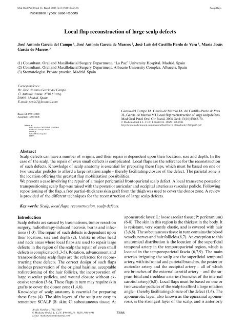
Med Oral Patol Oral Cir Bucal. 2008 Oct1;13(10):E666-70. Scalp <strong>flap</strong>s<br />
<strong>Local</strong> <strong>flap</strong> <strong>reconstruction</strong> <strong>of</strong> <strong>large</strong> <strong>scalp</strong> <strong>defects</strong><br />
José Antonio García del Campo 1 , José Antonio García de Marcos 2 , José Luis del Castillo Pardo de Vera 1 , María Jesús<br />
García de Marcos 3<br />
(1) Consultant. Oral and Maxill<strong>of</strong>acial Surgery Department. “La Paz” University Hospital. Madrid, Spain<br />
(2) Consultant. Oral and Maxill<strong>of</strong>acial Surgery Department. Albacete University Complex. Albacete, Spain<br />
(3) Stomatologist. Private practice. Madrid. Spain<br />
Correspondence:<br />
Dr. José Antonio García del Campo<br />
C/ Antonio Acuña. Nº10.5ºAizq.<br />
28009. Madrid, Spain<br />
E-mail: pepio2@hotmail.<strong>com</strong><br />
Received: 09/01/2008<br />
Accepted: 16/05/2008<br />
Publication Types: Case Reports<br />
Indexed in:<br />
-Index Medicus / MEDLINE / PubMed<br />
-EMBASE, Excerpta Medica<br />
-SCOPUS<br />
-Indice Médico Español<br />
-IBECS<br />
García-del Campo JA, García-de Marcos JA, del Castillo-Pardo de Vera<br />
JL, García-de Marcos MJ. <strong>Local</strong> <strong>flap</strong> <strong>reconstruction</strong> <strong>of</strong> <strong>large</strong> <strong>scalp</strong> <strong>defects</strong>.<br />
Med Oral Patol Oral Cir Bucal. 2008 Oct1;13(10):E666-70.<br />
© Medicina Oral S. L. C.I.F. B 96689336 - ISSN 1698-6946<br />
http://www.medicinaoral.<strong>com</strong>/medoralfree01/v13i10/medoralv13i10p666.pdf<br />
Abstract<br />
Scalp <strong>defects</strong> can have a number <strong>of</strong> origins, and their repair is dependent upon their location, size and depth. In the<br />
case <strong>of</strong> the <strong>scalp</strong>, the repair <strong>of</strong> even small <strong>defects</strong> is <strong>com</strong>plicated. <strong>Local</strong> <strong>flap</strong>s are the reference for the <strong>reconstruction</strong><br />
<strong>of</strong> such <strong>defects</strong>. Knowledge <strong>of</strong> <strong>scalp</strong> anatomy is essential for preparing these <strong>flap</strong>s, which must be based on one or<br />
two vascular pedicles to afford a <strong>large</strong> rotation angle – thereby facilitating closure <strong>of</strong> the defect. The parietal zone is<br />
the location <strong>of</strong>fering the greatest <strong>flap</strong> mobilization possibilities.<br />
We present a case involving the repair <strong>of</strong> a major pericranial frontoparietal <strong>scalp</strong> defect. A local transverse posterior<br />
transpositioning <strong>scalp</strong> <strong>flap</strong> was raised with the posterior auricular and occipital arteries as vascular pedicle. Following<br />
repositioning <strong>of</strong> the <strong>flap</strong>, a free partial-thickness skin graft from the thigh was used to cover the donor zone. A review<br />
is provided <strong>of</strong> the different techniques for the <strong>reconstruction</strong> <strong>of</strong> <strong>large</strong> <strong>scalp</strong> <strong>defects</strong>.<br />
Key words: Scalp, local <strong>flap</strong>s, <strong>reconstruction</strong>, <strong>scalp</strong> <strong>defects</strong>.<br />
Introduction<br />
Scalp <strong>defects</strong> are caused by traumatisms, tumor resection<br />
surgery, radiotherapy-induced necrosis, burns and infections<br />
(1-3). The repair <strong>of</strong> such <strong>defects</strong> is dependent upon<br />
their location, size and depth (2). Unlike in other head<br />
and neck areas where local <strong>flap</strong>s are used to repair <strong>large</strong><br />
<strong>defects</strong>, in the region <strong>of</strong> the <strong>scalp</strong> the repair <strong>of</strong> even small<br />
<strong>defects</strong> is <strong>com</strong>plicated (1,3-5). Rotation, advancement and<br />
transpositioning <strong>scalp</strong> <strong>flap</strong>s are the reference for reconstructing<br />
these <strong>defects</strong>. The correct design <strong>of</strong> such <strong>flap</strong>s<br />
includes preservation <strong>of</strong> the original hairline, acceptable<br />
redirectioning <strong>of</strong> the hair follicles, the incorporation <strong>of</strong><br />
<strong>large</strong> vascular pedicles, and wound closure without excessive<br />
tension (3-6). These <strong>flap</strong>s in turn may require skin<br />
grafts to cover the donor zone (1,4,6).<br />
Knowledge <strong>of</strong> <strong>scalp</strong> anatomy is essential for preparing<br />
these <strong>flap</strong>s (4). The skin layers <strong>of</strong> the <strong>scalp</strong> are easy to<br />
remember: SCALP (S: skin; C: subcutaneous tissue; A:<br />
Article Number: 1111111619<br />
© Medicina Oral S. L. C.I.F. B 96689336 - ISSN 1698-6946<br />
eMail: medicina@medicinaoral.<strong>com</strong><br />
E666<br />
aponeurotic layer; L: loose areolar tissue; P: pericranium)<br />
(6-8). The skin in this region is the thickest in the body. It<br />
is resistant, very scantly elastic, and is covered with hair<br />
(3,6,8). The subcutaneous tissue in turn contains the blood<br />
vessels, nerves and hair follicles (6,7). An exception to this<br />
anatomical distribution is the location <strong>of</strong> the superficial<br />
temporal artery in the temporoparietal region, which is<br />
located in the temporoparietal fascia (6,7,9). The main<br />
arteries irrigating the <strong>scalp</strong> are the superficial temporal<br />
artery, with its frontal and parietal branches, the posterior<br />
auricular artery and the occipital artery – all <strong>of</strong> which<br />
are branches <strong>of</strong> the external carotid artery – and the supraorbital<br />
and trochlear arteries (branches <strong>of</strong> the internal<br />
carotid artery)(6,8). <strong>Local</strong> <strong>flap</strong>s must be based on one or<br />
two vascular pedicles <strong>of</strong> the <strong>scalp</strong> to afford a <strong>large</strong> rotation<br />
angle – thereby facilitating closure <strong>of</strong> the defect (1,6). The<br />
aponeurotic layer, also known as the epicranial aponeurosis,<br />
is the strongest layer <strong>of</strong> the <strong>scalp</strong>, and is anteriorly
Med Oral Patol Oral Cir Bucal. 2008 Oct1;13(10):E666-70. Scalp <strong>flap</strong>s<br />
contiguous to the frontal muscle; posteriorly to the occipital<br />
muscles; and laterally to the temporoparietal fascia<br />
(6,8). As a result <strong>of</strong> its scant elasticity, the aponeurosis<br />
opposes desired <strong>flap</strong> advancement. This can be over<strong>com</strong>e<br />
by making incisions perpendicular to the direction <strong>of</strong> <strong>flap</strong><br />
movement (5,6,8). Correct closure with these <strong>flap</strong>s requires<br />
suturing the aponeurotic layer (6). <strong>Local</strong> <strong>scalp</strong> <strong>flap</strong>s are<br />
<strong>com</strong>posed <strong>of</strong> skin, subcutaneous tissue and aponeurosis,<br />
intimately connected by fibrous trabeculae. The loose<br />
areolar tissue – also known as the subaponeurotic fascia,<br />
fascia innominata, and subaponeurotic plane – allows<br />
mobility <strong>of</strong> the <strong>scalp</strong> (6,8). In this context, the parietal<br />
zone is the most mobile <strong>scalp</strong> region. The pericranium<br />
is intimately adhered to the skull. It must be left intact<br />
as far as possible, since free skin grafts can be applied to<br />
it – thereby facilitation <strong>reconstruction</strong> (6).<br />
There are a number <strong>of</strong> local <strong>flap</strong> techniques: single or<br />
multiple, and with or without skin grafting to close the<br />
donor <strong>defects</strong>. Single <strong>flap</strong>s in turn <strong>com</strong>prise rotation, advancement,<br />
transpositioning, bipedicle transpositioning,<br />
VY advancement and rhomboidal <strong>flap</strong>s, etc. (1,5,8,9).<br />
Multiple <strong>flap</strong>s in turn include the triple-<strong>flap</strong> technique<br />
described by Orticochea, the triple rotation (pinwheel)<br />
<strong>flap</strong>, double rotation <strong>flap</strong>, triple rhomboid <strong>flap</strong>, and V-Y-S<br />
<strong>scalp</strong> plasty technique, etc. (1,2,5). The patients at greatest<br />
risk <strong>of</strong> <strong>com</strong>plications after <strong>scalp</strong> <strong>reconstruction</strong> are those<br />
previously subjected to radiotherapy, patients receiving<br />
adjuvant or postoperative chemotherapy, those with<br />
cerebrospinal fluid fistulas, and patients with anteriorly<br />
located <strong>defects</strong> (4).<br />
We report the case <strong>of</strong> a patient with a recurrent <strong>scalp</strong> melanoma.<br />
Following resection <strong>of</strong> the lesion, a <strong>large</strong> frontoparietal<br />
defect including the pericranium remained. The<br />
defect was repaired by raising a local transverse posterior<br />
transpositioning <strong>scalp</strong> <strong>flap</strong> with the posterior auricular<br />
and occipital arteries as vascular pedicle. Following repositioning<br />
<strong>of</strong> the <strong>flap</strong>, a free partial-thickness skin graft<br />
from the thigh was used to cover the donor zone.<br />
Clinical Case<br />
An 82-year-old man was referred to our Service for evaluation<br />
and treatment <strong>of</strong> a long-evolving excrescent lesion<br />
in the frontoparietal region, measuring 2 x 2 x 1.2 cm in<br />
size. The growth was <strong>of</strong> s<strong>of</strong>t consistency, occasionally<br />
hemorrhagic, and was not adhered to deep layers. The personal<br />
history <strong>com</strong>prised surgery one month before for the<br />
removal <strong>of</strong> a basal cell epithelioma in the left nasogenian<br />
sulcus, and arterial hypertension treated with amiloride<br />
and hydrochlorothiazide.<br />
The lesion was removed under local anesthesia, with<br />
safety margins and preserving the periosteum. The resulting<br />
defect was repaired with a full-thickness skin graft<br />
from the supraclavicular region. The histopathological<br />
diagnosis was ulcerated polypoid nodular melanoma,<br />
corresponding to Level III <strong>of</strong> the Clark classification, a<br />
E667<br />
Breslow maximum thickness <strong>of</strong> 12 mm, and with tumorfree<br />
surgical margins.<br />
Twenty days after the operation the patient presented a 2.5<br />
x 2 cm ulcerated lesion in the operated zone, surrounded by<br />
multiple elevated nodular lesions – the <strong>large</strong>st measuring<br />
5 mm in diameter (Fig. 1A). Recurrent melanoma with<br />
satellitosis was diagnosed. The physical examination revealed<br />
no evidence if distant disease spread. The <strong>com</strong>plete<br />
blood count and biochemistry parameters were normal. A<br />
<strong>com</strong>puted tomography scan from head to abdomen for the<br />
evaluation <strong>of</strong> possible metastases proved negative. Under<br />
general anesthesia, radical resection <strong>of</strong> the frontoparietal<br />
<strong>scalp</strong> lesions was carried out, including the pericranium<br />
(Fig. 1B). The resulting post-resection defect measured 12<br />
x 8.5 x 0.4 cm in size. A local transverse posterior transpositioning<br />
<strong>scalp</strong> <strong>flap</strong> with preservation <strong>of</strong> the pericranium<br />
in the region <strong>of</strong> the <strong>flap</strong> was used to repair the defect (Fig.<br />
1C). Following repositioning <strong>of</strong> the <strong>flap</strong>, a free partialthickness<br />
skin graft from the right thigh was used to cover<br />
the donor zone (Fig. 1D and E). An aspirating drain was<br />
placed, and the wound was subjected to layered suturing.<br />
In the region <strong>of</strong> the local <strong>flap</strong> we placed a moderate <strong>com</strong>pression<br />
bandage, while gauze impregnated in antibiotic<br />
cream was placed over the skin graft zone, affixed with silk,<br />
and <strong>com</strong>pressing the graft to avoid hematoma formation.<br />
The histological study confirmed nodular melanoma metastasis,<br />
with disease-free resection margins (Fig. 1F). One<br />
and a half months after the operation, a <strong>flap</strong> scar plasty<br />
was performed, removing the “dog ear” left after surgery.<br />
The esthetic out<strong>com</strong>e was satisfactory (Fig. 2A and B).<br />
Five years after the last oncological operation, no evidence<br />
<strong>of</strong> local or regional disease recurrence is observed.<br />
Discussion<br />
There are many options for repairing <strong>large</strong> <strong>scalp</strong> <strong>defects</strong><br />
(1,9). The use <strong>of</strong> tissue expanders facilitates direct defect<br />
closure (1,6). This latter technique yields a <strong>large</strong>r amount<br />
<strong>of</strong> <strong>scalp</strong> tissue by placing an expanding device in the loose<br />
areolar tissue – with good esthetic results (4). Expansion<br />
preferably should be carried out before removal <strong>of</strong> the<br />
lesion, and constitutes an excellent repair option for the<br />
<strong>reconstruction</strong> <strong>of</strong> <strong>defects</strong> following the resection <strong>of</strong> benign<br />
lesions (1,4).<br />
Healing by second intention is a possibility for repairing<br />
<strong>scalp</strong> <strong>defects</strong>, though this option is not applicable in the<br />
presence <strong>of</strong> <strong>large</strong> <strong>defects</strong> with a lack <strong>of</strong> pericranium (1).<br />
The use <strong>of</strong> free skin grafts for <strong>reconstruction</strong> requires an<br />
intact pericranium to supply vascularization to the repaired<br />
zone. In addition, careful hemostasia <strong>of</strong> the receptor<br />
bed is necessary, since the formation <strong>of</strong> hematomas would<br />
<strong>com</strong>promise graft success. In the absence <strong>of</strong> a pericranium,<br />
and if grafting is the only repair option, perforations in<br />
the external cortical layer <strong>of</strong> the skull allow the formation<br />
<strong>of</strong> granulation tissue that would improve the prognosis <strong>of</strong><br />
the second-step free skin graft (1,4).
Med Oral Patol Oral Cir Bucal. 2008 Oct1;13(10):E666-70. Scalp <strong>flap</strong>s<br />
Fig. 1. A: Preoperative view <strong>of</strong> the lesion, showing recurrence <strong>of</strong> the melanoma with satellitosis; B: Defect after tumor resection, in the frontoparietal<br />
region; C: Transverse posterior transpositioning <strong>flap</strong>, preserving the pericranium in the region <strong>of</strong> the <strong>flap</strong>; D: Partial-thickness skin graft from<br />
the thigh applied to the pericranial zone to cover the donor site; E: Final appearance. Note the “dog ear” in the <strong>flap</strong> rotation zone; F: View <strong>of</strong> the<br />
surgical piece.<br />
E668
Med Oral Patol Oral Cir Bucal. 2008 Oct1;13(10):E666-70. Scalp <strong>flap</strong>s<br />
Fig. 2. Postoperative view. A: Four months after oncological surgery; B: Ten months after oncological surgery.<br />
The island graft is a local graft variant <strong>com</strong>posed <strong>of</strong> a<br />
<strong>scalp</strong> island without pericranium, with a vascular pedicle<br />
surrounded by subcutaneous tissue and an aponeurotic<br />
layer. It is mobilized to cover <strong>scalp</strong> <strong>defects</strong> (3).<br />
Pericranial and fascial <strong>flap</strong>s have also been described in<br />
which the pericranium, the frontal aponeurosis or temporoparietal<br />
fascia are mobilized below the donor zone<br />
and are rotated. Posteriorly, a skin graft is placed over the<br />
vascularized tissue (1).<br />
Pedicled myocutaneous <strong>flap</strong>s from other anatomical<br />
regions have been used to reconstruct inferior temporal<br />
or occipital <strong>scalp</strong> <strong>defects</strong> (e.g., from the latissimus dorsi,<br />
trapezius and pectoral muscle regions) (1,7). In our patient<br />
the defect was located in the frontoparietal region, which<br />
precluded the use <strong>of</strong> such <strong>flap</strong>s.<br />
Microvascularized <strong>flap</strong>s have be<strong>com</strong>e the treatment <strong>of</strong><br />
choice for repairing extensive <strong>scalp</strong> <strong>defects</strong> – particularly<br />
in the presence <strong>of</strong> an associated bone defect (1,6). This technique<br />
can be applied to previously irradiated or infected<br />
tissues, or in areas with a traumatized bed, where the use<br />
<strong>of</strong> local <strong>flap</strong>s would not be indicated (10). Most authors<br />
agree that the most <strong>com</strong>monly used microvascularized<br />
<strong>flap</strong>s are the radial fasciocutaneous <strong>flap</strong>, the latissimus<br />
dorsi myocutaneous <strong>flap</strong>, serratus major, rectus abdominalis<br />
myocutaneous <strong>flap</strong>, and the omentum <strong>flap</strong> (1,6,7).<br />
The advantage <strong>of</strong> these microvascularized <strong>flap</strong>s is that they<br />
allow the repair <strong>of</strong> <strong>large</strong> <strong>defects</strong> with highly vascularized<br />
tissue, in a single surgical step (1). The disadvantages are<br />
a prolongation <strong>of</strong> the surgical time, the possibility <strong>of</strong><br />
total <strong>flap</strong> loss, morbidity in the donor zone, the fact that<br />
some <strong>flap</strong>s do not have enough skin to reconstruct <strong>large</strong><br />
defect, and the absence <strong>of</strong> hair on the donor skin (1). It is<br />
advisable to reserve these <strong>flap</strong>s for cases where local <strong>flap</strong><br />
placement, skin grafting or healing by second intention<br />
are not possible.<br />
A basic principle in surgery is to first use the most simple<br />
E669<br />
technique available (2). In our case we presented a simple<br />
local <strong>flap</strong> procedure for repairing a <strong>large</strong> <strong>scalp</strong> defect that<br />
included pericranium.<br />
When these <strong>flap</strong>s are applied to the <strong>scalp</strong> region, the edges<br />
<strong>of</strong> the wound must be carefully treated, taking care to<br />
avoid excessive tension at the extremities, since localized<br />
ischemia with involution <strong>of</strong> the hair follicles could result.<br />
Prior local anesthetic infiltration with epinephrine reduces<br />
bleeding at the wound edges, and facilitates dissection <strong>of</strong><br />
the loose areolar plane (6). Electrocauterization is to be<br />
avoided, in order to reduce thermal damage to the hair<br />
follicles. The use <strong>of</strong> these <strong>flap</strong>s generally leaves “dog ears”<br />
at the end <strong>of</strong> surgery. When this happens, the temptation to<br />
resect them in the same surgical procedure is to be avoided,<br />
since they tend to disappear over time, and removal during<br />
surgery would increase tension upon the <strong>flap</strong> – thereby<br />
placing the extremities <strong>of</strong> the latter at risk. If the esthetic<br />
deformity persists after a certain period <strong>of</strong> time, it can<br />
be corrected by means <strong>of</strong> a small operation under local<br />
anesthesia (6). In our patient, “dog ear” plasty under local<br />
anesthesia was performed one and a half months after the<br />
initial operation, without <strong>com</strong>plications. The anteroposterior<br />
midline <strong>of</strong> the <strong>scalp</strong> is a zone where the vascular<br />
territories <strong>of</strong> both sides <strong>of</strong> the cranium converge, forming<br />
a physiological barrier. It is advisable not to surpass this<br />
zone too much when preparing the <strong>flap</strong>, particularly in the<br />
region <strong>of</strong> the nape <strong>of</strong> the neck, where skin circulation is<br />
less pronounced that at <strong>scalp</strong> level. When the transverse<br />
posterior <strong>flap</strong> is fully located within the <strong>scalp</strong> region, as<br />
in our case, circulation is better and the risk <strong>of</strong> necrosis is<br />
reduced as a result. In the immediate postoperative period<br />
it is advisable to apply slight pressure to the <strong>flap</strong>, in order<br />
to avoid hematoma formation. Excessive pressure is to be<br />
avoided, however, since ischemia secondary to <strong>com</strong>pression<br />
<strong>of</strong> the vascular pedicles may result (8).
Med Oral Patol Oral Cir Bucal. 2008 Oct1;13(10):E666-70. Scalp <strong>flap</strong>s<br />
Conclusion<br />
The present case describes a simple procedure for repairing<br />
a <strong>large</strong> <strong>scalp</strong> defect including the pericranium, by means<br />
<strong>of</strong> a local <strong>flap</strong> and partial-thickness skin graft. The shortening<br />
<strong>of</strong> surgical time <strong>com</strong>pared with other techniques, the<br />
simplicity <strong>of</strong> the surgical procedure, minimum morbidity<br />
in the free skin graft donor zone, and the satisfactory esthetic<br />
out<strong>com</strong>e make this an adequate option for repairing<br />
<strong>defects</strong> <strong>of</strong> this kind.<br />
References<br />
1. Frodel JL Jr, Ahlstrom K. Reconstruction <strong>of</strong> <strong>com</strong>plex <strong>scalp</strong> <strong>defects</strong>: the<br />
“Banana Peel” revisited. Arch Facial Plast Surg. 2004 Jan-Feb;6(1):54-<br />
60.<br />
2. Demir Z, Velidedeoğlu H, Celebioğlu S. V-Y-S plasty for <strong>scalp</strong> <strong>defects</strong>.<br />
Plast Reconstr Surg. 2003 Sep 15;112(4):1054-8.<br />
3. Mehrotra S, Nanda V, Shar RK. The islanded <strong>scalp</strong> <strong>flap</strong>: a better<br />
regional alternative to traditional <strong>flap</strong>s. Plast Reconstr Surg. 2005<br />
Dec;116(7):2039-40.<br />
4. Newman MI, Hanasono MM, Disa JJ, Cordeiro PG, Mehrara<br />
BJ. Scalp <strong>reconstruction</strong>: a 15-year experience. Ann Plast Surg. 2004<br />
May;52(5):501-6.<br />
5. Michaelidis IG, Stefanopoulos PK, Papadimitriou GA. The triple rotation<br />
<strong>scalp</strong> <strong>flap</strong> revisited: a case <strong>of</strong> <strong>reconstruction</strong> <strong>of</strong> cicatricial pressure<br />
alopecia. Int J Oral Maxill<strong>of</strong>ac Surg. 2006 Dec;35(12):1153-5.<br />
6. Leedy JE, Janis JE, Rohrich RJ. Reconstruction <strong>of</strong> acquired <strong>scalp</strong><br />
<strong>defects</strong>: an algorithmic approach. Plast Reconstr Surg. 2005 Sep<br />
15;116(4):54e-72e.<br />
7. Tellioğlu AT, Cimen K, Açar HI, Karaeminoğullar G, Tekdemir I.<br />
Scalp <strong>reconstruction</strong> with island hair-bearing <strong>flap</strong>s. Plast Reconstr Surg.<br />
2005 Apr 15;115(5):1366-71.<br />
8. Orticochea M. Colgajos de revestimiento cutáneo del cráneo. In: Grabb<br />
WC, Myers MB, ed. Colgajos cutáneos. Barcelona: Salvat Editores,<br />
S.A; 1982. p. 149-75.<br />
9. Onishi K, Maruyama Y, Hayashi A, Inami K. Repair <strong>of</strong> <strong>scalp</strong> defect<br />
using a superficial temporal fascia pedicle VY advancement <strong>scalp</strong> <strong>flap</strong>.<br />
Br J Plast Surg. 2005 Jul;58(5):676-80.<br />
10. Patel MP, Spinelli HM. The <strong>scalp</strong>ing <strong>flap</strong> for <strong>reconstruction</strong> <strong>of</strong><br />
upper cranial and cranial base <strong>defects</strong>. Plast Reconstr Surg. 2004<br />
Jul;114(1):186-9.<br />
E670