05 Biologia Celular y Molecular.pdf 31770KB Jun 26 2013 01:15:52 ...
05 Biologia Celular y Molecular.pdf 31770KB Jun 26 2013 01:15:52 ...
05 Biologia Celular y Molecular.pdf 31770KB Jun 26 2013 01:15:52 ...

Create successful ePaper yourself
Turn your PDF publications into a flip-book with our unique Google optimized e-Paper software.
overview<br />
The Department of Cellular and <strong>Molecular</strong> Biology is<br />
scaffolded by a multidisciplinary scientific project focused on<br />
the role that the cell, and the way it functions at the molecular<br />
level, plays as core element of Biology.<br />
The Department understands as Cell Biology not only the study of the cell as<br />
isolated entity but also its integration into tissues and into the development of<br />
organs and organisms. To that aim it involves research groups interested in a<br />
variety of model systems, including microbes, be they prokaryotic or eukaryotic,<br />
invertebrates and vertebrates. These groups exploit a range of methodologies,<br />
such as genetics, physiology, molecular biology and biochemistry, biophysics,<br />
optical microscopy and omics. In addition, it includes groups decidedly<br />
exploring ‘bottom-up’ synthetic approaches to reconstruct minimal<br />
cytomimetic systems by means of the controlled assembly of proteins or<br />
nucleic acids in defined structures, thus allowing their integration into<br />
lipid vesicles and other cytomimetic compartments. These objectives<br />
have clear implications for Nanotechnology and Biotechnology.<br />
Miguel Ángel Peñalva<br />
department head<br />
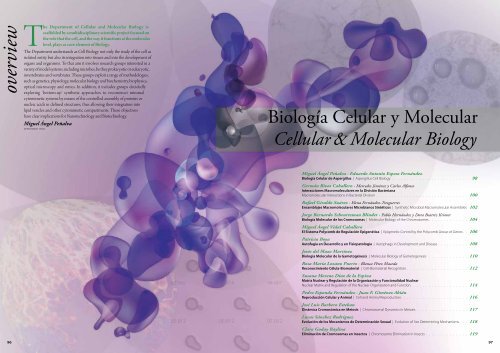
Biología <strong>Celular</strong> y <strong>Molecular</strong><br />
Cellular & <strong>Molecular</strong> Biology<br />
Miguel Ángel Peñalva · Eduardo Antonio Espeso Fernández<br />
Biología <strong>Celular</strong> de Aspergillus | Aspergillus Cell Biology 98<br />
Germán Rivas Caballero · Mercedes Jiménez y Carlos Alfonso<br />
Interacciones Macromoleculares en la División Bacteriana<br />
Macromolecular Interactions in Bacterial Division 100<br />
Rafael Giraldo Suárez · Elena Fernández-Tresguerres<br />
Ensamblajes Macromoleculares Microbianos Sintéticos | Synthetic Microbial Macromolecular Assemblies 102<br />
Jorge Bernardo Schvartzman Blinder · Pablo Hernández y Dora Beatriz Krimer<br />
Biología <strong>Molecular</strong> de los Cromosomas | <strong>Molecular</strong> Biology of the Chromosomes 104<br />
Miguel Ángel Vidal Caballero<br />
El Sistema Polycomb de Regulación Epigenética | Epigenetic Control by the Polycomb Group of Genes 106<br />
Patricia Boya<br />
Autofagia en Desarrollo y en Fisiopatología | Autophagy in Development and Disease 108<br />
Jesús del Mazo Martínez<br />
Biología <strong>Molecular</strong> de la Gametogénesis | <strong>Molecular</strong> Biology of Gametogenesis 110<br />
Rosa María Lozano Puerto · Blanca Pérez Maceda<br />
Reconocimiento Célula-Biomaterial | Cell-Biomaterial Recognition 112<br />
Susana Moreno Díaz de la Espina<br />
Matriz Nuclear y Regulación de la Organización y Funcionalidad Nuclear<br />
Nuclear Matrix and Regulation of the Nuclear Organization and Function 114<br />
Pedro Esponda Fernández · Juan F. Giménez Abián<br />
Reproducción <strong>Celular</strong> y Animal | Cell and Animal Reproduction 116<br />
José Luis Barbero Esteban<br />
Dinámica Cromosómica en Meiosis | Chromosomal Dynamics in Meiosis 117<br />
Lucas Sánchez Rodríguez<br />
Evolución de los Mecanismos de Determinación Sexual | Evolution of Sex Determining Mechanisms 118<br />
Clara Goday Baylina<br />
Eliminación de Cromosomas en Insectos | Chromosome Elimination in Insects 119<br />
96 97
Miguel Ángel Peñalva<br />
Profesora de Investigación |<br />
penalva@cib.csic.es<br />
PhD 1982<br />
Universidad Autónoma de Madrid<br />
Postdoctoral<br />
Antibióticos SA (Madrid) e Institut de Genetique<br />
et Microbiologie, Universidad de París, Orsay<br />
Científico Titular y Jefe de grupo CIB, 1987<br />
Profesor de Investigación desde 20<strong>01</strong><br />
CIB, CSIC<br />
Visiting Scientist, 20<strong>05</strong>-2006<br />
MRC Laboratory of <strong>Molecular</strong> Biology<br />
Cambridge (UK)<br />
Elegido miembro en 2000<br />
de EMBO<br />
Eduardo Antonio Espeso Fernández<br />
Científico Titular |<br />
eespeso@cib.csic.es<br />
PhD 1989<br />
Universidad Complutense de Madrid<br />
Postdoctoral, 1997-1999<br />
Imperial College London<br />
EMBO-Postdoctoral Fellow<br />
Contratado Ramón y Cajal 20<strong>01</strong>-2004<br />
Científico Titular y Jefe de grupo CIB, 2004<br />
CIB, CSIC<br />
Secretario, 2004-2008<br />
Grupo Especializado de Hongos Filamentosos y<br />
Levaduras (SEM)<br />
Investigadora del equipo | Staff scientist:<br />
Elena Reoyo<br />
Otros miembros | Other lab members:<br />
Areti Pantazopoulou<br />
Antonio Galindo Revilla<br />
Mario Pinar Sala<br />
Manuel Sánchez López-Berges<br />
María Villarino Pérez<br />
Juan Francisco Abenza Martínez<br />
http://www.cib.csic.es/es/grupo.php?idgrupo=8<br />
Ane Marquina Iñarrairaegui<br />
Erika Herrero García<br />
Laura Mellado Maroñas<br />
Daniel Lucena Agell<br />
Patricia Hernández Ortiz<br />
Publicaciones Seleccionadas<br />
Selected Publications<br />
A. Pantazopoulou and Miguel A. Peñalva. Characterization of Aspergillus<br />
nidulans RabCRab6. Traffic. 12:386-406, 2<strong>01</strong>1.<br />
Zhang, J., X. Yao, L. Fischer, J. F. Abenza, M. A. Peñalva and X. Xiang. The<br />
p25 subunit of the dynactin complex is required for dynein-early endosome<br />
interaction. J. Cell Biol. 193: 1245-1255, 2<strong>01</strong>1.<br />
Cortese M. S., Etxebeste O., Garzia A., Espeso E. A., Ugalde U. [2<strong>01</strong>1] Elucidation of<br />
functional markers from Aspergillus nidulans developmental regulator FlbB and<br />
their phylogenetic distribution. PLoS One. 6:e175<strong>05</strong>, 2<strong>01</strong>1.<br />
Herrero-García E., Garzia A., Cordobés S., Espeso E. A., Ugalde U. 8-Carbon oxylipins<br />
inhibit germination and growth, and stimulate aerial conidiation in Aspergillus<br />
nidulans. Fungal Biol. 1<strong>15</strong>, 393-400, 2<strong>01</strong>1.<br />
Calcagno-Pizarelli, A. M., Hervás-Aguilar, A., Galindo, A., Abenza, J. F., Peñalva, M. A.,<br />
and Arst, H. N., Jr. Rescue of Aspergillus nidulans severely debilitating null mutations<br />
in ESCRT-0, I, II and III genes by inactivation of a salt-tolerance pathway allows<br />
examination of ESCRT gene roles in pH signalling. J. Cell Science 124, 1-13, 2<strong>01</strong>1.<br />
Markina-Iñarrairaegui A., Etxebeste O., Herrero-García E., Araújo-Bazán L.,<br />
Fernández-Martínez J., Flores J. A., Osmani S. A., Espeso E. A. [2<strong>01</strong>1] Nuclear<br />
transporters in a multinucleated organism: functional and localization analyses<br />
in Aspergillus nidulans. Mol. Biol. Cell. 22:3874-86.<br />
Alkan N., Espeso E. A., Prusky D. [2<strong>01</strong>2] Virulence Regulation of Phytopathogenic<br />
Fungi by pH. Antioxid. Redox Signal. 2<strong>01</strong>2, PMID: 23249178 (e-published Dec 2<strong>01</strong>2).<br />
Abenza, J. F., Galindo, A., Pinar, M., Pantazopoulou, A., de los Ríos, V., and Peñalva, M. A.<br />
(2<strong>01</strong>2). Endosomal maturation by Rab conversion in Aspergillus nidulans is coupled to<br />
dynein-mediated basipetal movement. Mol. Biol. Cell 23, 1889-19<strong>01</strong>, 2<strong>01</strong>2.<br />
Etxebeste O., Herrero-García E., Cortese M. S., Garzia A., Oiartzabal-Arano E., de los Ríos<br />
V., Ugalde U., Espeso E. A. GmcA is a putative glucose-methanol-choline oxidoreductase<br />
required for the induction of asexual development in Aspergillus nidulans. PLoS One;<br />
7:e40292, 2<strong>01</strong>2.<br />
Galindo, A., Calcagno-Pizarelli, A. M., Arst, H. N., Jr., and Peñalva, M. A. An ordered pathway<br />
for the assembly of ESCRT-containing fungal ambient pH signalling complexes at the<br />
plasma membrane. J. Cell Science 125: 1784-1795, 2<strong>01</strong>2.<br />
Biología <strong>Celular</strong> y <strong>Molecular</strong> | Cellular & <strong>Molecular</strong> Biology<br />
Financiación | Funding<br />
Comunidad de Madrid, S2<strong>01</strong>0/BMD-2414<br />
Programación de circuitos microbianos en medicina protectiva y terapeútica. Desde: 2<strong>01</strong>2.<br />
Ministerio de Ciencia e Innovación: BIO2009-07281<br />
El receptor de pH ambiental alcalino de hongos filamentosos: activacion y tráfico<br />
endosomal. Desde 2009 hasta 2<strong>01</strong>2.<br />
Ministerio de Ciencia e Innovación: BFU2009-087<strong>01</strong><br />
Mecanismos transcripcionales, señalizadores y de localización celular que controlan la<br />
respuesta a estrés de cationes y su implicación en la patogenicidad de Aspergillus.<br />
Desde 2009 hasta 2<strong>01</strong>2.<br />
Ministerio de Economía y Competitividad: Impacto IPT-2<strong>01</strong>1-07<strong>52</strong>-900000<br />
Policétidos marinos en oncología. Desarrollo de bioprocesos para suministro de compuestos<br />
antitumorales de origen marino mediante biotecnología. Desde: 2<strong>01</strong>1.<br />
Figure 1 | Figura 1:<br />
Model of the pH signaling<br />
pathway. PalH activates the<br />
arrestin-like PalF, which recruits<br />
the ESCRT machinery to cortical<br />
structures. ESCRTs scaffold the<br />
assembly of the remaining pH signaling<br />
proteins. [Reproduced with permission of<br />
The Journal of Cell Science, The Company of<br />
Biologists Ltd, J. Cell Sci. 125, 1784–1795 (2<strong>01</strong>2)].<br />
Modelo de la ruta de señalización por pH. PalH activa<br />
la ‘arrestin-like’ PalF, que recluta la maquinaria ESCRT a estructuras corticales. Los ESCRTs<br />
facilitan el ensamblaje de las demás proteínas de la ruta. [reproducido con permiso<br />
de Journal of Cell Science, The Company of Biologists Ltd, J. Cell Sci. 125,<br />
1784–1795 (2<strong>01</strong>2)].<br />
Biología <strong>Celular</strong> de<br />
Aspergillus<br />
A. nidulans es un modelo genético para el estudio del<br />
crecimiento celular polarizado y el transporte a larga<br />
distancia (http://en.wikipedia.org/wiki/Model_organism).<br />
Sus hifas crecen exclusivamente por extensión apical,<br />
formando células tubulares multinucleadas en las que el<br />
tráfico intracelular está adaptado a las grandes distancias<br />
existentes entre distintas regiones o núcleos de la célula. La<br />
regulación de la transcripción por pH ambiental media la<br />
adaptación de los hongos a ambientes con distinto pH.<br />
Mediante abordajes genéticos y microscopía<br />
multidimensional in vivo, estudiamos la organización y<br />
los determinantes de identidad del Golgi y del sistema<br />
endovacuolar, centrándonos en GTPasas Rab y sus efectores. El Golgi<br />
de Aspergillus está formado por cisternas dispersas que pueden<br />
resolverse por microscopía óptica. Los endosomas tempranos son<br />
característicamente mótiles a grandes distancias sobre microtúbulos.<br />
La internalización endocítica y la maduración endosomal son<br />
esenciales. Pretendemos comprender los mecanismos de maduración<br />
de cisternas del Golgi, las rutas entre éste y los endosomas y el papel<br />
no canónico que los complejos ESCRT tienen en la organización<br />
de complejos de transducción de señal de pH en la membrana<br />
plasmática, que incluyen una arrestina especializada.<br />
Una gran variabilidad genética permite a los hongos la capacidad de<br />
medrar en ambientes extremos. La eficiente regulación transcripcional<br />
y la perfecta comunicación entre el núcleo y el citoplasma, donde se<br />
sintetizan los factores transcripcionales y concurren muchos de los<br />
sistemas de señalización, aseguran la correcta expresión de aquellos<br />
genes cuyos productos se necesitan para que el hongo se desarrolle<br />
en estas condiciones ambientales. Estudiamos la maquinaria molecular<br />
que participa en la homeostasis de cationes, mono y divalentes,<br />
y la que asegura el transporte núcleo-citoplásmico de factores<br />
transcripcionales de alta jerarquía que permiten afrontar los diferentes<br />
estreses abióticos. Así mismo, pretendemos establecer la forma en<br />
la que estos procesos participan en la diferenciación celular que<br />
ocurre durante el ciclo de reproducción asexual en Aspergillus y en la<br />
patogenicidad de este hongo en mamíferos.<br />
Aspergillus Cell Biology<br />
A. nidulans is a genetic model for studying polarised cell<br />
growth and long-distance transport (http://en.wikipedia.<br />
org/wiki/Model_organism). Hyphal tip cells grow<br />
exclusively by apical extension, leading to long, tubular<br />
multinucleated cells. Intracellular traffic is tailored to the<br />
needs imposed by the large distances separating apical<br />
and distal regions or the evenly spaced nuclei of the cell.<br />
Regulation of gene expression by ambient pH mediates<br />
adaptation of fungi to environments with different pH values.<br />
By combining genetic approaches with in vivo multidimensional<br />
microscopy, we are studying the organization and dynamics of<br />
the Golgi and endovacuolar systems, focusing on membrane<br />
domain-organising Rab GTPases and their effectors. The Aspergillus<br />
Golgi is formed by non-stacked early and late Golgi cisternae that<br />
can be resolved by optical microscopy. Early endosomes display<br />
characteristic long-distance movement on microtubule tracks.<br />
Endocytic internalization and endosomal maturation are essential. We<br />
are studying the mechanisms of cisternal maturation in the Golgi, the<br />
pathways connecting Golgi and endosomes and the ‘non-endosomal’<br />
role that ESCRT complexes play in scaffolding arrestin-depenedent<br />
ambient pH signal transduction complexes at the plasma membrane.<br />
The genetic versatility of fungi allows growth of these organisms in<br />
extreme environments. An efficient transcriptional regulation and<br />
a correct communication between nucleus and cytoplasm, where<br />
transcription factors are synthesised and most of signalling pathways<br />
play their roles, ensure the proper expression of those genes whose<br />
products are needed by the fungus to develop under such extreme<br />
ambient conditions. We study the molecular machineries participating<br />
in the homeostasis of mono and divalent cations, and in nucleocytoplasmic<br />
trafficking of high hierarchy transcription factors which<br />
allow the fungus to confront diverse abiotic stresses. In addition, we<br />
attempt to establish how these processes contribute to the cellular<br />
differentiation during the asexual reproductive cycle of Aspergillus and<br />
to its pathogenicity in mammals.<br />
Figure 3 | Figura 3:<br />
We are studying the responses to cation and alkaline pH<br />
stress, focusing on the Slt and Calcineurin-CrzA pathways<br />
(Spielvogel et al., 2008, Biochem J. 414:419). The CrzA<br />
pathway, conserved from fungi to mammals, involves the<br />
activities of calmodulin/calcineurin and protein kinases<br />
determining the phosphorylation status of CrzA.<br />
Estudiamos la respuesta a estrés por cationes y pH alcalino,<br />
centrándonos en el estudio de las rutas Slt y calcineurina-<br />
CrzA (Spielvogel et al. 2008, Biochem J. 414:419). La ruta CrzA,<br />
conservada desde hongos a mamíferos, implica las actividades<br />
de calmodulina/calcineurina y proteína-quinasas que<br />
determinan el estatus de fosforilación de CrzA.<br />
Figure 2 | Figura 2:<br />
RabS Rab7 recruits HOPS. GST-RabB Rab5 or GST-RabS Rab7 beads were used as baits in<br />
pull-downs of protein extracts containing HA-tagged preys. [With permission of the<br />
American Society of Cell Biology, MBoC 23: 1889-19<strong>01</strong> (2<strong>01</strong>2)].<br />
RabS Rab7 recluta HOPS. Perlas de Sepharosa cargadas con GST-RabB Rab5 o GST-<br />
RabS Rab7 se usaron como anzuelos en experimentos de ‘pull-down’ con extractos de<br />
células que contenían proteínas presa etiquetadas con HA. [Con permiso de la<br />
American Society of Cell Biology, MBoC 23: 1889-19<strong>01</strong> (2<strong>01</strong>2)].<br />
98 99
Germán Rivas Caballero<br />
Investigador Científico |<br />
grivas@cib.csic.es<br />
Doctorado en Química, 1989<br />
Universidad Autónoma de Madrid<br />
Postdoctoral, 1990-1992<br />
NIH, Bethesda, USA<br />
Postdoctoral, 1993<br />
Biozentrum, Univ. Basilea, CH<br />
Investigador, 1994<br />
Científico titular, 1995<br />
Jefe de grupo,1996<br />
Investigador científico, 2006<br />
CIB, CSIC<br />
Investigadores del equipo | Staff scientists:<br />
Carlos Alfonso Botello<br />
Otros miembros | Other lab members:<br />
Elisa Jiménez Cabré<br />
Begoña Monterroso Marco<br />
Concepción García Montañés<br />
Noelia Ropero de Torres<br />
Mercedes Jiménez Sarmiento<br />
Víctor Hernández Rocamora<br />
Rubén Ahijado Guzmán<br />
Ariadna Martos Sánchez<br />
Financiación | Funding<br />
2009-<strong>2<strong>01</strong>3</strong>: HEALTH-F3-2009-223432. Comunidad Europea /<br />
European Commission<br />
2<strong>01</strong>1-2<strong>01</strong>4: RGP0<strong>05</strong>0-2<strong>01</strong>0. Human Frontier Science Program<br />
2<strong>01</strong>2-2<strong>01</strong>4: BIO2<strong>01</strong>1-28941-C03. MINECO<br />
Publicaciones Seleccionadas<br />
Selected Publications<br />
Biología <strong>Celular</strong> y <strong>Molecular</strong> | Cellular & <strong>Molecular</strong> Biology<br />
Interacciones<br />
Macromoleculares en la<br />
División Bacteriana<br />
La investigación en nuestro grupo tiene como objetivo<br />
la reconstrucción de un conjunto mínimo de proteínas que<br />
reproduzca las etapas iniciales de la división bacteriana en<br />
compartimentos citomiméticos. Estudiamos los elementos<br />
individuales del primer complejo macromolecular del<br />
divisoma (el proto-anillo), sus interacciones funcionales,<br />
así como su organización espacio-temporal, mediante<br />
tecnologías bioquímicas, biofísicas y de imagen. Esta<br />
estrategia integradora, que combina enfoques cuantitativos<br />
y sintéticos, ayudará a obtener nuevos conocimientos sobre<br />
los mecanismos que operan en la división bacteriana, que<br />
podrían resultar en aplicaciones en el descubrimiento de<br />
fármacos antimicrobianos.<br />
Bioquímica sintética - Reconstrucción de divisomas mínimos<br />
en el tubo de ensayo: En Escherichia coli, la proteína de<br />
división celular FtsZ se ancla a la membrana citoplásmica por<br />
la acción de la proteína de membrana ZipA y la proteína anfitrópica<br />
FtsA, formando el ensamblaje inicial de la maquinaria de división, el<br />
proto-anillo, que dispara la citocinesis en el centro de la célula, donde<br />
polímeros de FtsZ forman un anillo-Z activo en división. Estudiamos<br />
las actividades, interacciones y propiedades de ensamblaje de FtsZ en<br />
reconstrucciones mínimas del proto-anillo estructuradas en sistemas<br />
miméticos de membrana, como nanodiscos, microesferas, bicapas,<br />
vesículas y microgotas. Investigamos la acción de las proteínas Min<br />
y estructuras que se asemejan al nucleoide (para reproducir los<br />
mecanismos de posicionamiento del anillo-Z) sobre las propiedades<br />
de divisomas mínimos. Estos estudios contribuirán a definir<br />
condiciones precisas para construir, con un mínimo de elementos,<br />
ensamblajes funcionales de división en ausencia de células; también<br />
serán utilizados para diseñar ensayos sistemáticos de búsqueda de<br />
nuevos antimicrobianos.<br />
http://www.cib.csic.es/es/grupo.grivas<br />
Figure 1 | Figura 1:<br />
Diagram illustrating the position of essential divisome elements in the living cell<br />
and reconstruction of proto-rings in biomimetic membrane systems. The protoring<br />
components are coloured while the other elements of the divisome are shown<br />
in dark grey.<br />
Diagrama que ilustra la posición de los elementos esenciales del divisoma en<br />
la célula y en reconstrucciones del proto-anillo en sistemas biomiméticos de<br />
membrana. Los componentes del proto-anillo están coloreados mientras que los otros<br />
elementos del divisoma se muestran en gris oscuro.<br />
Bioquímica citomimética – reactividad de proteínas en entornos<br />
aglomerados: El ensamblaje del proto-anillo in vivo tiene lugar en<br />
entornos caracterizados por la presencia de altas concentraciones de<br />
macromoléculas, estructuradas frecuentemente como redes dinámicas,<br />
así como de membranas y superficies. Diseñamos reconstrucciones<br />
sintéticas de estos microentornos (cuyo tamaño, forma y composición<br />
se ajustan de forma controlada) para investigar su influencia sobre la<br />
reactividad y la organización espacio-temporal (mediada por procesos<br />
de separación de fase) de ensamblajes mínimos del divisoma.<br />
Bioquímica física: Investigamos la energética funcional de reacciones<br />
de asociación y ensamblaje de FtsZ mediadas por ligandos, incluyendo<br />
las interacciones dinámicas con otros elementos del divisoma, mediante<br />
la aplicación combinada de ultracentrifugación analítica, dispersión de<br />
luz dinámica y estática, y espectroscopías de fluorescencia.<br />
Figure 2 | Figura 2:<br />
Mg 2+ -linked FtsZ assembly determined by sedimentation velocity (SV) and static light<br />
scattering (SLS). Mg 2+ concentrations are 0.1 (black) and 5 mM (red). Blue trace corresponds to<br />
unassembled species in the presence of EDTA. Binding of ZipA reconstructed in nanodiscs (Nd-<br />
ZipA) to FtsZ polymers, followed by SV and fluorescence correlation spectroscopy (FCS). Grey,<br />
free Nd-ZipA; green, complex with FtsZ polymers; red, free polymer.<br />
Determinación del ensamblaje de FtsZ ligado a Mg 2+ por velocidad de sedimentación (SV)<br />
y dispersión de luz estática (SLS). Negro, 0.1 mM Mg 2+ ; rojo, 5 mM Mg 2+ ; azul, especie monomérica<br />
en 2 mM EDTA. Unión de ZipA reconstituida en nanodiscos (Nd-ZipA) a los polímeros de<br />
FtsZ caracterizada por SV y espectroscopía de correlación de fluorescencia (FCS). Gris, Nd-<br />
ZipA libre; verde, complejo con FtsZ; rojo, polímero libre.<br />
Macromolecular Interactions<br />
in Bacterial Division<br />
Research in our group aims at reconstructing a minimal protein<br />
set that reproduces the initial steps of bacterial division in cell-like<br />
compartments. Individual elements of the first molecular assembly<br />
of the divisome (the proto-ring), their functional interactions<br />
and organization (in time and space) are studied by biochemical,<br />
biophysical and imaging technologies. This integrative strategy,<br />
combining quantitative and synthetic approaches, will help to gain<br />
new mechanistic insights on how bacterial division works, leading to<br />
applications in antimicrobial drug discovery.<br />
Synthetic biochemistry - Reconstruction of minimal divisomes<br />
in the test tube: In Eschericia coli, the cell division protein<br />
FtsZ is anchored to the cytoplasmic membrane by the action<br />
of the membrane protein ZipA and the amphitropic protein FtsA,<br />
forming the initial assembly of the division machinery, the proto-ring,<br />
which drives cytokinesis at mid-cell, where FtsZ polymers form a<br />
dynamic Z-ring active in division. We study the activities, interactions<br />
and assembly properties of FtsZ in minimal reconstructions of the<br />
proto-ring structured in membrane-like systems, such as nanodics,<br />
microbeads, bilayers, vesicles and microdroplets. We investigate the<br />
action of Min proteins and nucleoid-like structures (to reproduce<br />
Z-ring positioning mechanisms) on the properties of minimal<br />
divisomes. These studies will contribute to define the precise<br />
conditions to build, with a minimum set of elements, functional<br />
division assemblies in the absence of cells; they will also be exploited<br />
to design screening assays for new antimicrobials.<br />
del Castillo U., Alfonso C., Acebrón S. P., Martos A., Rivas G.*, Muga<br />
A.* (2<strong>01</strong>1). A quantitative analysis of the effect of nucleotides and<br />
M domain on the association equilibrium of ClpB. Biochemistry<br />
50:1991-2003.<br />
Jiménez M.*, Martos A., Vicente M., Rivas G.* (2<strong>01</strong>1).<br />
Reconstitution and organization of E. coli proto-ring elements<br />
(FtsZ and FtsA) inside giant unilamellar vesicles obtained from<br />
bacterial inner membranes. J. Biol. Chem. 286:11236-11241.<br />
Reija B., Monterroso B., Jiménez M., Vicente M., Rivas G.*, Zorrilla<br />
S.*. (2<strong>01</strong>1). Development of a homogeneous fluorescence<br />
anisotropy assay to monitor and measure FtsZ assembly in<br />
solution. Anal. Biochem. 418:89-96.<br />
Rivas G.*, Minton A. P.* (2<strong>01</strong>1). Beyond the second virial coefficient:<br />
sedimentation equilibrium in highly non-ideal solutions.<br />
Methods 54:167-174.<br />
Ahijado-Guzmán R., Gómez-Puertas P., Álvarez-Puebla R.,<br />
Rivas G.*, Liz-Marzán L.* (2<strong>01</strong>2). SERS-based detection of the<br />
interactions between the essential cell division FtsZ protein and<br />
bacterial membrane elements. ACS Nano 6:7514-7<strong>52</strong>0.<br />
Hernández-Rocamora V. M., Reija B., García-Montañés C., Natale<br />
P., Alfonso C., Minton A. P., Zorrilla S., Rivas G.*, Vicente M.*<br />
(2<strong>01</strong>2). Dynamic interaction of the Escherichia coli cell<br />
division ZipA and FtsZ proteins evidenced in nanodiscs. J.<br />
Biol. Chem. 287:30097-3<strong>01</strong>04.<br />
Hernández-Rocamora V. M., García-Montañés C.,<br />
Rivas G.*, Llorca O.* (2<strong>01</strong>2). Structural organization<br />
of the Escherichia coli cell division ZipA in nanodiscs<br />
complexed to FtsZ polymers as revealed by electron<br />
microscopy. J. Struct. Biol. 180:531-538.<br />
Martos A., Jiménez M., Rivas G.*, Schwille P.* (2<strong>01</strong>2).<br />
Towards a bottom-up reconstitution of bacterial cell<br />
division. Trends Cell Biol. 22:634-643.<br />
Martos A., Monterroso B., Zorrilla S., Reija B., Alfonso C.,<br />
Mingorance J., Rivas G.*, Jiménez M.* (2<strong>01</strong>2). Isolation,<br />
characterization and lipid-binding properties of the recalcitrant<br />
FtsA division protein from Escherichia coli. PloS One.<br />
7(6):e39829.<br />
Monterroso B., Ahijado-Guzmán R., Alfonso C., Reija B., Zorrilla S., Minton A.<br />
P., Rivas G.* (2<strong>01</strong>2). Mg 2+ -linked self-assembly of FtsZ in the presence of GTP<br />
and a GTP analog involves the concerted formation of a narrow distribution<br />
of oligomeric species. Biochemistry 51:4541-4550.<br />
Monterroso B., Alfonso C., Zorrilla S., Rivas G.* (<strong>2<strong>01</strong>3</strong>). Combined analytical<br />
ultracentrifugation, light scattering, and fluorescence correlation<br />
spectroscopy studies on the associations and assembly of the Escherichia<br />
coli cell division FtsZ protein. Methods (doi:10.1<strong>01</strong>6/j.ymeth.2<strong>01</strong>2.12.<strong>01</strong>4).<br />
Cytomimetic biochemistry – protein reactivity in crowded<br />
environments: Proto-ring assembly in vivo takes place in<br />
environments characterized by the presence of high concentrations<br />
of macromolecules, often structured as dynamic networks,<br />
surfaces and membranes. Synthetic reconstructions of these<br />
microenvironments (whose shape, size and composition is adjusted<br />
in a controlled manner) are being designed to investigate their<br />
influence on the reactivity, and organization in time and space<br />
(mediated by phase separation processes) of minimal divisome<br />
assemblies.<br />
Physical biochemistry: We investigate the functional energetics<br />
of ligand-linked FtsZ association and assembly reactions, including<br />
dynamic interactions with companion division elements, by combined<br />
measurements of analytical ultracentrifugation, static/dynamic light<br />
scattering and fluorescence spectroscopies.<br />
100 1<strong>01</strong>
Rafael Giraldo Suárez<br />
Profesor de Investigación |<br />
rgiraldo@cib.csic.es<br />
PhD en C.C. Biológicas, 1991<br />
Universidad Complutense de Madrid<br />
Postdoctoral, 1992-1994<br />
División de Estudios Estructurales del Laboratorio<br />
de Biología <strong>Molecular</strong> del MRC, Cambridge (UK)<br />
Investigador Contratado MEC, 1995-1999<br />
Científico Titular, 2000-2008<br />
Investigador Científico, 2008-2009<br />
Profesor de Investigación, desde 2<strong>01</strong>0<br />
CIB, CSIC<br />
Miembro de la Academia Europea, 2<strong>01</strong>0<br />
Investigadora del equipo | Staff scientist:<br />
M. Elena Fernández-Tresguerres Rodríguez-Vigil<br />
Otros miembros | Other lab members:<br />
Ana María Serrano López<br />
María Moreno del Álamo<br />
Cristina Fernández Fernández<br />
Fátima Gasset Rosa<br />
Laura Molina García.<br />
http://www.cib.csic.es/es/grupo.php?idgrupo=61<br />
Biología <strong>Celular</strong> y <strong>Molecular</strong> | Cellular & <strong>Molecular</strong> Biology<br />
Ensamblajes<br />
Macromoleculares<br />
Microbianos<br />
Sintéticos<br />
Los microorganismos ofrecen sistemas modelos de gran<br />
versatilidad para el estudio estructural y la reconstrucción de<br />
complejos de proteínas con funcionalidades nuevas. Como<br />
evolución de nuestros anteriores estudios sobre la proteína<br />
RepA en la replicación del DNA del plásmido pPS10, en<br />
nuestro grupo estamos desarrollando, con aproximaciones<br />
propias de la Biología Sintética, módulos derivados de<br />
dominios WH que permitan controlar el ensamblaje<br />
amiloide de proteínas en condiciones cuasi-fisiológicas.<br />
Financiación<br />
Funding<br />
PRODESTECH: Estructura y dinámica de proteínas<br />
aplicadas al desarrollo de nuevas enzimas, agentes<br />
terapéuticos, y máquinas macromoleculares sintéticas<br />
(CSD2009-00088).<br />
Módulos replicativos y amiloides sintéticos basados<br />
en dispositivos conmutadores WH microbianos<br />
(BIO2009-069<strong>52</strong>).<br />
PRODESTECH: From protein structure and dynamics<br />
to tailored enzymes, therapeutics, and synthetic<br />
macromolecular devices (CSD2009-00088).<br />
Synthetic replicative and amyloid modules built on<br />
microbial WH switch devices (BIO2009-069<strong>52</strong>).<br />
Nuestro grupo estudia los cambios conformacionales que<br />
experimentan dominios del tipo Winged-Helix (WH) al unirse<br />
al DNA, facultándoles para iniciar la replicación de plásmidos<br />
bacterianos (proteína RepA). Hemos descubierto que el ensamblaje de<br />
complejos RepA funcionales está modulado por secuencias específicas<br />
de DNA, que actúan como efectores alostéricos, y por chaperonas de<br />
la familia Hsp70 (DnaK). La interacción in vitro del dominio N-terminal<br />
de RepA (WH1) con el DNA promueve el ensamblaje de la proteína<br />
en fibras amiloides, de manera análoga a la transformación estructural<br />
amiloide descrita en las proteínas implicadas en las encefalopatías<br />
espongiformes y en la enfermedad de Parkinson. Hemos hallado<br />
moléculas que inhiben la amiloidosis de WH1 al unirse en esta proteína<br />
a los residuos que reconocen al DNA. La expresión en E. coli de fusiones<br />
de RepA-WH1 con una proteína marcadora fluorescente roja da lugar<br />
a inclusiones amiloides en el citoplasma observables al microscopio.<br />
La presencia de estos agregados amiloides genera un fenotipo<br />
compatible con el envejecimiento de las bacterias, que aumentan<br />
significativamente su tiempo de generación. Las inclusiones amiloides<br />
se transmiten estocásticamente durante la división celular a las células<br />
hijas resultantes. RepA-WH1 se comporta pues como un prionoide,<br />
siendo el modelo más sencillo del que se dispone para el estudio de las<br />
bases moleculares comunes a las proteinopatías amiloides y abriendo<br />
nuevas perspectivas a la ingeniería de nano-ensamblajes amiloides.<br />
Publicaciones Seleccionadas<br />
Selected Publications<br />
Giraldo, R., (2007). Defined DNA sequences promote the assembly of a bacterial protein into distinct<br />
amyloid nanostructures. Proc. Natl. Acad. Sci. USA 104, 17388-17393.<br />
Gasset-Rosa, F., Maté, M. J., Dávila-Fajardo, C., Bravo, J., and Giraldo, R., (2008). Binding of sulphonated indigo<br />
derivatives to RepA-WH1 inhibits DNA-induced protein amyloidogenesis. Nucleic Acids Res. 36, 2249-2256.<br />
Gasset-Rosa, F., Díaz-López, T., Lurz, R., Prieto, A., Fernández-Tresguerres, M. E., and Giraldo, R., (2008).<br />
Negative regulation of pPS10 plasmid replication: Origin pairing by zipping-up DNA-bound RepA<br />
monomers. Mol. Microbiol. 68, 560-572.<br />
Moreno-del Álamo, M., Sánchez-Gorostiaga, A., Serrano, A. M., Prieto, A., Cuellar, J., Martín-Benito, J.,<br />
Valpuesta, J. M., and Giraldo, R., (2<strong>01</strong>0). Structural analysis of the interactions between Hsp70 chaperones<br />
and the yeast DNA replication protein Orc4p. J. Mol. Biol. 403, 24-39.<br />
Fernández-Tresguerres, M. E., Moreno-Díaz de la Espina, S., Gasset-Rosa, F., Giraldo, R., (2<strong>01</strong>0). A DNApromoted<br />
amyloid proteinopathy in Escherichia coli. Mol. Microbiol. 77, 1456-1469.<br />
Giraldo, R., (2<strong>01</strong>0). Amyloid assemblies: Protein Legos at a crossroads in bottom-up synthetic biology.<br />
ChemBioChem 11, 2347-2357.<br />
Giraldo, R., Moreno-Díaz de la Espina, S., Fernández-Tresguerres, M. E., Gasset-Rosa, F., (2<strong>01</strong>1). RepA prionoid:<br />
A synthetic amyloid proteinopathy in a minimalist host. Prion 5, 60-64.<br />
Synthetic Microbial<br />
Macromolecular<br />
Assemblies<br />
Micro-organisms are versatile model systems for<br />
studying the structure and assembly of natural protein<br />
complexes, allowing to attempt the reconstruction of<br />
these assemblies with altered or novel functionalities.<br />
In a bottom-up Synthetic Biology task, evolved from<br />
our previous studies on the role of the RepA protein<br />
in the replication of pPS10 plasmid, we are currently<br />
developing modules derived from WH domains that<br />
allow for the control and engineering of amyloid<br />
assembly under quasi-physiological conditions.<br />
Figure 1 | Figura 1:<br />
Scheme of RepA-WH1 amyloidogenesis. Transient binding to DNA in vitro promotes<br />
substantial conformational changes (arrows) in extended regions (dark blue) of the<br />
dimeric WH1 domain. This would thus generate a monomeric metastable intermediate<br />
in which an α-helix (red; spheres: the hyper-amyloidogenic Val31 residue) would<br />
be unfolded, resulting in the assembly of the protein into amyloid (cross-β sheets.<br />
When expressed in E. coli as a fusion to a red fluorescent protein marker, RepA-WH1<br />
aggregates as amyloid inclusions that severely reduce cell fitness (right).<br />
Our group studies the conformational changes<br />
experienced by Winged-Helix (WH) protein<br />
domains upon binding to DNA, which enable<br />
these proteins to initiate DNA replication of bacterial<br />
plasmids (RepA protein). We have discovered that the<br />
assembly of RepA functional complexes is modulated by<br />
specific DNA sequences, which thus act as allosteric effectors,<br />
and by chaperones of the Hsp70 family (DnaK). In the case<br />
of the N-terminal domain in RepA (WH1), its binding to<br />
DNA promotes the assembly of the protein as amyloid fibers<br />
in vitro, as reported for the proteins involved in spongiform<br />
encephalopathies and in Parkinson’s disease. We have found<br />
molecules that inhibit WH1 amyloidosis by binding to the<br />
amino acid residues in the protein which should interact with<br />
DNA. Expression in E. coli of fusions between RepA-WH1 and a<br />
red fluorescent protein gives way to amyloid inclusions in the<br />
cytoplasm, which are readily visualized under the microscope.<br />
These amyloid aggregates are stochastically transmitted to the<br />
daughter cells, coupled to cell division. RepA-WH1 thus behaves<br />
as a prionoid, constituting the simplest model available for<br />
the study of the molecular basis common to all amyloid<br />
proteinopathies and opening new perspectives to<br />
engineer protein amyloid nano-scaffolds.<br />
Esquema de la amiloidogénesis de RepA-WH1. In vitro la unión transitoria al DNA promueve<br />
cambios conformacionales substanciales (flechas) en extensas regiones (azul oscuro) del<br />
dominio dimérico WH1. Se generaría así un intermediario monomérico metaestable en<br />
el que una hélice-α (rojo; esferas: el residuo hiperamiloidogénico Val31), al desplegarse,<br />
desencadenaría el ensamblaje de la proteína en una lámina-β cruzada amiloide. Cuando<br />
se expresa RepA-WH1 fusionada a un marcador fluorescente rojo en E. coli, se forman<br />
inclusiones amiloides que disminuyen drásticamente la viabilidad celular (derecha).<br />
102 103
Jorge Bernardo Schvartzman Blinder<br />
Profesor de Investigación | schvartzman@cib.csic.es<br />
PhD, 1979<br />
Universidad Politécnica de Madrid, España<br />
Postdoctoral, 1980-1982<br />
Brookhaven National Laboratory, New York (USA)<br />
Fullbright Fellow, 1987-1989<br />
Albert Einstein College of Medicine, New York (USA)<br />
Científico Titular, 1985<br />
Investigador Científico, 2002<br />
Profesor de Investigación, 2007<br />
CSIC<br />
Investigadores del equipo | Staff scientists:<br />
Pablo Hernández ValenzuelaDora<br />
Otros miembros | Other lab members:<br />
Jorge Cebrián Castillo<br />
Vanessa Fernández Calleja<br />
María José Fernández Nestosa<br />
Maridian José Kadomatsu<br />
Hermosa<br />
Virginia López Martínez<br />
Beatriz Krimer Smunis<br />
María Luisa Martínez Robles<br />
María Estefanía Monturus de<br />
Carandini<br />
Joaquín Rodrigo Otero Asman<br />
María Rodríguez López<br />
María Tenorio Gómez<br />
Financiación | Funding<br />
BFU2<strong>01</strong>1-22489 (MINECO)<br />
AP/038170/11 (AECID)<br />
I-COOP0009 (CSIC)<br />
Biología <strong>Celular</strong> y <strong>Molecular</strong> | Cellular & <strong>Molecular</strong> Biology<br />
http://www.cib.csic.es/es/grupo.php?idgrupo=2<br />
Biología <strong>Molecular</strong> de los Cromosomas<br />
Nos interesa la interrelación y coordinación de los procesos biológicos en los que está involucrado el DNA: replicación,<br />
transcripción, reparación y recombinación, cómo están regulados y cómo modifican o son afectados por factores<br />
genéticos, epigenéticos y ambientales como la topología del DNA, la organización de la cromatina y el estrés nutricional.<br />
Topología de la replicación del DNA. Estudiamos el<br />
superenrollamiento, encadenamiento y anudamiento del DNA<br />
durante la replicación y segregación de plásmidos bacterianos<br />
y minicromosomas de levaduras. La electroforesis bidimensional en<br />
geles de agarosa de alta resolución, la microscopía de fuerza atómica<br />
(AFM) y la simulación de MonteCarlo nos han permitido demostrar<br />
que el superenrollamiento (-) en procariotas y (+) en eucariotas juegan<br />
un papel fundamental en el desencadenamiento de las cromátidas<br />
hermanas por la Topo IV y la topoisomerasa II, respectivamente;<br />
Diferenciación en células eritroleucémicas. La integración del<br />
complejo vírico Friend cercana al promotor del gen Sfpi provoca<br />
la activación del factor de transcripción PU.1, probable causa del<br />
Figure 1 | Figura 1:<br />
A) Supercoiling, knotting and catenation of<br />
bacterial plasmids analyzed by high resolution<br />
two-dimensional agarose gel electrophoresis.<br />
Controlled nicking discerns relaxed and knotted<br />
forms as well as replication intermediates and<br />
catenanes. B) Partially replicated and nicked<br />
molecule corresponding to the bacterial plasmid<br />
pBR18-TerE@AatII covered with recA and<br />
visualized by Atomic Force Microscopy (AFM).<br />
A) Análisis del superenrollamiento,<br />
encadenamiento y anudamiento del DNA<br />
de plásmidos bacterianos por electroforesis<br />
bidimensional en geles de agarosa de alta<br />
resolución. La introducción controlada de<br />
roturas de cadena sencilla permite distinguir las<br />
formas monoméricas relajadas y anudadas de<br />
los intermediarios de replicación y las moléculas<br />
encadenadas. B) Molécula de DNA parcialmente<br />
replicada con una rotura de cadena sencilla<br />
correspondiente al plásmido bacteriano pBR18-<br />
TerE@AatII, recubierta con la proteína recA<br />
visualizada por microscopía de fuerza atómica<br />
(AFM).<br />
fenotipo eritroleucémico. Estudiamos la expresión de PU.1 y cambios<br />
en el patrón de replicación del DNA en líneas parentales y resistentes<br />
a agentes inductores tanto en células proliferantes como a lo largo de<br />
la diferenciación inducida;<br />
Barreras para las horquillas de replicación e inestabilidad del<br />
DNA. La parada accidental de las horquillas de replicación es una<br />
causa importante de inestabilidad genómica. También existen paradas<br />
naturales de las horquillas en sitios específicos, como en los genes del<br />
rRNA, cuya alta conservación indica que juegan un papel relevante<br />
aún no muy bien conocido. Estudiamos la regulación de estas barreras<br />
naturales y su papel en la recombinación homóloga, la progresión del<br />
ciclo celular y su relación con la respuesta al estrés nutricional.<br />
<strong>Molecular</strong> Biology of the Chromosomes<br />
We are interested in the relationships and coordination between biological processes where DNA is involved: replication,<br />
transcription, repair and recombination, how are they regulated and how they alter or are affected by genetic, epigenetic<br />
and environmental factors such as DNA topology, chromatin organization and nutritional stress.<br />
DNA Topology during Replication. We characterized the<br />
interplay of supercoiling, decatenation and unknotting of<br />
DNA during replication and segregation of sister duplexes<br />
in bacterial plasmids and yeast minichromosomes. We used high<br />
resolution two-dimensional agarose gel electrophoresis, atomic force<br />
microscopy (AFM) and MonteCarlo numerical simulations to show<br />
that (-) supercoiling in prokaryotes and (+) supercoiling in eukaryotes<br />
plays a fundamental role in the decatenation of newly replicated DNA<br />
molecules by Topo IV and topoisomerase II, respectively;<br />
Terminal Differentiation in Erythroleukemia Cells. Integration of<br />
the Friend complex virus upstream of the promoter of the Sfpi-1<br />
gene leads to the activation of the transcription factor PU.1. This<br />
Publicaciones Seleccionadas<br />
Selected Publications<br />
Baxter, J., N. Sen, V. López-Martínez, M. E. Monturus De Carandini, J.<br />
B. Schvartzman, J. F. X. Diffley and L. Aragón: Positive supercoiling of<br />
mitotic DNA drives de-catenation by topoisomerase II in eukaryotes.<br />
Science 331: 1328-1332 (2<strong>01</strong>1).<br />
Rodríguez-Sánchez, L., M. Rodríguez-López, Z. García, M. Tenorio-<br />
Gómez, J. B. Schvartzman, D. B. Krimer and P. Hernández: Fission<br />
yeast rDNA-binding protein Reb1 regulates G1 phase under<br />
nutritional stress. J Cell Science 124: 25-34 (2<strong>01</strong>1).<br />
López, V., M. L. Martínez-Robles, P. Hernández, D. B. Krimer and J. B.<br />
Schvartzman: Topo IV is the topoisomerase that knots and unknots<br />
sister duplexes during DNA replication. Nucleic Acids Res 40: 3563-<br />
3573 (2<strong>01</strong>2).<br />
Monturus, M. E., O. Ganier, P. Hernández, J. B. Schvartzman, M.<br />
Méchali and D. B. Krimer: DNA replication fading as proliferating<br />
cells advance in their commitment to terminal differentiation.<br />
Scientific Reports 2, 279 (2<strong>01</strong>2).<br />
Schvartzman, J. B., M. L. Martínez-Robles, V. López, P. Hernández and<br />
D. B. Krimer: 2D gels and their third-dimension potential. Methods<br />
57: 170-178 (2<strong>01</strong>2).<br />
Figure 2 | Figura 2:<br />
A) EMSA assays showing that protein Reb1 of S. pombe binds to<br />
the promoter of ste9+ gene and regulates the expession of this<br />
activator of APC (anaphase promotong complex/cyclosome)<br />
during the G1 phase of the cell cycle. B) DNA isolated from MEL<br />
cells stretched by DNA combing. The tracks sequentially labeled<br />
with IdU (red) and CldU (green) allow identification of replication<br />
origins and calculation of the rate of replication fork progression.<br />
A) Ensayos EMSA en los que se muestra que la proteína Reb1 se une<br />
al promotor de ste9+, regulando la expresión de este activador de<br />
APC (anaphase promoting complex/cyclosome) en S. pombe, durante<br />
la fase G1 del ciclo celular. B) DNA de células MEL extendidas por<br />
peinado molecular. Los tramos marcados secuencialmente con IdU<br />
(en rojo) y CldU (en verde) permiten identificar orígenes de replicación<br />
y la velocidad de progreso de las horquillas.<br />
could be responsible for the transformed phenotype. We analyze the<br />
expression of PU.1 and changes in the DNA replication pattern in<br />
parental and resistant cell lines during proliferation as well as along<br />
the cell differentiation process;<br />
Replication Fork Barriers and DNA Instability. Accidental arrest of<br />
replication forks is an important cause of genome instability. Natural<br />
barriers have also been identified at specific sites, like the rRNAcoding<br />
genes. Their high conservation indicates they play a relevant<br />
role, which is still poorly understood. We study the regulation of these<br />
natural barriers and their role in homologous recombination, cell<br />
cycle progression and cell response to nutritional stress.<br />
104 1<strong>05</strong>
Biología <strong>Celular</strong> y <strong>Molecular</strong> | Cellular & <strong>Molecular</strong> Biology<br />
Miguel Ángel Vidal Caballero<br />
Investigador Científico |<br />
mvidal@cib.csic.es<br />
PhD, 1985<br />
Universidad Complutense<br />
https://www.cib.csic.es/es/grupo.php?idgrupo=14<br />
Postdoctoral<br />
Medical Research Council, London (UK)<br />
Científico Titular,1991<br />
Jefe de grupo CIB, 1991<br />
Investigador Científico, 2008<br />
CIB, CSIC<br />
Otros miembros | Other lab members:<br />
Josué Alvaro Blanco<br />
Mónica Bravo Madrigal<br />
Katazyna Starowitz<br />
Fabio Nicolini<br />
Zahira Corrales<br />
El Sistema Polycomb de Regulación Epigenética<br />
El grupo investiga aspectos de la regulación epigenética mediada por el grupo de genes Polycomb (PcG). Los<br />
productos PcG se presentan en complejos de subunidades con dominios evolutivamente conservados que les confieren<br />
actividad(es) modificadora(s) de cromatina. El ámbito funcional del sistema incluye funciones de control transcripcional y<br />
otras de relevancia en la autorenovación de células madre y el desarrollo tumoral.<br />
La monoubiquitinación de la histona H2A depende,<br />
esencialmente, de Ring1A y Ring1B, subunidades PcG<br />
originalmente identificadas en nuestro laboratorio. Ring1A y<br />
Ring1B forman parte de todos los complejos PRC1, una de las dos<br />
clases de complejos Polycomb. También identificada en el laboratorio,<br />
RYBP caracteriza a la colección más heterogénea de complejos<br />
PRC1. Utilizamos modelos murinos de pérdida de función y otros<br />
que expresan subunidades marcadas para análisis funcionales y<br />
bioquímicos.<br />
Por un lado investigamos las contribuciones de las proteínas Ring1<br />
y RYBP en la homeostasis de los compartimentos hematopoyéticos<br />
y neurales, así como en mantenimiento del estado pluripotente<br />
de células ES. Los experimentos de pérdida de función muestran<br />
respuestas hipo e hiperproliferativas, dependiendo del estado<br />
diferenciativo y de tipo de tejido. En el sistema hematopoyético, en<br />
colaboración con C. Calés (IIB/UAM), investigamos las funciones de<br />
regulación negativa de proliferación (células inmaduras) así como<br />
las de regulación positiva de proliferación en células diferenciadas<br />
y otras funciones específicas en la diferenciación de progenitores<br />
linfoides. El compartimento neural se investiga en un modelo<br />
de células madre/progenitoras neurales. Aquí, la actividad de<br />
autorenovación dependiente de las proteínas Ring1 afecta tanto al<br />
estado proliferativo como al mantenimiento del estado indiferenciado<br />
y queremos determinar su probable interdependencia.<br />
Por otro lado, para caracterizar la diversidad de complejos PcG,<br />
utilizamos un modelo murino que expresa una forma de Ring1B que<br />
facilita el aislamiento de complejos de una variedad de tipos celulares,<br />
en una etapa preliminar de identificación proteómica.<br />
Figure 2 | Figura 2:<br />
Mitotic defects in Ring1-deficient cells. Example of binucleated cell, appearing often<br />
among normal cells in a culture of primary fibroblasts, soon after Ring1 inactivation.<br />
These cells form at the end of failed citokinesis. Nuclei, stained with DAPI and cytoplasm,<br />
as defined by the α-tubulin citoskeleton, visualized by immunofluorescence. It could<br />
indicate an unanticipated, transcription-independent, function for Ring1 proteins.<br />
Mitosis defectuosas de células deficientes en proteínas Ring1. Ejemplo de célula<br />
binucleada frecuentemente observado en cultivos de fibroblastos primarios; se producen<br />
como consecuencia de mitosis fallida por citocinesis abortada. Núcleos, teñidos con DAPI,<br />
y citoplasma, definido por el esqueleto de α-tubulina visualizado por inmunofluorescencia.<br />
Puede ser evidencia de una función no transcripcional de proteínas Ring1.<br />
Epigenetic Control by the Polycomb<br />
Group of Genes<br />
Ring1A and Ring1B are responsible for most of the<br />
monoubiquitylated histone H2A (lysine 119). Ring1A and Ring1B<br />
are part of the core of PRC1 complexes, one of the two major<br />
categories of Polycomb assembles. Ring1A and Ring1B were identified<br />
some time ago in the lab, just as RYBP, a subunit that defines the more<br />
heterogenous subset of PRC1 complexes. We use loss-of-function<br />
mouse models as well as models that express tagged variants for<br />
functional and biochemical analysis.<br />
On the one hand, we investigate roles of Ring1 and RYBP proteins<br />
in hematopoyetic and neural homeostasis and in pluripotency of ES<br />
cells. Loss-of-function experiments showed both hipo- and hyperproliferative<br />
responses, depending on differentiation status and<br />
tissue. The study of mutant hematopoyesis, in collaboration with C.<br />
Publicaciones Seleccionadas<br />
Selected Publications<br />
The lab focuses on epigenetic control by the Polycomb group (PcG) of genes. PcG<br />
associate in complexes displaying chromatin modifying activities endowed by evolutionary<br />
conserved protein motifs. Initially known as developmental regulators, over time their<br />
functions have expanded to include a wide range of processes essential for maintenance of<br />
stem cell and cancer development.<br />
Endoh, M., Endo, T. A., Tamie Endoh, T., Isono, K., Sharif, J., Ohara, O., Toyoda, T., Ito, T.,<br />
Eskeland, R., Bickmore, W. A., Vidal, M., Bernstein, B. E. and Koseki, H. (2<strong>01</strong>2) Histone<br />
H2A mono-ubiquitination is a crucial step to mediate PRC1 dependent repression of<br />
developmental genes to maintain ES cell identity. PLoS Genetics 7: e1002774.<br />
Posfai, E., Kunzmann, R., Brochard, V., Salvaing, J., Cabuy, E., Roloff, T. C., Liu, Z., Tardat, M.,<br />
van Lohuizen, M., Vidal, M., Beaujean, N., and Peters, A. (2<strong>01</strong>2) Polycomb function during<br />
oogenesis is required for mouse embryonic development. Genes and Dev. <strong>26</strong>: 920-932.<br />
Hisada, K., Sánchez, C., Endo, T., Endoh, M., Román-Trufero, M., Sharif, J., Koseki, H. and<br />
Vidal, M. (2<strong>01</strong>2) RYBP represses endogenous retroviruses, preimplantation- and germlinespecific<br />
genes in mouse embryonic stem cells. Mol. Cell. Biol. 32:1139-1149.<br />
Tavares, L., Dimitrova, E., Oxley, D., Webster, J., Poot, R., Demmers, J., Bezstarosti, K., Taylor,<br />
S., Ura, H., Koide, H., Wutz, A, Vidal, M., Elderkin, S. and Brockdorff, N. (2<strong>01</strong>2) RYBP mediates<br />
H3K27me3 independent recruitment of H2A ubiquitylation activity at polycomb target<br />
loci in ES cells and on the inactive X chromosome. Cell 148: 664-678.<br />
Konuma, T., Nakamura, S., Miyagi, S., Negishi, M., Chiba, T., Oguro, H., Yuan, J., M.<br />
Mochizuki-Kashio, M., Ichikawa, H., Miyoshi, H., Vidal, M. and Iwama, A. (2<strong>01</strong>1) Forced<br />
expression of the histone demethylase Fbxl10 maintains self-renewing hematopoietic<br />
stem cells. Exp. Hematology 39: 697-709.<br />
García-Tuñon, I., D. Guallar, D., Alonso-Martín, S., Benito, A. A., Benítez, A., Pérez-Palacios, R.,<br />
Muniesa, P., Climent, M., Sánchez, M., Vidal, M. and Schoorlemmer. J. (2<strong>01</strong>1) Association of<br />
Rex-1 to target genes support its interaction with polycomb function. Stem Cell Res. 7:1-16.<br />
Financiación | Funding<br />
BFU2<strong>01</strong>0-18146 (MICINN)<br />
Fundación Areces<br />
Oncocycle S2<strong>01</strong>0/BMD2470 (CAM)<br />
FP7-People-2<strong>01</strong>1-ITN<br />
Calés (IIB-UAM), approaches anti-proliferative activities<br />
(immature cells), promotion of proliferation (differentiating<br />
cells) and differentiation control of lymphoid progenitors.<br />
Neural homeostasis is being investigated in a model of neural<br />
stem/progenitor cells. The results showed Ring1A and Ring1B roles<br />
supporting cell proliferation and repression of neural differentiation<br />
programs as key elements in self-renewal maintenance. Mediators of<br />
these activities and their possible coupling are under investigation.<br />
On the other hand, in an attempt to describe the biochemical<br />
heterogeneity of PRC1 complexes we use a mouse model with a<br />
knocked-in tagged-Ring1 protein. This facilitates the isolation of the<br />
complexes from a variety of cell types, as a preliminary step in<br />
their identification by mass spectrometry.<br />
Figure 1 | Figura 1:<br />
106<br />
RYBP-independent expression of the pluripotency program of embryonic stem (ES)<br />
cells. Expression of stem cell markers in ethanol-treated (wild-type) or tamoxifentreated<br />
(RYBP-KO) ES cells identified by immunofluorescence (Oct4 and SSEA1) or<br />
immunohistochemistry (alkaline phosphatase [AP]). RYBP immunofluorescence was<br />
included as a control of gene inactivation. In contrast, RYBP is essential for repression of<br />
endogenous retroviral elements.<br />
RYBP no es esencial para el mantenimiento del programa de pluripotencia de células<br />
madre embrionarias (ES). Expresión de marcadores de pluripotencia en células ES<br />
control (wild type, tratadas con etanol) y mutantes (RYBP KO, tratadas con el inductor<br />
tamoxifen). Oct4 y SSEA1 se detectaron por immunofluorescencia y la fosfatasa alcalina<br />
(AP) mediante histoquímica. El análisis de la expresión de RYBP se muestra como control<br />
de inactivación. RYBP, en cambio silencia elementos retrovirales.<br />
107
Biología <strong>Celular</strong> y <strong>Molecular</strong> | Cellular & <strong>Molecular</strong> Biology<br />
Patricia Boya<br />
Científica Titular |<br />
pboya@cib.csic.es<br />
PhD en Biología, 2000<br />
Universidad de Navarra<br />
Postdoctoral, 20<strong>01</strong>-2004<br />
CNRS, París (Francia)<br />
Postdoctoral, 20<strong>05</strong><br />
Universisty of Cambridge (UK)<br />
Contrato Ramón y Cajal, 20<strong>05</strong>-2009<br />
Científica titular, 2009<br />
CIB, CSIC<br />
Otros miembros | Other lab members:<br />
Lorena Esteban Martínez<br />
Lucía García Ledo<br />
Esther Seco Martín<br />
Ana Serrano Puebla<br />
Marta Mauro Lizcano<br />
Fengjuan Wang<br />
http://www.cib.csic.es/en/grupo.php?idgrupo=73<br />
Autofagia en Desarrollo y en Fisiopatología<br />
En nuestro laboratorio utilizamos modelos celulares<br />
y animales para comprender el papel de la autofagia en<br />
la fisiología y la patología de los organismos. Este es<br />
un proceso de degradación intracelular que permite la<br />
eliminación y el reciclaje de componentes celulares. Es una<br />
importante respuesta frente al ayuno nutricional, participa<br />
en la degradación de orgánulos celulares y permite la<br />
supervivencia en situaciones de estrés.<br />
El interés de nuestro laboratorio se centra en entender por<br />
que el proceso de la autofagia es esencial para mantener<br />
la homeostasis de las células y qué patologías subyacen a<br />
alteraciones de este mecanismo de degradación intracelular.<br />
La importancia del proceso de autofagia queda patente por la<br />
letalidad embrionaria de animales deficientes en algunos de sus<br />
reguladores, los genes Atg. En nuestro grupo estudiamos la relación<br />
de la autofagia con procesos esenciales para las células como la<br />
proliferación, diferenciación y la muerte celular. Hemos demostrado<br />
este proceso es importante para la diferenciación neuronal ya que<br />
animales deficientes de autofagia no generan neuronas maduras<br />
y poseen defectos en neuritogénesis. Por otro lado y en trabajos<br />
recientes hemos demostrado que la inducción temprana de la<br />
autofagia durante procesos neurodegenerativos supone una respuesta<br />
citoprotectora. Daño axonal producido in vivo en animales deficientes<br />
de autofagia aumenta los niveles de muerte celular y por el contrario<br />
la inducción farmacológica de este proceso retrasa el proceso de<br />
neurodegeneración. Por último estamos interesados en la relación de la<br />
autofagia con procesos de envejecimiento del sistema nervioso.<br />
Además y estrecha colaboración con empresas españolas estamos<br />
buscando nuevos productos que sean capaces de modular este<br />
proceso y que puedan luego ser aplicados a la terapia para<br />
enfermedades humanas. Hemos puesto a punto varios métodos de<br />
screening para la determinación de nuevos compuestos que<br />
induzcan o bloquen el proceso de autofagia. Por último y también en<br />
colaboración con la empresa WIMASIS estamos desarrollando de un<br />
sistema de análisis cuantitativo de imágenes para la evaluación<br />
automatizada de los procesos de autofagia y apoptosis tanto en<br />
células como en tejido.<br />
Figure 1 | Figura 1:<br />
3T3 fibroblasts stained with<br />
accridine orange. This dye stains<br />
bright orange lysosomes and other<br />
acidic organelles.<br />
Fibroblastos 3T3 teñidos con la<br />
sonda naranja de acridina que marca<br />
de naranja intenso los lisosomas y<br />
orgánulos ácidos.<br />
Publicaciones Seleccionadas<br />
Selected Publications<br />
Rodríguez-Muela N., Koga H., García-Ledo L., de la Villa P., de la Rosa E. J., Cuervo A. M.,<br />
Boya P. Balance between autophagic pathways preserves retinal homeostasis. Aging<br />
Cell. <strong>2<strong>01</strong>3</strong> Mar 22. doi: 10.1111/acel.12072.<br />
Boya P., Reggiori F., Codogno P. Emerging regulation and functions of Autophagy.<br />
Nature Cell Biology. <strong>2<strong>01</strong>3</strong>, in press.<br />
Rodríguez-Muela N, Germain F, Mariño G, Fitze PS, Boya P. Autophagy promotes<br />
survival of retinal ganglion cells after optic nerve axotomy in mice. Cell Death Differ.<br />
2<strong>01</strong>2;19(1):162-9. doi: 10.1038/cdd.2<strong>01</strong>1.88.<br />
Boya P. Lysosomal function and dysfunction: mechanism and disease. Antioxid Redox<br />
Signal. 2<strong>01</strong>2;17(5):766-74. doi: 10.1089/ars.2<strong>01</strong>1.44<strong>05</strong>.<br />
Autophagy in Development and Disease<br />
We want to understand why the process of autophagy<br />
is essential to maintain cellular homeostasis and how<br />
deregulations in this mechanism can influence several<br />
pathological situations.<br />
Animals deficient for several autophagy regulators, the Atg genes,<br />
die during embryonic development revealing the importance of this<br />
process to maintain cellular homeostasis. In our group we study the<br />
relationship of autophagy with essential processes of proliferation,<br />
differentiation and cell death. We have recently demonstrated that<br />
autophagy is essential for neuronal differentiation since autophagydeficient<br />
animals generate reduced numbers of neurons in vitro and<br />
have defects in neuritogenesis. We have also shown that autophagy<br />
Figure 2 | Figura 2:<br />
Image analysis for automated quantification of<br />
autophagosomal number per cell using the algorithm<br />
developed with WIMASIS. HeLa cells were transfected<br />
with shRNA for the autophagy regulator Atg7 coupled<br />
to GFP. Red dots display autophagosomes labelled<br />
with anti-LC3 immunostaining. The inset shows the<br />
image after image analysis where the nuclei, cytoplasm<br />
and autophagosomes are detected. Note that cells 8,13<br />
and 17 that not have GFP and thus Atg7 has not been<br />
downregulated show many autophagosomes.<br />
Proceso de análisis de imagen para la cuantificación<br />
del número de autofagosomas por célula utilizando el<br />
algoritmo desarrollado con WIMASIS. Se muestra la imagen<br />
sin procesar de células HeLa transfectadas con un shRNA<br />
de Atg7-GFP y teñidas con anti-LC3 (rojo) para detectar los<br />
autofagosomas. El inserto muestra la misma imagen tras<br />
en análisis de imagen donde se identifican los núcleos,<br />
citoplasma y autofagosomas. Obsérvese que las células<br />
8, 13 y 17 que no tienen GFP (no transfectadas y por lo<br />
tanto siguen expresando Atg7) poseen numerosos<br />
autofagosomas en el citoplasma.<br />
Financiación<br />
Funding<br />
SAF2009-08086 (MICNN)<br />
INNPACTO IPT-<strong>01</strong>0000-2<strong>01</strong>0-48 (MINECO) en<br />
colaboración con WIMASIS<br />
SAF2<strong>01</strong>2-36079 (MINECO)<br />
Vázquez P., Arroba A. I., Cecconi F., de la Rosa E. J., Boya P.*, de Pablo F.*.<br />
Atg5 and Ambra1 differentially modulate neurogenesis in neural stem cells.<br />
Autophagy. 2<strong>01</strong>2;8(2):187-99. doi: 10.4161/auto.8.2.18535.<br />
Rodríguez-Muela N., Boya P. Axonal damage, autophagy and neuronal survival.<br />
Autophagy. 2<strong>01</strong>2;8(2):286-8. doi: 10.4161/auto.8.2.18982.<br />
Boya P., Codogno P. Micronucleophagy: A new mechanism to protect against<br />
chromosomal instability? Cell Cycle. 2<strong>01</strong>2;11(4).<br />
Klionsky D. J., …, Boya P. Guidelines for the use and interpretation of assays for<br />
monitoring autophagy. Autophagy. 2<strong>01</strong>2;8(4):445-544.<br />
Oeste C. L., Seco E., Patton W. F., Boya P., Pérez-Sala D. Interactions between autophagic<br />
and endo-lysosomal markers in endothelial cells. Histochem Cell Biol. 2<strong>01</strong>2 Dec 1.<br />
Vila M., Bove J., Dehay B., Rodríguez-Muela N., Boya P. Lysosomal membrane<br />
permeabilization in Parkinson disease. Autophagy. 2<strong>01</strong>1 Jan;7(1):98-100.<br />
In our group we use cellular and animal models to understand the role of autophagy in the physiology and pathology<br />
of organisms. Autophagy is an intracellular degradative process that allows the elimination and recycling of cellular<br />
constituents. This process is induced in many stress situations acting as a cytoprotective response.<br />
is an early cytoprotective response during several neurodegenerative<br />
conditions. Axonal damage in autophagy-deficient animals increases<br />
cell death and conversely, pharmacological upregulation of this<br />
process increases neuronal survival. Currently, we are also interested in<br />
the role of autophagy during the aging process in the nervous system.<br />
We also collaborate with several companies in the search of new<br />
autophagy regulators that could we used as new therapies for the<br />
treatment of human diseases. We have developed several screening<br />
methods to find new autophagy inducers and inhibitors. Moreover<br />
together with WIMASIS we are developing new tools for quantitative<br />
image analysis for evaluation of autophagy and apoptosis in cells<br />
and tissues.<br />
108<br />
109
Jesús del Mazo Martínez<br />
Investigador Científico |<br />
jdelmazo@cib.csic.es<br />
PhD, 1978<br />
Universidad Complutense de Madrid<br />
Postdoctoral<br />
CALTECH, Pasadena L.A. , California USA<br />
Científico Titular, 1981<br />
Jefe de Grupo CIB, 1984<br />
Investigador Científico, 2006<br />
CIB, CSIC<br />
Otros miembros | Other lab members:<br />
Jesús García López<br />
Miguel Angel Brieño Enríquez<br />
Juan de Dios Hourcade<br />
David B. Cárdenas<br />
Sergio Martínez Fernández<br />
Gerardo Martín Oresti<br />
<strong>Molecular</strong> Biology of Gametogenesis<br />
Gametogenesis involves complex gene regulation. Fertilization implies the reprogramming of gene<br />
expression. Our group is characterizing the regulation of genes involved in these processes. Currently,<br />
using various experimental approaches including deep sequencing of RNAs, we are analyzing the role of<br />
mRNAs, microRNAs, piRNAs and endo-siRNAs in gametogenesis and preimplantation embryogenesis.<br />
Biología <strong>Celular</strong> y <strong>Molecular</strong> | Cellular & <strong>Molecular</strong> Biology<br />
http://www.cib.csic.es/es/grupo.php?idgrupo=18<br />
Biología <strong>Molecular</strong> de la Gametogénesis<br />
La formación de gametos implica una regulación génica compleja. La fecundación supone la reprogramación de la<br />
expresión génica. Nuestro grupo caracteriza la regulación de genes implicados en tales procesos. Actualmente, usando<br />
diversas aproximaciones experimentales, incluida la secuenciación masiva de RNAs, estamos analizando el papel tanto<br />
de mRNAs como de microRNAs, piRNAs y endo-siRNAs en la gametogénesis y en la embriogénesis preimplantacional.<br />
Nuestro grupo está estudiando la regulación génica implicada en<br />
la gametogénesis en distintas etapas desde la fase embrionaria,<br />
desde la especificación de células primordiales germinales<br />
(PGCs) a la generación de ovocitos o espermatozoides viables en<br />
mamíferos adultos. Distintos abordajes moleculares y celulares nos están<br />
permitiendo precisar la función génica y su regulación en tales procesos.<br />
En este período, nos hemos centrado en mecanismos de regulación<br />
postranscripcional mediados por RNAs pequeños no codificantes<br />
(sRNAs) tales como miRNAs (microRNAs), piRNAs (piwi-interacting RNAs)<br />
y endo-siRNAs tanto en la gametogénesis como en su implicación<br />
en estadios postzigóticos hasta blastocisto, previo a su implantación<br />
uterina. Pretendemos valorar sus funciones específicas tanto en la<br />
regulación de mRNAs diana como en los cruciales mecanismos de<br />
protección frente a elementos transponibles de la línea germinal. Usando<br />
ratón como modelo experimental y combinando microarrays de alta<br />
densidad, secuenciación masiva de RNAs, herramientas bioinformáticas<br />
y otros abordajes celulares y moleculares estamos caracterizando las<br />
vías de regulación génica que operan en la diferenciación de PGCs en<br />
ambos sexos, su especificación meiótica o mitótica en el desarrollo<br />
y su progresión. Alteraciones de tales regulaciones pueden inducir<br />
patologías y consecuentemente infertilidad. Por ello, hemos proseguido<br />
paralelamente con la evaluación del efecto de compuestos conocidos<br />
como reprotóxicos ambientales. Este estudio nos está llevando a<br />
perfilar no solo el efecto en individuos directamente afectados sino su<br />
transmisión transgeneracional, mediante modificaciones epigenéticas en<br />
las que los sRNAs parecen jugar un papel importante, tal como estamos<br />
detectando en nuestros recientes resultados.<br />
We are progressing in the study of gene<br />
regulation operating in various stages of<br />
gametogenesis from the fate of primordial<br />
germ cells (PGCs) in the embryo to generate viable<br />
oocytes or sperm in adult mammals. Different molecular<br />
and cellular approaches are allowing us to precise the<br />
entailment of specific gene function and its regulation in<br />
such processes. In this period, we have focused on posttranscriptional<br />
regulatory mechanisms mediated by small<br />
non-coding RNAs (sRNAs) such as miRNAs (microRNAs),<br />
piRNAs (Piwi-interacting RNAs) and endo-siRNAs in both<br />
gametogenesis and in their involvement in postzigotic<br />
stages to the blastocyst, prior to uterine implantation.<br />
We aim to evaluate their specific functions in regulating<br />
both mRNA targets and the critical mechanisms of defence<br />
against transposable elements in germline. Using mouse as<br />
experimental model and combining high-density microarrays,<br />
deep of sequencing RNAs, bioinformatics tools and other cellular<br />
and molecular techniques we are characterizing gene regulatory<br />
pathways operating in the differentiation of PGCs in both<br />
sexes, their mitotic or meiotic fate in the development and its<br />
progression. Alterations of such regulatory processes may induce<br />
functional pathologies and consequently infertility. Therefore, we<br />
have continued in parallel with the evaluation of the effect of<br />
compounds known as environmental reprotoxicants. The study is<br />
is leading us to profile not only the impact on individuals directly<br />
affected but also by transgenerational transmission mediated<br />
by epigenetic modifications in which the sRNAs seem to play an<br />
important role, as we detected in our recent results.<br />
Publicaciones Seleccionadas<br />
Selected Publications<br />
P. P. López-Casas, S. C. Mizrak, L. A. López-Fernández, M. Paz, D.G. de Rooij, J. del Mazo.<br />
The effects of different endocrine disruptors defining compound-specific alterations of<br />
gene expression profiles in the developing testis. Reprod. Toxicol. 33, 106-1<strong>15</strong> (2<strong>01</strong>2).<br />
J. García-López and J. del Mazo. Functional dynamics of microRNA biogenesis during<br />
preimplantation mouse development. Biochim. Biophys. Acta-Gene Reg. Mech. 1819<br />
:847–854 (2<strong>01</strong>2).<br />
J. del Mazo, M. A. Brieño-Enríquez, J. García-López, L.A. López-Fernandez and M. De<br />
Felici. Endocrine disruptors, gene deregulation and male germ cell tumors. Int J. Dev.<br />
Biol. (doi: ijdb.130042jd.)<br />
J. García-López, JdD. Hourcade and J. del Mazo. Reprogramming of microRNAs by adenosineto-inosine<br />
editing and the selective elimination of edited microRNA precursors in mouse<br />
oocytes and preimplantation embryos. Nucleic Acids Res. (doi: I 10.1093/nar/gkt247).<br />
Financiación | Funding<br />
FIS (PI071007) (MSC)<br />
CEFIC-LRI (LRI-EMSG49). CEFIC (European Chemical Industry Council) Long-range<br />
Research Inititative [joining CEFIC (Europe), ACC (USA), and JCIA (Japan)]; Sep 2008-<br />
2<strong>01</strong>1<br />
PIE CSIC (2<strong>01</strong>020E<strong>01</strong>6)<br />
Ministère de l’Ecologie, du Developpment Durable, des Transports et du Logement.<br />
France (11-MRES-PNRPE-9-CVS-072-Nº210064934)<br />
Premio | Award<br />
Jesús del Mazo ha sido elegido miembro del “International Members Committee” de<br />
la “Society for the Study of Reproduction (SSR)” (U.S.A.).<br />
Figure 1 | Figura 1:<br />
Example of chromosome mapping of piRNAs from mouse oocyte RNAs identified by<br />
deep sequencing.<br />
Ejemplo del mapeo cromosómico de piRNAs identificados por secuenciación masiva de<br />
RNAs procedentes de ovocito de ratón.<br />
Figure 2 | Figura 2:<br />
Immunolocalization and dynamic<br />
distribution of the protein TUDOR-SN<br />
(red). This protein is involved in<br />
degradation of hyperedited (A- I) miRNA<br />
precursors during mouse preimplantation<br />
stages. This protein is organized in<br />
structures termed “T-bodies”. Chromatin<br />
was stained by DAPI.<br />
Inmunolocalización y dinámica de<br />
distribución de la proteína TUDOR-SN<br />
(rojo). Esta proteína está implicada en la<br />
degradación de precursores de miRNAs<br />
hipereditados (A-I) formando estructuras<br />
que denominamos “cuerpos-T”, durante las<br />
etapas del embrión preimplantacional de<br />
ratón. Cromatina teñida con DAPI.<br />
110<br />
111
Rosa María Lozano Puerto<br />
Científica titular |<br />
rlozano@cib.csic.es<br />
PhD Ciencias Biológicas, 1990<br />
Universidad Autónoma de Madrid<br />
Postdoctoral, 1991-1993<br />
University of California, Berkeley (USA)<br />
Investigadora contratada, 1994-20<strong>01</strong><br />
Científica Titular, 20<strong>01</strong><br />
Jefa de Grupo, 2008<br />
CIB, CSIC<br />
Investigadora del equipo | Staff scientist:<br />
Blanca Teresa Pérez Maceda<br />
Otros miembros | Other lab members:<br />
María Encarnación López Fernández<br />
Florencia Alvarez<br />
Biología <strong>Celular</strong> y <strong>Molecular</strong> | Cellular & <strong>Molecular</strong> Biology<br />
http:// www.cib.csic.es/en/grupo.php?idgrupo=67<br />
Reconocimiento Célula-Biomaterial<br />
El grupo “Reconocimiento Célula-Biomaterial” estudia la respuesta celular y molecular<br />
que se desencadena en la célula al interaccionar con ciertos biomateriales metálicos y<br />
cerámicos para su aplicación en aquellas situaciones clínicas en las que son necesarias la<br />
reparación del tejido óseo. Esta investigación proporciona conocimiento e información<br />
acerca de la biocompatibilidad y de la posible aplicabilidad de los biomateriales en estudio.<br />
Nuestra investigación tiene como objetivo general el estudio<br />
y la caracterización de la interacción de la célula con<br />
biomateriales de naturaleza metálica y cerámica para su<br />
aplicación en reparación de tejido óseo.<br />
Entre los materiales metálicos, se estudian aleaciones de Cobalto-<br />
Cromo con alto contenido en carbono y materiales biodegradables<br />
de base Magnesio y, entre los cerámicos, las hidroxiapatitas sustituidas<br />
con silicio. En el caso de los metálicos además, se estudian los efectos<br />
sobre las células de las partículas que aparecen como consecuencia<br />
del proceso de fricción-desgaste del propio material en el lugar del<br />
implante. Además, se investigan nuevos tratamientos que permitan<br />
controlar la velocidad de degradación de los materiales metálicos de<br />
base magnesio para conseguir que éstos tengan una reabsorción<br />
controlada y sincronizada con la regeneración del tejido óseo a reparar;<br />
y se diseñan nuevas superficies<br />
en los materiales cerámicos<br />
mediante la funcionalización de<br />
éstos con distintas proteínas, como son los factores de crecimiento<br />
para fibroblastos que favorecen la vascularización del tejido y que<br />
pueden estimular la actividad biológica de otros péptidos implicados<br />
en el crecimiento del tejido óseo.<br />
Para estudiar la interacción de la célula con los materiales y las<br />
partículas derivadas de éstos se realizan estudios celulares y<br />
bioquímicos con aquellas líneas celulares que están en el entorno del<br />
implante. Se analizan los perfiles proteómicos de las células una vez<br />
que interaccionan con el material y las partículas, dedicando especial<br />
atención a las proteínas que participan en aquellos procesos que son<br />
esenciales para la regeneración del tejido óseo.<br />
Cell-Biomaterial<br />
Recognition<br />
The Cell-Biomaterial Recognition group studies the<br />
molecular and cell response that takes place in the cell<br />
upon biomaterial interaction, paying special attention to<br />
certain metallic and ceramic biomaterials with application<br />
in those clinic situations when is necessary a bone tissue<br />
repair. This research gives the knowledge and an useful<br />
information about biomaterial biocompatibility and the<br />
possible applicability of the biomaterials under study.<br />
The main focus of our research is the study and the<br />
characterization of the interaction of the cell with metallic and<br />
ceramic biomaterials for bone tissue repair applications.<br />
Among metallic materials, our research is mainly focus in the study<br />
of the cobalt-chromium alloys with high-carbon content and in<br />
biodegradable magnesium-based materials, and among the ceramic<br />
materials the silicon substituted hydroxyapatites are object of our<br />
study. In the case of metallic materials, the effect on the cell biology<br />
of the metal wear debris particles is a main a topic of our research.<br />
New treatments will be also developed to control the degradation<br />
rate of the magnesium-based materials with the final purpose<br />
to design new biomaterials with a biodegradation kinetic rate<br />
similar to bone tissue regeneration. A research topic is the design<br />
of new surfaces on the silicon substituted hydroxyapatites upon<br />
functionalization with different proteins as are the fibroblast growth<br />
factors that are involved in tissue vascularization that can enhance the<br />
biological activity of other peptides involved in growth of bone tissue.<br />
In our aim to understand the mechanism of cell-biomaterial and<br />
the cell-metallic wear debris particles interactions, different cell and<br />
biochemical studies are performed with those cell lines that appear<br />
in the implant environment. Proteomic expression profiles of cells<br />
cultured in the presence of biomaterials and in the presence of metal<br />
wear debris particles are characterized with a special focus on those<br />
proteins with a relevant role in bone tissue repair.<br />
Figure 1 | Figura 1:<br />
Osteoblastic cells MC3T3-E1 cultured in presence of the magnesium-microparticles<br />
(dark particles). When magnesium material is implanted in the body, a corrosion<br />
reaction takes place releasing microparticles of magnesium to the medium. Cell<br />
culture. Inverted microscope image.<br />
Células osteoblásticas MC3T3-E1 cultivadas en presencia de micropartículas de<br />
magnesio (partículas negras). El magnesio implantado en el organismo se corroe<br />
liberando al medio partículas de tamaño micrométrico. Imagen obtenida en el<br />
microscopio invertido.<br />
Publicaciones Seleccionadas<br />
Selected Publications<br />
Feito, M. J., Lozano, R. M., Alcalde, M., Ramírez-Santillán, C., Arcos, D., Vallet-Regí, M. and<br />
Portolés, M. T. (2<strong>01</strong>1). Immobilization and bioactivity evaluation of FGF-1 and FGF-2 on<br />
powdered silicon-doped hydroxyapatite and their scaffolds for bone tissue engineering.<br />
J. Mater. Sci: Mater. Med. 22, 4<strong>05</strong>-416.<br />
Feito, M. J, Jiménez, M., Fernández-Cabrera, C., Rivas, G., Giménez-Gallego, G. and Lozano<br />
R. M. (2<strong>01</strong>1). Strategy for fluorescent labeling of human aFGF without impairment of its<br />
mitogenic activity: A bonafide tracer. Anal. Biochem. 411, 1-9.<br />
Carboneras, M., Pérez-Maceda, B-T, del Valle, J. A., García-Alonso, M. C., Lozano, R. M. and<br />
Escudero, M. L. (2<strong>01</strong>1). In vitro performance of magnesium processed by different routes<br />
for bone regeneration applications. Materials Letters 65, 3020-3023.<br />
Carboneras, M., Iglesias, C., Pérez-Maceda, B. T., del Valle, J. A., García-Alonso, M. C., Alobera,<br />
M. A., Clemente, C., Rubio, J. C., Escudero, M. L. and Lozano, R. M. (2<strong>01</strong>1). Corrosion behaviour<br />
and in vitro/in vivo biocompatibility of surface-modified AZ31 alloy. Rev. Metal. 47, 212-<br />
223.<br />
Domínguez-Soto A., Sierra-Filardi E., Puig-Kröger A., Pérez-Maceda B., Gómez-Aguado F.,<br />
Corcuera M. T., Sánchez-Mateos P. and Corbí A. L. (2<strong>01</strong>1). Dendritic cell-specific ICAM-3-<br />
grabbing nonintegrin expression on M2-polarized and tumor-associated macrophages<br />
is macrophage-CSF dependent and enhanced by tumor-derived IL-6 and IL-10. J.<br />
Immunol.186, 2192-2200.<br />
Matesanz, M. C., Feito, M. J., Ramírez-Santillán, C., Lozano, R. M., Sánchez-Salcedo, S.,<br />
Arcos, D., Vallet-Regí, M. and Portolés, M. T. (2<strong>01</strong>2). Signaling pathways of immobilized<br />
FGF-2 on silicon substituted hydroxyapatite. Macromol. Biosc. 12, 446-453.<br />
Lozano, D., Feito, M. J., Portal-Núñez, S., Lozano, R. M., Matesanz, M. C., Serrano, M. C.,<br />
Vellet-Regí, M., Portolés, M. T. and Esbrit, P. (2<strong>01</strong>2) Osteostatin improves the osteogenic<br />
activity of fibroblast growth factor-2 immobilized in Si-doped hydroxyapatite in<br />
osteoblastic cells. Acta Biomater. 8, 2770-2777.<br />
Lozano* R. M., Pérez-Maceda B. T., Carboneras M., Onofre-Bustamante E., García-Alonso<br />
M. C., Escudero M. L. (2<strong>01</strong>2) Response of MC3T3-E1 osteoblasts, L929 fibroblasts and<br />
J774 macrophages to fluoride surface-modified AZ31 magnesium alloy. Journal of<br />
Biomedical Materials Research: Part A doi: 10.1002/jbm.a.34579.<br />
Financiación | Funding<br />
MAT2008-06719-C03-02/MAT. MICINN.<br />
S2009/MAT1472. CAM. Participación como Grupo Asociado al Grupo de la Dra.<br />
María Teresa Portolés Pérez (Dpto. Bioquímica. Facultad Químicas. Universidad<br />
Complutense Madrid) .<br />
MAT2<strong>01</strong>1-29<strong>15</strong>2-C02-02. MICINN.<br />
Patentes | Patents<br />
Inventores: Cuevas Sánchez , P., Giménez-Gallego, G., Sáenz de Tejada Gorman I., Angulo<br />
Frutos, J., Valverde López, S., Romero Garrido, A. and Lozano Puerto, R. M.<br />
2,5-dihydroxybenzene compounds for the treatment of skin cancer.<br />
Patent Number: 81<strong>01</strong>660. 24 Enero 2<strong>01</strong>2.WO patent application: 2008/020027 Pub.<br />
Date:21-02-2008 PCT/EP2007/<strong>05</strong>8440. Transferida a Amderma Pharmaceuticals LLC<br />
(Bridgewater, NJ, USA) Antes ACTION MEDICINES, S.L.<br />
Inventores: Cuevas Sánchez , P., Giménez-Gallego, G., Sáenz de Tejada Gorman, I., Angulo<br />
Frutos, J.,Moreno Nuncio, F. J., Rivas López, L. I. Fernández Jaen, T. F., Lozano Puerto, R. M,<br />
Romero Garrido, A and Valverde López, S.<br />
Use of 2,5-dihydroxybenzene sulfonic acid derivatives for tissue reactive diseases<br />
Patent Number: EP2<strong>05</strong>68<strong>05</strong> B1. Fecha patente: 25/5/2<strong>01</strong>1. WO patent application:<br />
2008/020042.Pub.Date:21-02-2008 PCT/EP2007/<strong>05</strong>8456. Transferida a Amderma<br />
Pharmaceuticals LLC (Bridgewater, NJ, USA) Antes ACTION MEDICINES, S.L.<br />
112 113
Susana Moreno Díaz de la Espina<br />
Investigadora Científica |<br />
smoreno@cib.csic.es<br />
PhD, 1975<br />
Universidad Complutense Madrid<br />
Postdoctoral<br />
DKFZ, Heidelberg (Alemania)<br />
NCI/NIH, Frederick MD (USA)<br />
Científica Titular, 1979<br />
Investigadora Científica, 2002<br />
CIB, CSIC<br />
Investigadora del equipo | Staff scientist:<br />
Consuelo de la Torre García Quintana (Ad Honorem)<br />
Otros miembros | Other lab members:<br />
Consuelo de la Torre García-Quintana<br />
Malgorzata Ciska<br />
Mª Candelaria Pérez Munive<br />
Ana Ugidos Valladares<br />
Carlos Gaona López<br />
Mercedes Carnota Romero<br />
http://www.cib.csic.es/es/grupo.php?idgrupo=31<br />
Publicaciones Seleccionadas<br />
Selected Publications<br />
Ciska, M., Masuda, K., Moreno Díaz de la Espina, S. (<strong>2<strong>01</strong>3</strong>) Lamin-like analogues in<br />
plants: the characterization of AcNMCP1. J Exp Bot DOI:10.1093/jxb/ert020.<br />
Pérez Munive, C., Blumenthal, S. S. D, Moreno Díaz de la Espina, S. (2<strong>01</strong>2)<br />
Characterization of a 65 kDa NIF in the nuclear matrix of the monocot Allium cepa<br />
that interacts with nuclear spectrin-like proteins. Cell Biol Int 36, 1097-11<strong>05</strong>.<br />
Giraldo, R., Moreno Díaz de la Espina, S., Fernández Tresguerres, M. E., Gasset<br />
Rosa, F (2<strong>01</strong>1) RepA-WH1 prionoid. A synthetic amyloid proteinopathy in a<br />
minimalist host. Prion 5, 60-64.<br />
Pérez Munive, C., Moreno Díaz de la Espina, S. (2<strong>01</strong>1) Nuclear Spectrin-like<br />
proteins are structural actin binding proteins in plants. Biol Cell 103, 145-<strong>15</strong>7.<br />
Financiación | Funding<br />
Biología <strong>Celular</strong> y <strong>Molecular</strong> | Cellular & <strong>Molecular</strong> Biology<br />
Matriz Nuclear y<br />
Regulación de la<br />
Organización y<br />
Funcionalidad Nuclear<br />
Estudiamos el núcleoesqueleto de plantas, que carecen<br />
de homólogos de las principales proteínas del de metazoos.<br />
Hemos caracterizado proteínas específicas (NIFs, MFP1<br />
y NMCP) y conservadas (actina y NMI) del mismo.<br />
Recientemente hemos caracterizado la familia de proteínas<br />
NMCP que presenta muchas similitudes con láminas<br />
y estamos analizando su interacción con proteínas<br />
conservadas que se asocian a láminas como SUN y actina.<br />
Hemos caracterizado la familia proteínica NMCP en base a las<br />
relaciones filogenéticas, similitud de secuencia, estructura<br />
y distribución de regiones conservadas, clasificando sus<br />
proteínas en dos tipos: NMCP1 (dos genes en dicotiledóneas y uno<br />
en monocotiledóneas) y NMCP2 (un solo gen). Todas ellas tienen una<br />
estructura tripartita muy conservada y similar a la de láminas con un<br />
dominio central coiled coil con extremos muy conservados como<br />
lo es la región C-terminal de la proteína; pero también presentan<br />
regiones conservadas específicas de la familia (Fig 1). Además hemos<br />
caracterizado la proteína NMCP1 en Allium cepa y determinado<br />
su secuencia, estructura, propiedades bioquímicas, distribución<br />
subnuclear y niveles en los diferentes tejidos de la raíz (Ciska et<br />
al, JXB <strong>2<strong>01</strong>3</strong>; Fig 2). Nuestros resultados constituyen la primera<br />
caracterización exhaustiva de la familia de proteínas NMCP, revelan<br />
muchas semejanzas de estas proteínas con las láminas y sugieren que<br />
podrían corresponder a sus homólogos funcionales en plantas.<br />
Nuestro objetivo es avanzar en la caracterización y funcionalidad<br />
de las proteínas NMCP e investigar si son realmente homólogos<br />
funcionales de laminas. A corto plazo analizaremos si NMCP1 es<br />
un componente de los filamentos de la lámina mediante ME de<br />
emisión de campo (feSEM) e inmunomarcado de alta resolución.<br />
Analizaremos también si NMCP1 interacciona en el núcleo<br />
y núcleoesqueleto con las proteínas de unión a láminas<br />
conservadas en plantas: proteínas con dominios SUN<br />
y actina nuclear, mediante co-inmunoprecipitación<br />
y microscopía de fluorescencia. El grupo está<br />
integrado en el International Plant Nucleus<br />
Consortium (Graumann et al. Nucleus 4, <strong>2<strong>01</strong>3</strong>).<br />
BFU2<strong>01</strong>0-<strong>15</strong>900 (MICINN)<br />
Nuclear Matrix and Regulation of the Nuclear<br />
Organization and Function<br />
PIE 2<strong>01</strong>020E<strong>01</strong>9 (CSIC)<br />
We study the nucleoskeleton of plants that lack homologues of the main proteins of the metazoan<br />
nucleoskeleton. We have characterized plant specific proteins (NIFs, MFP1 and NMCP1) and conserved<br />
ones (actin, NMI). Recently we have characterized the NMCP protein family which presents many<br />
similarities with lamins, and are currently analysing their relationships with conserved lamin-binding<br />
proteins such as SUNs and actin.<br />
Figure 2 | Figura 2:<br />
Localization of AcNMCP1 in the lamina<br />
(lam) revealed by confocal microscopy<br />
(A, B) and immuno-electronmicroscopy<br />
(C). A´, B´: Overlays of anti-AcNMCP1<br />
and DAPI. Condensed chromatin (chr).<br />
Interchromatin domains (id). Bar in C =<br />
100 nm.<br />
Localización de AcNMCP1 en la lámina<br />
(lam) revelada mediante microscopía<br />
confocal (A, B) e inmunomicroscopía<br />
electrónica (C). A´, B´: Superposición<br />
de anti-AcNMCP1 y DAPI. Cromatina<br />
condensada (chr). Dominios<br />
intercromatínicos (id). Barra en C= 100 nm.<br />
Figure 1 | Figura 1:<br />
Evolutionary relationships and classification<br />
of NMCP proteins (A) and predicted protein<br />
structures (B): coiled coil domains (orange boxes),<br />
specific conserved regions (green bars), NLS (green<br />
boxes); phosphorylation sites (red bar: cdK1; gray<br />
bar: PKA/PKG).<br />
Relaciones filogenéticas y clasificación de las<br />
proteínas NMCP (A) y predicción de su estructura<br />
secundaria (B): dominios coiled-coil (cajas naranja),<br />
regiones conservadas específicas (barras verdes), NLS<br />
(cajas verdes); sitios de fosforilación (barras: roja, cdk1;<br />
gris, PKA/PKG).<br />
We have characterized the NMCP protein family based on<br />
phylogenetic relationships, sequence similarity, structure<br />
and distribution of conserved regions, and have classified<br />
the proteins in two types: NMCP1 (two genes in dicots and one<br />
in monocots) and NMCP2 (one gene). All NMCPs have a highly<br />
conserved tripartite structure similar to that of lamins with a central<br />
coiled-coil domain whose ends are highly conserved, as is the<br />
C-terminus of the protein, but they also present conserved regions<br />
specific of the family (Fig 1). We have also characterized NMCP1 in<br />
Allium cepa and determined its sequence, structure, biochemical<br />
properties, subnuclear distribution and levels in different root tissues<br />
(Ciska et al, JXB <strong>2<strong>01</strong>3</strong>; Fig 2). Our results provided the first exhaustive<br />
characterization of the NMCP protein family, reveal many similarities<br />
of them with lamins and suggest that they could correspond to<br />
functional homologues of lamins in plants.<br />
Our aim is to further advance in the characterization<br />
and functionality of NMCP proteins and to investigate<br />
if they are really functional homologs of lamins. In a<br />
short term we will analyse if NMCP1 is a component<br />
of the filaments of the lamina by field emission<br />
electron microscopy (feSEM) and high resolution<br />
immunolabelling. We will also analyze if NMCP1 interacts<br />
in the nucleus and nucleoskeleton with the lamin-binding<br />
proteins conserved in plants: SUN domain proteins and<br />
nuclear actin by co-immunoprecipitation and fluorescence<br />
microscopy. The group belongs to the International Plant<br />
Nucleus Consortium (Graumann et al. Nucleus 4, <strong>2<strong>01</strong>3</strong>).<br />
114<br />
1<strong>15</strong>
Pedro Esponda Fernández<br />
Juan F. Giménez Abián<br />
José Luis Barbero Esteban<br />
Investigador Científico |<br />
esponda@cib.csic.es<br />
Científico Titular |<br />
gimenezjf@cib.csic.es<br />
Investigador Científico |<br />
jlbarbero@cib.csic.es<br />
PhD in Biochemistry, 1981<br />
Universidad Complutense de Madrid<br />
Associate Research, 1984<br />
NYU Medical Center. Pathology Department. Dr.<br />
Angel Pellicer laboratory. New York (USA)<br />
Researcher, 1983-1996<br />
Pharmacia/Antibioticos Pharma<br />
Group Leader, 1996-2006<br />
Pharmacia/Department of Immunology and<br />
Oncology, Centro Nacional de Biotecnología<br />
Investigador Científico, 2006<br />
CIB, CSIC<br />
PhD, 1979<br />
Universidad de Córdoba<br />
PhD, 1992<br />
Universidad Autónoma de Madrid<br />
Otros miembros | Other lab members:<br />
Profesor Agregado Contratado, 1973-1981<br />
Universidad Autonoma Madrid<br />
Investigador Científico, 1981<br />
CSIC<br />
Research Associate, 1982-1985<br />
Cornell University Medical College (USA)<br />
Profesor Asociado, 1990-1999<br />
Universidad de Alcalá de Henares<br />
Postdoctoral, 1992-1994,<br />
University of Cambridge (UK)<br />
Guest Scientist, 20<strong>01</strong>-2003<br />
IMP, Viena (Austria)<br />
Guest Scientist, 2004-2006<br />
University of Minnesota, Minneapolis (USA)<br />
Científico Titular, 2008<br />
CSIC<br />
Adela Calvente Arroyo<br />
Ana Cuadrado García<br />
http://www.cib.csic.es/es/grupo.php?idgrupo=64<br />
Biología <strong>Celular</strong> y <strong>Molecular</strong> | Cellular & <strong>Molecular</strong> Biology<br />
http://www.cib.csic.es/es/grupo.php?idgrupo=19<br />
Reproducción <strong>Celular</strong> y<br />
Animal<br />
Nuestro grupo estudia los mecanismos de reproducción<br />
tanto a nivel celular como a nivel animal. Estudiamos<br />
la apoptosis y proteínas de stress en el tracto genital<br />
masculino. Respecto a proliferación celular analizamos los<br />
mecanismos de cohesión y segregación cromosómica.<br />
A) Reproducción Animal.<br />
Se estudian diversos aspectos del proceso reproductivo en algunos<br />
animales. Estos estudios se han centrado en dos temas:<br />
1) Cambios apoptóticos y de algunas proteínas de Stress provocados<br />
por cambios hormonales en el tracto reproductor masculino de<br />
mamíferos.<br />
2) Transferencia génica in vivo del tracto reproductor masculino.<br />
Se transfectó in vivo el epidídimo del ratón. Se observó que<br />
numerosas células principales del epitelio expresaban el transgen, y<br />
que las secreciones de este órgano también podían modificarse. Esto<br />
es importante ya que estas secreciones controlan la capacidad fértil<br />
del espermatozoide de mamíferos.<br />
B) Reproducción <strong>Celular</strong>.<br />
Estudiamos los diferentes mecanismos de cohesión entre cromátidas<br />
hermanas y su pérdida regulada en la anafase mitótica. Analizamos<br />
tanto la ruta de liberación de cohesinas y su dependencia del punto<br />
de chequeo del huso, como los mecanismos de control celular del<br />
proceso de decatenación de cromátidas hermanas por parte de la<br />
topoisomerasa II.<br />
Cell and Animal Reproduction<br />
Otros miembros | Other lab members:<br />
Ascensión González Díaz<br />
Figure 1 | Figura 1:<br />
A) Control metaphase, polar view, of a Hela cell. B) Polar view metaphase<br />
in a Hela cell after 30 minutes of topoisomerase II inhibition (ICRF-193).<br />
Chromosome spreads and Giemsa staining.<br />
A) Metafase control en vista polar en células Hela. B) Metafase en vista<br />
polar de célula Hela tras 30 minutos de inhibión de la topoisomerasa II<br />
mediante ICRF-193 (esparcidos y Giemsa).<br />
Publicaciones Seleccionadas<br />
Selected Publications<br />
Esponda P. Gene transfer to the mammalian reproductive tract. Zygote,<br />
2<strong>01</strong>1, 19: 287-295.<br />
Financiación | Funding<br />
Ministerio de Ciencia e Innovación<br />
(VICT) BFU2008-03579/BMC (2009-2<strong>01</strong>1)<br />
Different aspects of cell and animal Reproduction are being analyzed by our group. Changes in apoptosis and stress<br />
proteins have been analyzed in the male genital tract. On Cell Proliferation we focus on the mechanisms for chromosome<br />
cohesion and segregation.<br />
Dinámica Cromosómica<br />
en Meiosis<br />
El complejo de cohesinas y el control de la dinámica<br />
de dicho complejo en la cromatina son esenciales para la<br />
correcta segregación cromosómica. Errores en estos<br />
mecanismos conducen a la muerte celular, patologías<br />
como el síndrome de Down, la formación de tumores o<br />
la infertilidad.<br />
En colaboración con los grupos de AM Pendás (Centro de<br />
Investigación del Cáncer, Salamanca) y de JA Suja (Universidad<br />
Autónoma de Madrid) se ha identificado y caracterizado<br />
una nueva subunidad del complejo de cohesinas específica de<br />
meisois denominada RAD21L [4], así como su requerimiento para<br />
la fertilidad “in vivo” en un modelo de ratón deficiente para dicha<br />
subunidad [5] y la contribución de los complejos de cohesinas en la<br />
estructura del complejo sinaptonémico [8].<br />
La investigación actual de nuestro grupo se centra en el estudio<br />
de diferentes proteínas denominadas “cohesin-regulators” que<br />
controlan la dinámica del complejo de cohesinas, no solo durante<br />
la segregación cromosómica, sino también en procesos de<br />
control de la expresión génica en los que las cohesinas actúan<br />
modelando la estructura de ciertas regiones de los cromosomas.<br />
Mas concretamente en el estudio de las acetiltransferasas ESCO1 y<br />
ESCO2 y de la histona quinasa haspin en mamíferos.<br />
Chromosomal Dynamics<br />
in Meiosis<br />
Are there link between human syndromes with physical<br />
and mental problems, a tumor growing out of control<br />
and the incapability to contribute to next generation?<br />
This question can be answered if we look at the biological<br />
functions of a protein complex, named cohesin.<br />
Publicaciones Seleccionadas<br />
Selected Publications<br />
Barbero J. L. [2<strong>01</strong>1] Review: Sister chromatid cohesion control and aneuploidy. Cytogen.<br />
Genome Res. 133:223-233.<br />
Del Cacho E., Gallego M., Pagés M., Barbero J. L., Monteagudo L., Sánchez-Acedo C. [2<strong>01</strong>1]<br />
Location of RAD51-like protein during meiotic prophase in Eimeria tenella. Vet. Parasitol.<br />
178:77-85.<br />
Barbero J. L. [2<strong>01</strong>1] Cohesins, Genomic Stability and Cancer. In:Aneuploidy: Etiology,<br />
Disorders and Risk Factors. Editors: Salvatore de Rossi and Filippo Bianchi. Nova Science<br />
Publisher, Inc. New York. USA . ISBN: 978-1-62100-070-9.<br />
Gutiérrez-Caballero C., Herrán Y., Sánchez-Martín M., Suja J. A., Barbero J. L., Llano E.,<br />
Pendás A. M. [2<strong>01</strong>1] Identification and molecular characterization of the mammalian<br />
α-kleisin RAD21L. Cell Cycle 10:1477-1487.<br />
Herrán Y., Gutiérrez-Caballero C., Sánchez Martín M., Hérnandez T., Viera A., Barbero J. L.,<br />
de Álava E., de Rooij D. G., Suja J. A., Llano E., Pendás A. M. [2<strong>01</strong>1] The cohesin subunit<br />
RAD21L functions in meiotic synapsis and exhibits sexual dimorphism in fertility. EMBO J.<br />
30:3091-31<strong>05</strong>.<br />
Barbero J. L. [2<strong>01</strong>1] Cohesins and cohesin-regulators: Role in Chromosome Segregation/<br />
Repair and Potential in Tumorigenesis. Atlas Genet Cytogenet Oncol Haematol.<br />
September 2<strong>01</strong>1. URL : http://AtlasGeneticsOncology.org/Deep/CohesinsID2<strong>01</strong>00.html.<br />
Calvente, A. and Barbero, J. L. [2<strong>01</strong>2] Cohesins and Cohesin-Regulators in Meiosis, Meiosis<br />
- <strong>Molecular</strong> Mechanisms and Cytogenetic Diversity, Andrew Swan (Ed.), ISBN: 978-953-51-<br />
<strong>01</strong>18-5, InTech Publisher.<br />
Llano, E., Herrán, Y., García-Tuñón, I., Gutiérrez-Caballero, C., de Álava, E., Barbero, J. L., Schimenti,<br />
J., de Rooij, D. G., Sánchez-Martín, M. and Pendás, A. M. [2<strong>01</strong>2] Meiotic cohesin complexes are<br />
essential for the formation of the axial element in mice. J Cell Biol 197: 877-885.<br />
Calvente A., Viera A., Parra M. T., de la Fuente R., Suja J. A., Page J., Santos J. L., García de<br />
la Vega C., Barbero J. L , Rufas J. S. [<strong>2<strong>01</strong>3</strong>] Dynamics of cohesin subunits in grasshopper<br />
meiotic divisions. Chromosoma (DOI 10.1007/s00412-<strong>01</strong>2-0393-6).<br />
Financiación | Funding<br />
PIE. 2<strong>01</strong>120E02. CSIC.<br />
AP 98712<strong>01</strong>2. FUNDACIÓN DE INVESTIGACIÓN MÉDICA MUTUA MADRILEÑA.<br />
Figure 1 | Figura 1:<br />
ESCO2 location during prophase I in mammalian meiotic cells.<br />
Inmunodetección de la proteína reguladora del complejo de cohesinas ESCO2 durante<br />
la profase I en células meióticas de mamífero.<br />
A) Animal Reproduction.<br />
Studies on the reproductive mechanism of some animals has been<br />
focussed in two different topics.<br />
1) Apoptotic changes and stress proteins are induced by hormonal<br />
changes in the male reproductive tract.<br />
We have analyzed the Testosterone dependence of apoptotic<br />
changes and the presence of Stress Protein HSP70 in the male<br />
reproductive tract.<br />
2) In vivo gene transfer to the mammalian reproductive tract.<br />
The mouse epididymis was transfected. Numerous epithelial cells and<br />
secretions were transfected. Modifications of secretion proteins is<br />
important because they regulate sperm fertility.<br />
B) Cell Reproduction.<br />
Different mechanisms for sister chromatid cohesion and the<br />
regulation of loss of cohesion at anaphase onset are studied. We<br />
analyze the biochemical pathway which removes cohesin from<br />
chromosomes depending on the spindle assembly checkpoint as<br />
well as the cell cycle mechanisms which regulates topoisomerase<br />
II-mediated decatenation of sister chromatids.<br />
In collaboration with AM Pendás (CIC, Salamanca) and JA Suja<br />
(Universidad Autónoma de Madrid) laboratories, we identified<br />
and characterized a new meiosis-specific cohesin subunit<br />
RAD21L [4], and the function of RAD21L in mammalian meiosis<br />
in vivo studying the phenotype of the RAD21L deficient mouse<br />
generated in the AM Pendás laboratory [5, 8].<br />
Currently, we are studying the role of cohesin-regulators ESCO1 and<br />
ESCO2 acetyltransferases and the histone kinase haspin in mouse<br />
meiosis in order to understand the molecular mechanisms that control<br />
the association/dissociation of cohesin complexes to chromatin and<br />
chromosome segregation during mammalian germ cell development.<br />
116 117
Lucas Sánchez Rodríguez<br />
Clara Goday Baylina<br />
Profesor de Investigación |<br />
lsanchez@cib.csic.es<br />
PhD, 1976<br />
Universidad Complutense de Madrid.<br />
Postdoctoral, 1977-1979<br />
Zoological Institute, University of Zürich (Suiza)<br />
Investigador Asociado, 1979-1981<br />
Zoological Institute, University of Zürich (Suiza)<br />
Jefe de grupo, 1981-1984<br />
European <strong>Molecular</strong> Biology Laboratory (E.M.B.L.),<br />
Heidelberg (Alemania)<br />
Científico Titular, 1985<br />
Investigador Científico, 1989<br />
Profesor de Investigación, 2004<br />
CIB, CSIC<br />
Otros miembros | Other lab members:<br />
M. Fernanda Ruiz Lorenzo<br />
Investigadora Científica |<br />
claragoday@cib.csic.es<br />
PhD, 1980,<br />
Universidad Complutense de Madrid.<br />
Postdoctoral,<br />
Universidad de “La Sapienza” Roma. Italia.<br />
Científico Titular, 1986,<br />
Investigador Científico, 2008,<br />
CIB-CSIC.<br />
Otros miembros | Other lab members:<br />
María del Carmen Escribá Pérez<br />
Cecilia Giardini<br />
https://www.cib.csic.es/es/grupo.php?idgrupo=17<br />
http://www.cib.csic.es/es/grupo.php?idgrupo=<strong>15</strong><br />
Biología <strong>Celular</strong> y <strong>Molecular</strong> | Cellular & <strong>Molecular</strong> Biology<br />
Evolución de los Mecanismos de Determinación Sexual<br />
La determinación sexual es un proceso de desarrollo que da lugar a hembras y machos, los cuales son distintos desde<br />
el punto de vista morfológico, fisiológico y de conducta. Nuestro proyecto analiza cómo los mecanismos de determinación<br />
sexual han evolucionado.<br />
Los genes tra-2 de Sciara han sido aislados y caracterizados.<br />
Codifican una única proteína en ambos sexos que posee<br />
las características de las proteínas Tra2 de otros insectos. La<br />
conservación funcional de las proteínas Tra2 de Sciara y Anastrepha<br />
para controlar la determinación sexual en Drosophila ha sido<br />
analizada. La ORF correspondientes a esas proteínas han sido<br />
clonadas en el vector pUAST y se han generado moscas transgénicas<br />
de Drosophila. La expresión de las proteínas transgénicas se ha<br />
Evolution of Sex<br />
Determining Mechanisms<br />
Sex determination is the developmental process that<br />
gives rise to males and females, which are different at the<br />
morphological, physiological, and behavioural levels.<br />
The goal of our project is the knowledge of how the sex<br />
determination mechanisms evolved.<br />
The tra-2 genes of Sciara species have been isolated and<br />
characterised. They encode a single protein in both sexes that<br />
shares the characteristics of the Tra2 proteins of other insects.<br />
The functional conservation of the Sciara and the tephritid Anastrepha<br />
Tra2 proteins to direct the sexual development in Drosophila has been<br />
analysed. The ORF corresponding to those proteins have been cloned<br />
in the pUAST vector and Drosophila transgenic flies were generated.<br />
To express the transgenic Tra-2 proteins, the GAL4-UAS system was<br />
used. Both Sciara and Anastrepha Tra2 proteins supply tra-2 function<br />
in Drosophila; that is, the transgenic proteins form a complex with the<br />
endogenous Drosophila Tra protein to promote the female-specific<br />
splicing of the Drosophila dsx pre-mRNA. However, this complex is less<br />
effective at inducing the female-specific splicing of the endogenous<br />
Drosophila dsx pre-mRNA than the DrosophilaTra-Tra2 complex. This<br />
suggests the existence of species-specific molecular co-evolution of<br />
the Tra and Tra2 proteins.<br />
llevado a cabo mediante el sistema GAL4-UAS. Tanto la proteína<br />
Tra2 de Sciara como la de Anastrepha proveen la function tra-2 en<br />
Drosophila: ambas proteínas forman un complejo con la proteína<br />
Tra endógena de Drosophila, el cual promueve el procesamiento<br />
específico de hembra del dsx pre-mRNA endógeno, aunque dicho<br />
complejo es menos efectivo que el propio complejo Tra-Tra2 de<br />
Drosophila. Esto sugiere que ha existido una coevolución molecular<br />
específica de especie entre las proteínas Tra y Tra2.<br />
Publicaciones Seleccionadas<br />
Selected Publications<br />
Martín, I., Ruiz, M. F. and Sánchez, L. (2<strong>01</strong>1) The gene transformer-2 of Sciara<br />
(Diptera, Nematocera) and its effect on Drosophila sexual development. BMC<br />
Developmental Biology 11: 19.<br />
Sarno, F., Ruiz, M. F. and Sánchez, L. (2<strong>01</strong>1) Effect of the gene transformer-2 of<br />
Anastrepha on the somatic sexual development of Drosophila. Int J Dev Biol<br />
55: 975-979.<br />
Financiación<br />
Funding<br />
2009-2<strong>01</strong>1: BFU2008-00474<br />
2<strong>01</strong>2: BFU2<strong>01</strong>1-24569<br />
Eliminación de<br />
Cromosomas en Insectos<br />
El díptero Sciara constituye un ejemplo de eliminación<br />
programada de cromosomas y de impronta genómica.<br />
Durante el desarrollo se eliminan cromosomas<br />
exclusivamente paternos. Investigamos modificaciones<br />
epigenéticas de la cromatina implicadas en alteraciones en<br />
la segregación cromósomica mitótica y meiótica.<br />
Los procesos biológicos específicos de Sciara son: (1)<br />
eliminación selectiva de cromosomas paternos del soma<br />
embrionario (2) eliminación del set paterno en meiosis I<br />
masculina y (3) no-disyunción del cromosoma X materno durante la<br />
meiosis II masculina. Investigamos, mediante inmunocitoquímica, las<br />
modificaciones covalentes de la cromatina (acetilación, metilación y<br />
fosforilación de las histonas H3 y H4) asociadas al origen parental de<br />
los cromosomas y a la eliminación cromosómica en Sciara durante los<br />
procesos (1-3).<br />
Los procesos de eliminación (1) y (3) están regulados por un<br />
“elemento controlador” (CE), aún desconocido, que actúa en cis<br />
y que reside en la heterocromatina adyacente al centrómero del<br />
cromosoma X. En colaboración con A Villasante (CBM) estamos<br />
caracterizando la organización molecular de la heterocromatina de la<br />
región del centrómero en el cromosoma X de Sciara.<br />
Publicaciones Seleccionadas<br />
Selected Publications<br />
Escribá, M. C, Cecilia Giardini, C. and Goday, C. Histone H3 phosphorylation and<br />
non-disjunction of maternal X chromosome during male meiosis in sciarid flies. J.<br />
Cell Sci. 124(10): 17<strong>15</strong>-25 (2<strong>01</strong>1).<br />
Escribá M. C., Greciano P. G., Méndez-Lago M., de Pablos B., Trifonov V. A., Ferguson-<br />
Smith M. A., Goday C., Villasante A. <strong>Molecular</strong> and cytological characterization<br />
of repetitive DNA sequences from the centromeric heterochromatin of Sciara<br />
coprophila. Chromosoma. 120(4): 387-97 (2<strong>01</strong>1).<br />
Escribá, M. C. and Goday, C. Histone H3 phosphorylation and elimination of paternal<br />
X chromosomes at early cleavages in sciarid flies. J. Cell Sci. 000, 1–9 (<strong>2<strong>01</strong>3</strong>).<br />
Financiación | Funding<br />
BFU2008-02047-C02/MMC<br />
Chromosome Elimination<br />
in Insects<br />
Sciara (Diptera) constitutes a classic example of<br />
programmed chromosome and genomic imprinting.<br />
During development chromosomes of paternal origin<br />
are discarded from the genome. We analyze epigenetic<br />
modifications of chromatin involved in chromosome<br />
segregation alterations, both at mitosis and meiosis.<br />
Among the unusual biological processes found in sciarids<br />
there are: (1) the selective elimination of paternal<br />
X-chromosome/s from the embryo soma (2) the elimination<br />
of the paternal set at male meiosis I and (3) the non-disjunction of<br />
the maternal X-chromosome at male meiosis II. We investigate, by<br />
immunocytochemistry, covalent chromatin modifications (histone<br />
H3/H4 acetylation, methylation and phosphorylation) associated to<br />
the parental origin of chromosomes and to chromosome elimination<br />
in Sciara (1-3) processes.<br />
A cis-acting “controlling element” (CE) regulates the chromosome<br />
elimination processes Sciara (1) and (3). The CE, still undetermined,<br />
locates at the heterochromatin adjacent to the X- centromere. We are<br />
investigating the molecular organization of heterochromatin at the<br />
X-centromere region (collaboration with A Villasante, CBM).<br />
118<br />
119