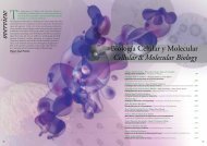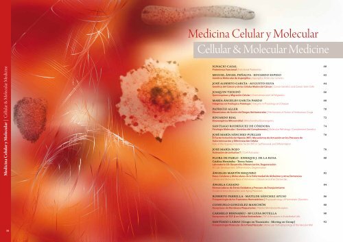
03 Medicina Celular y Molecular.pdf 5474KB May - Centro de ...
03 Medicina Celular y Molecular.pdf 5474KB May - Centro de ...
03 Medicina Celular y Molecular.pdf 5474KB May - Centro de ...

You also want an ePaper? Increase the reach of your titles
YUMPU automatically turns print PDFs into web optimized ePapers that Google loves.
<strong>Medicina</strong> <strong>Celular</strong> y <strong>Molecular</strong> | Cellular & <strong>Molecular</strong> Medicine<br />
58<br />
<strong>Medicina</strong> <strong>Celular</strong> y <strong>Molecular</strong><br />
Cellular & <strong>Molecular</strong> Medicine<br />
iGnaCio CasaL 60<br />
Proteómica Funcional | Functional Proteomics<br />
miGueL ánGeL PeñaLva · eduardo esPeso 62<br />
Genética <strong>Molecular</strong> <strong>de</strong> Aspergillus | Aspergillus <strong>Molecular</strong> Genetics<br />
José aLBerTo GarCía · auGusTo siLva 64<br />
Genética <strong>de</strong>l Cáncer y <strong>de</strong> las Células Madre <strong>de</strong>l Cáncer | Cancer Genetics and Cancer Stem Cells<br />
Joaquin TeixidÓ 66<br />
Quimioquinas y Migración <strong>Celular</strong> | Chemokines and Cell Migration<br />
maría ánGeLes GarCía Pardo 68<br />
Integrinas en Fisología y Patología | Integrins in Physiology and Disease<br />
PaTriCio aLLer 70<br />
Mecanismos <strong>de</strong> Acción <strong>de</strong> Drogas Antitumorales | Mechanisms of Action of Antitumour Drugs<br />
eduardo riaL 72<br />
Bioenergética Mitocondrial | Mitochondrial Bioenergetics<br />
sanTiaGo rodríGuez <strong>de</strong> CÓrdoBa 74<br />
Patología <strong>Molecular</strong> / Genética <strong>de</strong>l Complemento | <strong>Molecular</strong> Pathology / Complement Genetics<br />
José maría sánChez-PueLLes 76<br />
El Factor Inducible <strong>de</strong> Hipoxia (HIF); Mecanismos <strong>de</strong> Actuación en los Procesos <strong>de</strong><br />
Autorrenovación y Diferenciación <strong>Celular</strong><br />
Role of the Hypoxia Inducible Factor (HIF) in Self Renewal and Differentiation<br />
Jose maría roJo 78<br />
Activación <strong>de</strong> Linfocitos T | T Cell Activation<br />
FLora <strong>de</strong> PaBLo · enrique J. <strong>de</strong> La rosa 80<br />
Catalina hernán<strong>de</strong>z · Teresa suárez<br />
Laboratorio 3D: Desarrollo, Diferenciación, Degeneración<br />
3D Lab: Development, Differentiation, Degeneration<br />
ánGeLes marTín requero 82<br />
Bases <strong>Celular</strong>es y <strong>Molecular</strong>es <strong>de</strong> la Enfermedad <strong>de</strong> Alzheimer y otras Demencias<br />
Cellular and <strong>Molecular</strong> Basis of Alzheimer´s Disease and other Dementias<br />
ánGeLa Casado 84<br />
Biomarcadores <strong>de</strong> Estrés Oxidativo y Procesos <strong>de</strong> Envejecimiento<br />
Oxidative Stress Biomarkers and Aging Processes<br />
roBerTo ParriLLa · maTiL<strong>de</strong> sánChez ayuso 86<br />
Fisiopatología <strong>de</strong> los Trastornos Hemostáticos | Physiopathology of Hemostatic Disor<strong>de</strong>rs<br />
ConsueLo GonzáLez manChÓn 88<br />
Receptores <strong>de</strong> Membrana Plaquetarios | Platelet Membrane Receptors<br />
CarmeLo BernaBeu · mª Luisa BoTeLLa 90<br />
Receptores <strong>de</strong> TGF-β en Células Endoteliales | TGF-β Receptors in Endothelial Cells<br />
sanTiaGo Lamas (Grupo en Transición - moving on Group) 92<br />
Fisiopatología <strong>Molecular</strong> <strong>de</strong> la Pared Vascular | <strong>Molecular</strong> Pathophysiology of the Vascular Wall
Presentación<br />
<strong>Medicina</strong> <strong>Celular</strong> y <strong>Molecular</strong><br />
La investigación <strong>de</strong> los 16 grupos adscritos al <strong>de</strong>partamento <strong>de</strong> <strong>Medicina</strong><br />
<strong>Celular</strong> y <strong>Molecular</strong> preten<strong>de</strong> profundizar en las bases moleculares<br />
<strong>de</strong> la patología humana.<br />
Así, tiene como objetivos la i<strong>de</strong>ntificación <strong>de</strong> dianas diagnósticas y terapéuticas y la implementación<br />
<strong>de</strong> estrategias encaminadas al <strong>de</strong>scubrimiento <strong>de</strong> nuevos fármacos en diversas<br />
patologías humanas como cáncer (melanoma, leucemia, a<strong>de</strong>nocarcinoma <strong>de</strong> colon…),<br />
patologías relacionadas con la pared vascular y el miocardio y estados alterados <strong>de</strong>l sistema <strong>de</strong><br />
complemento y <strong>de</strong> la hemostasia. Por otro lado, abordamos el estudio <strong>de</strong> procesos <strong>de</strong> <strong>de</strong>sarrollo,<br />
<strong>de</strong>generación y envejecimiento para enten<strong>de</strong>r los mecanismos patogénicos <strong>de</strong> enfermeda<strong>de</strong>s<br />
como la retinitis pigmentosa, enfermedad <strong>de</strong> Lafora o el Alzheimer, a<strong>de</strong>más <strong>de</strong> la genética<br />
molecular <strong>de</strong> hongos. En los últimos años se ha hecho un esfuerzo particularmente<br />
importante en la introducción <strong>de</strong> tecnologías <strong>de</strong> alto rendimiento (Proteómica, Genómica,<br />
Farmacología <strong>Molecular</strong>…) cada vez más necesarias en el ámbito <strong>de</strong> la Biomedicina.<br />
El Departamento impulsa activamente las colaboraciones entre sus miembros, así como<br />
con grupos <strong>de</strong> otros Departamentos <strong>de</strong>l CIB, para <strong>de</strong>sarrollar una investigación biomédica<br />
interdisciplinaria <strong>de</strong> alta calidad que aumente la competitividad nacional e<br />
internacional <strong>de</strong> los grupos <strong>de</strong> investigación individuales. Nuestro programa apoya,<br />
incluso en tiempos restrictivos, la incorporación <strong>de</strong> nuevas tecnologías e infraestructuras<br />
experimentales y el <strong>de</strong>sarrollo <strong>de</strong> investigaciones pioneras en biomedicina<br />
mediante la incorporación y promoción <strong>de</strong> investigadores jóvenes. El Departamento<br />
también participa en iniciativas <strong>de</strong> transferencia <strong>de</strong> tecnología y anima<br />
a la creación <strong>de</strong> valor añadido para la investigación translacional, protegiendo<br />
a<strong>de</strong>cuadamente los resultados y reactivos producidos por los grupos <strong>de</strong> investigación.<br />
Ese valor añadido ha dado ya lugar a, y facilitará, la generación <strong>de</strong><br />
“spin-off” biotecnológicas. Por último, el Departamento <strong>de</strong>sea reconocer y<br />
agra<strong>de</strong>cer la fructífera y extensa labor investigadora realizada por Santiago<br />
Lamas y Augusto Silva en nuestro <strong>Centro</strong> y que finalizó el pasado bienio.<br />
Nuestros mejores <strong>de</strong>seos a ambos en su nueva andadura.<br />
iGnaCio CasaL<br />
JEFE DE DEPARTAMENTO<br />
Cellular and<br />
<strong>Molecular</strong> Medicine<br />
The research performed by the 16 groups of the Department of Cellular<br />
and <strong>Molecular</strong> Medicine aims to the characterization of the molecular<br />
basis of human physiopathology.<br />
As main objectives, we can mention the i<strong>de</strong>ntification of diagnostic markers and the implementation<br />
of targeted strategies for the discovery of new drugs. Diverse human pathologies<br />
are being characterized, such as cancer (melanoma, leukemia, colon a<strong>de</strong>nocarcinoma<br />
…), pathologies related with the vascular wall and the myocardium and disturbed<br />
states of the complement system and hemostasia. Also, the processes involved in Development,<br />
Degeneration and Aging are being studied to un<strong>de</strong>rstand the molecular mechanisms<br />
of pathogenesis of human disor<strong>de</strong>rs like retinitis pigmentosa, Lafora disease or Alzheimer’s<br />
disease, together with the molecular genetics of fungi. Moreover, in the last years, a particularly<br />
important effort has been ma<strong>de</strong> to introduce high-throughput technologies such as Proteomics,<br />
Genomics or <strong>Molecular</strong> Pharmacology, increasingly relevant in the Biomedicine field.<br />
Collaborations across laboratories and with other Departments within the CIB are actively encouraged<br />
to strengthen high quality interdisciplinary biomedical research in the Centre and to increase<br />
the national and international competitiveness of the individual research groups. Our program seeks<br />
the incorporation of the highest level of technological <strong>de</strong>velopments and experimental infrastructures,<br />
even in this restrictive period, and the promotion of pioneering research by attracting bright<br />
young researchers in Biomedicine. The Department also promotes technology transfer initiatives<br />
and encourages the creation of ad<strong>de</strong>d value for translational research through the a<strong>de</strong>quate protection<br />
of the scientific results and reagents produced by the individual groups. Such ad<strong>de</strong>d value<br />
has already resulted in the generation of biotechnology spin-offs, and will continue supporting new<br />
ones. Last, our Department would like to recognize and acknowledge the fruitful and extensive<br />
research work carried out by Santiago Lamas and Augusto Silva in our Centre, which finalized last<br />
biennium. Our best wishes to both in their new venture.<br />
iGnaCio CasaL<br />
DEPARTMENT HEAD<br />
Overview<br />
60 61
<strong>Medicina</strong> <strong>Celular</strong> y <strong>Molecular</strong> | Cellular and <strong>Molecular</strong> Medicine<br />
62<br />
José ignacio Casal alvarez<br />
Investigador Científico | icasal@cib.csic.es<br />
Proteómica Funcional<br />
I<strong>de</strong>ntificación <strong>de</strong> marcadores diagnósticos<br />
en cáncer colorrectal mediante<br />
<strong>de</strong>tección <strong>de</strong> autoanticuerpos y/o<br />
antígenos asociados a tumores (AATs).<br />
Existen claras evi<strong>de</strong>ncias <strong>de</strong> la existencia<br />
<strong>de</strong> una respuesta inmune a cáncer en<br />
humanos, como se ha <strong>de</strong>mostrado por la<br />
presencia <strong>de</strong> autoanticuerpos en sueros<br />
<strong>de</strong> pacientes con cáncer. Nuestro grupo<br />
utiliza diversas estrategias proteómicas<br />
para la i<strong>de</strong>ntificación <strong>de</strong> AATs específicos<br />
<strong>de</strong> cáncer colorrectal. La caracterización <strong>de</strong><br />
los mecanismos que subyacen la inducción<br />
<strong>de</strong> la respuesta <strong>de</strong> anticuerpos frente a<br />
tumores y los tipos <strong>de</strong> mutaciones presente<br />
en los AATs están siendo analizados. A partir<br />
PhD, 1984,<br />
<strong>Centro</strong> <strong>de</strong> Biología <strong>Molecular</strong>, CSIC-Universidad<br />
Autónoma <strong>de</strong> Madrid.<br />
Post-doctoral Fellow (1985-1986), Massachusetts<br />
Institute of Technology (MIT), Cambridge, MA, USA.<br />
Jefe <strong>de</strong> Proyecto, 1986-1997,<br />
Director <strong>de</strong> Investigación, 1997-2001,<br />
INGENASA.<br />
Director Programa <strong>de</strong> Biotecnología, 2001-2008,<br />
<strong>Centro</strong> Nacional <strong>de</strong> Investigaciones Oncológicas<br />
(CNIO).<br />
Investigador Científico 2007, CSIC.<br />
Incorporación 2008, CIB.<br />
Nuestro grupo utiliza las tecnologías proteómicas<br />
como herramienta para i) la i<strong>de</strong>ntificación <strong>de</strong><br />
biomarcadores diagnósticos y ii) el estudio <strong>de</strong><br />
los mecanismos moleculares implicados en la<br />
metástasis <strong>de</strong> cáncer colorrectal y su relación con<br />
el componente inflamatorio asociado a tumores.<br />
Estrategias proteómicas empleadas para la i<strong>de</strong>ntificación <strong>de</strong> biomarcadores<br />
en cáncer <strong>de</strong> colon. Para <strong>de</strong>talles ver Bar<strong>de</strong>ras et al. Proteomics Clin Appl. 2010<br />
Feb;4(2):159-78.<br />
Proteomic strategies used for the i<strong>de</strong>ntification of colon cancer biomarkers. See<br />
Bar<strong>de</strong>ras et al. Proteomics Clin Appl. 2010 Feb;4(2):159-78 for <strong>de</strong>tails.<br />
<strong>de</strong> los AATs i<strong>de</strong>ntificados, nuestro grupo<br />
está <strong>de</strong>sarrollando inmunoensayos tipo<br />
ELISA para el diagnóstico, preferentemente<br />
temprano, <strong>de</strong>l cáncer <strong>de</strong> colon. Los<br />
resultados han sido patentados y licenciados.<br />
Efecto <strong>de</strong>l estroma en progresión tumoral<br />
y caracterización <strong>de</strong> la metástasis en<br />
cáncer <strong>de</strong> colon. El proceso <strong>de</strong> invasión<br />
tumoral va acompañado por cambios<br />
en la morfología y fenotipo celular, este<br />
proceso es conocido como transición<br />
epitelio-mesénquima. Actualmente estamos<br />
estudiando las alteraciones proteómicas<br />
inducidas por factores clave en la regulación<br />
<strong>de</strong> la proliferación celular y el fenotipo<br />
epitelial como SNAIL, TWIST o Vitamina D<br />
Otros miembros | Other lab members:<br />
Rodrigo Bar<strong>de</strong>ras<br />
Ingrid Babel<br />
Alberto Peláez<br />
Roi Villar<br />
María Fernan<strong>de</strong>z<br />
www.cib.csic.es/es/grupo.php?idgrupo=66<br />
Esto incluye el análisis <strong>de</strong> patrones <strong>de</strong> proteínas y<br />
la caracterización <strong>de</strong> vías <strong>de</strong> señalización y rutas<br />
metabólicas alteradas durante la metástasis hepática.<br />
El laboratorio participa en una activa transferencia<br />
tecnológica, particularmente en el campo <strong>de</strong>l<br />
diagnóstico molecular.<br />
tanto en células tumorales como estromales<br />
adyacentes.<br />
Por otro lado, estamos utilizando proteómica<br />
cuantitativa para estudiar y caracterizar<br />
el proteoma <strong>de</strong> dos líneas <strong>de</strong> cáncer <strong>de</strong><br />
colon, KM12C y KM12SM, que difieren<br />
en propieda<strong>de</strong>s metastáticas. Hemos<br />
i<strong>de</strong>ntificado alteraciones tanto en moléculas<br />
implicadas en adhesión y migración celular<br />
como en los patrones <strong>de</strong> quimioquinas<br />
asociadas a metástasis. Hemos analizado su<br />
relación con el componente inflamatorio<br />
y el reclutamiento <strong>de</strong> células mielocíticas.<br />
Finalmente, hemos <strong>de</strong>tectado cambios<br />
a nivel metabólico que están siendo<br />
caracterizados actualmente.<br />
ADH-GFP ADH-SN<br />
Imagen representativa <strong>de</strong> un microarray <strong>de</strong> citoquinas incubado con medio<br />
condicionado proce<strong>de</strong>nte <strong>de</strong> células <strong>de</strong> cáncer <strong>de</strong> colon transformadas con SNAIL y sus<br />
correspondientes controles. Para <strong>de</strong>talles ver Peña et al. Oncogene 2009 28, 4375-4385.<br />
Representative image of a cytokine microarray incubated with conditioned medium from<br />
SNAIL-transformed and mock colon cancer cells. See Peña et al. Oncogene 2009 28, 4375-<br />
4385 for <strong>de</strong>tails.<br />
Functional Proteomics<br />
Our group uses the proteomic technologies as a<br />
tool for i) the i<strong>de</strong>ntification of diagnostic biomarkers<br />
and ii) the study of the molecular mechanisms<br />
involved in colorectal cancer metastasis and their<br />
relationship with the inflammatory component<br />
associated to tumors. This inclu<strong>de</strong>s the analysis of<br />
protein expression patterns and the characterization<br />
of signalling and metabolic pathways altered during<br />
the metastasis in liver. The laboratory participates<br />
actively in technology transfer, particularly in the<br />
field of molecular diagnosis.<br />
I<strong>de</strong>ntification of diagnostic markers in colorectal cancer<br />
by autoantibody and/or tumor-associated antigens (TAAs)<br />
<strong>de</strong>tection. There is mounting evi<strong>de</strong>nce of the existence of<br />
an immune response to cancer in humans, as <strong>de</strong>monstrated by<br />
the presence of autoantibodies in cancer sera. Our group uses<br />
different proteomic strategies for the i<strong>de</strong>ntification of colorectal<br />
cancer-specific TAAs. The characterization of the mechanisms that<br />
un<strong>de</strong>rlie the induction of autoantibodies in cancer and the type of<br />
mutations present in the AATs are being characterized. Based on<br />
the i<strong>de</strong>ntified TAAs, our group is <strong>de</strong>veloping ELISA immunoassays<br />
for the, preferentially early, diagnosis of colon cancer. The results<br />
have been patented and licensed .<br />
Effect of the stroma in tumoral progression and characterization<br />
of the metastasis in colon cancer. The invasion process in cancer is<br />
accompanied by changes in cell morphology and phenotype, this<br />
process is known as epithelium-mesenchymal transition. Currently,<br />
we are studying proteomic alterations induced by key factors in<br />
the regulation of cell proliferation and epithelial phenotype, such<br />
as SNAIL, TWIST or 1,25 dihydroxyvitamin D3 in cancer cells and<br />
adjoining stromal cells.<br />
In addition, we are using quantitative proteomics to study and<br />
characterize the proteome of two colon cell lines, KM12C and<br />
KM12SM, differing in metastatic properties. We have i<strong>de</strong>ntified<br />
alterations in adhesion and cell migration-related molecules as<br />
well as in chemokine expression patterns associated to metastasis.<br />
We have analyzed their relationship with the inflammatory<br />
component and the recruitment of mielocytic cells. Finally, we<br />
have <strong>de</strong>tected changes at the metabolic level that are being<br />
currently characterized.<br />
Publicaciones Seleccionadas<br />
Selected Publications<br />
Babel, I., Bar<strong>de</strong>ras R., Diaz-Uriarte, R., Martínez-Torrecuadrada JL,<br />
Sánchez-Carbayo M and JI Casal [2009]. I<strong>de</strong>ntification of tumorassociated<br />
autoantigens for the diagnosis of colorectal cancer in<br />
serum using high-<strong>de</strong>nsity protein microarrays. Mol Cell Proteomics<br />
8, 2382-2395.<br />
Casado-Vela, J., Martinez-Torrecuadrada JL and JI Casal [2009].<br />
Differential phosphorylation patterns between the Cyclin-a2/CDK2<br />
complex and their monomers. Protein Expr Purif 66, 15-21.<br />
Timmerman P, Bar<strong>de</strong>ras R, Desmet J, Altschuh D, Shochat S, Hollestelle<br />
MJ, ßHöppener JWM, Monasterio A, Casal JI*, Meloen RH [2009].<br />
A combinatorial approach for the <strong>de</strong>sign of complementarity<br />
<strong>de</strong>termining regions (CDR)-<strong>de</strong>rived peptidomimetics with in vitro<br />
anti-tumoral activity. J Biol Chem 284: 34126-34134 (*, corresponding<br />
author).<br />
Peña C, García JM, Larriba MJ, Bar<strong>de</strong>ras R, Garcia V, Silva J, Dominguez<br />
G, Rodriguez R, Cuevas J, Garcia <strong>de</strong> Herreros A., Casal JI, Muñoz A,<br />
Bonilla F [2009]. SNAI1 expression in colon cancer related with CDH1<br />
and VDR downregulation in normal adjacent tissue. Oncogene 28,<br />
4375-4385.<br />
Bar<strong>de</strong>ras R, Babel I and Casal JI [2010]. Colorectal cancer proteomics,<br />
molecular characterization and biomarker discovery. Proteomics Clin<br />
Appl 4, 159-178.<br />
Luque-Garcia JL, Martínez-Torrecuadrada JL, Epifanio C, Cañamero<br />
M, Babel I and Casal JI [2010]. Differential protein expression on the<br />
cell surface of colorectal cancer cells associated to tumor metastasis.<br />
Proteomics 10, 940-952.<br />
Larriba MJ, Casado-Vela J, Pendás-Franco N, Peña R., García <strong>de</strong> Herreros<br />
A, Berciano MT, Lafarga M, Casal JI, Muñoz A [2010]. Novel Snail1 Target<br />
Proteins in Human Colon Cancer I<strong>de</strong>ntified by Proteomic Analysis.<br />
PLOS One 5, e10221.<br />
Guillou E, Ibarra A, Coulon V, Casado J, Rico D, Casal I, Schwob S,<br />
Losada A and Mén<strong>de</strong>z J [2010]. Cohesin organizes chromatin loops at<br />
DNA replication factories. Genes Dev 24, 2812-2822.<br />
Patentes<br />
Patents<br />
Babel, R. Bar<strong>de</strong>ras y J.I. Casal [2009]. Método <strong>de</strong> obtención <strong>de</strong> datos útiles<br />
para el diagnóstico o el pronóstico <strong>de</strong>l cáncer colorrectal.<br />
Patent Number: P2009302<strong>03</strong>. PCT-Treaty. Licensed.<br />
Babel, R. Bar<strong>de</strong>ras y J.I. Casal [2010]. Método <strong>de</strong> diagnóstico/pronóstico<br />
<strong>de</strong>l cáncer colorrectal basado en la <strong>de</strong>tección en suero <strong>de</strong> anticuerpos<br />
frente a autoantígenos tumorales.<br />
Patent Number: P201<strong>03</strong>0708. PCT-Treaty. Licensed.<br />
63
<strong>Medicina</strong> <strong>Celular</strong> y <strong>Molecular</strong> | Cellular and <strong>Molecular</strong> Medicine<br />
64<br />
miguel a. Peñalva<br />
Profesor <strong>de</strong> Investigación | penalva@cib.csic.es<br />
PhD, 1982.<br />
Universidad Autónoma <strong>de</strong> Madrid.<br />
Postdoctoral,<br />
Antibióticos SA (Madrid) e Institut <strong>de</strong> Genetique et<br />
Microbiologie, Universidad <strong>de</strong> París, Orsay.<br />
Científico Titular y Jefe <strong>de</strong> grupo, 1987,<br />
Profesor <strong>de</strong> Investigación <strong>de</strong>s<strong>de</strong> 2001.<br />
CIB, CSIC.<br />
Visiting Scientist, 2005-2006.<br />
MRC Laboratory of <strong>Molecular</strong> Biology, Cambridge UK.<br />
Elegido miembro <strong>de</strong> EMBO en 2000.<br />
Genética <strong>Molecular</strong> <strong>de</strong> Aspergillus<br />
A. nidulans es un mo<strong>de</strong>lo genético para el estudio<br />
<strong>de</strong>l crecimiento celular polarizado y el transporte<br />
a larga distancia (http://en.wikipedia.org/wiki/<br />
Mo<strong>de</strong>l_organism). Sus hifas crecen exclusivamente<br />
por extensión apical, formando células tubulares<br />
Publicaciones Seleccionadas<br />
Selected Publications<br />
Abenza JF, Pantazopoulou A, Rodríguez JM, Galindo<br />
A, Peñalva MA [2009] Long-distance movement of<br />
Aspergillus nidulans early endosomes on microtubule<br />
tracks. Traffic 10: 57-75.<br />
Rodríguez-Galán O, Galindo A, Hervás-Aguilar A, Arst<br />
HN, Jr., Peñalva MA [2009] Physiological involvement<br />
in pH signalling of Vps24-mediated recruitment of<br />
Aspergillus PalB cysteine protease to ESCRT-III. J Biol<br />
Chem 284: 4404-4412.<br />
Pantazopoulou A, Peñalva MA [2009] The<br />
organization and dynamics of the Aspergillus<br />
nidulans Golgi during apical extension and mitosis.<br />
Mol Biol Cell 20: 4335-4337.<br />
Hervás-Aguilar A, Galindo A, Peñalva MA [2010]<br />
Receptor-in<strong>de</strong>pen<strong>de</strong>nt Ambient pH signaling by<br />
ubiquitin attachment to fungal arrestin-like PalF. J<br />
Biol Chem 285: 18095-18102.<br />
Abenza JF, Galindo A, Pantazopoulou A, Gil C, <strong>de</strong><br />
Los Ríos V, Peñalva MA [2010] Aspergillus RabB/Rab5<br />
Integrates Acquisition of Degradative I<strong>de</strong>ntity with<br />
the Long Distance Movement of Early Endosomes.<br />
Mol Biol Cell. 21:2756-2769.<br />
Etxebeste O, Markina-Iñarrairaegui A, Garzia A,<br />
Herrero-García E, Ugal<strong>de</strong> U, Espeso EA. [2009] KapI, a<br />
non-essential member of the Pse1p/Imp5 karyopherin<br />
family, controls colonial and asexual <strong>de</strong>velopment in<br />
Aspergillus nidulans. Microbiology. 155:3934-45.<br />
Etxebeste O, Herrero-García E, Araújo-Bazán L,<br />
Rodríguez-Urra AB, Garzia A, Ugal<strong>de</strong> U, Espeso EA.<br />
[2009] The bZIP-type transcription factor FlbB regulates<br />
distinct morphogenetic stages of colony formation in<br />
Aspergillus nidulans. Mol Microbiol. 73:775-89.<br />
Garzia A, Etxebeste O, Herrero-García E, Ugal<strong>de</strong> U,<br />
Espeso EA. [2010] The concerted action of bZip and<br />
cMyb transcription factors FlbB and FlbD induces<br />
brlA expression and asexual <strong>de</strong>velopment in<br />
Aspergillus nidulans. Mol Microbiol. 75:1314-24.<br />
Etxebeste O, Garzia A, Espeso EA, Ugal<strong>de</strong> U. [2010]<br />
Aspergillus nidulans asexual <strong>de</strong>velopment: making the<br />
most of cellular modules. Trends Microbiol. 18:569-76.<br />
Etxebeste O, Ugal<strong>de</strong> U, Espeso EA. [2010] Adaptative<br />
and <strong>de</strong>velopmental responses to stress in Aspergillus<br />
nidulans. Curr Protein Pept Sci. 11:704-18.<br />
eduardo a. espeso<br />
Científico Titular | eespeso@cib.csic.es<br />
Un aspecto clave <strong>de</strong> los hongos<br />
es su invasividad, asociada con<br />
su rápido crecimiento apical.<br />
Combinando abordajes genéticos con<br />
microscopía multidimensional, estudiamos<br />
la organización <strong>de</strong> Golgi y endosomas<br />
en el contexto <strong>de</strong>l tráfico intracelular<br />
polarizado, centrándonos en GTPasas<br />
Rab y sus efectores. El Golgi <strong>de</strong> Aspergillus<br />
está formado por cisternas dispersas<br />
(Golgi equivalents) que pue<strong>de</strong>n resolverse<br />
por microscopía óptica. Los endosomas<br />
tempranos son característicamente mótiles<br />
a gran<strong>de</strong>s distancias sobre microtúbulos.<br />
La internalización endocítica y la<br />
maduración endosomal son esenciales.<br />
Preten<strong>de</strong>mos compren<strong>de</strong>r los mecanismos<br />
<strong>de</strong> maduración <strong>de</strong> cisternas <strong>de</strong>l Golgi, las<br />
rutas entre éste y los endosomas y el papel<br />
no canónico que los complejos ESCRT<br />
tienen en la organización <strong>de</strong> complejos<br />
<strong>de</strong> transducción <strong>de</strong> señal <strong>de</strong> pH en la<br />
membrana plasmática.<br />
Early and late<br />
cisternae of the nonstacked<br />
Golgi os<br />
Aspergillus. nidulans<br />
can be resolved by<br />
in vivo fluorescence<br />
microscopy, allowing<br />
investigation of<br />
cisternal maturation.<br />
Pantazopoulou<br />
& Peñalva,<br />
“Characterization of<br />
Arpergillus nidulans<br />
RabC/Rab6’ Traffic<br />
12:386 (2011).<br />
PhD, 1989.<br />
Universidad Complutense <strong>de</strong> Madrid.<br />
Postdoctoral, 1997-1999,<br />
EMBO-Postdoctoral Fellow.<br />
Imperial College London.<br />
Contratado Ramón y Cajal, 2001-2004,<br />
Científico Titular y Jefe <strong>de</strong> grupo, 2004.<br />
CIB, CSIC.<br />
Secretario, 2004-2008.<br />
Grupo Especializado <strong>de</strong> Hongos Filamentosos y<br />
Levaduras (SEM).<br />
multinucleadas en las que el tráfico intracelular está<br />
adaptado a las gran<strong>de</strong>s distancias existentes entre<br />
distintas regiones o núcleos <strong>de</strong> la célula. La regulación<br />
<strong>de</strong> la transcripción por pH ambiental media la<br />
adaptación <strong>de</strong> los hongos a ambientes con distinto pH.<br />
Una gran variabilidad genética permite<br />
a los hongos la capacidad <strong>de</strong> medrar en<br />
ambientes extremos. La eficiente regulación<br />
transcripcional y la perfecta comunicación<br />
entre el núcleo y el citoplasma, don<strong>de</strong> se<br />
sintetizan los factores transcripcionales<br />
y concurren muchos <strong>de</strong> los sistemas <strong>de</strong><br />
señalización, aseguran la correcta expresión<br />
<strong>de</strong> aquellos genes cuyos productos se<br />
necesitan para que el hongo se <strong>de</strong>sarrolle<br />
en estas condiciones ambientales.<br />
Estudiamos la maquinaria molecular que<br />
participa en la homeostasis <strong>de</strong> cationes,<br />
mono y divalentes, y la que asegura el<br />
transporte núcleo-citoplásmico <strong>de</strong> factores<br />
transcripcionales <strong>de</strong> alta jerarquía que<br />
permiten afrontar los diferentes estreses<br />
abióticos. Así mismo, preten<strong>de</strong>mos<br />
establecer la forma en la que estos procesos<br />
participan en la diferenciación celular que<br />
ocurre durante el ciclo <strong>de</strong> reproducción<br />
asexual en Aspergillus y en la patogenicidad<br />
<strong>de</strong> este hongo en mamíferos.<br />
Las cisternas tempranas y tardías <strong>de</strong>l Golgi no apilado <strong>de</strong> Aspergillus nidulans pue<strong>de</strong>n<br />
resolverse por microscopía <strong>de</strong> fluorescencia in vivo, lo que permite investigar la<br />
maduración <strong>de</strong> cisternas.<br />
Otros miembros | Other lab members:<br />
Dr. Areti Pantazopoulou Dr. Antonio Galindo Revilla<br />
Dr. Mario Pinar Sala Elena Reoyo<br />
Juan Francisco Abenza Martínez Laura Cobeño Fariñas<br />
Ane Marquina Iñarrairaegui Erika Herrero García<br />
Laura Mellado Maroñas<br />
Patricia Hernán<strong>de</strong>z Ortiz<br />
Daniel Lucena Agell.<br />
www.cib.csic.es/es/grupo.php?idgrupo=8<br />
Fotografías que<br />
muestran la carioferina<br />
kapH-GFP (en ver<strong>de</strong>)<br />
asociada al uso mitótico<br />
durante mitosis cerrada. La<br />
proteina Nud1-mCherry (en<br />
magenta) marca la posición <strong>de</strong>l<br />
“spindle-pole-body” (centrosoma<br />
fúngico). Parte inferior izquierda una<br />
placa maestra con diferentes cepas<br />
y mutantes en el color <strong>de</strong> esporas en<br />
Aspergillus. <strong>Centro</strong> y <strong>de</strong>recha, imágenes<br />
<strong>de</strong> las portadas en Microbiology y Trends in<br />
Microbiology que ilustran el ejemplar don<strong>de</strong> se<br />
han publicado parte <strong>de</strong>l trabajo realizado en el<br />
bienio 09-10.<br />
Insets showing the association of GFP tagged<br />
kariopherin KapH (in green) with the mitotic spindle<br />
during the closed mitosis. Protein Nud1-mCherry (in magenta) labels the position of the spindle pole body, the fungal centrosome. Lower left panel shows different<br />
Aspergillus strains carrying mutations affecting the colour of conidiospores. Centre and right, cover pages of Microbiology and Trends in Microbiology issues, respectively,<br />
where part of our work has been published during years 2009 and 2010.<br />
Aspergillus <strong>Molecular</strong> Genetics<br />
A. nidulans is a genetic mo<strong>de</strong>l for studying polarised<br />
cell growth and long distance transport (http://<br />
en.wikipedia.org/wiki/Mo<strong>de</strong>l_organism). Hyphal tip<br />
cells grow exclusively by apical extension, leading<br />
to tubular multinucleated cells where intracellular<br />
traffic is tailored to the needs imposed by the<br />
relatively large distances between apical and distal<br />
regions and between the different nuclei of the<br />
cell. Regulation of gene expression by ambient pH<br />
mediates fungal adaptation to environments with<br />
different values of pH.<br />
A<br />
key aspect of fungi is their invasiveness, associated with<br />
rapid apical extension. By combining genetic approaches<br />
with multidimensional microscopy, we are studying the<br />
organization of the Golgi and endosomal systems in the context<br />
of polarized traffic, focusing on membrane domain-organising<br />
Rab GTPases and their effectors. The Aspergillus Golgi is formed<br />
by scattered ‘Golgi equivalents’, such that early and late cisternae<br />
can be resolved by optical microscopy. Early endosomes are<br />
characteristically motile for long distances, riding on microtubules.<br />
Endocytic internalization and the maturation of EEs are essential.<br />
We are trying to un<strong>de</strong>rstand the mechanisms of cisternal<br />
maturation in the Golgi, the pathways connecting Golgi and<br />
endosomes and the ‘non-endosomal’ role that ESCRT complexes<br />
play in scaffolding ambient pH signal transduction complexes at<br />
the plasma membrane.<br />
The genetic versatility of fungi allows growth of these organisms in<br />
extreme environments. An efficient transcriptional regulation and<br />
a correct communication between nucleus and cytoplasm, where<br />
transcription factors are synthesised and most of signalling pathways<br />
play their roles, ensure the proper expression of those genes<br />
whose products are nee<strong>de</strong>d by the fungus to <strong>de</strong>velop un<strong>de</strong>r such<br />
extreme ambient conditions. We study the molecular machineries<br />
participating in the homeostasis of mono and divalent cations, and<br />
in nucleo-cytoplasmic trafficking of high hierarchy transcription<br />
factors which allow the fungus to confront diverse abiotic stresses. In<br />
addition, we attempt to establish how these processes contribute to<br />
the cellular differentiation during the asexual reproductive cycle of<br />
Aspergillus and to its pathogenicity in mammals.<br />
65
<strong>Medicina</strong> <strong>Celular</strong> y <strong>Molecular</strong> | Cellular and <strong>Molecular</strong> Medicine<br />
66<br />
José alberto García-sanz<br />
Científico Titular | jasanz@cib.csic.es<br />
Genética <strong>de</strong>l Cancer y <strong>de</strong> las Células Madre <strong>de</strong>l Cáncer<br />
Células Madre <strong>de</strong> Gándula mamaria:<br />
(E. Diaz-Guerra, Mª A. Lillo, Jose A.<br />
Garcia-Sanz). La glándula mamaria<br />
tiene la mayor parte <strong>de</strong> su <strong>de</strong>sarrollo tras el<br />
nacimiento, don<strong>de</strong> ocurren gran<strong>de</strong>s cambios<br />
en arquitectura durante la pubertad y con<br />
cada ciclo reproductivo. Lás células madre<br />
<strong>de</strong> glándula mamaria tienen un fenotipo<br />
CD24+CD29+ y una sola célula con dicho<br />
fenotipo es capaz, al ser transplantada, <strong>de</strong><br />
regenerar una glándula mamaria funcional.<br />
Hemos analizado la celularidad y numero<br />
<strong>de</strong> células madre durante la pubertad y<br />
preñez. Los datos obtenidos nos llevaron a<br />
proponer un mo<strong>de</strong>lo <strong>de</strong> control <strong>de</strong> tamaño<br />
<strong>de</strong>l órgano con importantes implicaciones<br />
en tumorigenesis mamaria. Actualmente<br />
intentamos verificar in vivo las implicaciones<br />
PhD, 1987.<br />
Universidad <strong>de</strong> Barcelona.<br />
Postdoctoral,<br />
1987-1989, ISREC,<br />
1989-1991, Universidad <strong>de</strong> Miami,<br />
1991-1996, Miembro Científico Basel Institute for<br />
Immunology.<br />
Investigador contratado, 1997-20<strong>03</strong>.<br />
CNB, CSIC.<br />
Investigador, 20<strong>03</strong>-2008.<br />
Ramon y Cajal.<br />
Científico Titular, 2008.<br />
CIB, CSIC.<br />
La homeostasis <strong>de</strong> los tejidos durante la vida <strong>de</strong><br />
un individuo es mantenida por las células madre<br />
adultas. Estamos interesados en compren<strong>de</strong>r varios<br />
aspectos <strong>de</strong> la biologia <strong>de</strong> dichas células y su<br />
relación con las células madre <strong>de</strong>l cáncer. Para ello<br />
Publicaciones Seleccionadas<br />
Selected Publications<br />
Armesilla-Diaz, A., Bragado, P., Del Valle, I., Cuevas, E., Lazaro, I., Martin, C., Cigudosa,<br />
J.C., and Silva, A. (2009). p53 regulates the self-renewal and differentiation of neural<br />
precursors. Neuroscience 158, 1378-1389.<br />
Armesilla-Diaz, A., Elvira, G., and Silva, A. (2009). p53 regulates the proliferation, differentiation<br />
and spontaneous transformation of mesenchymal stem cells. Exp Cell Res 315, 3598-3610.<br />
Dutzan, N., Gamonal, J., Silva, A., Sanz, M., and Vernal, R. (2009). Over-expression of<br />
forkhead box P3 and its association with receptor activator of nuclear factor-kappa<br />
B ligand, interleukin (IL) -17, IL-10 and transforming growth factor-beta during the<br />
progression of chronic periodontitis. J Clin Periodontol 36, 396-4<strong>03</strong>.<br />
Juanes, M.E., Elvira, G., Garcia-Gran<strong>de</strong>, A., Calero, M., and Gasset, M. (2009). Biosynthesis of<br />
prion protein nucleocytoplasmic isoforms by alternative initiation of translation. J Biol<br />
Chem 284, 2787-2794.<br />
<strong>de</strong>l mo<strong>de</strong>lo en tumorigenesis mamaria.<br />
Characterización <strong>de</strong> anticuerpos<br />
monoclonales frente a células madre y<br />
progenitores neurales tempranos. (G.<br />
Elvira, P. Perez, A. Silva, Jose A. Garcia-<br />
Sanz). Hemos generado nuevos anticuerpos<br />
monoclonales que i<strong>de</strong>ntifican células<br />
madre neurales y progenitores tempranos<br />
(Nilo1, 2, 3 y 8). Estamos trabajando en la<br />
i<strong>de</strong>ntificación <strong>de</strong> los antigenos reconocidos<br />
por dichos anticuerpos, su distribución<br />
en distinas poblaciones <strong>de</strong> células madre<br />
y progenitores, así como en su efecto<br />
inhibidor bloqueando la diferenciación <strong>de</strong> las<br />
células madre. Dichos anticuerpos unidos a<br />
nanoparticulas paramagnétics se han usado<br />
para i<strong>de</strong>ntificar in vivo las células madre<br />
y progenitores endógenos en su nicho, y<br />
augusto silva González<br />
Investigador Científico | asilvagonzalez@celgene.com<br />
PhD, 1981, Group Lea<strong>de</strong>r, 1984.<br />
Clinica Puerta <strong>de</strong> Hierro, UCM.<br />
Visiting Fellow 1978 NIH, 1979, UCLA.<br />
Postdoctoral Fellow, 1981, ISREC.<br />
Colaborador Científico, 1986,<br />
Investigador Científico, 2008.<br />
CIB, CSIC.<br />
Subdirector Gral. ISCIII, 1992,<br />
Director General Terapias Avanzadas y<br />
Transplantes, 2008, Ministerio <strong>de</strong> Sanidad.<br />
Director of Biological Science, Celgene Institute<br />
of Translational Research Europe. (En exce<strong>de</strong>ncia<br />
temporal <strong>de</strong>l CIB).<br />
estamos analizando aspectos <strong>de</strong> las células madre <strong>de</strong><br />
glándula mamaria, sistema nerviosos central y células<br />
mesenquimales (este trabajo se ha llevado a cabo<br />
<strong>de</strong>ntro <strong>de</strong> la red <strong>de</strong> terapia celular <strong>de</strong>l ISCIII).<br />
<strong>de</strong>terminar su dinámica <strong>de</strong> migración en<br />
respuesta a daño.<br />
Characterización <strong>de</strong> células madre<br />
mesenquimales humanas para<br />
regeneración periodontal (S. Santamaria,<br />
Jose A. Garcia-Sanz, en colaboración con<br />
M. Sanz, UCM). Estamos usando células<br />
madre mesenquimales humanas para<br />
caracterizar la población <strong>de</strong> células madre<br />
<strong>de</strong>ntro <strong>de</strong> esa población compleja, en<br />
particular su capacidad <strong>de</strong> autorenovación y<br />
diferenciación a hueso, cartilago y grasa, así<br />
como caracterizar las divisiones simétricas<br />
y asimétricas <strong>de</strong> las mismas intentando<br />
compren<strong>de</strong>r mejor la biologia <strong>de</strong> dichas<br />
celulas y <strong>de</strong>terminar las posiblida<strong>de</strong>s <strong>de</strong> su<br />
uso para regeneración <strong>de</strong> tejido oseo para<br />
regeneraciones periodontales.<br />
Vernal, R., Leon, R., Silva, A., van Winkelhoff, A.J., Garcia-Sanz, J.A., and Sanz, M. (2009).<br />
Differential cytokine expression by human <strong>de</strong>ndritic cells in response to different<br />
Porphyromonas gingivalis capsular serotypes. J Clin Periodontol 36, 823-829.<br />
Del Valle, I., Elvira, G., Garcia-Benzaquen, L., Armesilla-Diaz, A., Kremer, L., Garcia-Sanz, J.A.,<br />
Martinez, S., and Silva, A. (2010). Characterization of novel monoclonal antibodies able<br />
to i<strong>de</strong>ntify neurogenic niches and arrest neurosphere proliferation and differentiation.<br />
Neuroscience 169, 1473-1485.<br />
García-Gómez, I., Elvira, G., Zapata, A.G., Lamana, M.L., Ramírez, M., Garcia Castro, J., García<br />
Arranz, M., Vicente, A., Bueren, J., and García-Olmo, D. (2010). Mesenchymal stem cells:<br />
biological properties and clinical applications. Expert Opin Biol Ther 10, 1-16.<br />
Elvira, G., Moreno, B., <strong>de</strong>lValle, I., Garcia–Sanz, J.A., Canillas, M., Chinarro, E., Jurado, J.R., and<br />
Silva, A. (2010). Targeting Neural Stem Cells with Titanium Oxi<strong>de</strong> Nanoparticles Coupled<br />
to Specific Monoclonal Antibodies. J Biomat Appl DOI: 10.1177/088532821<strong>03</strong>93294.<br />
Otros miembros | Other lab members:<br />
Gema Elvira Serrano<br />
Patricia Pérez Arnaiz<br />
Alejandro Armesilla Díaz<br />
Rolando Vernal Astudillo<br />
Eva Díaz Guerra<br />
Mª Angeles Lillo Osuna<br />
Silvia Santamaría García-Minguillán<br />
www.cib.csic.es/es/grupo.php?idgrupo=26<br />
Cancer Genetics and<br />
Cancer Stem Cells<br />
Tissue homeostasis through an individual’s life<br />
is maintained by tissue-specific stem cells. We<br />
are interested on un<strong>de</strong>rstanding several aspects<br />
of adult stem cell biology and their possible<br />
relation with cancer stem cells. For this purpose<br />
we are currently analyzing different aspects of<br />
adult stem cells from the mammary gland, the<br />
nervous central system and mesenchymal stem<br />
cells (this work has been done in the context of<br />
the ISCIII network of Cell Therapy).<br />
Mammary Stem Cells: (E. Diaz-Guerra, Mª A.<br />
Lillo, Jose A. Garcia-Sanz). The mammary gland<br />
un<strong>de</strong>rgoes most of its <strong>de</strong>velopment after birth,<br />
and large remo<strong>de</strong>ling and architectural changes occur<br />
with each reproductive cycle. The mammary stem cells are<br />
CD24+CD29+ cells, and a single CD24+CD29+ cell is able,<br />
after transplantation to regenerate a functional mammary<br />
gland. We have analyzed mammary gland cellularity and stem<br />
cell number during puberty and the reproductive cycle. The<br />
data obtained allowed us to propose a mo<strong>de</strong>l for organ size<br />
control with clear implications for mammary tumorigenesis.<br />
Our current efforts aim to verify in vivo the implications of this<br />
mo<strong>de</strong>l on mammary tumorigenesis.<br />
Characterization of new antibodies recognizing neural stem<br />
and early progenitor cells. (G. Elvira, P. Perez, A. Silva and<br />
Jose A. Garcia-Sanz). We have generated new monoclonal<br />
antibodies i<strong>de</strong>ntifying adult neural stem and early progenitor<br />
cells (Nilo1, 2, 3 and 8). We are working on the i<strong>de</strong>ntification<br />
of the antigens recognized by these antibodies, their<br />
distribution in several stem and early progenitor cell<br />
populations and their inhibitory effect blocking neural<br />
stem cell differentiation. These antibodies, coupled to<br />
magnetic nanoparticles have been used to in vivo i<strong>de</strong>ntify<br />
the endogenous stem and early progenitor cell niches,<br />
<strong>de</strong>termining their dynamics during pathological transitions.<br />
Characterization of human mesenchymal stem cells for<br />
periodontal regeneration (S. Santamaria and Jose A. Garcia-<br />
Sanz, in collaboration with M. Sanz, UCM). We are using<br />
human mesenchymal stem cells to characterize the stem cells<br />
on this complex population, in particular their self renewal<br />
ability and their ability to differentiate into bone, cartilage and<br />
fat, as well as to characterize their type of divisions (symmetric<br />
and asymmetric), aiming as a long term goal to better<br />
un<strong>de</strong>rstand this cell population and <strong>de</strong>termine the possibilities<br />
to use it to regenerate bone tissue for periodontal regeneration.<br />
Tinción <strong>de</strong> neuroesferas con<br />
los mAbs Nilo. (A). Neuroesferas<br />
<strong>de</strong>rivadas <strong>de</strong> la ZSV se tiñieron<br />
doblemente con Nilo 1 o Nilo 2<br />
y anticuerpos para Nestina o GFAP.<br />
(B) Análisis <strong>de</strong> citometría <strong>de</strong> flujo <strong>de</strong><br />
una suspensión <strong>de</strong> células <strong>de</strong>rivadas <strong>de</strong><br />
neuroesferas y teñidas con anticuerpos<br />
Nilo1-FITC y Nilo2-PE. (C) Células individuales<br />
<strong>de</strong>rivadas <strong>de</strong> neuroesferas y <strong>de</strong>positadas en<br />
Matrigel® fueron posteriormente fijadas y teñidas<br />
con anticuerpos Nilo1-FITC y Nilo2-PE y analizadas al<br />
microscopio confocal.<br />
Neurosphere staining with Nilo mAbs. (A) SVZ-<strong>de</strong>rived<br />
neurospheres were double stained with Nilo1 or Nilo2<br />
antibodies and either nestin or GFAP antibodies. (B) FACS analysis<br />
of a neurosphere-<strong>de</strong>rived single cell suspension labeled with<br />
Nilo1-FITC and Nilo2-PE antibodies. (C) Neurosphere-<strong>de</strong>rived single<br />
cell suspensions see<strong>de</strong>d in Matrigel® were fixed and stained with Nilo1-<br />
FITC and Nilo2-PE antibodies and analyzed by confocal microscopy.<br />
67
<strong>Medicina</strong> <strong>Celular</strong> y <strong>Molecular</strong> | Cellular and <strong>Molecular</strong> Medicine<br />
Joaquin Teixidó Calvo<br />
Profesor <strong>de</strong> Investigación | joaquint@cib.csic.es<br />
Quimioquinas y Migración <strong>Celular</strong><br />
La caracterización <strong>de</strong> la señalización<br />
inducida por quimioquinas es clave<br />
para relacionar activación y respuestas<br />
celulares, tanto en condiciones fisiológicas<br />
como en procesos patológicos. Nuestros<br />
mo<strong>de</strong>los celulares incluyen la migración y<br />
activación <strong>de</strong> linfocitos T y células tumorales.<br />
En el primer mo<strong>de</strong>lo estudiamos moléculas<br />
activadas por quimioquinas requeridas para<br />
la estimulación <strong>de</strong> la adhesión <strong>de</strong> células<br />
T mediada por la integrina α4β1, un paso<br />
clave en el tráfico linfocitario hacia lugares<br />
<strong>de</strong> inflamación durante la respuesta inmune.<br />
Nuestro mo<strong>de</strong>lo propone que cambios<br />
dinámicos estimulados por quimioquinas en<br />
una plataforma <strong>de</strong> señalización compuesta<br />
PhD, 1985,<br />
Max Plank Institute für Molekulare Genetik, Berlin,<br />
and <strong>Centro</strong> <strong>de</strong> Biología <strong>Molecular</strong>, Universidad<br />
Autónoma <strong>de</strong> Madrid.<br />
Postdoctoral,<br />
University of Massachusetts; Dana Farber Cancer<br />
Institute, Boston, USA.<br />
Científico Titular 1992,<br />
Incorporación, 1994,<br />
Jefe Grupo, 1994,<br />
Investigador Científico, 20<strong>03</strong>,<br />
Profesor <strong>de</strong> Investigación, 2007,<br />
CIB, CSIC.<br />
Nuestra investigación se centra en la caracterización<br />
<strong>de</strong> la señalización intracelular requerida para<br />
la adhesión y migración celular en respuesta a<br />
quimioquinas. Procesos fisiológicos que comportan<br />
migración y activación celular, tales como respuesta<br />
inmune y recirculación linfocitaria, así como<br />
por talin y Vav1 es esencial en este proceso.<br />
Los resultados se han obtenido utilizando<br />
linfocitos T humanos, y el siguiente paso<br />
utilizaremos mo<strong>de</strong>los inflamatorios en<br />
ratón y diferentes ratones knock-out para<br />
investigar <strong>de</strong> un modo más fisiológico el<br />
papel <strong>de</strong> esta plataforma <strong>de</strong> señalización<br />
durante la respuesta inmune mediada por<br />
linfocitos T.<br />
En el segundo mo<strong>de</strong>lo, estamos estudiando<br />
el papel <strong>de</strong> las quimioquinas en el tráfico<br />
<strong>de</strong> células <strong>de</strong> mieloma y en la metástasis<br />
<strong>de</strong> células <strong>de</strong> melanoma. La caracterización<br />
<strong>de</strong> los mecanismos que controlan la<br />
migración <strong>de</strong> las células <strong>de</strong> mieloma hacia la<br />
médula ósea y la metástasis <strong>de</strong> melanoma<br />
Otros miembros | Other lab members:<br />
Rubén A. Bartolomé Con<strong>de</strong><br />
David García Bernal<br />
Georgina P. Colo<br />
Pablo Hernán<strong>de</strong>z Varas<br />
Ana Dios Esponera<br />
Ana Serrano Somavilla<br />
Nohemí Arellano Sánchez<br />
Soledad Isern <strong>de</strong> Val<br />
www.cib.csic.es/es/grupo.php?idgrupo=27<br />
patologías tales como inflamación y cáncer, están<br />
controlados por quimioquinas. Tras su unión a sus<br />
receptores acoplados a proteínas G, las quimioquinas<br />
activan moléculas señalizadoras para migración<br />
celular, expresión génica y proliferación, como<br />
diferentes GTPasas y fosfatidilinositol 3-quinasa.<br />
es importante para un futuro <strong>de</strong>sarrollo<br />
<strong>de</strong> mejores terapias. Nuestros resultados<br />
han <strong>de</strong>mostrado que la activación <strong>de</strong> Vav-<br />
Rho y <strong>de</strong> MT1-MMP, así como <strong>de</strong> la vía <strong>de</strong><br />
activación <strong>de</strong>pendiente <strong>de</strong> Gα 13 -regula la<br />
invasión <strong>de</strong> células <strong>de</strong> melanoma. Asimismo,<br />
nuestros resultados recientes indican que el<br />
lípido S1P controla el tráfico <strong>de</strong> las células<br />
<strong>de</strong> mieloma a la médula ósea en respuesta<br />
a quimioquinas. Basándonos en nuestros<br />
presentes resultados, estudiaremos el<br />
papel <strong>de</strong> Src quinasas y <strong>de</strong> proteínas <strong>de</strong> las<br />
adhesiones focales en la invasión <strong>de</strong> células<br />
<strong>de</strong> melanoma, y analizaremos en <strong>de</strong>talle los<br />
mecanismos que controlan el tráfico <strong>de</strong> las<br />
células <strong>de</strong> mieloma.<br />
La plataforma <strong>de</strong> señalización<br />
Vav1/talina juega un papel<br />
clave en la estimulación por<br />
quimioquinas <strong>de</strong> la adhesión<br />
<strong>de</strong> linfocitos T humanos<br />
<strong>de</strong>pendiente <strong>de</strong> α4β1. (Para<br />
<strong>de</strong>talles, ver García-Bernal<br />
et al., 2009. Immunity 31,<br />
953-964 ).<br />
The Vav1/talin signaling<br />
platform plays a crucial role<br />
for chemokine-stimulated<br />
human T lymphocyte adhesion<br />
mediated by α4β1. (See García-<br />
Bernal et al., 2009. Immunity 31,<br />
953-964, for <strong>de</strong>tails).<br />
Chemokines and Cell Migration<br />
Our main research concerns the characterization<br />
of intracellular signalling required for cell adhesion<br />
and migration in response to chemokines.<br />
Physiologic processes involving cell migration<br />
and activation including immune response and<br />
lymphocyte surveillance, as well as pathologic<br />
conditions such as inflammation and cancer,<br />
Characterization of chemokine-induced signalling is<br />
fundamental to relate cell activation with cell responses,<br />
both in physiologic and pathologic processes. Our cellular<br />
mo<strong>de</strong>ls are T lymphocyte and tumor cell migration and activation.<br />
In the first mo<strong>de</strong>l, we study the molecules activated by chemokines<br />
that lead to α4β1 integrin activation and upregulation of T cell<br />
adhesion, a key step in lymphocyte trafficking to inflammatory sites<br />
during the immune response. Our work proposes that chemokinepromoted<br />
dynamic changes in a signalling platform formed by Vav1<br />
and talin are essential in this process. This has been achieved using<br />
human T lymphocytes, and our next step is to use mouse mo<strong>de</strong>ls<br />
of inflammation and already available knock-out mice to further<br />
investigate in a more physiologic set-up the role of this signalling<br />
platform during T lymphocyte-mediated immune response.<br />
In the second mo<strong>de</strong>l, we are studying the role of chemokines<br />
in the homing and metastasis of myeloma and melanoma<br />
cells. Characterization of mechanisms controlling myeloma cell<br />
homing to bone marrow and the metastasis of melanoma cells<br />
is important for <strong>de</strong>velopment of improved therapies. Our work<br />
has <strong>de</strong>monstrated that activation of Vav-RhoGTPase and MT1-<br />
MMP, as well as Gα -<strong>de</strong>pen<strong>de</strong>nt signalling pathways, regulate<br />
13<br />
melanoma cell invasion. Furthermore, our recent data indicate that<br />
the lipid S1P controls chemokine-stimulated homing of myeloma<br />
cells to the bone marrow. Based on our present results, we will<br />
next study the involvement of Src kinases and focal adhesion<br />
Publicaciones Seleccionadas<br />
Selected Publications<br />
Bartolomé, RA., Wright, N., Molina-Ortiz, I., Sánchez-Luque, FJ. and Teixidó, J. Activated<br />
Gα impairs cell invasiveness through p190RhoGAP-mediated inhibition of RhoA<br />
13<br />
activity. 2008. Cancer Res. 68, 8221-8230.<br />
Marrero-Diaz, R., Bravo-Cor<strong>de</strong>ro, JJ., Megías, D., García, MA., Bartolomé, RA., Teixido, J,<br />
and Montoya, MC. Polarized MT1-MMP-CD44 interaction and CD44 cleavage reveals<br />
an essential role for MT1-MMP in CD44-mediated invasion. 2009, Cell Motility<br />
Cytosk. 66:48-61.<br />
Bartolomé, RA, Ferreiro,S., Miquilena-Colina, ME., Martínez-Prats, L., Soto-Montenegro,<br />
MJ., García-Bernal, D., Vaquero, JJ., Agami, R., Delgado, R., Desco, M., Sánchez-Mateos,<br />
P. and Teixidó, J. The chemokine receptor CXCR4 and the metalloproteinase mt1mmp<br />
are mutually required during melanoma metastasis to lungs. 2009. Amer. J.<br />
Pathol. 174:602-12.<br />
are governed by chemokines.<br />
Upon binding to their G proteincoupled<br />
receptors, chemokines<br />
activate signalling pathways for<br />
cell migration, gene expression and<br />
proliferation, including GTPases and<br />
phosphatidylinositol 3-kinases.<br />
proteins in melanoma cell invasion, and will further dissect the<br />
mechanisms required for the trafficking of myeloma cells.<br />
Mo<strong>de</strong>lo <strong>de</strong> la regulación <strong>de</strong>pendiente <strong>de</strong> Rho <strong>de</strong> la invasión <strong>de</strong> células <strong>de</strong> melanoma<br />
en respuesta a estimulación <strong>de</strong> receptores acoplados a proteínas G. (Para <strong>de</strong>talles ver<br />
Bartolomé et al. Cancer Res. 68, 8221-8230, 2008).<br />
Mo<strong>de</strong>l for Rho-<strong>de</strong>pen<strong>de</strong>nt regulation of melanoma cell invasion in response to G-protein<br />
coupled receptor stimulation. (See Bartolomé et al. Cancer Res. 68, 8221-8230, 2008, for<br />
<strong>de</strong>tails).<br />
Molina-Ortiz, I., Bartolomé, RA, Hernán<strong>de</strong>z-Varas, P. Colo, GP and Teixidó, J.<br />
Overexpression of E-cadherin on melanoma cells inhibits chemokine-promoted<br />
invasion involving p190RhoGAP/p120ctn-<strong>de</strong>pen<strong>de</strong>nt inactivation of RhoA. 2009. J.<br />
Biol. Chem. 284: 15147-57.<br />
Estecha, A., Sánchez-Martín, L., Puig-Kröger, A., Bartolomé, RA, Teixido, J., Samaniego,<br />
R. and Sánchez-Mateos, P. Moesin orchestrates cortical polarity of melanoma tumour<br />
cells to initiate 3D. 2009, J. Cell Sci. 122, 3492-3501.<br />
García-Bernal, D., Parmo-Cabañas, M., Dios-Esponera-A, Samaniego, R., Hernán-P <strong>de</strong><br />
la Ossa, D. and Teixidó, J. Chemokine-induced Zap70 kinase-mediated dissociation of<br />
the Vav1-Talin complex activates α4β1 integrin for T cell adhesion. 2009. Immunity<br />
31, 953-964.<br />
Gonzalo, P., Guadañillas, MC, Hernán<strong>de</strong>z-Riquer, MV, Pollán, A., Gran<strong>de</strong>-García, A.,<br />
Bartolomé, RA., Vasanji, A., Ambrosio, C., Chiarle, R., Teixido, J., Risteli, J., Apte, SS., <strong>de</strong>l<br />
Pozo, MA and Arroyo, AG. MT1-MMP is required for myeloid cell fusion via regulation<br />
of Rac1 signaling. 2010. Dev. Cell. 18, 77-89.<br />
68 69
<strong>Medicina</strong> <strong>Celular</strong> y <strong>Molecular</strong> | Cellular and <strong>Molecular</strong> Medicine<br />
70<br />
maria Ángeles García Pardo<br />
Profesora <strong>de</strong> Investigación | agarciapardo@cib.csic.es<br />
PhD, 1976.<br />
Fundación Jiménez Díaz, UAM.<br />
Assistant Research Scientist 1974-76.<br />
New York University Medical Center, NY, USA.<br />
Research Associate 1977-78<br />
Massachusetts Institute of Technology,<br />
Cambridge, MA, USA y New York University<br />
Medical Center, NY, USA.<br />
Adjunto, 1978-81.<br />
Fundación Jiménez Díaz, Madrid.<br />
Research Assistant Professor 1981-86.<br />
New York University Medical Center, NY, USA<br />
Assistant Investigator 1986-89.<br />
The New York Blood Center, NY, USA<br />
Integrinas en Fisiología y Patología<br />
Un objetivo esencial <strong>de</strong>l laboratorio ha sido la<br />
caracterización <strong>de</strong>l papel <strong>de</strong> las integrinas (expresión,<br />
regulación, interacción con ligandos, señalización<br />
intracelular) en la fisiología y patología <strong>de</strong> los<br />
linfocitos. Nos hemos centrado en el papel <strong>de</strong> la<br />
Nuestro objetivo general es caracterizar los mecanismos<br />
implicados en la progresión <strong>de</strong> la LLC-B, la leucemia más<br />
común en países occi<strong>de</strong>ntales. En concreto estudiamos: 1)<br />
moléculas implicadas en adhesión y migración, particularmente la<br />
metaloproteinasa <strong>de</strong> matriz-9 (MMP-9); moléculas que contribuyen<br />
a la supervivencia <strong>de</strong> células LLC-B; 3) nuevos agentes inductores<br />
<strong>de</strong> apoptosis que pudieran servir para el tratamiento <strong>de</strong> la LLC-B.<br />
Hemos mostrado previamente que la MMP-9 está regulada por<br />
moléculas implicadas en adhesión y migración cellular (integrina<br />
α4β1, quimioquinas CXCL12/CCL21, VEGF) (Fig. 1) y se une a células<br />
LLC-B via α4β1 y CD44v, un nuevo complejo <strong>de</strong> anclaje para MMP-9.<br />
Recientemente, hemos establecido que la interacción <strong>de</strong> la MMP-9<br />
con sus receptores celulares inhibe la migración celular, y estamos<br />
confirmando este aspecto con ensayos in vivo en ratones. Otra función<br />
<strong>de</strong> la MMP-9 asociada a superficie cellular es la inducción <strong>de</strong> una ruta<br />
<strong>de</strong> señalización que conduce a supervivencia celular (Fig. 2). Esta ruta es<br />
operativa en pacientes con LLC-B, sugiriendo que la MMP-9 contribuye<br />
a la patología <strong>de</strong> la LLC-B y constituye una diana terapéutica para esta<br />
enfermedad. Actualmente estamos caracterizando los dominios <strong>de</strong><br />
MMP-9 implicados en estos procesos y probando compuestos que<br />
modulen la función <strong>de</strong> MMP-9 en la LLC-B.<br />
El mieloma múltiple se caracteriza por la acumulación <strong>de</strong> células<br />
plasmáticas malignas en la médula ósea, produciendo lesiones<br />
osteolíticas e inmuno<strong>de</strong>ficiencia. En colaboración con el Dr. Joaquín<br />
Assistant Professor 1990. Columbia<br />
University, NY, USA<br />
Científica Titular, 1987,<br />
Incorporación CIB, 1991,<br />
Investigadora Científica, 1993,<br />
Profesora <strong>de</strong> Investigación, 2007.<br />
CIB, CSIC.<br />
Otros miembros | Other lab members:<br />
M a Irene Amigo Jiménez<br />
Elvira Bailón Fernán<strong>de</strong>z<br />
Merce<strong>de</strong>s Hernán<strong>de</strong>z <strong>de</strong>l Cerro<br />
Javier Redondo Muñoz<br />
Estefanía Ugarte Berzal<br />
www.cib.csic.es/es/grupo.php?idgrupo=24<br />
integrina α4β1 y <strong>de</strong> otras moléculas implicadas en<br />
procesos <strong>de</strong> adhesión y migración en patologías <strong>de</strong><br />
células B, fundamentalmente la leucemia linfocítica<br />
crónica B (LLC-B) y el mieloma múltiple.<br />
Teixidó estamos estudiando los mecanismos moleculares implicados<br />
en estos procesos, con énfasis en los ejes CXCR4/CXCL12 y S1P/S1P1.<br />
El mejor conocimiento <strong>de</strong> estos mecanismos ayudará a diseñar dianas<br />
apropiadas en el myeloma.<br />
Regulación dual <strong>de</strong> MMP-9 inducida por la ligación <strong>de</strong> la integrina α4β1, <strong>de</strong><br />
receptores <strong>de</strong> quimioquinas, o <strong>de</strong> VEGF en células LLC-B, que se traduce en efectos<br />
opuestos en migración celular. Ver también Blood 115, 846-849 (2010).<br />
Dual regulation of MMP-9 by α4β1 integrin, chemokine receptors, or VEGF in B-CLL cells,<br />
leading to opposite effects on cell migration. See also Blood 115, 846-849 (2010).<br />
Integrins in<br />
Physiology and Disease<br />
A major focus of the laboratory has been to characterize the role<br />
of integrins (expression, regulation, ligand interaction, intracellular<br />
signaling), in the physiology and pathology of lymphocytes. We have<br />
focused on the role of α4β1 integrin and other molecules involved<br />
in adhesion and migration events on B-cell malignancies, mostly B cell<br />
chronic lymphocytic leukemia (B-CLL) and multiple myeloma.<br />
Our general aim is to characterize the mechanisms involved<br />
in the progression of B-CLL, the most common leukemia<br />
in Western countries. We focus on the following aspects: 1)<br />
molecules involved in cell adhesion and migration, particularly matrix<br />
metalloproteinase-9 (MMP-9); 2) molecules contributing to the survival<br />
of B-CLL cells; 3) novel apoptosis-inducing compounds which might<br />
be useful in the clinical treatment of B-CLL. We previously showed<br />
that MMP-9 is regulated by molecules involved in cell adhesion and<br />
migration (α4β1 integrin, CXCL12/CCL21 chemokines, VEGF) (Fig.<br />
1), and binds to B-CLL cells via α4β1 integrin and CD44v, a novel<br />
docking complex for MMP-9. We have recently established that MMP-9<br />
Publicaciones Seleccionadas<br />
Selected Publications<br />
Redondo-Muñoz, J., Ugarte-Berzal, E., Terol, M.J., Van <strong>de</strong>n Steen, P.E., Hernán<strong>de</strong>z <strong>de</strong>l Cerro,<br />
M., Ro<strong>de</strong>rfeld, M., Roeb, E., Op<strong>de</strong>nakker, G., García-Marco, J.A., and Garcia-Pardo, A. (2010).<br />
Matrix metalloproteinase-9 (MMP-9) promotes chronic lymphocytic leucemia B-cell survival<br />
through its hemopexin domain. Cancer Cell 17, 160-172.<br />
Ugarte-Berzal, E., Redondo-Muñoz, J., Eroles, P., Hernán<strong>de</strong>z <strong>de</strong>l Cerro, M., García-Marco,<br />
J.A., Terol, M.J., and García-Pardo, A. (2010). VEGF/VEGFR2 interaction downregulates<br />
matrix metalloproteinase-9 via STAT1 activation and inhibits B chronic lymphocytic<br />
leukemia cell migration. Blood, 115, 846-849.<br />
Kamarajan, P., García-Pardo, A., D´Silva N.J., and Kapila, Y. (2010). The CS1 segment of<br />
fibronectin is involved in human OSCC pathogenesis by mediating OSCC cell spreading,<br />
migration and invasion. BMC Cancer 10, 330.<br />
Redondo-Muñoz, J*., Escobar-Díaz, E*., Hernán<strong>de</strong>z <strong>de</strong>l Cerro, M., Pandiella, A., Terol,<br />
M.J., García-Marco, A., and García-Pardo, A. (2010). Induction of B-chronic lymphocytic<br />
interaction with its surface<br />
receptors inhibits cell migration,<br />
and we are confirming this by<br />
doing in vivo studies in mice.<br />
Another function of surface-bound<br />
MMP-9 is the induction of a signaling<br />
pathway that leads to cell survival (Fig.<br />
1). This pathway operates in B-CLL patients,<br />
suggesting that MMP-9 contributes to B-CLL<br />
pathology and constitutes a therapeutic target<br />
for this disease. We are currently characterizing<br />
the MMP-9 domains involved in these events and<br />
testing compounds which may modulate the function<br />
of MMP-9 in B-CLL.<br />
Multiple myeloma is characterized by the accumulation<br />
of malignant plasma cells in the bone marrow, leading<br />
to osteolytic bone lesions and immuno<strong>de</strong>ficiency. In<br />
collaboration with Dr. Joaquín Teixidó we are studying the<br />
molecular mechanisms involved in these events, with a major<br />
focus on the CXCR4/CXCL12 and S1P/S1P1 axes. A better<br />
knowledge of these mechanisms should help <strong>de</strong>signed<br />
appropriate targets for myeloma.<br />
Ruta <strong>de</strong> señalización inducida tras la interacción <strong>de</strong> MMP-9 con la<br />
integrina α4β1 en células LLC-B. Comparación con la ruta inducida por<br />
VCAM-1, otro ligando <strong>de</strong> α4β1. Para <strong>de</strong>talles ver Cancer Cell 17, 160-172<br />
(2010).<br />
Cell survival signaling pathway induced by MMP-9 interaction with α4β1<br />
integrin in B-CLL cells. Comparison with the pathway induced by VCAM-1,<br />
another α4β1 ligand. See Redondo-Muñoz et al., Cancer Cell 17, 160-172<br />
(2010) for <strong>de</strong>tails.<br />
leukemia cell apoptosis by arsenic trioxi<strong>de</strong> involves suppression of the PI3K/Akt<br />
pathway via JNK activation and PTEN upregulation. Clin. Cancer Res. 16: 4382-<br />
4391. *equal contribution. Editorial comment: Goussetis, D.J., and Platanias, L.C.<br />
(2010). Arsenic trioxi<strong>de</strong> and the phosphoinositi<strong>de</strong> 3-kinase/Akt pathway in chronic<br />
lymphocytic leukemia. Clin. Cancer Res. 16, 4311.<br />
Redondo-Muñoz, J., Terol, M.J., García-Marco, J.A., and Garcia-Pardo, A. (2008). MMP-9 is<br />
upregulated by CCL21/CCR7 interaction via ERK1/2 signaling and is involved in CCL21driven<br />
B-CLL cell invasion and migration. Blood, 111: 383-386.<br />
Redondo-Muñoz, J., Ugarte-Berzal, E., García-Marco, J.A., Hernán<strong>de</strong>z <strong>de</strong>l Cerro, M., Van<br />
<strong>de</strong>n Steen, P.E., Op<strong>de</strong>nakker, G., Terol, M.J., and Garcia-Pardo, A. (2008). α4β1 integrin<br />
and 190 kDa CD44v constitute a cell surface docking complex for gelatinase-B/MMP-9<br />
in chronic leukemic but not in normal B cells. Blood, 112: 169-178. Editorial comment:<br />
Nowakowski, G.S. (2008). Gang of 3 in aggressive CLL? Blood, 112: 5-6.<br />
71
<strong>Medicina</strong> <strong>Celular</strong> y <strong>Molecular</strong> | Cellular and <strong>Molecular</strong> Medicine<br />
72<br />
Patricio aller Tresguerres<br />
Profesor <strong>de</strong> Investigación | aller@cib.csic.es<br />
Mecanismos <strong>de</strong> Acción <strong>de</strong> Drogas Antitumorales<br />
Nuestro interés se centra<br />
especialmente en la acción<br />
<strong>de</strong>l trióxido <strong>de</strong> arsénico (ATO,<br />
TrisenoxTM), droga usada clínicamente<br />
frente a la leucemia promielocítica aguda,<br />
y objeto <strong>de</strong> gran investigación frente a<br />
otras neoplasias. Estudiamos también la<br />
acción <strong>de</strong> otras drogas mitocondriotóxicas<br />
como la lonidamina, ya utilizada frente<br />
algunos tumores sólidos. Como objetivo<br />
general, intentamos diseñar estrategias<br />
<strong>de</strong> sensibilización que incrementen la<br />
eficacia apoptótica y reduzcan la toxicidad<br />
colateral <strong>de</strong> estos compuestos. Entre<br />
otros aspectos, investigamos: (1) Acción<br />
quimio-sensibilizadora <strong>de</strong> agentes fenólicos<br />
PhD, 1979.<br />
Universidad Complutense <strong>de</strong> Madrid.<br />
Postdoctoral, 1983.<br />
Department of Pathology, Temple University,<br />
Phila<strong>de</strong>lphia, USA.<br />
Profesor Titular, 1985.<br />
Universidad <strong>de</strong> Córdoba.<br />
Científico Titular, 1986,<br />
Investigador Científico, 2002,<br />
Profesor <strong>de</strong> Investigación, 2009.<br />
CIB, CSIC.<br />
El laboratorio se constituyó hace aproximadamente<br />
veinte años, para el estudio <strong>de</strong> aspectos relativos<br />
al control <strong>de</strong> la diferenciación, muerte, y respuesta<br />
<strong>de</strong> estrés celular. Nuestro interés actual se centra en<br />
los mecanismos <strong>de</strong> regulación <strong>de</strong> la proliferación<br />
dietéticos (p.e., flavonoi<strong>de</strong>s, curcumina,<br />
resveratrol), en su capacidad <strong>de</strong> afectar el<br />
ciclo <strong>de</strong> proliferación, potenciar apoptosis, y<br />
ocasionalmente favorecer la diferenciación.<br />
(2) Acción quimio-sensibilizadora (proapoptótica)<br />
<strong>de</strong> agentes anti-glucolíticos.<br />
(3) Correlación entre apoptosis y estrés<br />
oxidativo, medido por sobre-producción <strong>de</strong><br />
especies reactivas <strong>de</strong> oxígeno y alteraciones<br />
en glutation intracelular, y acción protectora<br />
<strong>de</strong> agentes anti-oxidantes. (4) Señalización<br />
por proteínas quinasas tales como JNK y<br />
p38-MAPK, ruta Raf-1/MEK/ERK, ruta PI3K/<br />
Akt/mTOR, AMPK, JAK/STAT; efecto <strong>de</strong><br />
moduladores farmacológicos y siRNAs frente<br />
a estas quinasas; y alteraciones en la ruta IKK/<br />
Otros miembros | Other lab members:<br />
Yolanda Sánchez Delgado<br />
Eva Calviño Venegas<br />
Gloria Pilar Simón García <strong>de</strong> Mora<br />
Mª Cristina Estañ Omaña<br />
Elena <strong>de</strong> Blas Brotons<br />
www.cib.csic.es/es/grupo.php?idgrupo=13<br />
y muerte celular (apoptosis y necrosis) inducida<br />
por agentes antitumorales, especialmente el<br />
trióxido <strong>de</strong> arsénico (TRISENOX) y otras drogas<br />
mitocondriotóxicas, y en el diseño <strong>de</strong> estrategias que<br />
podrían mejorar su eficacia clínica.<br />
IκB-NF-κB. (5) Mecanismos <strong>de</strong> activación/<br />
ejecución <strong>de</strong> la muerte celular, incluyendo<br />
estrés <strong>de</strong> retículo endoplásmico, activación<br />
<strong>de</strong> la vía apoptótica intrínseca (mitocondrial)<br />
y secundariamente extrínseca (mediada<br />
por receptor), y selección entre muerte<br />
apoptótica y necrótica. Empleamos mo<strong>de</strong>los<br />
<strong>de</strong> células leucémicas humanas mieloi<strong>de</strong>s<br />
(p.e. U937, HL60, NB4) y linfoi<strong>de</strong>s (p.e.,Jurkat),<br />
que exten<strong>de</strong>remos a células leucemicas<br />
primarias, y ocasionalmente mo<strong>de</strong>los <strong>de</strong><br />
cáncer <strong>de</strong> próstata (PC3 y LNCaP). Asimismo<br />
mantenemos colaboración con el laboratorio<br />
<strong>de</strong>l Dr. E. Rial (CIB), en su investigación sobre<br />
la proteína mitocondrial UCP2 como diana<br />
terapéutica.<br />
La figura ilustra algunos efectos <strong>de</strong> la<br />
isoflavona <strong>de</strong> soja genisteína en mo<strong>de</strong>los <strong>de</strong><br />
células leucémicas mieloi<strong>de</strong>s agudas (AML)<br />
humanas. A concentraciones subcitotóxicas,<br />
la genisteína (Gen) potencia fuertemente la<br />
acción apoptótica <strong>de</strong> la droga antileucémica<br />
trióxido <strong>de</strong> arsénico (ATO) (A); provoca sobreproducción<br />
<strong>de</strong> especies reactivas <strong>de</strong> oxigeno<br />
(ROS), medida con la sonda fluorescente<br />
H2DCFDA (B), y provoca fosforilación/<br />
activación <strong>de</strong> las proteínas quinasas p38-<br />
MAPK, AMPK y ERK (C). La producción <strong>de</strong> ROS<br />
y activación <strong>de</strong> p38-MAPK y AMPK median la<br />
producción <strong>de</strong> apoptosis, según <strong>de</strong>muestra el<br />
efecto protector <strong>de</strong>l anti-oxidante N-acetil-Lcisteína<br />
(NAC) y <strong>de</strong> los inhibidores <strong>de</strong> quinasas<br />
SB2<strong>03</strong>580 (SB) y “compound C” (CC), mientras<br />
que la activación <strong>de</strong> MEK/ERK probablemente<br />
ejerce un efecto protector, a juzgar por el<br />
incremento en toxicidad provocado por los<br />
inhibidores PD98059 (PD) y U0126 (U) (A) o<br />
siRNAs específicos para dichas quinasas (no<br />
mostrado). Por otro lado la genisteína inhibe<br />
la transición G2 – M en el ciclo <strong>de</strong> proliferación<br />
(D) y altera la expresión o fosforilación/<br />
activación <strong>de</strong> proteínas reguladoras <strong>de</strong> dicha<br />
transición (E), y asimismo induce diferenciación<br />
celular, medida por sobre-expresión en<br />
superficie celular <strong>de</strong> CD11b/CD18 (F). Ambos<br />
Mechanisms of<br />
Action of Antitumour Drugs<br />
The laboratory was established approximately twenty years ago,<br />
to investigate different aspects of the control of cell differentiation,<br />
cell <strong>de</strong>ath, and the stress response. Our present interest is mainly<br />
focussed on the mechanism of regulation of cell proliferation and <strong>de</strong>ath<br />
(apoptotic and necrotic) induced by arsenic trioxi<strong>de</strong> (TRISENOX) and other<br />
mitochondriotoxic drugs, and the generation of strategies which might<br />
allow to improve their clinical efficacy.<br />
Our research is mainly focussed on the action of arsenic<br />
trioxi<strong>de</strong> (ATO, TrisenoxTM), clinically used in the cure<br />
of acute promielocítica leukaemia and un<strong>de</strong>r intensive<br />
investigation in other neoplastic diseases. Nonetheless, we are also<br />
interested on the action of other mitochondriotoxic drugs such<br />
as lonidamine, used against some solid tumours. The major goal<br />
consists in the generation of strategies to improve the apoptotic<br />
efficacy and <strong>de</strong>crease the si<strong>de</strong> toxicity of those agents. Among<br />
other aspects, we investigate: (1) Chemo-sensitization by dietary<br />
phenolic agents (such as flavonoi<strong>de</strong>s, curcumin, resveratrol),<br />
measured by alterations in cell growth cycle, <strong>de</strong>ath generation<br />
(apoptosis, necrosis), and occasionally differentiation induction.<br />
(2) Chemo-sensitization by glycolytic inhibitors. (3) Relationship<br />
between apoptosis and oxidative stress, as <strong>de</strong>termined by reactive<br />
oxygen species (ROS) over-production, alterations in intracellular<br />
glutathione, and protection by antioxidant agents. (4) Signalling<br />
by protein kinases such as JNK and p38-MAPK, Raf-1/MEK/ERK and<br />
efectos, parada <strong>de</strong> ciclo y diferenciación, son regulados por MEK/ERK, a juzgar<br />
por la atenuación provocada por PD9859 y U0126 o siRNAs apropiados (no<br />
mostrado), pero son in<strong>de</strong>pendientes <strong>de</strong> ROS y <strong>de</strong> p38-MAPK, a juzgar por la<br />
falta <strong>de</strong> efecto <strong>de</strong> NAC y SB2<strong>03</strong>580 ((G) y resultados no mostrados). Un esquema<br />
integrador <strong>de</strong> los efectos <strong>de</strong> la genisteína sobre apoptosis, ciclo celular y<br />
diferenciación, y sus interacciones, se muestra en (H). Estos resultados avalan el<br />
potencial interés clínico <strong>de</strong> la genisteína (y quizás otros flavonoi<strong>de</strong>s) en terapias<br />
basadas en apoptosis y diferenciación celular.<br />
The figure shows several effects of the soy isoflavone genistein in human acute<br />
myeloid leukaemia (AML) cell mo<strong>de</strong>ls. At sub-cytotoxic concentrations, genistein<br />
(Gen) greatly potentiates apoptosis induction by the anti-leukaemic agent arsenic<br />
trioxi<strong>de</strong> (ATO) (A); causes reactive oxygen species (ROS) over-production, as measured<br />
by fluorescent probe H2DCFDA (B), and stimulates phosphorylation/activation<br />
of protein kinases p38-MAPK, AMPK and ERKs (C). ROS and p38-MAPK and AMPK<br />
activation mediates apoptosis generation, as indicated by the protective action<br />
of the antioxidant N-acetyl-L- cysteine (NAC) and the kinase inhibitors SB2<strong>03</strong>580<br />
(SB) and “compound C” (CC), while MEK/ERK activation likely exerts a protective<br />
action, as judged by increase in toxicity caused by kinase inhibitors PD98059 (PD)<br />
and U0126 (U) (A) or appropriate specific siRNAs (not shown). In addition, genistein<br />
inhibits G2 – M cell cycle transition (D) and alters the expression or phosphorylation/<br />
activation of transition regulatory proteins (E), and also promotes cell differentiation,<br />
as measured y cell surface expression of CD11b/CD18 (F). Both effects, cycle arrest<br />
and differentiation, are regulated by MEK/ERK, as judged by the attenuation caused<br />
by PD98059 or U0126 or by appropriate siRNAs (not shown), but are ROS- and<br />
p38-MAPK-in<strong>de</strong>pen<strong>de</strong>nt, as judged by the lack of effects of NAC and SB2<strong>03</strong>580 ((G)<br />
and not-shown results). An integrative scheme showing the effects of genistein on<br />
apoptosis, cell cycle and differentiation, and their interactions, is shown in (H). These<br />
results support the potential interest of genistein (and perhaps other flavonoi<strong>de</strong>s) for<br />
apoptosis- and differentiation- based anti-tumour therapies.<br />
PI3K/Akt/mTOR pathways, AMPK;<br />
effects pharmacologic modulators<br />
and siRNAs; and alterations in IKK/IκB/<br />
NF-κB pathway. (5) Events responsible<br />
for cell <strong>de</strong>ath activation and execution,<br />
including endoplasmic reticulum stress,<br />
activation of the “intrinsic” (mitochondrial)<br />
executioner pathway, and secondarily of the<br />
“extrinsic” (<strong>de</strong>ath receptor-mediated) pathway, and<br />
selection between apoptotic and necrotic mo<strong>de</strong>s<br />
of <strong>de</strong>ath. We currently use human myeloid (e.g., HL60,<br />
U937, NB4) and lymphoid (e.g., Jurkat) leukaemia cell<br />
mo<strong>de</strong>ls, which will be exten<strong>de</strong>d to primary leukaemia cells,<br />
and occasionally cancer prostate-<strong>de</strong>rived cells (LNCaP and<br />
PC3). As a complement, we maintain collaborative work with Dr.<br />
E. Rial’s laboratory, focused on the mitochondrial protein UCP2 as<br />
a therapeutic target.<br />
Publicaciones Seleccionadas<br />
Selected Publications<br />
Rial, E., Rodríguez-Sánchez L., Aller, P., Guisado, A., González-Barroso, M., Gallardo-Vara,<br />
E., Redondo-Horcajo, M., Castellanos, E., Fernán<strong>de</strong>z <strong>de</strong> la Pradilla, R., and Viso, A. (2010).<br />
Development of chromanes as novel inhibitors of the uncoupling proteins. Chem.<br />
Biol. DOI: 10.1016/j.chembiol.2010.12.012.<br />
Sánchez, Y., Simón, G.P., Calviño, E., <strong>de</strong> Blas, E., and Aller, P., (2010). Curcumin stimulates<br />
reactive oxygen species production and potentiates apoptosis induction by the<br />
antitumour drugs arsenic trioxi<strong>de</strong> and lonidamine in human myeloid leukaemia cell<br />
lines. J. Pharmacol. Exp. Ther. 335, 114-123.<br />
Sánchez Y, Amrán D., <strong>de</strong> Blas, E., and Aller, P., (2010). Arsenic trioxi<strong>de</strong> as an anti-tumour<br />
agent. Mechanisms of action and strategies of sensitization. J. Appl. Biomed. 8, 199-208.<br />
Sánchez, Y., Calle, C., <strong>de</strong> Blas, E., and Aller, P., (2009). Modulation of arsenic trioxi<strong>de</strong>induced<br />
apoptosis by genistein and functionally related agents in U937 human<br />
leukaemia cells. Regulation by ROS and mitogen-activated protein kinases. Chem.<br />
Biol. Interact. 182, 37-44.<br />
Rodríguez-García, M.E., Quiroga, A.G., Castro, J., Ortiz, A., Aller, P., and Mata, F., (2009).<br />
Inhibition of p38-MAPK potentiates cisplatin-induced apoptosis via GSH <strong>de</strong>pletion<br />
and increased intracellular drug accumulation in growth-arrested kidney tubular<br />
epithelial cells. Toxicol. Sci. 111, 413-423,<br />
Sánchez, Y., Amrán, D., <strong>de</strong> Blas, E., and Aller, P., (2009). Regulation of genistein-induced<br />
differentiation in human acute myeloid leukaemia cells (HL60, NB4). Protein kinase<br />
modulation and reactive oxygen species generation. Biochem. Pharmacol. 77, 384-396.<br />
73
<strong>Medicina</strong> <strong>Celular</strong> y <strong>Molecular</strong> | Cellular and <strong>Molecular</strong> Medicine<br />
74<br />
eduardo rial zueco<br />
Investigador Científico | rial@cib.csic.es<br />
Bioenergética Mitocondrial<br />
Las UCPs disminuyen la eficiencia<br />
energética <strong>de</strong> la fosforilación oxidativa<br />
mitocondrial permitiendo la reentrada<br />
a la matriz <strong>de</strong> los protones bombeados<br />
durante la respiración. La UCP1 es exclusiva<br />
<strong>de</strong>l tejido adiposo pardo <strong>de</strong> mamíferos y su<br />
papel fisiológico es la producción <strong>de</strong> calor<br />
como mecanismo <strong>de</strong> <strong>de</strong>fensa frente al frío o<br />
para eliminar un exceso <strong>de</strong> calorías ingeridas.<br />
Des<strong>de</strong> hace más <strong>de</strong> dos décadas, trabajamos<br />
en el esclarecimiento <strong>de</strong> su estructura,<br />
mecanismo molecular <strong>de</strong> transporte y<br />
regulación. A<strong>de</strong>más, mantenemos un<br />
activo programa <strong>de</strong> búsqueda <strong>de</strong> nuevos<br />
Doctor en Ciencias Biológicas, 1984.<br />
Facultad <strong>de</strong> Ciencias, Universidad <strong>de</strong>l País Vasco.<br />
Estudiante Doctorado , 1982-1984.<br />
Universidad <strong>de</strong> Dun<strong>de</strong>e (Escocia).<br />
Research Assistant, 1984-1987.<br />
Universidad <strong>de</strong> Dun<strong>de</strong>e (Escocia).<br />
Científico Titular, 1988,<br />
Investigador Científico, 2004.<br />
CIB, CSIC.<br />
Las proteínas <strong>de</strong>sacoplantes (UCPs) son<br />
transportadores mitocondriales implicadas en<br />
procesos tan diversos como la termogénesis<br />
adaptativa, la regulación <strong>de</strong> la secreción <strong>de</strong> insulina<br />
o el control <strong>de</strong> la producción <strong>de</strong> especies reactivas<br />
compuestos que puedan modular la<br />
actividad <strong>de</strong> las UCPs.<br />
El grupo estudia también la UCP2, proteína<br />
que se expresa en muchos tejidos y cuyo<br />
papel principal parece ser la <strong>de</strong>fensa frente al<br />
estrés oxidativo, disminuyendo la producción<br />
mitocondrial <strong>de</strong> especies reactivas. La<br />
investigación se centra en su papel en las<br />
células tumorales aunque adipocitos blancos<br />
y células β-pancreáticas son también objeto<br />
<strong>de</strong> estudio. Las células tumorales presentan<br />
un alto estrés oxidativo intrínseco y se<br />
adaptan a esta situación aumentando los<br />
Otros miembros | Other lab members:<br />
María <strong>de</strong>l Mar González Barroso<br />
Eva Moyano Blázquez<br />
Mariano Redondo Horcajo<br />
Leonor Rodríguez Sánchez<br />
Andrea Anedda<br />
Eunate Gallardo Vara<br />
Pilar Zaragoza Jiménez<br />
www.cib.csic.es/es/grupo.php?idgrupo=39<br />
<strong>de</strong>l oxígeno. Nuestro grupo investiga el papel<br />
fisiológico y regulación <strong>de</strong> las UCPs y <strong>de</strong>sarrolla<br />
nuevos reguladores que podrían utilizarse para tratar<br />
patologías como la diabetes o la resistencia a la<br />
quimioterapia <strong>de</strong> células tumorales.<br />
sistemas <strong>de</strong> <strong>de</strong>fensa antioxidante, entre los<br />
que se encuentra la UCP2. Niveles elevados<br />
<strong>de</strong> UCP2 se han asociado incluso con la<br />
resistencia a la quimioterapia. El grupo está<br />
interesado en el papel fisiológico <strong>de</strong> la UCP2<br />
en líneas celulares tumorales centrándose en<br />
los efectos que causa su inhibición actividad.<br />
El programa <strong>de</strong> búsqueda <strong>de</strong> nuevos<br />
reguladores ha llevado a la i<strong>de</strong>ntificación<br />
<strong>de</strong> <strong>de</strong>rivados <strong>de</strong> cromano que inhiben la<br />
UCP2 y sensibilizan a la célula tumoral frente<br />
a drogas que causan estrés oxidativo. Se<br />
analiza el uso <strong>de</strong> estos compuestos como<br />
nuevos agentes antitumorales.<br />
The uncoupling proteins (UCPs) catalyze the reentry into the matrix of the<br />
protons that have been pumped by the respiratory chain. The activity of the<br />
UCPs lower the energetic efficiency of the oxidative phosphorylation, dissipating<br />
the energy of the proton gradient as heat and lowering the mitochondrial<br />
production of reactive oxygen species. Chromane <strong>de</strong>rivatives inhibit the UCPs.<br />
Las proteínas <strong>de</strong>sacoplantes (UCPs) catalizan la reentrada a la matriz <strong>de</strong><br />
los protones que han sido bombeados por la ca<strong>de</strong>na respiratoria. La<br />
actividad <strong>de</strong> las UCPs disminuye la eficiencia <strong>de</strong> la fosforilación<br />
oxidativa, disipando la energía <strong>de</strong>l gradiente <strong>de</strong> protones en<br />
forma <strong>de</strong> calor y reduciendo la producción mitocondrial <strong>de</strong><br />
especies reactivas <strong>de</strong>l oxígeno. Determinados <strong>de</strong>rivados <strong>de</strong><br />
cromano inhiben las UCPs.<br />
Publicaciones Seleccionadas<br />
Selected Publications<br />
Rial, E., and Zardoya, R. (2009) Oxidative stress, thermogenesis and evolution of uncoupling proteins. J.<br />
Biol. 8: 58.1-58.5.<br />
Delás, J., Notari, M., Forés, J., Pechuan, J., Porcar, M., Navarro, E., Montagud, A., Baguena, M., Peretó, J., Córdoba,<br />
P.F., González-Barroso, M.M., Rial, E., Moya, A., and Urchueguía, J. (2009) Yeast cultures with UCP1 uncoupling<br />
activity as a heating <strong>de</strong>vice. N. Biotechnol. 26: 300-306.<br />
Luévano-Martínez, L.A., Moyano, E., García <strong>de</strong> Lacoba, M., Rial, E., and Uribe-Carvajal, S. (2009) I<strong>de</strong>ntification of the<br />
mitochondrial carrier that provi<strong>de</strong>s Yarrowia lipolytica with a fatty acid-induced nucleoti<strong>de</strong>-sensitive uncoupling<br />
protein-like activity. Biochim. Biophys. Acta Bioenerg. 1797: 81-88.<br />
Alonso-Monge, R., Carvaihlo, S., Nombela, C., Rial, E., and Pla, J. (2009) The Hog1 MAP kinase controls respiratory metabolism<br />
in the fungal pathogen Candida albicans. Microbiology - SGM 155: 413-23.<br />
González-Barroso, M.M., and Rial, E. (2009) The role of fatty acids in the activity of the uncoupling proteins. Curr. Chem. Biol. 3:<br />
180-188.<br />
Redondo-Horcajo, M., Romero, N., Martínez-Acedo, P., Martínez-Ruiz, A., Quijano, C., Lourenço, C.F., Movilla, N., Enríquez, J.A., Rodríguez-<br />
Pascual, F., Rial, E., Radi, R., Vázquez, J., and Lamas, S. (2010) Cyclosporine A-induced nitration of tyrosine 34 MnSOD in endothelial cells:<br />
role of mitochondrial superoxi<strong>de</strong>. Cardiovasc Res 87: 356-365.<br />
Rial, E., Rodríguez-Sánchez, L., Gallardo-Vara, E., Zaragoza, P., Moyano, E., and González-Barroso, M.M. (2010) Lipotoxicity, fatty acid uncoupling<br />
and mitochondrial carrier function. Biochim. Biophys. Acta Bioenerg. 1797: 800-806.<br />
Rial, E., Rodríguez-Sánchez, L., Aller, P., Guisado, A., González-Barroso, M.M., Gallardo-Vara, E., Redondo-Horcajo, M., Castellanos, E., Fernán<strong>de</strong>z <strong>de</strong> la<br />
Pradilla, R., and Viso, A. (2011) Development of chromanes as novel inhibitors of the uncoupling proteins. Chemistry & Biology 18: 264-274.<br />
Mitochondrial Bioenergetics<br />
The uncoupling proteins (UCPs) are mitochondrial transporters involved in processes such as the regulation<br />
of insulin secretion, adaptive thermogenesis or the control of the production of reactive oxygen species.<br />
Our group investigates the physiological role and regulation of the UCPs and works on the <strong>de</strong>velopment of<br />
new regulators that could be used to treat pathologies like diabetes or the chemoresistance of tumour cells.<br />
UCPs <strong>de</strong>crease the efficiency of mitochondrial oxidative<br />
phosphorylation by allowing the reentry into the matrix<br />
of the protons pumped during respiration. UCP1 is only<br />
expressed in the brown adipose tissue of mammals and its<br />
physiological role is the production of heat either as a mechanism<br />
of <strong>de</strong>fense against cold or to eliminate excess calories ingested. For<br />
over two <strong>de</strong>ca<strong>de</strong>s, the group has been working on the structure,<br />
molecular mechanism of transport and regulation of UCP1. We have<br />
an active program to search for new chemical compounds that may<br />
modulate the activity of the UCPs.<br />
The group is also working on UCP2, a UCP expressed in many tissues<br />
and whose main role appears to be the <strong>de</strong>fence against oxidative<br />
stress by <strong>de</strong>creasing the mitochondrial production of reactive species.<br />
Our research focuses mainly on its role in tumour cells although<br />
white adipocytes and pancreatic β-cells are also investigated. Tumour<br />
cells present elevated intrinsic oxidative stress (probably responsible<br />
for their oncogenic transformation) and cells adapt to this situation<br />
by increasing the antioxidant <strong>de</strong>fence mechanisms, including<br />
the upregulation of UCP2. Increased UCP2 levels have even been<br />
associated with resistance to chemotherapeutic agents. The group<br />
is interested in the physiological role of UCP2 in cancer cell lines<br />
and analyzes the consequences of its inhibition. Thus, the search<br />
for new UCP regulators has led to the i<strong>de</strong>ntification of chromane<br />
<strong>de</strong>rivatives that inhibit UCP2 and sensitize tumour cells to drugs that<br />
cause oxidative stress. We are analyzing the potential use of these<br />
compounds as novel anticancer agents.<br />
75
<strong>Medicina</strong> <strong>Celular</strong> y <strong>Molecular</strong> | Cellular and <strong>Molecular</strong> Medicine<br />
76<br />
santiago rodríguez <strong>de</strong> Córdoba<br />
Profesor <strong>de</strong> Investigación | sr<strong>de</strong>cordoba@cib.csic.es<br />
Patología <strong>Molecular</strong> / Genética <strong>de</strong>l Complemento<br />
Tras i<strong>de</strong>ntificar varios genes causantes<br />
<strong>de</strong> enfermeda<strong>de</strong>s, nuestro trabajo<br />
actual se centra en:<br />
Complemento y enfermedad. El<br />
complemento es esencial en la inmunidad<br />
innata con papeles fundamentales en la<br />
infección, la eliminación <strong>de</strong> restos celulares<br />
y complejos inmunes y la modulación <strong>de</strong> la<br />
inmunidad adquirida. Sin embargo, es una<br />
espada <strong>de</strong> doble filo ya que su activación<br />
<strong>de</strong>scontrolada se asocia con muchas<br />
enfermeda<strong>de</strong>s. La manera exacta por la<br />
que el complemento cambia <strong>de</strong> proteger<br />
a <strong>de</strong>struir no se entien<strong>de</strong> totalmente. Para<br />
compren<strong>de</strong>rlo estamos caracterizando las<br />
PhD, 1981.<br />
Hospital Ramón y Cajal, UCM.<br />
Visiting Scientist 1981,<br />
Associate Investigator 1985.<br />
The New York Blood Center, NY, USA.<br />
Científico Titular, 1986,<br />
Incorporación CIB, 1989,<br />
Investigador Científico, 1990.<br />
CIB, CSIC.<br />
Director Unidad <strong>de</strong> Patología <strong>Molecular</strong>, 1996-2002.<br />
Fundación Jiménez Díaz, Madrid.<br />
Profesor <strong>de</strong> Investigación, 2000.<br />
CIB, CSIC.<br />
Utilizamos técnicas genómicas y <strong>de</strong> biología molecular<br />
con el objetivo <strong>de</strong> <strong>de</strong>scifrar las bases moleculares <strong>de</strong><br />
enfermeda<strong>de</strong>s humanas. Nuestra actividad incluye<br />
i<strong>de</strong>ntificar los genes que las causan y caracterizar<br />
funcionalmente sus variantes patogénicas mediante<br />
consecuencias funcionales <strong>de</strong> variaciones<br />
genéticas <strong>de</strong> los genes <strong>de</strong>l complemento<br />
asociados al síndrome hemolítico urémico<br />
atípico, a la enfermedad por <strong>de</strong>pósitos<br />
<strong>de</strong>nsos y a la <strong>de</strong>generación macular asociada<br />
a la edad.<br />
Enfermedad <strong>de</strong> Lafora (LD). LD es una<br />
enfermedad neuro<strong>de</strong>generativa fatal y la<br />
causa más frecuente <strong>de</strong> epilepsia progresiva<br />
mioclónica en Europa meridional. Dos<br />
genes causan LD, EPM2A, que codifica una<br />
tirosina-fosfatasa dual <strong>de</strong>nominada laforina<br />
y EPM2B, que codifica una E3-ubicuitina<br />
ligasa llamada malina. Después <strong>de</strong> “clonar”<br />
el gen EPM2A y mostrar que laforina<br />
Otros miembros | Other lab members:<br />
Tamara Montes Fernán<strong>de</strong>z<br />
Silvana Mouron<br />
Arturo Jiménez Periañez<br />
Rubén Martínez Barricarte<br />
Agustín Tortajada Alonso<br />
Olga Criado García<br />
Sheila Pinto García<br />
Ángela Ruiz Sánchez<br />
www.cib.csic.es/es/grupo.php?idgrupo=21<br />
Laura Blanco Sobero<br />
Javier Gayarre Navarro<br />
Lara Durán Trío<br />
Sonia Alejo Martín<br />
el análisis bioquímico, celular y estructural <strong>de</strong><br />
las proteínas que codifican. A<strong>de</strong>más, generamos<br />
mo<strong>de</strong>los in vitro e in vivo <strong>de</strong> estas enfermeda<strong>de</strong>s con<br />
el objetivo <strong>de</strong> enten<strong>de</strong>r los mecanismos patogénicos<br />
y <strong>de</strong>sarrollar estrategias diagnósticas y terapéuticas.<br />
interacciona con proteínas <strong>de</strong>l metabolismo<br />
<strong>de</strong>l glucógeno, hemos <strong>de</strong>mostrado<br />
que el complejo laforina-malina regula<br />
la síntesis <strong>de</strong>l glucógeno a través <strong>de</strong> la<br />
ubicuitinilación y <strong>de</strong>gradación proteosomal<br />
<strong>de</strong> glucógeno Sintasa y <strong>de</strong> PTG. También<br />
hemos <strong>de</strong>mostrado que laforina juega un<br />
papel en autofagia. Usando una colección<br />
<strong>de</strong> mo<strong>de</strong>los animales generados en<br />
el laboratorio estamos <strong>de</strong>scifrando los<br />
mecanismos patogénicos <strong>de</strong> la LD.<br />
Transferencia <strong>de</strong> tecnología. Hemos<br />
generado una compañía spin-off <strong>de</strong>l CIB<br />
(www.secugen.es) <strong>de</strong>dicada a secuenciación<br />
<strong>de</strong> DNA y diagnóstico molecular.<br />
La figura muestra el árbol genealógico <strong>de</strong>l primer caso <strong>de</strong> enfermedad<br />
por <strong>de</strong>pósitos <strong>de</strong>nsos (EDD) <strong>de</strong>bido a una mutación en el gen <strong>de</strong> C3.<br />
La caracterización funcional <strong>de</strong> esta mutación ha aportado pruebas<br />
concluyentes en humanos indicando que la <strong>de</strong>sregulación <strong>de</strong>l complemento<br />
en plasma, con la generación continua <strong>de</strong> C3b, juega un papel importante<br />
en la patogénesis <strong>de</strong> EDD. El análisis <strong>de</strong> esta excepcional mutación ha<br />
mostrado a<strong>de</strong>más algunos <strong>de</strong> los requisitos estructurales que subyacen en<br />
el reconocimiento <strong>de</strong>l sustrato y a los mecanismos <strong>de</strong> regulación por fH, DAF<br />
y MCP, aumentando nuestro conocimiento <strong>de</strong> la activación y la regulación<br />
<strong>de</strong> la convertasa <strong>de</strong>l C3 <strong>de</strong> la vía alternativa (Véase Martínez-Barricarte et al.,<br />
J Clin Invest. 120:3702-3712 (2010) para más <strong>de</strong>talles).<br />
The figure shows the pedigree of the first case of <strong>de</strong>nse <strong>de</strong>posit disease<br />
(DDD) due to a mutation in the C3 gene. The functional characterization of<br />
this mutation provi<strong>de</strong>d conclusive evi<strong>de</strong>nce in humans indicating that fluid<br />
phase-restricted AP dysregulation, resulting in the continuous generation of<br />
C3b in plasma, plays a major role in DDD pathogenesis. Furthermore, analysis<br />
of this exceptional C3 mutation illustrates some of the structural requirements<br />
un<strong>de</strong>rlying the substrate recognition and regulatory activities of fH, DAF and<br />
MCP, advancing our un<strong>de</strong>rstanding of the activation and regulation of the<br />
AP C3 convertase. See Martínez-Barricarte et al., J Clin Invest. 120:3702-3712<br />
(2010) for <strong>de</strong>tails.<br />
<strong>Molecular</strong> Pathology /<br />
Complement Genetics<br />
We perform research in genomics and molecular genetics with<br />
a specific interest in the molecular bases of human disease.<br />
Our activities involve gene i<strong>de</strong>ntification, mutation <strong>de</strong>tection and<br />
biochemical, cellular and structural analysis of the proteins enco<strong>de</strong>d by<br />
the disease-associated genes. A major objective is generating in vitro and<br />
in vivo mo<strong>de</strong>ls to increase our un<strong>de</strong>rstanding of pathogenic mechanisms, to<br />
improve diagnostics and to <strong>de</strong>velop preventive and therapeutic strategies.<br />
After previous successes in mapping and i<strong>de</strong>ntifying various<br />
disease-causing genes our current work focused on:<br />
Disor<strong>de</strong>rs due to dysregulation of the complement system.<br />
Complement is central to innate immunity with roles in bacterial killing,<br />
apoptotic cell clearance, immune complex handling and modulating<br />
adaptive immunity. Complement is a double-edged sword, with<br />
uncontrolled activation contributing to pathology in many diseases.<br />
However, the precise way in which complement shifts from protective<br />
to <strong>de</strong>structive roles is not completely un<strong>de</strong>rstood. Using in vitro and<br />
in vivo mo<strong>de</strong>ls, we are currently addressing the study of the functional<br />
consequences of different genetic variations of the complement<br />
genes associated to atypical hemolytic uremic syndrome, <strong>de</strong>nse<br />
<strong>de</strong>posit disease and age-related macular <strong>de</strong>generation.<br />
Lafora Disease (LD). LD is a fatal neuro<strong>de</strong>generative disor<strong>de</strong>r and<br />
the most frequent cause of progressive myoclonus epilepsy in<br />
Southern Europe. Two genes cause LD, EPM2A encoding laforin, a<br />
dual protein tyrosine phosphatise and EPM2B encoding malin, a<br />
E3-ubiquiting ligase. We cloned EPM2A and later <strong>de</strong>monstrated that<br />
laforin interacts with proteins implicated in glycogen metabolism.<br />
Recently we showed that the complex formed by laforin and malin<br />
La figura muestra el análisis mediante microscopia confocal <strong>de</strong> las proteínas asociadas<br />
a los Cuerpos <strong>de</strong> Lafora (LB) en ratones Epm2B -/-, un mo<strong>de</strong>lo <strong>de</strong> Enfermedad <strong>de</strong><br />
Lafora. Los ratones Epm2B -/- presentan dos tipos <strong>de</strong> LBs, gran<strong>de</strong>s redondos y<br />
uniformes y pequeños e irregulares. La reconstrucción con imágenes seriadas <strong>de</strong><br />
los LBs en el tronco cerebral <strong>de</strong> estos ratones <strong>de</strong>muestra que los LBs irregulares<br />
pequeños se tiñen uniformemente para laforina y ubiquitina (A), tal vez sugiriendo<br />
un intento <strong>de</strong> eliminar los LBs en su etapa inicial <strong>de</strong> formación. La ubiquitina está<br />
ausente en los LBs gran<strong>de</strong>s don<strong>de</strong>, a<strong>de</strong>más <strong>de</strong> laforina, está presente la glucógeno<br />
sintasa (MGS) (B). En estos LBs, laforina se localiza predominantemente en el centro<br />
y MGS está en la superficie (C), lo que podría indicar que el glucógeno se acumula<br />
continuamente manteniendo estas estructuras en crecimiento.<br />
The figure shows the confocal microscope analysis of proteins associated with Lafora<br />
Bodies (LB) in the Epm2B-/- mouse, a mo<strong>de</strong>l of Lafora Disease. Epm2B-/- mice present<br />
two different types of LBs, big round uniform structures and small irregular ones. Confocal<br />
serial reconstruction of the LBs in the brainstem of these mice <strong>de</strong>monstrates that the<br />
small irregular LBs stain uniformly for laforin and ubiquitin (A), perhaps suggesting an<br />
attempt to eliminate the LBs at this early stage of formation. Ubiquitin is absent in the<br />
round LBs, where, in contrast skeletal muscle glycogen synthase (MGS) is present (B). In<br />
these LBs, laforin localizes predominantly at the core and MGS is in the surface (C), perhaps<br />
suggesting that glycogen is continuously accumulating in these growing structures.<br />
regulates glycogen metabolism<br />
by a novel mechanism involving<br />
the ubiquitinilation and proteosomal<br />
<strong>de</strong>gradation of GS and PTG proteins<br />
and have provi<strong>de</strong>d evi<strong>de</strong>nce that laforin<br />
has a role in autophagy. Using a collection<br />
of animal mo<strong>de</strong>ls generated in our lab we are<br />
currently exploring the pathogenic mechanisms<br />
un<strong>de</strong>rlying LD.<br />
Technology transfer. Our expertise in DNA<br />
sequencing and its applications contributed to<br />
generate a spin-off company in the CIB (www.secugen.es)<br />
<strong>de</strong>dicated to DNA sequencing and molecular diagnostics.<br />
Publicaciones Seleccionadas<br />
Selected Publications<br />
Martínez-Barricarte R.*, Heurich M.*, Val<strong>de</strong>s-Cañero F., Vázquez-Martul E.,<br />
Torreira E., Montes T., Tortajada A., Pinto S., López-Trascasa M., Morgan BP.,<br />
Llorca O., Harris CL. and Rodríguez <strong>de</strong> Córdoba S. Human C3 mutation<br />
reveals a mechanism of Dense Deposit Disease pathogenesis and provi<strong>de</strong>s<br />
insights into complement activation and regulation. J Clin Invest.<br />
120:3702-3712 (2010). (*Equal first author).<br />
Hakobyan S*, Tortajada A*, Harris CL, Rodríguez <strong>de</strong> Córdoba S and<br />
Morgan PB. Variant-specific quantification of factor H in plasma i<strong>de</strong>ntifies<br />
null alleles associated with atypical Hemolytic Uremic Syndrome. Kidney<br />
Int. 78:782-788 (2010). (*Equal first author).<br />
Rodríguez <strong>de</strong> Córdoba S. aHUS: a disor<strong>de</strong>r with many risk factors. Blood.<br />
115:158-160 (2010).<br />
Moreno-Navarrete JM, Martínez-Barricarte R, Catalán V, Sabater M, Gómez-<br />
Ambrosi J, Ortega FJ, Ricart W, Blüher M, Frühbeck G, Rodríguez <strong>de</strong> Córdoba<br />
S and Fernán<strong>de</strong>z-Real JM. Complement Factor H is expressed in adipose<br />
tissue in association with insulin resistance. Diabetes 59:200-9 (2010).<br />
Torreira E*, Tortajada A*, Montes T*, Rodríguez <strong>de</strong> Córdoba S (*#) and Llorca<br />
O (*#). Coexistence of closed and open conformations of complement factor<br />
B in the alternative pathway C3bB (Mg2+) proconvertase. J. Immunol.<br />
183:7347-7351 (2009) (*Equal first and last author, # correspon<strong>de</strong>nce author).<br />
Abarrategui-Garrido C*, Martínez Barricarte R*, López-Trascasa M,<br />
Rodríguez <strong>de</strong> Córdoba S and Sánchez-Corral P. Characterization of<br />
complement factor H-related (CFHR) proteins in plasma reveals novel<br />
genetic variations of CFHR1 associated with atypical Hemolytic Uremic<br />
Syndrome. Blood. 114: 4261-4271 (2009). (*Equal first author).<br />
Tortajada A, Montes T, Martínez-Barricarte R, Morgan PB, Harris CL and<br />
Rodríguez <strong>de</strong> Córdoba S. The disease-protective complement factor<br />
H allotypic variant Ile62 shows increased binding affinity for C3b and<br />
enhanced cofactor activity. Hum Mol Genet. 18:3452-61 (2009).<br />
Montes T, Tortajada A, Morgan BP, Rodríguez <strong>de</strong> Córdoba S and Harris CL.<br />
Functional basis of protection against age-related macular <strong>de</strong>generation<br />
conferred by a common polymorphism in complement factor B. Proc.<br />
Natl. Acad Sci USA. 106:4366-4371 (2009).<br />
Torreira E*, Tortajada A*, Montes T*, Rodríguez <strong>de</strong> Córdoba S (*#) and<br />
Llorca O (*#). 3D structure of the C3bB complex provi<strong>de</strong>s insights into<br />
the activation and regulation of the complement alternative pathway<br />
convertase. Proc. Natl. Acad Sci USA. 106:882-887 (2009). (*Equal first<br />
and last author, # correspon<strong>de</strong>nce author).<br />
Premio<br />
Award<br />
Premio Nacional <strong>de</strong> Genética por las contribuciones en investigación<br />
<strong>de</strong> la genética humana y sus aplicaciones (2009).<br />
77
<strong>Medicina</strong> <strong>Celular</strong> y <strong>Molecular</strong> | Cellular and <strong>Molecular</strong> Medicine<br />
78<br />
José maría sánchez-Puelles González-Carvajal<br />
Investigador Científico | jmspuelles@cib.csic.es<br />
Doctor en C.C. Biológicas, 1886, UCM.<br />
Investigador Postdoctoral, 1987-1988,<br />
Instituto Max-Plank en Tuebingen, Alemania.<br />
1988-1990, CSIC.<br />
Investigador Senior, 1990-1992.<br />
Merck, Sharp & Dohme.<br />
Director <strong>de</strong> Microbiología, 1992-1999.<br />
SmithKline Beecham<br />
Director <strong>de</strong> Investigación, 1999-20<strong>03</strong>. Pharmamar.<br />
Jefe <strong>de</strong> Dpto. <strong>de</strong> Farmacología <strong>Molecular</strong> y<br />
Coordinador <strong>de</strong> Descubrimiento <strong>de</strong> Fármacos, 2004-<br />
2008. <strong>Centro</strong> <strong>de</strong> Investigación Príncipe Felipe.<br />
Investigador Científico 2008, CIB, CSIC.<br />
El Factor Inducible <strong>de</strong> Hipoxia (HIF); mecanismos<br />
<strong>de</strong> actuación en los procesos <strong>de</strong> autorrenovación y<br />
diferenciación celular<br />
El objetivo <strong>de</strong>l grupo <strong>de</strong> Farmacología <strong>Molecular</strong><br />
es el estudio <strong>de</strong> las conexiones entre las proteínas<br />
HIFα y más específicamente HIF2α, con las rutas<br />
<strong>de</strong> señalización celular que controlan las funciones<br />
<strong>de</strong> troncalidad y diferenciación celular. Nuestros<br />
Publicaciones Seleccionadas<br />
Selected Publications<br />
Fernán<strong>de</strong>z Jimenez F.J., Moreno Manzano V., Lucas<br />
Dominguez R, Sánchez-Puelles J.M. “Hypoxia causes<br />
down-regulation of Mismatch Repair System and<br />
genomic instability in Stem Cells” 2008, Stem Cells<br />
26,8, 2052- 2062.<br />
Moreno Manzano V., Fernán<strong>de</strong>z Jimenez F.J., García-<br />
Roselló M., Laínez S., Erceg S., Calvo M.T., Lloret M.,<br />
Planell-Cases R., Sánchez-Puelles J.M., Stojkovic, M.<br />
“Activated spinal cord ependymal stem cells rescue<br />
neurological function”. 2009, Stem Cells. 27(3):733-<br />
743.<br />
Moreno Manzano V., Fernán<strong>de</strong>z Jimenez F.J., Aceña<br />
J.L., Fustero-Lardies, S., Erceg Dopazo J., Montaner<br />
D., Stojkovic, M., Sánchez-Puelles J.M. “FM19G11, a<br />
new HIF modulator, affects Stem Cell differentiation<br />
status” 2010, J. Biol. Chem. 285: 1333-1342.<br />
Fernán<strong>de</strong>z Jimenez F.J., Moreno Manzano V.,<br />
Mateos-Gregorio P., Royo I. Ercerg S., Murguía J.R.,<br />
Sánchez-Puelles J.M. “ FM19G11: A new modulator<br />
of HIF that links mTOR activation with the DNA<br />
damage checkpoint pathways”. 2010, Cell Cycle 9,<br />
14, 28<strong>03</strong>-2813.<br />
Patentes<br />
Patents<br />
Garijo R. (11 inventores), Sánchez-Puelles J.M.<br />
Composición farmacéutica para inhibir el factor <strong>de</strong><br />
transcripción inducible por hipoxia, moduladores <strong>de</strong><br />
procesos patológicos <strong>de</strong> angiogénesis, oncogénesis,<br />
inflamación, apoptosis y terapia celular. PCT<br />
ES2009/000135, propiedad <strong>Centro</strong> <strong>de</strong> Investigación<br />
Príncipe Felipe /ISCIII.<br />
Rodríguez-Jimenez F.J., Stojkovic M., Sánchez-Puelles<br />
J.M., Moreno-Manzano V. Utilización <strong>de</strong> composición<br />
farmacéutica para el tratamiento <strong>de</strong> diabetes, obesidad<br />
y lesiones en el sistema nervioso central. P20100<strong>03</strong>73,<br />
Fecha <strong>de</strong> prioridad <strong>May</strong>o 2010, propiedad <strong>Centro</strong> <strong>de</strong><br />
Investigación Principe Felipe /ISCIII.<br />
HIFα, Cancer y Troncalidad.<br />
Nuestros resultados <strong>de</strong>muestran<br />
que HIF2α controla la diferenciación<br />
<strong>de</strong> células troncales neurales <strong>de</strong> rata, por<br />
la regulación directa <strong>de</strong> Sox-2 y <strong>de</strong> Oct-4 e<br />
indirecta <strong>de</strong> Nanog y Tgf-α, posicionando<br />
esta isoforma <strong>de</strong> HIF en la más alta jerarquía<br />
<strong>de</strong> la troncalidad celular (stemness). La<br />
<strong>de</strong>mostrada inducción específica <strong>de</strong> estos<br />
factores <strong>de</strong> troncalidad por HIF2α pudiera<br />
explicar los resultados que asocian esta<br />
isoforma con las subpoblaciones tumorales<br />
mas indiferenciadas, que son los que<br />
presentan mayor resistencia frente a los<br />
tratamientos con quimio y radioterapias y<br />
son objeto <strong>de</strong> nuestro presente proyecto.<br />
Otros miembros | Other lab members:<br />
Inmaculada Royo González<br />
Almu<strong>de</strong>na Garrido Gallardo<br />
Nagore Marín Ramos<br />
Ana Isabel Muro<br />
www.cib.csic.es/es/grupo.php?idgrupo=65<br />
resultados <strong>de</strong>ben permitir la indicación <strong>de</strong> nuevos<br />
mecanismos para la actuación farmacológica<br />
sobre las isoformas α, que tutelan los procesos <strong>de</strong><br />
progresión tumoral por la generación o expansión <strong>de</strong><br />
las células iniciadoras <strong>de</strong>l tumor o cancer stem cells.<br />
El cabeza <strong>de</strong> serie FM19G11 <strong>de</strong> una nueva<br />
familia <strong>de</strong> inhibidores <strong>de</strong> HIFα, favorece<br />
la diferenciación <strong>de</strong> células neurales<br />
ependimarias <strong>de</strong> rata (epSPCs) utilizadas en<br />
el mo<strong>de</strong>lo preclínico <strong>de</strong> reparación <strong>de</strong>l daño<br />
en la medula espinal. FM19G11 muestra un<br />
novedoso modo <strong>de</strong> acción ya que induce<br />
la apoptosis celular tras la parada en la fase<br />
G1/S y la rápida activación <strong>de</strong> mTOR a partir<br />
<strong>de</strong> la vía <strong>de</strong> señalización <strong>de</strong>l daño a ADN<br />
(DDR), en células tumorales <strong>de</strong> colon p53<br />
proficientes. Esperamos que estos fármacos<br />
sean pronto objeto <strong>de</strong> licencia al sector<br />
privado, para completar su estudio en fases<br />
más avanzadas bajo estrictras condiciones<br />
regulatorias.<br />
Role of the Hypoxia<br />
Inducible Factor (HIF) in<br />
self-renewal and differentiation<br />
Hypoxia factors are commonly associated with bad prognosis in several<br />
cancer diseases. Recent literature endow HIF2α a pivotal role in the<br />
undifferentiated status of stem cells, thus expanding its biology from the<br />
normal angiogenic function, to the maintenance, the progression and the<br />
aetiology of tumor subpopulations known as cancer initiating cells or cancer<br />
stem cells. Our group focus on the role of HIF2α in the control of the self-renewal<br />
and differentiation processes, dissecting the connections with other pathways to<br />
approach pharmacological-oriented research<br />
HIFα, Cancer & Stemness.<br />
Hypoxia is associated with the undifferentiated status<br />
of stem cells and the function of HIFα proteins in<br />
maintaining multipotency was only found quite recently. We<br />
have recently <strong>de</strong>monstrated that Sox-2 is a target gene of HIF2α<br />
and this result put this isoform in the upper hierarchy of the<br />
factors that govern pluripotency. Our recent results show that<br />
HIF2α regulates the differentiation of ependymal neural stem<br />
cells (epSPCs) through direct regulation of Sox-2 and Oct4 and,<br />
undirectly, of Nanog and Tgf-α. We have recenlty <strong>de</strong>scribed a<br />
novel family of HIFα inhibitors with a novel mo<strong>de</strong> of action,<br />
based on the triggering of the DNA damage response (DDR),<br />
associated with G1/S-phase arrest in a p53-<strong>de</strong>pen<strong>de</strong>nt manner<br />
and due to rapid hyper-activation of the growth signaling<br />
pathway through mTOR. The discovery of new drugs that<br />
modulate this novel mTOR/DDR pathway may contribute to the<br />
better un<strong>de</strong>rstanding of the molecular mechanisms openings<br />
up new strategies for anti-tumor therapies. In addition, the<br />
availability of novel HIFα modulators permits to<br />
further characterize the use of epSPCs mo<strong>de</strong>l in<br />
the spinal cord injury regeneration un<strong>de</strong>r the strict<br />
regulatory rules.<br />
Los resultados obtenidos por el análisis FatiGO en las células <strong>de</strong> rata<br />
madre ependimarias (epSPC) <strong>de</strong>spués <strong>de</strong>l tratamiento con 0.5 μM<br />
FM19G11. Los genes que participan en dos grupos relacionados funcionales<br />
que incluyen procesos como el ciclo celular o división celular, así como la<br />
respuesta al daño <strong>de</strong>l ADN/replicación <strong>de</strong>l ADN se activaron encélulas epSPC<br />
tratadas con el fármaco. La intensidad <strong>de</strong>l color <strong>de</strong> las barras se representa según<br />
el tipo <strong>de</strong> puntuación obtenida.<br />
Influence of FM19G11, the new modulator of HIF, in cells grown un<strong>de</strong>r normoxia.<br />
Over/un<strong>de</strong>r term representation in gene ontology biological processes obtained by<br />
microarray technology. Results obtained by FatiGO analysis in rat ependymal stem<br />
cells (epSPC) after treatment with 0.5 μM FM19G11. Genes involved in two related<br />
functional groups including processes such as cell cycle/cell division as well as DNA<br />
damage response/DNA replication were upregulated in epSPC treated with the drug.<br />
The color intensity of bars is <strong>de</strong>picted according to the obtained score rate.<br />
79
<strong>Medicina</strong> <strong>Celular</strong> y <strong>Molecular</strong> | Cellular and <strong>Molecular</strong> Medicine<br />
80<br />
Jose maría rojo Hernán<strong>de</strong>z<br />
Investigador Científico | jmrojo@cib.csic.es<br />
Publicaciones Seleccionadas<br />
Selected Publications<br />
Bensi, T., Mele, F., Ferretti, M., Norelli, S., El Daker, S.,<br />
Chiocchetti, A., Rojo, J.M., Cauda, R., Dianzani, U.,<br />
Savarino, A. (2009) Evaluation of the antiretroviral<br />
effects of a PEG-conjugated pepti<strong>de</strong> <strong>de</strong>rived from<br />
human CD38. Expert Opin Ther. Tar. 14, 141-152.<br />
Bello, R., Feito, M.J., Ojeda, G., Portolés, P., and Rojo,<br />
J.M. (2009) N-terminal negatively charged residues<br />
in CD3ε chains is a phylogenetically conserved<br />
trait potentially yielding isoforms with different<br />
isoelectric points: Analysis of human CD3ε chains.<br />
Immunol. Lett. 126, 8-15.<br />
Mahtout, H., Chandad, F., Rojo, J.M., and Grenier,<br />
D. (2009) Porphyromonas gingivalis mediates the<br />
shedding and proteolysis of complement regulatory<br />
protein CD46 expressed by oral epithelial cells. Oral<br />
Microbiol. Immunol. 24, 396-400.<br />
Portolés, P., and Rojo, J.M. (2009) The TCR/<br />
CD3 complex: Opening the gate to successful<br />
vaccination. Curr. Pharm. Design 15, 3290-3300.<br />
Dianzani, D., Minelli, R., Mesturini, R., Chiocchetti, A.,<br />
Barrera, G., Sblattero, D., Yagi, J., Rojo, J.M., Fantozzi,<br />
R., and Dianzani, U. (2010) B7h triggering inhibits<br />
umbilical vascular endothelial cell adhesiveness to<br />
colon carcinoma cell lines and polymorphonuclear<br />
cells. J. Immunol. 185, 3970-3979.<br />
Mahtout, H., Chandad, F., Rojo, J., and Grenier,<br />
D. (2010) Fusobacterium nucleatum binding to<br />
complement regulatory protein CD46 modulates<br />
the expression and secretion of cytokines and<br />
metalloproteinases by oral epithelial cells. J.<br />
Periodontol (doi:10.1902/jop.2010.100458).<br />
Acosta, Y.Y., Zafra, M.P., Ojeda, G., Seren-Bernardone, I.,<br />
Dianzani, U., Portolés, P., and Rojo, J.M. (2010) Biased<br />
binding of Class IA phosphatidyl inositol 3-kinase<br />
subunits to Inducible Costimulator (CD278). Cell.<br />
Mol. Life Sci. (doi: 10.1007/s00018-010-0606-1).<br />
Doctorado, 1978.<br />
Universidad Complutense <strong>de</strong> Madrid.<br />
Postdoctoral Fellow, 1980-81.<br />
Institute of Animal Physiology, A.R.C. Cambridge, U.K.<br />
Research Associate and Fulbright Fellow, 1986-88.<br />
Yale University School of Medicine, New Haven, CT, U.S.A.<br />
Científico Titular y Jefe <strong>de</strong> grupo, 1988,<br />
Investigador Científico, 1990.<br />
CIB, CSIC.<br />
Activación <strong>de</strong> Linfocitos T<br />
Estamos analizando el papel <strong>de</strong>l<br />
coestimulador ICOS (H4, Inducible<br />
Costimulator, CD278) en la activación<br />
y adhesión <strong>de</strong> linfocitos T, su impacto en<br />
enfermeda<strong>de</strong>s inflamatorias autoinmunes<br />
como la encefalomielitis alérgica<br />
experimental (EAE) y su interés como diana<br />
terapéutica (Proyecto AES, PI10/00650 IS<br />
Carlos III). ICOS se expresa típicamente en<br />
células T activadas e interviene la generación<br />
<strong>de</strong> células T efectoras <strong>de</strong> diferentes linajes,<br />
especialmente Th2 y Tfh. A<strong>de</strong>más, interviene<br />
en la homeostasis <strong>de</strong> subpoblaciones <strong>de</strong><br />
linfocitos, incluyendo las células Treg FoxP3 + ,<br />
y otros linfocitos T reguladores. ICOS y CD28<br />
son estructural y funcionalmente semejantes,<br />
conservando motivos <strong>de</strong> secuencia YMxM<br />
implicados en la señalización celular por<br />
medio <strong>de</strong> PI-3 cinasa. Recientemente hemos<br />
observado que las subunida<strong>de</strong>s catalíticas<br />
p110α <strong>de</strong> la PI3 cinasa son prepon<strong>de</strong>rantes<br />
Otros miembros | Other lab members:<br />
Maria Luisa <strong>de</strong>l Pozo <strong>de</strong>l Campillo<br />
Yenny Acosta<br />
Maria Paz Zafra<br />
www.cib.csic.es/es/grupo.php?idgrupo=25<br />
Los linfocitos T son una población esencial para el <strong>de</strong>sarrollo <strong>de</strong><br />
respuestas inmunitarias celulares y humorales efectivas normales y<br />
autoinmunes. Nuestro grupo analiza la activación <strong>de</strong> estos linfocitos,<br />
estudiando la estructura y función <strong>de</strong>l complejo <strong>de</strong>l receptor para<br />
antígeno <strong>de</strong> los linfocitos T (complejo TCR/CD3), y <strong>de</strong> moléculas <strong>de</strong> la<br />
superficie <strong>de</strong> los linfocitos T como ICOS, capaces <strong>de</strong> aumentar y modificar<br />
las señales <strong>de</strong>l complejo TCR/CD3.<br />
en las señales <strong>de</strong> ICOS, aunque confirmando<br />
la función <strong>de</strong> p110δ en la activación <strong>de</strong><br />
linfocitos T <strong>de</strong>scrita previamente. Para<br />
profundizar en las funciones <strong>de</strong> ICOS<br />
estamos <strong>de</strong>sarrollando, por un lado, mo<strong>de</strong>los<br />
<strong>de</strong> expresión génica controlada en distintas<br />
subpoblaciones, por otro, el papel <strong>de</strong> la PI3<br />
cinasa p110α como diana para el control <strong>de</strong><br />
respuestas inmunitarias.<br />
En colaboración con otros grupos,<br />
continuamos estudiando la variabilidad <strong>de</strong><br />
las ca<strong>de</strong>nas CD3ε <strong>de</strong>l complejo TCR/CD3<br />
<strong>de</strong> linfocitos T (un fenómeno que hemos<br />
observado en distintas especies incluyendo<br />
la humana) en la señal <strong>de</strong>l receptor para<br />
antígeno, y en la diferenciación <strong>de</strong> linajes<br />
efectores <strong>de</strong> linfocitos T. Por último,<br />
nos interesa el papel <strong>de</strong> reguladores <strong>de</strong><br />
Complemento (MCP (CD46), Crry/p65) como<br />
coestimuladores <strong>de</strong> linfocitos T CD4 + o<br />
receptores <strong>de</strong> bacterias patógenas.<br />
T cell activation<br />
Our interest focuses on the activation of T lymphocytes, as a<br />
population essential to the <strong>de</strong>velopment of effective cell- and<br />
antibody-mediated adaptive immune responses to foreign antigens,<br />
but also in autoimmune diseases. We have analyzed the structure and<br />
functional mechanisms of the antigen receptor complex of T cells (TCR/CD3<br />
complex) as well as the effects of surface molecules of T lymphocytes, like<br />
ICOS, that enhance and/or modifiy TCR/CD3 complex signals.<br />
Our main interest is <strong>de</strong>termining the role of the Inducible<br />
Costimulator (ICOS, H4) in T lymphocyte activation<br />
and adhesion, as well as its impact on autoimmune<br />
inflammatory diseases including experimental autoimmune<br />
encephalitis (EAE), or its interest as a therapeutic target (Grant<br />
AES, PI10/00650 IS Carlos III). ICOS is expressed by activated T cells,<br />
taking part in the differentiation of effector T lineages, particularly<br />
Th2 and Tfh. It also plays a role in the homeostasis of lymphocyte<br />
subpopulations including FoxP3 + Treg and other regulatory T cells.<br />
ICOS and CD28 are structurally and functionally similar, sharing<br />
sequence motifs (YMxM) involved in PI3-kinase binding and<br />
signaling. Our recent results show that the p110α PI3-kinase<br />
catalytic subunits play a major role in ICOS downstream signaling,<br />
while confirming previous data on the role of p110δ catalytic<br />
subunits. With these data in mind, our aim is to <strong>de</strong>velop mo<strong>de</strong>ls that<br />
selectively express ICOS in <strong>de</strong>fined functional populations. At the<br />
same time, we are interested in exploring the p110α PI3-kinase as a<br />
target to control immune responses.<br />
In cooperation with other groups,<br />
we have continued our studies on<br />
the variability of the CD3ε chains within<br />
the TCR/CD3 antigen receptor complex<br />
of T lymphocytes. We have recently shown<br />
that this is general phenomenon in different<br />
vertebrate species including humans, and<br />
will go on to <strong>de</strong>termine its role in effector T cell<br />
lineage differentiation. Last, we have ma<strong>de</strong> further<br />
contributions to our previous studies on the role of<br />
complement regulatory proteins (MCP (CD46), Crry/p65)<br />
as costimulators of CD4 + T lympocytes and receptors of<br />
pathogenic bacteria.<br />
Los ligandos <strong>de</strong> la molécula coestimuladora<br />
ICOS adsorbidos a una matriz sólida inducen un<br />
alargamiento marcado <strong>de</strong> linfocitos T CD4 + ICOS + con<br />
aparición <strong>de</strong> numerosas microespículas o filopodios<br />
claramente <strong>de</strong>tectables al teñir los filamentos <strong>de</strong> actina<br />
(A). Tanto el alargamiento celular como la aparición <strong>de</strong><br />
microespículas <strong>de</strong>pen<strong>de</strong>n <strong>de</strong> la actividad PI3-cinasa,<br />
y concretamente, <strong>de</strong>pen<strong>de</strong>n <strong>de</strong> la isoforma catalítica<br />
p110α , pero no <strong>de</strong> la isoforma p100δ, como se<br />
muestra en la figura empleando inhibidores específicos<br />
<strong>de</strong> p110α (B) y p110δ (C) (Acosta et al., 2010).<br />
Ligands of the T lymphocyte costimulatory molecule<br />
ICOS adsorbed to solid surfaces induce a marked<br />
elongation of CD4 + ICOS + T cells, together with abundant<br />
microspikes or filopodia clearly <strong>de</strong>tectable upon stining<br />
of polymerized actin (A). Both cell elongation and<br />
microspike formation are <strong>de</strong>pen<strong>de</strong>nt on PI3-kinase<br />
activity, and specifically on p110α (but not p110δ)<br />
catalytic subunits, as shown in the figure using specific<br />
pharmacological inhibitors of p110α (B) and p110δ (C)<br />
isoforms (Acosta et al., 2010).<br />
81
<strong>Medicina</strong> <strong>Medicina</strong> <strong>Celular</strong> y <strong>Molecular</strong> | Cellular and <strong>Molecular</strong> Medicine<br />
82<br />
Flora <strong>de</strong> Pablo<br />
Profesora <strong>de</strong> Investigación | f<strong>de</strong>pablo@cib.csic.es<br />
Laboratorio 3D:<br />
Desarrollo, Diferenciación, Degeneración<br />
La muerte celular programada es un<br />
proceso fisiológico en el <strong>de</strong>sarrollo que<br />
se encuentra alterado en numerosas<br />
patologías. Hemos caracterizado su<br />
inci<strong>de</strong>ncia y regulación en el <strong>de</strong>sarrollo <strong>de</strong>l<br />
sistema nervioso, especialmente en la retina.<br />
Procesos <strong>de</strong> generación y reparación <strong>de</strong><br />
roturas <strong>de</strong> doble hebra en el DNA subyacen<br />
a la inci<strong>de</strong>ncia <strong>de</strong> apoptosis entre neuronas<br />
recién generadas. Por su parte, la proinsulina<br />
es un potente factor antiapoptótico durante<br />
el <strong>de</strong>sarrollo, capaz <strong>de</strong> activar varias vías <strong>de</strong><br />
supervivencia, por lo que hemos iniciado el<br />
estudio <strong>de</strong> su posible aplicación terapéutica.<br />
La proinsulina retrasa la apoptosis <strong>de</strong><br />
fotorreceptores en ratones mo<strong>de</strong>lo <strong>de</strong><br />
retinosis pigmentaria y prolonga su función<br />
MD, 1975, PhD, 1979. Universidad <strong>de</strong> Salamanca.<br />
MIR Endocrinología y Nutrición. Salamanca.<br />
Adjunta, Servicio <strong>de</strong> Endocrinología, 1982-84.<br />
Hospital Sta. Cruz y San Pablo. Barcelona.<br />
Postdoctoral Visiting Fellow, 1980-82 &<br />
Visiting, Scientist, 1984-91.<br />
National Institutes of Health. Bethesda, U.S.A.<br />
Visiting Scientist, 1996. California Institute of<br />
Technology. Pasa<strong>de</strong>na, California, U.S.A.<br />
Directora General, 2007.<br />
Instituto <strong>de</strong> Salud Carlos III, Madrid.<br />
Investigadora Científica, 1991,<br />
Profª. <strong>de</strong> Investigación, 20<strong>03</strong>. CIB, CSIC.<br />
Abordamos problemas biológicos básicos en sistemas<br />
mo<strong>de</strong>lo <strong>de</strong>l <strong>de</strong>sarrollo, con el objetivo <strong>de</strong> enten<strong>de</strong>r<br />
los mecanismos que subyacen a la formación <strong>de</strong><br />
varios tejidos. Utilizando diversos vertebrados<br />
(pollo, ratón), un eucariota inferior (Dictyostelium) y<br />
células pluripotentes, estudiamos los mecanismos<br />
Nuestro grupo se creó en 1991, inicialmente con el<br />
nombre <strong>de</strong> “Factores <strong>de</strong> Crecimiento en el Desarrollo<br />
<strong>de</strong> Vertebrados”, y pasó a llamarse “Lab 3D” en 2007,<br />
al consolidarse nuevas líneas temáticas. La foto<br />
correspon<strong>de</strong> a la Memoria <strong>de</strong>l CIB 1991-92.<br />
Our group was created in 1991, initially with the name<br />
of “Growth Factors in vertebrate <strong>de</strong>velopment” and was<br />
renamed “3D Lab” in 2007 when new research lines<br />
got consolidated. The picture corresponds to the CIB<br />
Scientific Report of 1991-92.<br />
enrique J. <strong>de</strong> la rosa<br />
Investigador Científico | ej<strong>de</strong>larosa@cib.csic.es<br />
visual. ProRetina Therapeutics S.L. (ver pag. 160),<br />
spin-off impulsada por nuestro grupo, está<br />
<strong>de</strong>sarrollando una posible terapia aplicable<br />
en clínica. También hemos abordado el<br />
papel <strong>de</strong> la autofagia durante el <strong>de</strong>sarrollo y<br />
la diferenciación neuronal. El trabajo en esta<br />
línea ha sido li<strong>de</strong>rado por Patricia Boya que,<br />
recientemente, ha sido promovida a Grupo<br />
emergente (ver pag. 136). Por otro lado,<br />
empleamos D. discoi<strong>de</strong>um para encontrar<br />
nuevas proteínas implicadas en los<br />
mecanismos universales <strong>de</strong> diferenciación<br />
celular. Hemos i<strong>de</strong>ntificado un nuevo gen<br />
que codifica un co-represor transcripcional,<br />
PadA y estamos i<strong>de</strong>ntificando los<br />
mecanismos moleculares que intervienen en<br />
la competición celular. Hemos conseguido,<br />
PhD en Ciencias Biológicas, 1984.<br />
Universidad Autónoma <strong>de</strong> Madrid y CBM, CSIC.<br />
Postdoctoral, 1986-1989.<br />
Instituto Max-Planck <strong>de</strong> Biología <strong>de</strong>l Desarrollo.<br />
Tübingen, Alemania.<br />
Postdoctoral, 1989-1992.<br />
Instituto Cajal. CSIC, Madrid.<br />
Científico Titular, 1993,<br />
Investigador Científico, 2002.<br />
CIB, CSIC, Madrid.<br />
www.cib.csic.es/es/grupo.php?idgrupo=20<br />
fisiológicos <strong>de</strong> regulación <strong>de</strong> la proliferación, la<br />
diferenciación, la competición y la muerte celulares,<br />
así como su <strong>de</strong>sregulación en situaciones patológicas.<br />
En particular, hemos planteado la posible traslación<br />
biomédica <strong>de</strong> nuestras observaciones en mo<strong>de</strong>los <strong>de</strong><br />
<strong>de</strong>generación <strong>de</strong>l sistema nervioso.<br />
en colaboración con físicos y químicos <strong>de</strong>l<br />
CSIC y la UAB, introducir microdispositivos<br />
<strong>de</strong> silicio en el interior <strong>de</strong> células <strong>de</strong> diversos<br />
linajes sin interferir con la supervivencia.<br />
Finalmente, utilizando células madre<br />
embrionarias <strong>de</strong> ratón, hemos <strong>de</strong>mostrado<br />
que la dopamina tiene efecto estimulante<br />
<strong>de</strong> la diferenciación durante la cardiogénesis<br />
y, en el embrión in vivo, el enzima iniciador<br />
<strong>de</strong> la síntesis <strong>de</strong> catecolaminas, la Tirosina<br />
Hidroxilasa, participa en la formación y<br />
regionalización <strong>de</strong> las cámaras cardiacas. En<br />
la actualidad estamos analizando a través<br />
<strong>de</strong> qué receptores y rutas <strong>de</strong> señalización<br />
pue<strong>de</strong> actuar la dopamina y la función <strong>de</strong> la<br />
TH en otros órganos en <strong>de</strong>sarrollo, como es<br />
el páncreas.<br />
Roturas <strong>de</strong> doble banda <strong>de</strong>l DNA, y su reparación<br />
activa, coexisten con la muerte celular en la retina<br />
embrionaria <strong>de</strong> ratón <strong>de</strong> E14.5. Las retinas recién<br />
disociadas se montaron en plano y se hizo co-tinción<br />
para la histona γH2AX y los marcadores <strong>de</strong> apoptosis<br />
TUNEL (a) o Anexina V (b). Las células doblemente<br />
marcadas están indicadas con una flecha. La<br />
distribución focal nuclear <strong>de</strong> γH2AX (c y f), DNA-PK<br />
(d) y pATM (e y f) se visualizó en células <strong>de</strong> retina<br />
disociada.<br />
DNA double strand breaks, their active repair and<br />
programmed cell <strong>de</strong>ath, coexist in the E14.5 mouse<br />
retina. Freshly dissected retinas were flat mounted and<br />
costained for γH2AX and for the apoptotic markers<br />
TUNEL (a) or Annexin V (b). Double-stained cells are<br />
indicated with an arrow. The focal nuclear distribution<br />
of γH2AX (c and f), DNA-PK (d) and pATM (e and f) was<br />
visualized in retinal dissociated cells.<br />
Investigadoras <strong>de</strong>l equipo | Staff Scientists:<br />
Patricia Boya (Grupo emergente 2011) Catalina Hernán<strong>de</strong>z Sánchez Teresa Suárez<br />
Otros miembros | Other lab members:<br />
José Mª Adrover<br />
Noemí Álvarez-Lindo<br />
Ana I Arroba<br />
Jimena Baleriola<br />
Marta Fierro<br />
Ane Garciandía<br />
3D Lab: Development,<br />
Differentiation, Degeneration<br />
We address basic biological problems in mo<strong>de</strong>l<br />
systems of <strong>de</strong>velopment with the aim to un<strong>de</strong>rstand<br />
the un<strong>de</strong>rlying mechanisms of formation of various<br />
tissues. We use vertebrate mo<strong>de</strong>ls (chick, mouse), a<br />
lower eukaryote (Dictyostelium) and pluripotent cells<br />
to study the physiological mechanisms of regulation<br />
of cell proliferation, differentiation, competition and<br />
<strong>de</strong>ath, together with the alteration of these processes<br />
in pathological situations. We have additionally<br />
given a translational focus to the study of mo<strong>de</strong>ls of<br />
nervous system <strong>de</strong>generation.<br />
Programmed cell <strong>de</strong>ath is a physiological process of <strong>de</strong>velopment<br />
that is also altered in different diseases. We have characterized<br />
its inci<strong>de</strong>nce and regulation in the <strong>de</strong>velopment of the nervous<br />
system, especially in the retina. Double DNA strand brakes are<br />
generated and repaired, and this process impacts on the newly<br />
generated neurons <strong>de</strong>ath. In turn, proinsulin (the molecular precursor<br />
Publicaciones Seleccionadas<br />
Selected Publications<br />
Violeta Gómez Vicente<br />
Alberto Hernán<strong>de</strong>z-Pinto<br />
Esther Hernán<strong>de</strong>z<br />
Enrique Martínez Campos<br />
José Luis Martínez<br />
Mª Ángeles Mellén<br />
López-Sánchez C/ Bártulos O, Martínez-Campos E, Gañán C, Valenciano AI, García-<br />
Martínez V, De Pablo F, Hernán<strong>de</strong>z-Sánchez C [2010] Tyrosine hydroxylase is expressed<br />
during early heart <strong>de</strong>velopment and is required for cardiac chamber formation.<br />
Cardiovasc. Res. 88:111-120. Editorial Comment: Thomas Brandt. Exciting news:<br />
Catecholamines in induction and regionalization of the heart. Cardiovasc. Res. 88,1-2.<br />
Galardi-Castilla M, Garciandía A, Suárez T, Sastre L [2010] The Dictyostelium discoi<strong>de</strong>um<br />
acaA gene is transcribed from alternative promoters during aggregation and<br />
multicellular <strong>de</strong>velopment. PLOS One 5(10):e13286.<br />
Baleriola J, Suárez T, <strong>de</strong> la Rosa EJ [2010] DNA-PK promotes the survival of young<br />
neurons in the embryonic mouse retina. Cell Death Differ. 17:1697-1706.<br />
Mellén MA, <strong>de</strong> la Rosa EJ/Boya P [2009] Autophagy is not universally required<br />
for phosphatidyl-serine exposure and apoptotic cell engulfment during neural<br />
<strong>de</strong>velopment. Autophagy 5:964-972.<br />
Salazar M, Carracedo A, Salanueva IJ, Hernán<strong>de</strong>z-Tiedra S, Lorente M, Egia A, Vázquez<br />
P, Blázquez C, Torres S, García S, Nowak J, Fimia GM, Piacentini M, Cecconi F, Pandolfi<br />
PP, González-Feria L, Iovanna JL, Guzmán M, Boya P, Velasco G [2009] Cannabinoid<br />
action induces autophagy-mediated cell <strong>de</strong>ath through stimulation of ER stress in<br />
human glioma cells. J. Clin. Invest. 119: 1359-1372.<br />
Cayetana Murillo<br />
Ana Mª Robles<br />
Natalia Rodríguez Muela<br />
Patricia Vázquez<br />
of insulin) is a potent antiapoptotic factor during<br />
<strong>de</strong>velopment, capable of activating several cell survival<br />
pathways and, thus, we have started to study its possible<br />
use in human therapy. Proinsulin <strong>de</strong>lays photoreceptors<br />
apoptosis in a mouse mo<strong>de</strong>l of Retinitis Pigmentosa and<br />
prolongs visual function. The spin-off created by our group,<br />
ProRetina Therapeutics S.L. (see page 160) is <strong>de</strong>veloping a<br />
possible therapy applicable in clinic. We have also analyzed the<br />
role of autophagy during neural <strong>de</strong>velopment and differentiation.<br />
This research line has been coordinated by Patricia Boya who,<br />
recently, has been promoted to new emerging Group (see page 136).<br />
Additionally, we use D. discoi<strong>de</strong>um to find new proteins involved in<br />
the universal mechanisms of cell differentiation. We have i<strong>de</strong>ntified a<br />
new gene that codifies a transcriptional co-represor, PadA, and we<br />
are i<strong>de</strong>ntifying the molecular mechanisms implicated in cellular<br />
competition. We have succee<strong>de</strong>d, in collaboration with physicists<br />
and chemists of the CSIC and the Autonomous University of<br />
Barcelona, in introducing silicon micro<strong>de</strong>vices intracellularly (in<br />
various types of cells) without interfering with cell survival. Finally,<br />
using mouse stem cells, we have <strong>de</strong>monstrated that dopamine has a<br />
stimulatory effect in cardiomyocites differentiation; in vivo, in chick<br />
embryos. Tyrosine Hydroxylase (TH, the rate limiting enzyme of<br />
catecholamines synthesis) participates in the regionalization of<br />
cardiac chambers. Now, we are studying through which receptor and<br />
intracellular signaling pathways may be acting dopamine, as well as<br />
the function of TH in other <strong>de</strong>veloping organs, such as the pancreas.<br />
Gómez-Martínez R/ Vázquez P, Duch M, Muriano A, Pinacho D, Sanvicens N, Sánchez-<br />
Baeza F, Boya P, <strong>de</strong> la Rosa EJ, Esteve J, Suárez T/ Plaza JA [2010] Intracellular Silicon<br />
Chips in Living Cells. Small 6:499-502.<br />
Hurtado-Chong A, Yusta-Boyo MJ, Vergaño-Vera E, Bulfone A, De Pablo F, Vicario-Abejón<br />
C [2009] IGF-I promotes neuronal migration and positioning in the olfactory bulb and<br />
the exit of neuroblasts from the subventricular zone. Eur. J. Neurosci. 30:742-755.<br />
Patentes y Premios | Patents & Awards<br />
C. Hernán<strong>de</strong>z-Sánchez, F. De Pablo Dávila, O. Bártulos Encinas and A. Aránega. Use of<br />
catecholamine for stem cell differentiation to cardiomyocites.<br />
International priority number: PCT ES09/07<strong>03</strong>39.<br />
E. J. <strong>de</strong> la Rosa Cano, F. <strong>de</strong> Pablo Dávila, P. Boya Tremoleda, S. Corrochano Sánchez, P.<br />
<strong>de</strong> la Villa Polo, R. Barhoum Tannous and F. Bosch Tubert. Uso <strong>de</strong> la proinsulina para la<br />
elaboración <strong>de</strong> una composición farmacéutica neuroprotectora, composición terapéutica<br />
que la contiene y sus aplicaciones.<br />
Número <strong>de</strong> patente Española: ES2331342B1. Concedida 21.9.2010. Solicitud <strong>de</strong><br />
extensión <strong>de</strong> PCT a EU, EEUU, Japón, Australia en 2009. En explotación por ProRetina S.L.<br />
Premio FIN-RETINA 2010 otorgado anualmente por la Asociación Retina Navarra a<br />
ProRetina S.L. Se ha premiado la apuesta <strong>de</strong> la empresa (www.proretina.com) por la<br />
investigación en la búsqueda <strong>de</strong> soluciones farmacológicas para la retinosis pigmentaria.<br />
83
<strong>Medicina</strong> <strong>Celular</strong> y <strong>Molecular</strong> | Cellular and <strong>Molecular</strong> Medicine<br />
84<br />
Ángeles martín requero<br />
Investigadora Científica | amrequero@cib.csic.es<br />
Bases celulares y <strong>Molecular</strong>es <strong>de</strong> la Enfermedad <strong>de</strong><br />
Alzheimer y otras Demencias<br />
Bases celulares y moleculares <strong>de</strong><br />
la enfermedad <strong>de</strong> Alzheimer.<br />
Alteraciones extraneurales.<br />
Una <strong>de</strong> las características <strong>de</strong> la enfermedad<br />
<strong>de</strong> Alzheimer (EA) es la <strong>de</strong>generación<br />
neuronal en regiones <strong>de</strong>l cerebro asociadas<br />
con las funciones cognitivas. En los<br />
últimos años se ha consi<strong>de</strong>rado que la<br />
entrada aberrante <strong>de</strong> ciertas neuronas en<br />
el ciclo celular induce muerte neuronal.<br />
Nos proponemos estudiar algunos <strong>de</strong> los<br />
mecanismos <strong>de</strong> control <strong>de</strong>l ciclo celular y/o<br />
supervivencia, que pue<strong>de</strong>n contribuir a la<br />
patología <strong>de</strong> EA, utilizando como mo<strong>de</strong>lo<br />
experimental linfocitos inmortalizados <strong>de</strong><br />
EA. Tratamos <strong>de</strong> <strong>de</strong>mostrar que los cambios<br />
que se observan en proteínas reguladoras<br />
<strong>de</strong>l ciclo celular en linfoblastos <strong>de</strong> pacientes<br />
<strong>de</strong> EA tienen interés diagnóstico. Por otra<br />
parte, utilizamos ratones dobles transgénicos<br />
(APP/PS1) para investigar 1) si los cambios<br />
funcionales observados en linfocitos son<br />
reflejo <strong>de</strong> alteraciones similares en células<br />
neuronales y 2) si los cambios en proteínas<br />
reguladoras <strong>de</strong>l ciclo celular ocurren antes<br />
<strong>de</strong>l <strong>de</strong>terioro cognitivo y/o la aparición <strong>de</strong><br />
placas <strong>de</strong> amiloi<strong>de</strong>.<br />
Déficit <strong>de</strong> progranulina y <strong>de</strong>mencia<br />
frontotemporal (FTLD-TDP).<br />
La <strong>de</strong>generación <strong>de</strong>l lóbulo frontotemporal<br />
(FTLD) <strong>de</strong>signa a un grupo heterogéneo<br />
<strong>de</strong> procesos neuro<strong>de</strong>generativos que<br />
comportan <strong>de</strong>terioro cognitivo asociado<br />
a sintomatología motora o <strong>de</strong> lenguaje,<br />
PhD. 1978.<br />
Universidad Complutense <strong>de</strong> Madrid.<br />
Postdoctoral, 1979-1982.<br />
University of Pennsylvania, Phila<strong>de</strong>lphia, USA.<br />
Profesora Titular, 1982-1983.<br />
Universidad <strong>de</strong> Extremadura.<br />
Científica Titular, 1985. CIB, CSIC.<br />
Visiting Scientist ,1996-1997.<br />
Medical School, University of California (UCLA),<br />
Los Angeles USA.<br />
Investigadora Científica, 2008.<br />
CIB, CSIC.<br />
Nuestro grupo preten<strong>de</strong> <strong>de</strong>svelar la posible<br />
contribución <strong>de</strong> fallos en el control <strong>de</strong>l ciclo celular a<br />
la neuro<strong>de</strong>generación en la enfermedad <strong>de</strong> Alzheimer<br />
y otras <strong>de</strong>mencias, así como en otros trastornos<br />
neuro<strong>de</strong>generativos como la Esclerosis Lateral<br />
Amiotrófica o Parkinson. Este campo <strong>de</strong> investigación<br />
se inició hace solamente unos pocos años pero se<br />
admite hoy en día que una entrada aberrante en ciclo<br />
trastornos <strong>de</strong> comportamiento y cambios<br />
<strong>de</strong> personalidad con preservación inicial <strong>de</strong><br />
la memoria. La forma más común <strong>de</strong> FTLD<br />
se caracteriza por presentar inclusiones<br />
neuronales citoplásmicas tau-negativas<br />
y ubiquitin-positivas, compuestas por<br />
la proteína TDP-43. La mayor parte <strong>de</strong><br />
los casos con historia familiar <strong>de</strong> FTLD-<br />
TDP presentan mutaciones en el gen <strong>de</strong><br />
Progranulina (PGRN). Estas mutaciones<br />
Otros miembros | Other lab members:<br />
Noemí Esteras Gallego<br />
Carolina Alquezar Burillo<br />
Fernando Bartolomé Robledo<br />
Javier Eizaguirre Ruiz<br />
www.cib.csic.es/es/grupo.php?idgrupo=57<br />
celular, por parte <strong>de</strong> ciertas neuronas, es una causa<br />
importante <strong>de</strong> la pérdida neuronal por apoptosis.<br />
La originalidad <strong>de</strong> nuestra propuesta radica en la<br />
utilización <strong>de</strong> células extraneurales <strong>de</strong> pacientes para<br />
estudiar los procesos <strong>de</strong> proliferación y apoptosis<br />
asociados a la neuro<strong>de</strong>generación. Estos estudios se<br />
complementan con cultivos <strong>de</strong> células neuronales y<br />
mo<strong>de</strong>los murinos.<br />
causan generalmente la haploinsuficiencia<br />
<strong>de</strong> PGRN. Estamos tratando <strong>de</strong> <strong>de</strong>svelar la<br />
influencia patogénica <strong>de</strong> mutaciones en<br />
el gen PGRN, en la <strong>de</strong>generación neuronal<br />
asociada a FTLD-TDP. Para este fin, utilizamos<br />
células periféricas <strong>de</strong> pacientes portadores<br />
<strong>de</strong> una mutación en PGRN, así como células<br />
neuronales <strong>de</strong> ratones <strong>de</strong>ficientes en PGRN y<br />
células SH-S5Y5 <strong>de</strong>spués <strong>de</strong> haber silenciado<br />
el gen PGRN.<br />
Publicaciones Seleccionadas | Selected Publications<br />
C. Alquezar, N. Esteras, F. Bartolomé, J.J. Merino, A. Alzual<strong>de</strong>, A. López<br />
<strong>de</strong> Munaín, A. Martin-Requero. Alteration in cell cycle-related proteins<br />
in lymphoblasts from Carriers of the c.709-1G>A PGRN mutation<br />
associated with FTLD-TDP <strong>de</strong>mentia. Neurobiol. Aging, 2011<br />
doi:10.1016/j.neurobiolaging.2010.11.020.<br />
F. Bartolomé, U. Muñoz, N. Esteras N, C. Alquezar, A. Collado, F. Bermejo-<br />
Pareja, A. Martín-Requero . Simvastain overcomes the resistance to<br />
serum withdrawal-induced apoptosis of lymphocytes fro Alzheimer’s<br />
disease patients. Cell Mol Life Sci. 67:4257-4268 (2010).<br />
N. Cuevas, U. Muñoz, Bartolomé F., N. Esteras, C. Alquezar, A. Martín-<br />
Requero. Cell cycle and Alzheimer¡s disease. Studies in nonneuronal<br />
cells. (Review). J. Appl. Biomed. 8:121-130 (2010).<br />
T. Vargas, MJ Bullido, A. Martinez-Garcia, D. Antequera, J. Clarimon, M,<br />
Rosich-Estrago, A. Martin-Requero, I. Mateo, E. Rodriguez-Rodriguez, E.<br />
Vilella-Cuadrada, A. Frank A, A- Lleo, L. Molina-Porcel, P. Blesa, O. Combarros,<br />
T. Gomez-Isla, F. Bermejo-Pareja, F. Valdivieso F, E. Carro. A megalin<br />
polymprphism associated with promoter activity and Alzheimer’s disease<br />
risk. Am J Med Genet B Neuropsychiatr Genet. 153 (B) 895-902 (2010).<br />
F. Bartolomé, U. Muñoz, N.<br />
Esteras, J. Esteban, F. Bermejo-<br />
Pareja, A. Martín-Requero. Distinct<br />
regulation of cell cycle and survival<br />
in lymphocytes from patients with<br />
Alzheimer’s disease and amyotrophic<br />
lateral sclerosis. Int. J. Clin. Exp. Pathol.2,<br />
390-398 (2009).<br />
U. Muñoz, F. Bartolomé, F. Bermejo, A. Martín-<br />
Requero. Enhanced proteasome-<strong>de</strong>pen<strong>de</strong>nt<br />
<strong>de</strong>gradation of the CDK inhibitor p27kip1 in<br />
immortalized lymphocytes from Alzheimer <strong>de</strong>mentia<br />
patients. Neurobiol. Aging 29, 1474-1484 (2008).<br />
F. Bartolomé, N. Cuevas, U. Muñoz, F. Bermejo, A. Martín-<br />
Requero. Impaired apoptosis induced by withdrawal of<br />
throphic factors in immortalized lymphocytes from Alzheimer’s<br />
disease patients. Cross-talk of ca2+/Calmodulin and ERK1/2<br />
signaling pathways. Cell. Mol. Life Sci. 64, 1437-1448 (2007).<br />
Cellular and <strong>Molecular</strong> Basis of Alzheimer´s<br />
Disease and other Dementias<br />
Our group is engaged in a research project aimed<br />
at evaluating the potential contribution of cell cycle<br />
control failure to neuro<strong>de</strong>generation in Alzheimer’s<br />
disease and other <strong>de</strong>mentias, as well as in other<br />
neuro<strong>de</strong>generative disor<strong>de</strong>rs such as Amiotrophic<br />
Lateral Sclerosis or Parkinson disease. The innovative<br />
aspect of this work is to consi<strong>de</strong>r non-neuronal<br />
cells from patients, as suitable mo<strong>de</strong>l to study<br />
cell proliferation and apoptosis events associated<br />
with neuro<strong>de</strong>generative pathogenesis. In addition<br />
neuronal cultures and murine mo<strong>de</strong>ls are used as<br />
experimental mo<strong>de</strong>ls.<br />
Cellular and molecular basis of of Alzheimer’s disease.<br />
Relevance of studies in non-neuronal cells.<br />
One of the characteristics of Alzheimer’s disease (AD) is a<br />
progressive neuronal loss in certain brain regions associated with<br />
cognitive functions. In the last years, it has been consi<strong>de</strong>red the<br />
hypothesis that the attempt of certain neurons to enter the cell<br />
cycle might cause neuronal <strong>de</strong>ath. The main goal of our research is<br />
to study some of the mechanisms that control cell cycle progression<br />
and cell survival using immortalized lymphocytes from control and<br />
AD patients, as experimental mo<strong>de</strong>l. We will try to <strong>de</strong>monstrate that<br />
changes in cell cycle related proteins found in human lymphocytes<br />
Esquema <strong>de</strong> las vias <strong>de</strong> señalización alteradas en linfoblastos <strong>de</strong> pacientes<br />
<strong>de</strong> Alzheimer.<br />
En los linfoblastos <strong>de</strong>rivados <strong>de</strong> pacientes <strong>de</strong> Alzheimer, el aumento en los<br />
niveles <strong>de</strong> CaM coopera con la estimulación por el suero produciendo la<br />
hiperactivación <strong>de</strong> PI3K/Akt, que a su vez induce la <strong>de</strong>gradación <strong>de</strong> p27,<br />
favoreciendo mediante este mecanismo la progresión <strong>de</strong> las células a través <strong>de</strong>l<br />
ciclo <strong>de</strong> division cellular. La CaMKII bloquea la activación <strong>de</strong> NF-κB y <strong>de</strong> ERK1/2,<br />
inducidas por la privación <strong>de</strong> suero, lo cual parece proteger a estas células <strong>de</strong> la<br />
apoptosis en ausencia <strong>de</strong> factores tróficos.<br />
Depiction of the proposed signaling pathways altered in AD lymphoblasts.<br />
In AD cells, increased levels of CaM synergy with serum stimulation and promote<br />
overactivation of PI3K/Akt leading to enhanced p27 <strong>de</strong>gradation, and activation of<br />
cyclin/CDK/pRb/E2F, therefore favoring, the progression of cells through the cell<br />
cycle. In the absence of serum, the Ca2+/CaM-binding protein, CaMKII, <strong>de</strong>creased<br />
the serum-<strong>de</strong>privation induced NF-κB and ERK1/2 activation in comparison with<br />
control cells, and increased the cellular content of p21, which then seems to protect<br />
AD lymphoblasts from apoptosis induced by the absence of trophic support.<br />
have diagnostic value. Moreover, we are using double<br />
transgenic mice (APP/PS1) to investigate: 1) whether<br />
functional changes in lymphocytes mirror similar changes<br />
in neuronal cells, and 2) whether cell cycle-related functional<br />
changes in lymphocytes and neurons appear before cognitive<br />
impairment and/or amyloid plaque <strong>de</strong>position.<br />
Progranulin haploinsufficiency and Frontotemporal Lobar<br />
Degeneration (FTLD-TDP).<br />
Frontotemporal lobar <strong>de</strong>generation (FTLD) is a genetically<br />
complex neuro<strong>de</strong>generative disor<strong>de</strong>r. The clinical symptoms<br />
associated with FTLD inclu<strong>de</strong> behavior and personality changes,<br />
language disor<strong>de</strong>rs of expression and comprehension, with early<br />
preservation of memory. The most common neuropathology<br />
associated with this type of <strong>de</strong>mentia is FTLD with ubiquitin and<br />
TAR DNA-binding protein 43 (TDP-43) positive inclusions (FTLD-<br />
TDP). Mutations in the Progranulin (PGRN) gene are the major cause<br />
of familial FTLD-TDP. Most of the pathogenic mutations <strong>de</strong>scribed<br />
result in null allele, suggesting that PGRN haploinsufficiency is a<br />
major pathogenic mechanism.<br />
The main goal of this research project is to evaluate the pathogenic<br />
influence of mutations on the progranulin (PGRN) gene, leading<br />
to haploinsufficiency of the protein, to neuro<strong>de</strong>generation in<br />
frontotemporal lobar <strong>de</strong>generation (FTLD-TDP). Peripheral cells from<br />
carriers of PGRN mutations, as well as neuronal cells from PGRN<br />
<strong>de</strong>ficient mice or SH-S5Y5 after silencing PGRN gene will be used as<br />
experimental mo<strong>de</strong>ls.<br />
85
<strong>Medicina</strong> <strong>Celular</strong> y <strong>Molecular</strong> | Cellular and <strong>Molecular</strong> Medicine<br />
86<br />
Ángela Casado moragón<br />
Científica Titular | acasado@cib.csic.es<br />
Biomarcadores <strong>de</strong> Estrés Oxidativo<br />
y Procesos <strong>de</strong> Envejecimiento<br />
Las especies reactivas <strong>de</strong> oxígeno (ROS)<br />
formadas durante el metabolismo<br />
celular están implicadas en el proceso<br />
<strong>de</strong> envejecimiento ya que provocan<br />
una ca<strong>de</strong>na <strong>de</strong> lesiones oxidativas en el<br />
interior celular, afectando a proteínas, DNA<br />
y lípidos. Se ha postulado que existe un<br />
equilibrio dinámico entre generación <strong>de</strong><br />
ROS y actividad <strong>de</strong> sistemas <strong>de</strong> <strong>de</strong>fensa<br />
antioxidante. Los antioxidantes protegen las<br />
estructuras celulares <strong>de</strong> la acción nociva <strong>de</strong><br />
ROS, <strong>de</strong> ahí su implicación en el proceso <strong>de</strong><br />
envejecimiento.<br />
Síndrome <strong>de</strong> Down (SD). SD, una alteración<br />
genética asociada a la presencia <strong>de</strong><br />
tres copias <strong>de</strong>l cromosoma 21 (HSA21),<br />
presenta envejecimiento prematuro<br />
<strong>de</strong>bido a la presencia <strong>de</strong> estrés oxidativo<br />
elevado, causado por sobreexpresión<br />
(como <strong>de</strong>mostramos hace años) <strong>de</strong>l gen<br />
Cu/ZnSOD codificado en cromosoma<br />
21, región 21q22.1. Como marcadores <strong>de</strong><br />
estrés oxidativo se analizan: 8OHdG (daño<br />
oxidativo a DNA); MDA, 8-isoprostanos<br />
(lípidos); total <strong>de</strong> nitritos y nitratos (NOx),<br />
Ditirosina y grupos carbonilo (proteínas), y<br />
capacidad antioxidante.<br />
Demencia Alzheimer (DA). El estrés oxidativo<br />
en DA es <strong>de</strong>bido a que el sistema nervioso<br />
central es particularmente vulnerable al<br />
efecto <strong>de</strong> ROS por su elevado consumo <strong>de</strong><br />
oxígeno, su abundante contenido <strong>de</strong> ácidos<br />
grasos poliinsaturados y la relativa precariedad<br />
<strong>de</strong> algunos enzimas antioxidantes, unido a la<br />
presencia (en cerebros <strong>de</strong> pacientes con DA)<br />
<strong>de</strong> hierro, mercurio y aluminio, que son<br />
potentes catalizadores para la producción <strong>de</strong><br />
PhD, 1972.<br />
Universidad Complutense <strong>de</strong> Madrid.<br />
Licenciada en Farmacia.<br />
Universidad Complutense <strong>de</strong> Madrid.<br />
Científica Titular, 1972.<br />
CIB,CSIC.<br />
Jefa <strong>de</strong>l Grupo <strong>de</strong> Radicales Libres y Procesos<br />
<strong>de</strong> Envejecimiento,1986.<br />
Nuestro grupo <strong>de</strong>termina, utilizando diversos<br />
marcadores <strong>de</strong> estrés oxidativo, el daño oxidativo<br />
al DNA, proteínas y lípidos producido por especies<br />
reactivas <strong>de</strong> oxígeno (ROS) y su implicación en el<br />
proceso <strong>de</strong> envejecimiento y patologías relacionadas<br />
ROS. Hemos valorado marcadores <strong>de</strong> estrés<br />
oxidativo en linfocitos y la respuesta a<br />
diferentes tratamientos antioxidantes<br />
mediante electroforesis en gel <strong>de</strong> células<br />
individuales.<br />
Publicaciones Seleccionadas<br />
Selected Publications<br />
Otros miembros | Other lab members:<br />
Mª Encarnación López Fernán<strong>de</strong>z<br />
Rodrigo Guzmán Martínez<br />
Carlos Campos Vaquero<br />
Rebeca López Froilán<br />
www.cib.csic.es/es/grupo.php?idgrupo=55<br />
(Alzheimer, Down). También analizamos en que<br />
medida la ingesta <strong>de</strong> antioxidantes (dieta o<br />
suplementos) pue<strong>de</strong> paliar el efecto nocivo <strong>de</strong> ROS, y<br />
el papel <strong>de</strong>l SH 2 en los efectos antioxidantes <strong>de</strong> aguas<br />
mineromedicinales sulfuradas y sulfatadas.<br />
Ángela Casado, Mª Encarnación López-Fernán<strong>de</strong>z,<br />
Rocío Ruíz. (2007). Lipid peroxidatión in Down<br />
syndrome caused by regular trisomy 21, trisomy 21<br />
by Robertsonian translocation and mosaic trisomy<br />
21. Clin Chem Lab Med. 45:59-62.<br />
Ángela Casado, Mª Encarnación López-Fernán<strong>de</strong>z,<br />
Mª Concepción Casado, Rosario <strong>de</strong> la Torre. (2008).<br />
Lipid peroxidatión and antioxidant enzyme activities<br />
in Vascular and Alzheimer <strong>de</strong>mentias. Neurochem<br />
Res. 33:450-458.<br />
Ángela Casado, Alberto Castellanos, Mª Encarnación<br />
López-Fernán<strong>de</strong>z, Rocío Ruíz, C García Aroca, F<br />
Noriega. (2008). Relationship between oxidative<br />
and occupational stress and aging in nurses of an<br />
intensive care unit. AGE. 30:229-239<br />
Carlos Campos, Rodrigo Guzmán, Mª Encarnación<br />
López-Fernán<strong>de</strong>z, Ángela Casado. (2009).<br />
Evaluation of the copper(II) reduction assay using<br />
bathocuproinedisulfonic acid disodium salt for the<br />
total antioxidant capacity assessment: The CUPRAC-<br />
BCS assay. Analytical Biochem.392:37-44.<br />
Ramón Giménez JR, Hernán<strong>de</strong>z Torres A,<br />
Casado Moragón A, Avellaneda Fernán<strong>de</strong>z A.<br />
Biogerontología Médica. Capítulo: VITAMINAS Y<br />
OTROS ANTIOXIDANTES. Sastre J, Pamplona R,<br />
Ramón JR Editores. Editorial Ergon, 2009. Pgs:<br />
313-326.<br />
Hernán<strong>de</strong>z Torres A, Ramón Giménez JR, Casado<br />
Moragón A, Cuenca Giral<strong>de</strong> E, Polo De Santos Mª M.<br />
Biogerontología Médica. Capítulo: AGUAS MINERO<br />
Efectos protectores <strong>de</strong> aguas mineromedicinales<br />
sulfuradas. Analizamos los efectos<br />
antioxidantes <strong>de</strong> aguas minero medicinales con<br />
niveles elevados <strong>de</strong> SH2 en diversas patologías<br />
relacionadas con el envejecimiento.<br />
MEDICINALES Y EFECTOS ANTIOXI-DANTES EN EL<br />
ENVEJECIMIENTO. Sastre J, Pamplona R, Ramón JR<br />
Editores. Editorial Ergon, 2009. Pgs: 327-343.<br />
Carlos Campos, Rodrigo Guzmán, Mª Encarnación<br />
López-Fernán<strong>de</strong>z, Ángela Casado. (2010). Urinary<br />
uric acid and antioxidant capacity in children<br />
and adults with Down syndrome. Clin. Biochem.<br />
43:228-233.<br />
Ángela Casado, Alberto Castellanos, Mª Encarnación<br />
López-Fernán<strong>de</strong>z, Rocío Ruiz, Eulalia López Imedio,<br />
Carmen Castillo, Ana María Fernán<strong>de</strong>z Prieto.<br />
Determination of oxidative and occupational stress<br />
in palliative care workers. Clin. Chem. Lab. Med.<br />
DOI 101515 CCLM. 2010061.<br />
Patentes<br />
Patents<br />
Ángela Casado, Mª Encarnación López-Fernán<strong>de</strong>z,<br />
Ántonio Hernán<strong>de</strong>z- Torres, Mar Polo <strong>de</strong> Santos,<br />
José Ramón Ramón, Mª Luisa Pérez. Procedimiento<br />
para <strong>de</strong>terminar po<strong>de</strong>r antioxidante en agua<br />
mineromedicinal.<br />
Fecha concesión 07/04/2010.<br />
Carlos Campos Vaquero, Rodrigo Guzmán Martínez,<br />
Mª Encarnación López-Fernán<strong>de</strong>z, Ángela Casado<br />
Moragón. Método para valorar la concentración y la<br />
capacidad antioxidante <strong>de</strong>l ácido úrico.<br />
Solicitud P201<strong>03</strong>0674. Fecha 07/05/2010.<br />
Oxidative Stress Biomarkers and Aging<br />
Processes<br />
We perform research in aging processes with a<br />
specific interest in end products resulting from<br />
oxidation of mitochondrial and nuclear DNA,<br />
lipid peroxidation, protein oxidation, and free<br />
radicals and ROS effects in human lymphocytes<br />
of Down syndrome and Alzheimer’s patients. We<br />
also <strong>de</strong>termine whether the intake of antioxidants<br />
may prevent from the risk of different age-related<br />
diseases, and the role of hydrogen sulphi<strong>de</strong> in<br />
antioxidants effects of crenotherapic treatment with<br />
sulphured and sulphated mineral waters.<br />
Reactive oxygen species (ROS) generated during cellular<br />
metabolism are involved in the aging process. ROS are<br />
unstable compounds which exert toxic effects by reacting<br />
with lipids, proteins and nucleoti<strong>de</strong>s to produce oxidiced<br />
compounds. It has been postulated that a dynamic equilibrium<br />
exists between the generation of ROS and the level of antioxidant<br />
<strong>de</strong>fences. Antioxidants protect biological system from oxidants and<br />
have been consi<strong>de</strong>red to act <strong>de</strong>laying aging.<br />
Visualización microscópica <strong>de</strong>l daño al DNA en linfocitos utilizando<br />
electroforesis en gel <strong>de</strong> células individuales (comet assay). A) linfocitos con daño<br />
en DNA: la estructura parece un cometa con una cabeza circular que correspon<strong>de</strong><br />
al DNA sin daño que permanece en la cavidad celular y una cola con DNA dañado.<br />
B) linfocitos sin daño: el DNA enrollado permanece <strong>de</strong>ntro <strong>de</strong> la célula.<br />
A B<br />
Down’s syndrome (DS). DS is a genetic<br />
abnormality associated with the presence of three<br />
copies of chromosome 21 (HSA21) and the presence<br />
of premature aging due to elevated oxidative stress<br />
caused by overexpression of Cu/ZnSOD gene (according<br />
to what we have <strong>de</strong>monstrated years ago), that is enco<strong>de</strong>d<br />
on chromosomal region 21q22.1. As oxidative/nitrosative<br />
stress markers we analysed: 8OHdG (DNA damage); MDA and<br />
8-isoprostane (lipids); total nitrite and nitrate (NOx), DiTyr and<br />
carbonyls (protein), as well as AGES and antioxidant capacity.<br />
Alzheimer Dementia (AD). Oxidative stress in AD focused on the<br />
following premise: brain requires elevated oxygen consumption,<br />
brain has a high content of easily peroxidizable unsatured<br />
fatty acids, brain is relatively poor in many antioxidants, such as<br />
catalase and this together with a high content of metals such as<br />
Fe, Hg or Al, which are strong catalytic for ROS production. We<br />
have evaluated oxidative stress markers in lymphocytes and the<br />
response to antioxidant treatments using the comet assay.<br />
Antioxidant effects of sulphurous waters. We analyse the protective<br />
effects of the administration of sulphurous mineral-medicinal waters<br />
against ROS damage in age-related diseases. Sulphurous waters may<br />
be valuable in preserving and enhancing antioxidant status.<br />
Microscopy visualization in peripheral lymphocytes of DNA damage using the<br />
Single Cell Gel Electrophoresis or Comet Assay (SCGE). A) Lymphocytes with DNA<br />
damage: the overall structure resembles a comet with a circular head corresponding<br />
to the undamaged DNA that remains in the cell cavity and a tail of damaged DNA. B)<br />
Lymphocytes without DNA damage: undamaged supercoiled DNA remains within the<br />
confines of the cell membrane.<br />
87
<strong>Medicina</strong> <strong>Celular</strong> y <strong>Molecular</strong> | Cellular and <strong>Molecular</strong> Medicine<br />
roberto Parrilla sánchez<br />
Profesor <strong>de</strong> Investigación | rparrilla@cib.csic.es<br />
Fisiopatología <strong>de</strong> los Trastornos Hemostáticos<br />
Durante los últimos años nuestro<br />
trabajo se ha centrado en la<br />
manipulación genética <strong>de</strong><br />
animales con el objetivo fundamental <strong>de</strong><br />
emular procesos patológicos humanos.<br />
Recientemente hemos <strong>de</strong>dicado nuestra<br />
atención preferente al estudio funcional<br />
<strong>de</strong> la podocalicina (PODXL) plaquetaria.<br />
La PODXL es una mucoproteína tipo<br />
I <strong>de</strong> membrana, <strong>de</strong> la familia CD34 <strong>de</strong><br />
proteínas. Con tal fin, hemos modificado<br />
genéticamente ratones para producir la<br />
ablación especifica <strong>de</strong> tejido <strong>de</strong>l gen <strong>de</strong> la<br />
podxl, mediante el sistema Cre-loxP.<br />
Hemos generado y analizado los siguientes<br />
tipos <strong>de</strong> animales:<br />
• Ratones con expresión plaquetaria<br />
restringida <strong>de</strong>l transgén recombinasa cre.<br />
• Ablación <strong>de</strong> PODXL restringida a plaquetas,<br />
células <strong>de</strong>l endotelio vascular y/o tejido<br />
neural.<br />
• Ratones con sobreexpresión <strong>de</strong> PODXL en<br />
megacariocitos y plaquetas<br />
Algunas <strong>de</strong> las conclusiones <strong>de</strong>l análisis<br />
funcional <strong>de</strong> estos animales han sido las<br />
siguientes:<br />
• Los ratones transgénicos con expresión<br />
restringida <strong>de</strong> recombinasa Cre en<br />
megacariocitos y plaquetas bajo el control<br />
<strong>de</strong>l promotor <strong>de</strong>l gen <strong>de</strong> la glucoproteína<br />
αIIb (mαIIb-cre) muestraron suficiente<br />
actividad recombinasa para producir<br />
la ablación <strong>de</strong> genes floxed, verificada<br />
mediante cruce con ratones Rosa26-LacZ y<br />
con ratones CD40L-floxed.<br />
• Los animales con ablación específica <strong>de</strong><br />
podocalicina en:<br />
• El tejido neural muestran un aumento<br />
significativo <strong>de</strong> los volúmenes ventriculares,<br />
MD, 1964 y PhD, 1968.<br />
Universidad Complutense <strong>de</strong> Madrid.<br />
Postdoctoral, 1969-1971 y 1971-1973.<br />
Universidad <strong>de</strong> Harvard y Universidad <strong>de</strong><br />
Pennsylvania, EE UU.<br />
Científico Titular, 1971,<br />
Investigador Científico, 1975.<br />
CSIC.<br />
Profesor Agregado Numerario, 1977.<br />
Universidad Autónoma <strong>de</strong> Madrid.<br />
Catedrático Numerario <strong>de</strong> Fisiología, 1981.<br />
Universidad <strong>de</strong> Extremadura.<br />
Profesor <strong>de</strong> Investigación, 1992. CIB, CSIC.<br />
Las plaquetas son fragmentos anucleados <strong>de</strong> megacariocitos, presentes<br />
en el torrente circulatorio, con activida<strong>de</strong>s multifuncionales. En<br />
condiciones normales, tanto el endotelio vascular como las plaquetas<br />
producen factores pro- y anti-coagulantes <strong>de</strong> cuya proporción <strong>de</strong>pen<strong>de</strong><br />
la lui<strong>de</strong>z <strong>de</strong>l torrente circulatorio. Las plaquetas, a<strong>de</strong>más <strong>de</strong> su función<br />
hemostática, tienen un papel importante en inflamación, trombosis,<br />
reconocimiento celular, reendotelización <strong>de</strong> células vasculares o la unión<br />
a células tumorales, contribuyendo a la formación <strong>de</strong> metastasis.<br />
engrosamiento <strong>de</strong> la arteria carótida<br />
principal y <strong>de</strong> los capilares coroi<strong>de</strong>os <strong>de</strong> los<br />
espacios ventriculares.<br />
• La ablación <strong>de</strong> PODXL en el endotelio<br />
vascular produjo trastornos hemostáticos<br />
manifestados principalmente en una<br />
disminución <strong>de</strong>l 50% en el tiempo <strong>de</strong><br />
hemorragia y disminución <strong>de</strong> similar <strong>de</strong>l<br />
tiempo <strong>de</strong> oclusión <strong>de</strong> la arteria carótida<br />
inducido por Cl Fe. 3<br />
• El análisis funcional <strong>de</strong> animales con<br />
sobreexpresión <strong>de</strong> PODXL en megacariocitos<br />
y plaquetas sugiere que la PODXL podría<br />
actuar como coestimulador <strong>de</strong> agonistas en<br />
la activación <strong>de</strong> plaquetas y la formación <strong>de</strong><br />
trombos estables.<br />
matil<strong>de</strong> sánchez ayuso<br />
Investigadora Científica | msayuso@cib.csic.es<br />
a) Esquema <strong>de</strong> la construcción <strong>de</strong>l transgén mαIIb-cre <strong>de</strong> 4.1<br />
Kb, microinyectado en zigotos. b) I<strong>de</strong>ntificación <strong>de</strong> Cre en<br />
megacariocitos y plaquetas. Los paneles superiores muestran<br />
frotis <strong>de</strong> medula ósea obtenidos <strong>de</strong> ratones doble transgénicos<br />
mαIIb-cre(A or B) / Rosa26. Para i<strong>de</strong>ntificar la presencia <strong>de</strong><br />
β-galactosidasa los frotis <strong>de</strong> medula ósea se tiñeron con X-Gal.<br />
Las preparaciones se analizaron en un microscopio Olympus<br />
IX50 provisto <strong>de</strong> una cámara digital DP70. Las fotografías se<br />
tomaron con objetivo 40X. Los paneles inferiores muestran la<br />
tinción con X-Gal <strong>de</strong> plaquetas obtenidas <strong>de</strong> ratones mαIIbcre(A<br />
or B) / Rosa26.<br />
a) Scheme of the 4.1Kb mαIIb-cre transgene construct<br />
microinjected into zygotes. b) I<strong>de</strong>ntification of Cre in<br />
megakaryocytes. The upper panels <strong>de</strong>pict bone marrow smears<br />
prepared from mαIIb-cre (A or B) / Rosa26 double transgenic mice.<br />
To in<strong>de</strong>ntify the presence of β-galactosidase, bone marrow smears<br />
were stained with X-Gal. The preparations were examined in an<br />
Olympus microscope IX50 provi<strong>de</strong>d with a digital DP70 camera.<br />
Pictures were taken at 40X magnification. The lower panels show<br />
the X-Gal staining of platelets smears from mαIIb-cre (A or B) /<br />
Rosa26 mice.<br />
PhD, 1969, Universidad Complutense <strong>de</strong> Madrid.<br />
Postdoctoral, 1969-1971 y 1971-1973.<br />
Universidad <strong>de</strong> Harvard y Universidad <strong>de</strong><br />
Pennsylvania, EE UU.<br />
Científica Titular, 1973<br />
Investigadora Científica, 1979. CSIC.<br />
Subdirectora Gral. Relaciones Internacionales,<br />
1996-2002. CSIC.<br />
Subdirectora Gral. Organismos y Programas<br />
Internacionales, 2002-20<strong>03</strong>. CSIC.<br />
Directora Gral. <strong>de</strong> Investigación, 20<strong>03</strong>-2004.<br />
Ministerio <strong>de</strong> Ciencia y Tecnología.<br />
Vocal Asesora, 2004. CSIC.<br />
Publicaciones Seleccionadas<br />
Selected Publications<br />
Nowakowski A, Alonso-Martín S, Arias-Salgado EG,<br />
Fernán<strong>de</strong>z D, Vilar M, Ayuso MS, Parrilla R (2011)<br />
Megakaryocyte gene targeting mediated by<br />
restricted expression of recombinase Cre. Thromb.<br />
Haemost., 105:138-144.<br />
Larrucea S, Butta N, Arias-Salgado EG, Alonso-<br />
Martin S, Ayuso MS, Parrilla R (2008) Expression of<br />
podocalyxin enhance the adherence, migration, and<br />
intercellular communication of cells, Exp. Cell Res.<br />
314: 2004-2015.<br />
Nowakowski A, Alonso S, González-Manchón C,<br />
Larrucea S, Fernán<strong>de</strong>z D, Vilar MP, Cerdán S, Ayuso<br />
MS, Parrilla R (2010) Ventricular Enlargement<br />
associated with the Panneural Ablation of the<br />
Podocalyxin Gene. Mol. Cell Neurosci. 43; 90-97.<br />
Otros miembros | Other lab members:<br />
Darío Fernan<strong>de</strong>z<br />
Tomás Fontela Casado<br />
Elena García Arias-Salgado<br />
A<strong>de</strong>la García Martín<br />
Adam Nowakowski<br />
Miguel Pericacho<br />
Victor Fernán<strong>de</strong>z Soria<br />
www.cib.csic.es/es/grupo.php?idgrupo=80<br />
Physiopathology of Hemostatic Disor<strong>de</strong>rs<br />
The platelets are anucleated fragments of mega karyocytes<br />
circulating in the blood stream, exhibiting<br />
multifunctional activities. Un<strong>de</strong>r normal conditions,<br />
the patency of blood vessels <strong>de</strong>pends on the balanced<br />
proportion of pro- and anti-coagulant factors produced<br />
in the vascular endothelium and the platelets. Besi<strong>de</strong>s<br />
their hemostatic function, the platelets play an<br />
important role in inflammation, thrombosis, cellular<br />
recognition, reendotheliasation of damaged<br />
endothelial vascular cells or in binding tumour cells<br />
contributing to the formation of metastasis.<br />
During the last few years, our work has been <strong>de</strong>voted to<br />
the genetic manipulation of experimental animals aiming<br />
at reproduce patterns of human diseases. More recently<br />
we focus our attention at analyzing the function of podocalyxin<br />
(PODXL) in megakaryocytes and platelets. PODXL is a type I<br />
membrane mucoprotein, member of the CD34 family of membrane<br />
proteins. To carry out this study we have generated mice with<br />
tissue restricted ablation of PODXL by the Cre-loxP gene targeting<br />
methodology.<br />
We have <strong>de</strong>veloped and analyzed the following genetically<br />
modified animals:<br />
• Mice with restricted expression of recombinase cre transgene in<br />
megakaryocytes and platelets,<br />
• Null-PODXL mice restricted to megakaryocytes, vascular<br />
endothelial cells or neural tissue.<br />
• Mice overexpressing PODXL in megakaryocytes and platelets.<br />
Alonso-Martin S, Nowakowski A, Larrucea S,<br />
Fernán<strong>de</strong>z D, Vilar-Egea MP, Ayuso MS, Parrilla<br />
R. (2010) Overexpression of podocalyxin in<br />
megakaryocytes and platelets <strong>de</strong>creases the<br />
bleeding time and enhances the agonist-induced<br />
aggregation of platelets. Thrombosis Res. Mar 9<br />
(DOI: 10.1016/j.thromres.2010.02.008).<br />
Wei C, Möller CC, Altintas MM, Li J, Schwarz K,<br />
Zacchigna S, Xie L, Henger A, Schmid H, Rastaldi MP,<br />
Cowan P, Kretzler M, Parrilla R, Bendayan M, Gupta<br />
V, Nikolic B, Kalluri R, Carmeliet P, Mun<strong>de</strong>l P, Reiser J<br />
(2008) Modification of kidney barrier function by the<br />
urokinase receptor. Nature Medicine;14:55-63.<br />
Larrucea, S., Butta, N., Rodriguez, R.B., Alonso-Martin,<br />
S., Arias-Salgado, E.G., Ayuso, M.S., Parrilla, R. (2007)<br />
Podocalyxin enhances the adherence of cells to<br />
platelets. Cell. Mol. Life Sci. 64: 2965-2974.<br />
The functional analysis of these groups of<br />
animals support the following conclusions:<br />
• Transgenic mice expressing Cre recombinase<br />
specifically in megakaryocytes and platelets<br />
un<strong>de</strong>r the control of the glucoprotein αIIb<br />
promoter (mαIIb-cre) have sufficient recombinase<br />
activity to excise floxed genes. This was verified by<br />
crossing our floxed mice with ROSA26-LacZ reporter<br />
and floxed CD40L mice confirming the excision of the<br />
corresponding genes restricted to megakaryocytes.<br />
• PODXL-null mice in:<br />
- Neural tissue show dilated ventricular spaces, engrossment of<br />
the main carotid artery and increased number of the choroidal<br />
capillaries in the ventricular spaces.<br />
- Vascular endothelium shows a 50% <strong>de</strong>crease of vascular<br />
occlusion induced by Cl3Fe and bleeding time.<br />
• The analysis of mice overexpressing PODXL in megakaryocytes<br />
and platelets indicates that this protein acts as a co-stimulator<br />
of agonists in the processes of platelets activation and thrombus<br />
formation.<br />
Imágenes <strong>de</strong> resonancia<br />
magnética <strong>de</strong> ratones normales<br />
y Podxl-/- . A) Imágenes <strong>de</strong><br />
resonancia magnética (MR)<br />
<strong>de</strong> cerebros <strong>de</strong> ratón; B) Los<br />
tamaños ventriculares se<br />
<strong>de</strong>terminaron utilizando el<br />
programa Image J. Las cifras<br />
representan la proporción entre<br />
el área ventricular y la <strong>de</strong>l cerebro<br />
total. Se analizaron al menos 8<br />
animales <strong>de</strong> cada grupo y los<br />
valores son medias ±SEM.<br />
Magnetic resonance images of<br />
normal and null-Podxl mouse<br />
brains. A) Representative magnetic<br />
resonance (MR) images of mouse<br />
brains; B) The ventricular sizes have been <strong>de</strong>termined using the software package Image J. The figures represent the<br />
ventricular to total brain area ratios. At least eight animals of each group were examined and the values are means ±SEM.<br />
88 89
<strong>Medicina</strong> <strong>Celular</strong> y <strong>Molecular</strong> | Cellular and <strong>Molecular</strong> Medicine<br />
90<br />
Consuelo González manchón<br />
Científica Titular | cgmanchon@cib.csic.es<br />
MD, 1983.<br />
Universidad <strong>de</strong> Extremadura.<br />
PhD, 1987.<br />
Universidad Complutense <strong>de</strong> Madrid.<br />
Postdoctoral, 1988.<br />
Salk Institute, la Jolla, CA, USA.<br />
Contratada, 1991,<br />
Científica Titular, 1999,<br />
Jefa <strong>de</strong> Grupo, 2005.<br />
CIB, CSIC.<br />
Receptores <strong>de</strong><br />
Membrana Plaquetarios<br />
El objetivo <strong>de</strong> nuestro trabajo es aclarar las bases<br />
genéticas y moleculares <strong>de</strong> trastornos hemostáticos<br />
humanos <strong>de</strong>bidos a alteraciones <strong>de</strong> las plaquetas.<br />
Utilizando plaquetas y mo<strong>de</strong>los celulares y<br />
animales diseñados en el laboratorio, investigamos<br />
las consecuencias funcionales <strong>de</strong> los cambios<br />
estructurales <strong>de</strong> receptores <strong>de</strong> membrana y otras<br />
moléculas plaquetarias, con el fin <strong>de</strong> avanzar en el<br />
conocimiento <strong>de</strong> los mecanismos implicados en la<br />
fisiopatología <strong>de</strong> la hemostasia.<br />
Receptores <strong>de</strong> membrana. Nuestro<br />
grupo ha realizado la caracterización<br />
molecular y funcional <strong>de</strong> la integrina<br />
αIIbβ3 (principal receptor <strong>de</strong> fibrinógeno)<br />
y el complejo GPIb-IX (receptor <strong>de</strong>l factor<br />
<strong>de</strong> von Willebrand) en numerosos casos <strong>de</strong><br />
tromboastenia <strong>de</strong> Glanzmann y síndrome <strong>de</strong><br />
Bernard-Soulier, respectivamente, <strong>de</strong>svelando<br />
importante información sobre la biología<br />
<strong>de</strong> dichos receptores. En la actualidad,<br />
estamos investigando los mecanismos<br />
moleculares por los que <strong>de</strong>terminadas<br />
mutaciones dominantes en estas moléculas<br />
dan lugar a síndromes no bien <strong>de</strong>finidos<br />
fenotípicamente que se asocian a<br />
alteraciones en el número y tamaño <strong>de</strong> las<br />
plaquetas. Uno <strong>de</strong> los principales objetivos<br />
<strong>de</strong> este trabajo es aclarar si la integrina<br />
normal participa en la formación <strong>de</strong> las<br />
plaquetas a partir <strong>de</strong>l megacariocito maduro<br />
o, alternativamente, sólo <strong>de</strong>terminadas<br />
mutantes pue<strong>de</strong>n alterar este proceso. Otro<br />
objetivo a <strong>de</strong>stacar es <strong>de</strong>scifrar el papel <strong>de</strong>l<br />
reagrupamiento (“clustering”) <strong>de</strong> receptores<br />
en la señalización mediada por la integrina<br />
αIIbβ3 y su relevancia in vivo.<br />
Factor XIII celular (FXIIIc). A<strong>de</strong>más <strong>de</strong>l<br />
factor XIII plasmático <strong>de</strong> la coagulación<br />
(heterodímero A2B2), existe una forma<br />
celular (homodímero A2) abundante en<br />
plaquetas y monocitos cuya función no<br />
se conoce bien. Utilizando células <strong>de</strong><br />
pacientes con <strong>de</strong>ficiencia <strong>de</strong> FXIII-A, hemos<br />
<strong>de</strong>svelado que la actividad transglutaminasa<br />
<strong>de</strong>l FXIII-A tiene un importante papel en la<br />
regulación <strong>de</strong> las funciones <strong>de</strong> las plaquetas<br />
y en la motilidad <strong>de</strong> las células <strong>de</strong>ndríticas<br />
<strong>de</strong>rivadas <strong>de</strong> monocitos. Actualmente,<br />
estamos investigando si estos efectos<br />
están relacionados con un posible papel<br />
<strong>de</strong> la actividad “crosslinking” <strong>de</strong>l FXIIIc en la<br />
oligomerización <strong>de</strong> receptores <strong>de</strong> agonistas<br />
plaquetarios.<br />
Otros miembros | Other lab members:<br />
Pedro Lastres Varo<br />
Isabel Con<strong>de</strong> Martín<br />
Juan Quintana Alcalá<br />
www.cib.csic.es/es/grupo.php?idgrupo=59<br />
Efecto <strong>de</strong> la sobreexpresión <strong>de</strong> FXIII-A en la<br />
migración <strong>de</strong> células CHO. (A) Microfotografías<br />
<strong>de</strong> un ensayo representativo <strong>de</strong> “cierre <strong>de</strong><br />
herida” don<strong>de</strong> se observa que la tasa <strong>de</strong><br />
cierre es mayor en las poblaciones celulares<br />
que sobreexpresan FXIII-A. (B) Protrusiones<br />
<strong>de</strong> membrana tipo “blebs” en células con<br />
sobreexpresión <strong>de</strong> FXIII-A migrando sobre una<br />
superficie recubierta con suero.<br />
Effect of FXIII-A overexpression on migration of<br />
CHO cells. (A) Microphotographs of a scratchwound<br />
assay showing that the rate of scratch<br />
closure is higher in clonal cell populations<br />
expressing FXIII-A. (B) Membrane bleb-like<br />
protrusions in FXIII-A-transfected cells migrating<br />
on a serum-coated surface.<br />
Publicaciones Seleccionadas<br />
Selected Publications<br />
Jayo, A., Con<strong>de</strong>, I., Lastres, P., Jiménez-Yuste, V., and González-Manchón, C. (2009) New insights<br />
into the expression and role of platelet factor XIII-A. J. Thromb. Haemost. 7, 1184-1191.<br />
Jayo, A., Arnold, E., González-Manchón, C., Green, D., and Lord, S.T. (2009) Hypodysfibrinogenemia<br />
causing mild bleeding and thrombotic complications in a compound heterozygote of AαIVS4+1G>T<br />
and Aa4841DelC truncation (AaPerth ). Thromb. Haemost. 101, 770-772.<br />
Jayo, A., Con<strong>de</strong>, I., Lastres, P., Jiménez-Yuste, V., and González-Manchón, C. (2009) Possible role of cellular FXIII<br />
in monocyte-<strong>de</strong>rived <strong>de</strong>ndritic cell motility. Eur. J. Cell Biol. 88, 423-431.<br />
Nowakowski, A., Alonso-Martín, S., González-Manchón, C., Larrucea, S., Fernán<strong>de</strong>z, D., Vilar, M., Cerdán, S., Ayuso,<br />
M.S., and Parrilla, R. (2010) Ventricular enlargement associated with the panneural ablation of the podocalyxin gene.<br />
Mol. Cell. Neurosci. 43, 90-97.<br />
Con<strong>de</strong>, I., Pabón, D., Jayo, A., Lastres, P., and González-Manchón, C. (2010) Involvement of ERK1/2, p38 and PI3K in<br />
megakaryocytic differentiation of K562 cells. Eur. J. Haematol. 84, 430-40.<br />
Jayo, A., Con<strong>de</strong>, I., Lastres, P., Martínez, C., Rivera, J., Vicente, V., and González-Manchón, C. (2010) L718P mutation in the membraneproximal<br />
cytoplasmic tail of β3 promotes abnormal αIIbβ3 clustering and lipid microdomain coalescence, and associates with<br />
thrombasthenia-like phenotype. Haematologica-The Hematology Journal. 95, 1158-1166.<br />
Platelet Membrane Receptors<br />
Our research is aimed at clarifying the genetic<br />
and molecular basis of haemostatic disor<strong>de</strong>rs,<br />
specifically those related to platelet dysfunctions.<br />
Using platelets and <strong>de</strong>signing cellular and animal<br />
mo<strong>de</strong>ls, we investigate the functional consequences<br />
of structural changes in membrane receptors and<br />
other platelet molecules, in or<strong>de</strong>r to gain insights<br />
into the molecular mechanisms involved in the<br />
pathophysiology of haemostasis.<br />
Membrane receptors. Our group has previously performed<br />
the molecular and functional analysis of the αIIbβ3<br />
integrin (the main receptor for fibrinogen) and the GPIbIX<br />
complex (von Willebrand factor receptor) in a number of cases<br />
of Glanzmann thrombasthenia and Bernard-Soulier syndrome,<br />
respectively, providing important new information about the<br />
biology of these receptors. We are now studying the molecular<br />
mechanisms by which some mutant receptors give rise to not<br />
well-<strong>de</strong>fined disor<strong>de</strong>rs associated to thrombocytopenia and<br />
alterations in platelet size. One of the main goals of this project is to<br />
clarify whether normal αIIbβ3 participates in platelet formation or,<br />
alternatively, only some mutant integrins might disturb that process.<br />
Another important objective is to elucidate the role of receptor<br />
clustering on αIIbβ3-mediated signalling as well as its relevance in<br />
vivo.<br />
Cellular factor XIII (cFXIII). The A subunit of plasma FXIII is highly<br />
expressed in platelets and monocytes, but the function of this<br />
cellular form of FXIII is not well known. By using platelets and<br />
monocytes obtained from FXIII-A-<strong>de</strong>ficient patients we have<br />
provi<strong>de</strong>d evi<strong>de</strong>nce for the direct involvement of FXIII-A-<strong>de</strong>rived<br />
transglutaminase activity in the regulation of platelet functions<br />
and motility of monocyte-<strong>de</strong>rived <strong>de</strong>ndritic cells. Currently, we<br />
are investigating whether these effects may be mediated by FXIIIc<br />
crosslinking activity on platelet agonist receptor oligomerization.<br />
Detección <strong>de</strong> la interacción <strong>de</strong> la quinasa Src con αIIbβ3 mediante<br />
ensayo <strong>de</strong> ligación por proximidad (PLA) in situ. Las células con<br />
expresión estable <strong>de</strong> αIIbβ3 se transfectaron <strong>de</strong> forma transitoria con<br />
c-Src y la interacción αIIbβ3-Src en el lado interno <strong>de</strong> la membrana<br />
plasmática se analizó mediante microscopía confocal.<br />
Detection of αIIbβ3-Src interaction using in situ proximity ligation<br />
assay (PLA). Cells expressing αIIbβ3 were transiently transfected with<br />
c-Src and the interaction αIIbβ3-Src on the inner si<strong>de</strong> of the plasma<br />
membrane was analyzed by confocal microscopy.<br />
91
<strong>Medicina</strong> <strong>Celular</strong> y <strong>Molecular</strong> | Cellular and <strong>Molecular</strong> Medicine<br />
92<br />
Carmelo Bernabeu Quirante<br />
Profesor <strong>de</strong> Investigación | bernabeu.c@cib.csic.es<br />
Receptores <strong>de</strong> TGF-β en Células Endoteliales<br />
Nuestro laboratorio estudia la expresión<br />
génica, estructura y función <strong>de</strong> los<br />
receptores <strong>de</strong> TGF-β endoglina y<br />
ALK1, tanto en la fisiología normal como en<br />
el contexto <strong>de</strong> la patología humana, y en<br />
especial en HHT. Mutaciones en los genes <strong>de</strong><br />
endoglina o ALK1 dan lugar a HHT1 y HHT2,<br />
respectivamente, las cuales están asociadas<br />
con frecuentes epistaxis, hemorragias<br />
gastrointestinales, telangiectasias cutáneas, y<br />
malformaciones arteriovenosas en pulmón,<br />
hígado y cerebro (OMIM #187300). Los estudios<br />
<strong>de</strong> expresión <strong>de</strong> las proteínas mutantes han<br />
establecido que la haploinsuficiencia explica<br />
las manifestaciones clínicas <strong>de</strong> la HHT, ya que<br />
la mayoría <strong>de</strong> las proteínas mutantes o bien<br />
no se expresan en la superficie celular, o no<br />
son funcionales. Entre los objetivos científicos<br />
<strong>de</strong>l grupo se incluyen: i) Llevar a cabo el<br />
diagnóstico molecular <strong>de</strong> HHT mediante<br />
secuenciación <strong>de</strong> los exones <strong>de</strong> endoglina y<br />
ALK1 en la población <strong>de</strong> pacientes españoles;<br />
PhD, 1977.<br />
Universidad Autónoma <strong>de</strong> Madrid.<br />
Postdoctoral Scholar, 1980-1981.<br />
<strong>Molecular</strong> Biology Institute, University of California,<br />
Los Angeles, USA.<br />
Research Fellow, 1982-1983.<br />
Dana-Farber Cancer Institute, Harvard Medical<br />
School, Boston, Massachusetts, USA.<br />
Científico Titular, 1985,<br />
Jefe <strong>de</strong> grupo, 1988,<br />
Investigador Científico, 1987,<br />
Profesor <strong>de</strong> Investigación, 20<strong>03</strong>.<br />
CIB, CSIC.<br />
Endoglina y ALK1 son miembros <strong>de</strong>l complejo receptor <strong>de</strong> TGF-β en células<br />
endoteliales con implicaciones en la fisiopatología vascular. Mutaciones<br />
en los genes <strong>de</strong> endoglina o ALK1 (ACVRL1) son responsables <strong>de</strong> la<br />
Telangiectasia Hemorrágica Hereditaria (HHT), una enfermedad vascular<br />
autosómica dominante. Endoglina y ALK1 regulan la angiogénesis y<br />
homeostasis vascular, y una forma soluble <strong>de</strong> endoglina tiene un papel<br />
patogénico en preeclampsia. A<strong>de</strong>más, endoglina es un marcador <strong>de</strong> la<br />
neoangiogénesis tumoral y es un supresor <strong>de</strong> tumores epiteliales.<br />
Estructura tridimensional <strong>de</strong> endoglina y ALK1<br />
y sus mutaciones en HHT. Las mutaciones<br />
missense y nonsense <strong>de</strong>scritas para la parte<br />
extracelular <strong>de</strong> endoglina y la parte intracelular<br />
<strong>de</strong> ALK1 se muestran como esferas ver<strong>de</strong>s<br />
y segmentos rojos, respectivamente. El<br />
volumen <strong>de</strong> las esferas ver<strong>de</strong>s está relacionado<br />
exclusivamente con el tamaño <strong>de</strong>l residuo<br />
mutado en la ca<strong>de</strong>na. El bucle L45, el dominio<br />
GS y el sitio <strong>de</strong> union <strong>de</strong> caveolina-1 en ALK1<br />
tambien se indican. Adaptado <strong>de</strong> Bernabeu et<br />
al. (2010) J. Applied. Biomed. 8: 169-177.<br />
Endoglin and ALK1 three dimensional structure<br />
and HHT mutations. The missense and nonsense<br />
mutations <strong>de</strong>scribed for the extracellular part<br />
of endoglin and the intracellular part of ALK1<br />
are shown as green spheres and red segments,<br />
respectively. The volume of the green spheres<br />
is exclusively related to the size of the mutated<br />
residue si<strong>de</strong> chain. The L45 loop, the GS domain<br />
and the caveolin-1 binding motif of ALK1 are also<br />
indicated. Adapted from Bernabeu et al. (2010) J.<br />
Applied. Biomed. 8: 169-177.<br />
ii) Buscar nuevos biomarcadores en HHT;<br />
iii) Analizar los mecanismos <strong>de</strong> regulación<br />
transcripcional <strong>de</strong> los genes <strong>de</strong> endoglina y<br />
ALK1; iv) Obtener cultivos primarios <strong>de</strong> células<br />
endoteliales y monocitos activados a partir<br />
<strong>de</strong> sangre periférica <strong>de</strong> pacientes HHT para<br />
estudios <strong>de</strong> expresión genica, bioquímicos, <strong>de</strong><br />
señalización celular y para ensayos <strong>de</strong> terapias<br />
célulo-génicas en ratones; v) Analizar la función<br />
y los patrones <strong>de</strong> expresión génica <strong>de</strong> celulas<br />
transfectantes que sobreexpresan las dos<br />
isoformas <strong>de</strong> endoglina; vi) generar ratones<br />
transgénicos que sobreexpresen endoglina<br />
soluble y analizar el impacto en la hipertensión<br />
arterial; vii) generar ratones KO condicionales<br />
<strong>de</strong> endoglina en el linaje mieloi<strong>de</strong> para analizar<br />
el impacto <strong>de</strong> HHT en el sistema inmune; viii)<br />
Buscar medicamentos huérfanos con posible<br />
aplicación en HHT y estudiar su mecanismo<br />
<strong>de</strong> acción; y ix) Estudiar la estructura<br />
tridimensional <strong>de</strong> los complejos <strong>de</strong> endoglina,<br />
ALK1 y miembros <strong>de</strong> la familia <strong>de</strong>l TGF-β.<br />
Luisa-maría Botella Cubells<br />
Investigadora Científica | cibluisa@cib.csic.es<br />
PhD, 1985.<br />
Universidad <strong>de</strong> Valencia.<br />
Visiting Scientist, post-doc, 1986-1988.<br />
Genetiska Institutionen, Wallenberg Laboratory,<br />
Universidad <strong>de</strong> Lund, Suecia.<br />
Científica Titular, 1989,<br />
Investigadora Científica, 2007.<br />
CIB, CSIC.<br />
Publicaciones Seleccionadas<br />
Selected Publications<br />
Bernabeu C, Lopez-Novoa JM, Quintanilla M.<br />
(2009). The emerging role of TGF-beta superfamily<br />
coreceptors in cancer. Biochim. Biophys. Acta.<br />
1792, 954-973.<br />
Botella LM, Sanz-Rodriguez F, Komi Y, Fernan<strong>de</strong>z-L<br />
A, Varela E, Garrido-Martin EM, Narla G, Friedman SL,<br />
Kojima S. (2009). TGF-beta regulates the expression<br />
of transcription factor KLF6 and its splice variants<br />
and promotes co-operative transactivation of<br />
common target genes through a Smad3-Sp1-KLF6<br />
interaction. Biochem J. 419, 485-495.<br />
Cristobal I, Blanco FJ, Garcia-Orti L, Marcotegui<br />
N, Vicente C, Rifon J, Novo FJ, Bandres E,<br />
Calasanz MJ, Bernabeu C, O<strong>de</strong>ro MD. (2010).<br />
SETBP1 overexpression is a novel leukemogenic<br />
mechanism that predicts adverse outcome in<br />
el<strong>de</strong>rly patients with acute myeloid leukemia.<br />
Blood. 115, 615-625.<br />
Albiñana V, Bernabeu-Herrero ME, Zarrabeitia R,<br />
Bernabeu C, Botella LM. (2010). Estrogen therapy<br />
for Hereditary Haemorrhagic Telangiectasia<br />
(HHT): Effects of Raloxifene, on Endoglin and<br />
ALK1 expression in endothelial cells. Thromb.<br />
Haemost. 1<strong>03</strong>, 525-534.<br />
Ojeda-Fernan<strong>de</strong>z L, Barrios L, Rodriguez-Barbero<br />
A, Recio-Poveda L, Bernabeu C, Botella LM. (2010).<br />
Reduced plasma levels of Ang-2 and sEng as novel<br />
biomarkers in Hereditary Hemorrhagic Telangiectasia<br />
(HHT). Clin. Chim. Acta. 411, 494-499.<br />
Garrido-Martin EM, Blanco FJ, Fernan<strong>de</strong>z-L A,<br />
Langa C, Vary CP, Lee UE, Friedman SL, Botella LM,<br />
Bernabeu C. (2010). Characterization of the human<br />
Activin-A receptor type II-like kinase 1 (ACVRL1)<br />
promoter and its regulation by Sp1. BMC Mol.<br />
Biol. 11(1): 51 (22 pags).<br />
Bernabeu C, Blanco FJ, Langa C, Garrido-Martin<br />
EM, Botella LM. (2010). Involvement of the TGFbeta<br />
superfamily signalling pathway in hereditary<br />
haemorrhagic telangiectasia. J. Applied Biomed.<br />
8, 169-177.<br />
Lopez-Novoa JM, Bernabeu C. (2010).<br />
The physiological role of endoglin in the<br />
cardiovascular system. Am. J. Physiol. Heart Circ.<br />
Physiol. 299, H959-974.<br />
Santibanez JF, Perez-Gomez E, Fernan<strong>de</strong>z-L A,<br />
Garrido-Martin, EM, Carnero A, Malumbres M,<br />
Vary CPH, Quintanilla M, Bernabeu C. (2010). The<br />
TGF-beta co-receptor endoglin modulates the<br />
expression and transforming potential of H-Ras.<br />
Carcinogenesis. 31, 2145-2154.<br />
Perez-Gomez E, Del Castillo G, Santibañez JF, Lopez-<br />
Novoa JM, Bernabeu C, Quintanilla M. (2010). The<br />
role of the TGF-beta coreceptor endoglin in cancer.<br />
ScientificWorldJournal. 10, 2367-2384.<br />
Otros miembros | Other lab members:<br />
Francisco Javier Blanco Lopez<br />
Elisa Rossi<br />
Eva María Garrido Martin<br />
Ana Cristina Valbuena Díaz<br />
María Luisa Ojeda Fernán<strong>de</strong>z<br />
Virginia Albiñana Díaz<br />
Mikel Aristorena San Adrian<br />
Carmen Langa Poza<br />
Lucia Recio Poveda<br />
www.cib.csic.es/es/grupo.php?idgrupo=22<br />
TGF-β Receptors in<br />
Endothelial Cells<br />
Endoglin and ALK1 are components of the TGF-β<br />
receptor complex in endothelial cells that are<br />
involved in vascular physiopathology. Mutations in<br />
endoglin or ALK1 (ACVRL1) genes give rise to the<br />
Hereditary Hemorrhagic Telangiectasia (HHT), a<br />
dominant autosomic vascular dysplasia. Endoglin<br />
and ALK1 regulate angiogenesis and vascular<br />
homeostasis, and a soluble form of endoglin has<br />
a pathogenic role in preeclampsia. In addition,<br />
endoglin is a marker of tumor neoangiogenesis and<br />
is a suppressor of epithelial tumors.<br />
Our laboratory studies the gene expression, structure and<br />
function of the TGF-β receptors endoglin and ALK1, in<br />
normal physiology and in the context f human pathology,<br />
with special emphasis in HHT. Mutations in endoglin or ALK1 genes<br />
give raise to HHT1 or HHT2, respectively, which are associated with<br />
Mo<strong>de</strong>lo hipotético <strong>de</strong>l papel <strong>de</strong> endoglina en la apoptosis <strong>de</strong> la célula<br />
endotelial y su relevancia en las lesiones vasculares <strong>de</strong> HHT. En presencia <strong>de</strong><br />
<strong>de</strong>terminados estímulos, tales como TGF-β e hipoxia, las células endoteliales<br />
sufren un proceso <strong>de</strong> apoptosis que es prevenido por la expresión<br />
inducida <strong>de</strong> endoglina en los individuos sanos. En los pacientes HHT, la<br />
haploinsuficiencia <strong>de</strong> endoglina pue<strong>de</strong> conducer a una apoptosis masiva<br />
frequent epistaxis, gastrointestinal<br />
hemorrhages, cutaneous telangiectasis<br />
and arteriovenous malformations in<br />
lung, liver and brain (OMIM #187300).<br />
Expression studies of mutant proteins<br />
have <strong>de</strong>monstrated that the mechanism of<br />
haploinsufficiency accounts for the clinical<br />
manifestations of HHT because most of the<br />
mutant proteins are either not expressed or are<br />
non functional. Among the scientific aims of our<br />
group are: i) To carry out the molecular diagnosis of<br />
HHT by sequencing endoglin and ALK1 exons from the<br />
Spanish HHT patients; ii) To search for new biomarkers in<br />
HHT; iii) Analyze the mechanisms of transcriptional regulation<br />
of endoglin and ALK1 genes; iv) To generate primary cultures<br />
of endothelial cells and activated monocytes <strong>de</strong>rived from<br />
blood samples of HHT patients for studies on gene expression,<br />
biochemistry, cellular signalling and assays of cellular-genetic<br />
therapies in mice; v) To analyze the function and gene expression<br />
patterns of transfectant cells overexpressing the two endoglin<br />
isoforms; vi) To generate transgenic mice overexpressing soluble<br />
endoglin and analyze the impact on arterial hypertension; vii) To<br />
generate a conditional Endoglin KO mice in the myeloid lineage to<br />
analyze the impact of HHT on the immune system; viii) To search<br />
for orphan drugs with potential application in HHT and study of<br />
their mechanisms of action; ix) To study the three dimensional<br />
structure of endoglin, ALK1 and members of the TGF-β family.<br />
apoptosis en las células endoteliales, don<strong>de</strong> la función <strong>de</strong> endoglina es<br />
requerida para la supervivencia, conduciendo a una regresión <strong>de</strong> los capilares.<br />
Se muestra una sección transversal <strong>de</strong> un vaso individual con celulas<br />
endoteliales en rojo, pericitos en azul y células endoteliales apoptóticas en<br />
rosa. Adaptado <strong>de</strong> Lopez-Novoa & Bernabeu (2010) Am. J. Physiol. Heart Circ.<br />
Physiol. 299: H959-974.<br />
Hypothetical mo<strong>de</strong>l for the role of<br />
endoglin in EC apoptosis, and its relevance<br />
in HHT vascular lesions. Un<strong>de</strong>r certain<br />
stimuli, such as TGF-β and hypoxia, ECs<br />
un<strong>de</strong>rgo apoptosis that is prevented<br />
by the induced expression of endoglin<br />
in healthy subjects. In HHT patients,<br />
endoglin haploinsufficiency may lead to a<br />
massive apoptosis in ECs where endoglin<br />
function is required for survival leading<br />
to capillary regression. A cross-section of<br />
an individual vessel is <strong>de</strong>picted with ECs<br />
in red, pericytes in blue and apoptotic<br />
ECs in pink. Adapted from Lopez-Novoa &<br />
Bernabeu (2010) Am. J. Physiol. Heart Circ.<br />
Physiol. 299, H959-974.<br />
93
MOVING ON GROUP GRUPO EN TRANSICIÓN<br />
<strong>Medicina</strong> <strong>Celular</strong> y <strong>Molecular</strong> | Cellular and <strong>Molecular</strong> Medicine<br />
94<br />
santiago Lamas<br />
Profesor <strong>de</strong> Investigación | slamas@cbm.uam.es<br />
Fisiopatología <strong>Molecular</strong> <strong>de</strong> la Pared<br />
Vascular<br />
Nuestro grupo investiga los<br />
mecanismos moleculares,<br />
bioquímicos y celulares que<br />
subyacen a la fisiopatología <strong>de</strong> la pared<br />
vascular. La disfunción endotelial es un<br />
Este grupo se trasladó el 1 <strong>de</strong> enero <strong>de</strong> 2010 al <strong>Centro</strong><br />
<strong>de</strong> Biología <strong>Molecular</strong> Severo Ochoa (CSIC, Madrid).<br />
This group moved to the <strong>Centro</strong> <strong>de</strong> Biología <strong>Molecular</strong><br />
Severo Ochoa (CSIC, Madrid) on January 1st, 2010.<br />
Publicaciones Seleccionadas<br />
Selected Publications<br />
Tello D, Tarin C, Ahicart P, Bretón-Romero R,<br />
Lamas S, Martinez-Ruiz A. A “fluorescence switch”<br />
technique increases the sensitivity of proteomic<br />
<strong>de</strong>tection and i<strong>de</strong>ntification of S-nitrosylated<br />
proteins. Proteomics. 2009, 9:5359-70<br />
Redondo-Horcajo M, Romero N, Martínez-Acedo<br />
P, Martínez-Ruiz A, Quijano C, Lourenço CF, Movilla<br />
N, Enríquez JA, Rodríguez-Pascual F, Rial E, Radi<br />
R, Vázquez J, Lamas S. Cyclosporine A-Induced<br />
Nitration of Tyrosine 24 MnSOD in Endothelial<br />
Cells: Role of Mitochondrial Superoxi<strong>de</strong>.<br />
Cardiovasc. Res. 2010, 87: 356-65.<br />
Lagares D, García-Fernán<strong>de</strong>z R.A., López Jiménez<br />
C, Magán-Marchal N, Busnadiego O, Lamas S,<br />
Rodríguez-Pascual F. Endothelin-1 contributes to<br />
the effect of transforming growth factor-β1 on<br />
wound repair and skin fibrosis. Arthritis Rheum.<br />
2010, 62: 878-89.<br />
Patente<br />
Patent<br />
Santiago Lamas, Patricia Rodriguez, M. Ángeles<br />
Higueras, Jorge Laborda. (CSIC y Universidad <strong>de</strong><br />
Castilla-La Mancha). Uso <strong>de</strong> Dlk-1 como inhibidor <strong>de</strong><br />
angiogénesis.<br />
Nº <strong>de</strong> solicitud española: P201<strong>03</strong>1547 (21-10-2010)<br />
MD, 1981. UAM, Spain.<br />
Especialista en Nefrologia, 1986.<br />
Ministerio <strong>de</strong> Sanidad y Consumo.<br />
PhD, 1989 (Premio Extraordinario).<br />
UAM, Spain.<br />
Fellow of Renal and Cardiovascular Division at<br />
Brigham & Women’s Hospital, 1990-1992.<br />
Harvard Medical School, Boston, USA.<br />
Cientifico Titular, 1993,<br />
Investigador Cientifico, 2000,<br />
Profesor <strong>de</strong> Investigación, 2005.<br />
CIB, CSIC.<br />
Investigador <strong>de</strong>l equipo | Staff Scientist:<br />
Fernando Rodríguez Pascual<br />
proceso consustancial al <strong>de</strong>sarrollo <strong>de</strong><br />
diversas patologías vasculares como<br />
la aterotrombosis, la hipertensión o la<br />
diabetes. En el <strong>de</strong>sarrollo <strong>de</strong> las mismas,<br />
es fundamental la <strong>de</strong>scompensación<br />
entre <strong>de</strong>pósito y <strong>de</strong>gradación <strong>de</strong> matriz<br />
extracelular que se produce en los procesos<br />
fibróticos. Parte <strong>de</strong> los mecanismos<br />
moleculares que llevan al <strong>de</strong>sarrollo <strong>de</strong> estos<br />
procesos están bien establecidos, pero no se<br />
sabe como <strong>de</strong>tener o suprimir este proceso.<br />
Para compren<strong>de</strong>r el papel <strong>de</strong> los agentes<br />
profibróticos y vasoconstrictores en el daño<br />
vascular hemos <strong>de</strong>sarrollado mo<strong>de</strong>los <strong>de</strong><br />
animales genéticamente modificados en<br />
relación con el eje TGFβ/ET-1. Estamos<br />
también interesados en la búsqueda <strong>de</strong><br />
nuevos mecanismos <strong>de</strong> regulación <strong>de</strong>l gen<br />
<strong>de</strong> la endotelina-1 a través <strong>de</strong> mecanismos<br />
<strong>de</strong> regulación postraduccional sensibles<br />
al estado redox. Nuestro laboratorio ha<br />
contribuido también durante este periodo<br />
a la i<strong>de</strong>ntificación <strong>de</strong> modificaciones<br />
post-traduccionales especificas<br />
(S-glutationilación, S- nitrosilación y nitración<br />
<strong>de</strong> tirosinas) en células endoteliales y en<br />
proteínas aisladas. Finalmente, estamos<br />
interesados en la búsqueda <strong>de</strong> nuevas<br />
estrategias antiangiogénicas con el fin <strong>de</strong><br />
reducir el crecimiento tumoral. En este<br />
sentido hemos i<strong>de</strong>ntificado al ligando no<br />
canónico <strong>de</strong> la ruta <strong>de</strong> Notch, dlk-1/Pref1,<br />
como un potente inhibidor <strong>de</strong> angiogénesis.<br />
Queremos aplicar estos conocimientos a<br />
mo<strong>de</strong>los in vivo fisiopatológicos utilizando<br />
células, animales y pacientes.<br />
Otros miembros | Other lab members:<br />
Mariano Redondo Horcajo Patricia Rodríguez Perez<br />
Maria Angeles Higueras López Óscar Busnadiego Prieto<br />
Clara López Jiménez David Lagares Salto<br />
Rosa Bretón Romero M. Estrella Soria López<br />
www.rosasnet.org/consoli<strong>de</strong>r/11<br />
<strong>Molecular</strong><br />
Pathophysiology of<br />
the Vascular Wall<br />
Our group investigates the molecular,<br />
biochemical, and cellular mechanisms<br />
that un<strong>de</strong>rlie the pathophysiology of<br />
the vascular wall. Endothelial dysfunction is an<br />
event inherently related to the <strong>de</strong>velopment of<br />
vascular pathology in humans and associated<br />
to disease entities such as atherotrombosis,<br />
hypertension or diabetic vascular disease. In the<br />
<strong>de</strong>velopment of vascular disease the balance<br />
between extracellular matrix <strong>de</strong>gradation<br />
and <strong>de</strong>position which ultimately leads to the<br />
establishment or resolution of a fibrotic process.<br />
Even when part of the molecular mechanisms<br />
leading to it are well known, no clues are<br />
available to effectively <strong>de</strong>fer or suppress this<br />
process. In or<strong>de</strong>r to un<strong>de</strong>rstand the role of<br />
pro-fibrotic and vasoconstrictive agents in<br />
vascular damage we are <strong>de</strong>veloping genetically<br />
modified animals addressing the role of the<br />
TGFβ-endothelin-1 axis. Our laboratory has<br />
contributed extensively to i<strong>de</strong>ntify specific<br />
posttranslational modifications promoted<br />
by reactive oxygen and nitrogen species in<br />
endothelial cells and in isolated proteins.<br />
Finally, we are interested in <strong>de</strong>veloping new<br />
antiangiogenic strategies in or<strong>de</strong>r to halt tumor<br />
growth. In this regard, we have i<strong>de</strong>ntified the<br />
non canonical Notch ligand, dlk-1/Pref1, as a<br />
potent angiogenesis inhibitor. We would now<br />
like to apply the relevance of this knowledge to<br />
in vivo pathophysiological mo<strong>de</strong>ls of disease,<br />
using cells, animals and patients.<br />
medicina<br />
cellular<br />
& celular molecular<br />
95