Monitoreo de las presiones de la arteria pulmonar Catéter de Swan ...
Monitoreo de las presiones de la arteria pulmonar Catéter de Swan ...
Monitoreo de las presiones de la arteria pulmonar Catéter de Swan ...

Create successful ePaper yourself
Turn your PDF publications into a flip-book with our unique Google optimized e-Paper software.
INSUFICIENCIA CARDIACA<br />
Vol. 2, Nº 1, 2007<br />
5 A Lauga ISSN: y A D’Ortencio 1850-1044<br />
Catéter <strong>de</strong> <strong>Swan</strong>-Ganz © 2007 Blood - Parte (SH) I<br />
<strong>Monitoreo</strong> <strong>de</strong> <strong><strong>la</strong>s</strong> <strong>presiones</strong> <strong>de</strong> <strong>la</strong> <strong>arteria</strong> <strong>pulmonar</strong><br />
Catéter <strong>de</strong> <strong>Swan</strong>-Ganz<br />
Parte I<br />
Aina Lauga* y Alfredo D’Ortencio**<br />
GUIA DE MONITOREO HEMODINAMICO<br />
*Jefa <strong>de</strong> Enfermería.<br />
Instituto Argentino <strong>de</strong> Diagnóstico y Tratamiento. Bs. As. Argentina.<br />
** Médico Cardiólogo. Jefe <strong>de</strong> Cardiología y Director UDH <strong>de</strong>l<br />
Instituto “Dr. Angel Roffo”. Facultad <strong>de</strong> Medicina. UBA.<br />
Buenos Aires. Rep. Argentina.<br />
Correspon<strong>de</strong>ncia: Lic. Aina Lauga<br />
Jefatura <strong>de</strong> Enfermería.<br />
Instituto Argentino <strong>de</strong> Diagnóstico y Tratamiento.<br />
Marcelo T <strong>de</strong> Alvear 2346.<br />
1122 - Buenos Aires. Rep. Argentina.<br />
E-mail: aina<strong>la</strong>u@yahoo.com.ar<br />
Trabajo recibido: 20/10/2006<br />
Trabajo aprobado: 07/03/2007<br />
Insuf Cardiac 2007; (Vol 2) 1:5-11<br />
Introducción<br />
Antes <strong>de</strong> <strong>la</strong> disponibilidad <strong>de</strong> los dispositivos para el monitoreo<br />
hemodinámico invasivo usados en <strong>la</strong> cabecera <strong>de</strong>l paciente,<br />
sólo contábamos con <strong>la</strong> clínica para <strong>de</strong>terminar <strong>la</strong><br />
función cardíaca y seleccionar el tratamiento.<br />
Los signos y síntomas son ex<strong>presiones</strong> <strong>de</strong> alteraciones fisiopatológicas<br />
que ocurren frente a una enfermedad, <strong>de</strong>biendo<br />
<strong>de</strong>scartarse los distintos diagnósticos diferenciales para<br />
llegar al diagnóstico <strong>de</strong>finitivo y <strong>de</strong>cidir <strong>la</strong> conducta terapéutica.<br />
Por ejemplo, disnea, taquipnea y sonidos <strong>pulmonar</strong>es<br />
adventicios pue<strong>de</strong>n sugerir una patología <strong>pulmonar</strong>, cardíaca<br />
o sanguínea. El e<strong>de</strong>ma <strong>pulmonar</strong> (EP) cardiogénico<br />
es <strong>la</strong> manifestación secundaria, y en ocasiones tardía, <strong>de</strong><br />
una disfunción ventricu<strong>la</strong>r izquierda severa. Pudiendo existir<br />
una diferencia <strong>de</strong> varias horas entre <strong>la</strong> insta<strong>la</strong>ción <strong>de</strong> <strong>la</strong><br />
patología y <strong>la</strong> presentación <strong>de</strong> los hal<strong>la</strong>zgos clínicos, como<br />
así también, <strong>la</strong> resolución <strong>de</strong>l problema y <strong>la</strong> <strong>de</strong>saparición <strong>de</strong><br />
los síntomas y signos. Por ejemplo, <strong>la</strong> evi<strong>de</strong>ncia auscultatoria<br />
y radiográfica <strong>de</strong> un EP cardiogénico pue<strong>de</strong> persistir por<br />
varias horas <strong>de</strong>spués <strong>de</strong> <strong>la</strong> reducción <strong>de</strong> <strong>la</strong> presión <strong>de</strong> aurícu<strong>la</strong><br />
izquierda (AI) y <strong>de</strong> <strong>la</strong> presión capi<strong>la</strong>r <strong>pulmonar</strong> (PCP)<br />
a sus valores normales, como expresión <strong>de</strong>l aumento <strong>de</strong> <strong>la</strong><br />
presión <strong>de</strong> fin <strong>de</strong> diástole (PFD) <strong>de</strong>l ventrículo izquierdo<br />
(VI).<br />
La diferencia entre el momento <strong>de</strong> aparición o resolución<br />
<strong>de</strong>l evento patológico, y <strong>la</strong> aparición <strong>de</strong> signos clínicos y<br />
síntomas es particu<strong>la</strong>rmente notable en los pacientes críticos,<br />
presentando por lo general cambios muy rápidos en su<br />
función cardiovascu<strong>la</strong>r <strong>de</strong>bido a su inestabilidad hemodinámica.<br />
En 1962, <strong>la</strong> introducción <strong>de</strong>l monitoreo <strong>de</strong> <strong>la</strong> presión venosa<br />
central (PVC) fue el primer paso en el control hemodinámico<br />
a <strong>la</strong> cabecera <strong>de</strong>l paciente 1 . En ausencia <strong>de</strong> patología<br />
<strong>de</strong> <strong>la</strong> válvu<strong>la</strong> tricúspi<strong>de</strong> (VT), <strong>la</strong> PVC se corre<strong>la</strong>ciona con <strong>la</strong><br />
PFD <strong>de</strong>l ventrículo <strong>de</strong>recho (VD). Por lo tanto, el estado<br />
<strong>de</strong>l volumen intravascu<strong>la</strong>r y <strong>la</strong> función <strong>de</strong>l VD pue<strong>de</strong>n ser<br />
evaluados certeramente a través <strong>de</strong> <strong>la</strong> medición continua o<br />
intermitente <strong>de</strong> <strong>la</strong> PVC (Fig. 1).<br />
Se asumió entonces, inicialmente que <strong>la</strong> PFDVI podría ser<br />
comparada con <strong><strong>la</strong>s</strong> medidas <strong>de</strong> <strong>la</strong> PVC, porque se suponía<br />
que había una re<strong>la</strong>ción muy cercana entre <strong><strong>la</strong>s</strong> <strong>presiones</strong> <strong>de</strong><br />
llenado <strong>de</strong> ambos ventrículos. Sin embargo, se <strong>de</strong>mostró posteriormente<br />
que <strong>la</strong> PVC se corre<strong>la</strong>cionaba escasamente con<br />
<strong>la</strong> PFDVI (Fig. 1); existía todo un sistema vascu<strong>la</strong>r entre<br />
ellos: <strong>arteria</strong> <strong>pulmonar</strong> (AP), arterio<strong><strong>la</strong>s</strong> <strong>pulmonar</strong>es, capi<strong>la</strong>res<br />
<strong>pulmonar</strong>es (CP), venas <strong>pulmonar</strong>es, AI, válvu<strong>la</strong> mitral<br />
(VM).<br />
Como el VI es el principal impulsor <strong>de</strong> <strong>la</strong> sangre, el monitoreo<br />
correcto <strong>de</strong> sus <strong>presiones</strong> <strong>de</strong> llenado y función son esenciales<br />
en el manejo <strong>de</strong> los pacientes críticos 2-5 .<br />
Entonces, para realizar un diagnóstico preciso, el cateterismo<br />
<strong>de</strong> <strong><strong>la</strong>s</strong> cavida<strong>de</strong>s cardíacas <strong>de</strong>pendió <strong>de</strong>l uso <strong>de</strong> catéteres<br />
semi-rígidos 6,7 que requerían control fluoroscópico 8-11 y experiencia<br />
en su manejo. Pero, posiciones no habituales o<br />
anóma<strong><strong>la</strong>s</strong> <strong>de</strong> los gran<strong>de</strong>s vasos, asociadas con di<strong>la</strong>taciones<br />
cardíacas, rotaciones o malformaciones congénitas p<strong>la</strong>nteaban<br />
dificulta<strong>de</strong>s, aún para hemodinamistas experimentados<br />
12,13 . Por ello, un escalón fundamental en el monitoreo<br />
hemodinámico fue el invento <strong>de</strong> los catéteres con un balón<br />
inf<strong>la</strong>ble en su extremo guiados por el flujo sanguíneo y diseñados<br />
para uso experimental 14 y clínico 15-19 sin fluoroscopía.<br />
Esta técnica, a<strong>de</strong>más <strong>de</strong> po<strong>de</strong>r realizarse sin <strong>de</strong>masiada<br />
complejidad, evita <strong>la</strong> formación <strong>de</strong> coágulos y <strong>la</strong> aparición<br />
<strong>de</strong> arritmias ventricu<strong>la</strong>res durante su introducción, a diferencia<br />
<strong>de</strong> otros métodos.<br />
Catéter <strong>de</strong> <strong>Swan</strong>-Ganz<br />
El <strong>de</strong>sarrollo y <strong>la</strong> aplicación clínica <strong>de</strong> un catéter en <strong>la</strong> AP<br />
dirigido por flujo y con un balón en <strong>la</strong> punta por <strong>Swan</strong> y<br />
Ganz 20 , en 1970, proporcionó un medio re<strong>la</strong>tivamente simple,<br />
seguro, rápido y preciso para medir <strong>la</strong> PFDVI, estimado<br />
por <strong>la</strong> presión <strong>de</strong> enc<strong>la</strong>vamiento en una arterio<strong>la</strong> pulmo-<br />
Disponible en http://www.insuficienciacardiaca.org
INSUFICIENCIA CARDIACA<br />
Vol. 2, Nº 1, 2007<br />
6 A Lauga y A D’Ortencio<br />
Catéter <strong>de</strong> <strong>Swan</strong>-Ganz - Parte I<br />
CATETER<br />
SWAN-GANZ<br />
AUTICULA<br />
DERECHA<br />
ARTERIA<br />
PULMONAR<br />
VALVULA<br />
TRICUSPIDE<br />
ABIERTA<br />
(DIASTOLE)<br />
SANGRE OXIGENADA<br />
SANGRE CARBOXIGENADA<br />
BALON INFLADO<br />
A B<br />
ORIFICIO DISTAL<br />
DEL CATETER<br />
VALVULA<br />
PULMONAR<br />
CERRADA<br />
(DIASTOLE)<br />
VENTRICULO<br />
DERECHO<br />
APARATO RESPIRATORIO<br />
ALVEOLO<br />
PULMONAR<br />
CAPILARES<br />
CIRCULACION<br />
PULMONAR<br />
VALVULA<br />
AORTICA<br />
CERRADA<br />
(DIASTOLE)<br />
VENTRICULO IZQUIERDO<br />
AURICULA IZQUIERDA<br />
VENA PULMONAR<br />
ARTERIOLA PULMONAR<br />
CAPILAR PULMONAR<br />
VENTRICULO DERECHO<br />
AURICULA DERECHA<br />
ARTERIA PULMONAR<br />
AORTA<br />
ARTERIAS SISTEMICAS<br />
APARATO RESPIRATORIO<br />
VENTRICULO<br />
IZQUIERDO<br />
VENA PULMONAR<br />
© INSUFICIENCIACARDIACA.ORG<br />
PRESION DE FIN DE DIASTOLE<br />
DEL VENTRICULO IZQUIERDO<br />
AURICULA<br />
IZQUIERDA<br />
VALVULA<br />
MITRAL ABIERTA<br />
(DIASTOLE)<br />
CAPILARES<br />
CIRCULACION SISTEMICA<br />
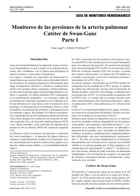
Figura 1. <strong>Monitoreo</strong> hemodinámico invasivo: posición <strong>de</strong>l catéter <strong>de</strong> <strong>arteria</strong> <strong>pulmonar</strong> en cavida<strong>de</strong>s <strong>de</strong>rechas. A: balón <strong>de</strong>l<br />
catéter inf<strong>la</strong>do en <strong>arteria</strong> <strong>pulmonar</strong>. B: Balón <strong>de</strong>l catéter inf<strong>la</strong>do en posición <strong>de</strong> enc<strong>la</strong>vamiento (wedge): el orificio distal <strong>de</strong>l<br />
catéter recibe información directa <strong>de</strong> <strong>la</strong> presión diastólica <strong>de</strong>l ventrículo izquierdo. La presión capi<strong>la</strong>r <strong>pulmonar</strong> (<strong>de</strong> enc<strong>la</strong>vamiento)<br />
refleja <strong>la</strong> presión <strong>de</strong> fin <strong>de</strong> diástole <strong>de</strong>l ventrículo izquierdo, siendo un indicador fundamental <strong>de</strong> <strong>la</strong> función cardíaca<br />
(precarga).<br />
nar (wedge) o PCP (en ausencia <strong>de</strong> valvulopatía mitral y <strong>de</strong><br />
estenosis <strong>de</strong> venas <strong>pulmonar</strong>es), así como <strong><strong>la</strong>s</strong> <strong>presiones</strong> sistólica<br />
y diastólica <strong>de</strong> <strong>la</strong> AP (Fig. 2).<br />
Las anomalías que afectan el corazón <strong>de</strong>recho y/o <strong>la</strong> circu<strong>la</strong>ción<br />
<strong>pulmonar</strong>, no impi<strong>de</strong>n <strong>la</strong> evaluación <strong>de</strong> <strong><strong>la</strong>s</strong> <strong>presiones</strong><br />
<strong>de</strong> llenado <strong>de</strong>l VI y su función. Por cierto, el catéter en <strong>la</strong><br />
AP, hace posible distinguir el EP cardiogénico <strong>de</strong>l no cardiogénico<br />
y establecer un diagnóstico hemodinámico diferencial<br />
para el tromboembolismo <strong>pulmonar</strong> (TEP) masivo.<br />
Se han producido muchas modificaciones al catéter origi-<br />
ECG<br />
AD VD AP PCP<br />
PRESION DE<br />
AURICULA DERECHA<br />
0-8 mm Hg<br />
PRESION DE<br />
VENTRICULO DERECHO<br />
Sistólica: 20-30 mm Hg<br />
Diastólica: 0-8 mm Hg<br />
PRESION DE<br />
ANTERIA PULMONAR<br />
Sistólica: 20-30 mm Hg<br />
Diastólica: 8-15 mm Hg<br />
PRESION DE<br />
CAPILAR PULMONAR<br />
(ENCLAVAMIENTO)<br />
8-12 mm Hg<br />
Figura 2. Características y valores normales <strong>de</strong> <strong><strong>la</strong>s</strong> <strong>presiones</strong> <strong>de</strong> cavida<strong>de</strong>s <strong>de</strong>rechas (AD, VD, AP y PCP) durante <strong>la</strong> introducción<br />
<strong>de</strong>l catéter <strong>de</strong> <strong>arteria</strong> <strong>pulmonar</strong> (<strong>Swan</strong>-Ganz). AD: Aurícu<strong>la</strong> <strong>de</strong>recha. VD: Ventrículo <strong>de</strong>recho. AP: Arteria <strong>pulmonar</strong>. PCP: Presión<br />
capi<strong>la</strong>r <strong>pulmonar</strong>.
INSUFICIENCIA CARDIACA<br />
Vol. 2, Nº 1, 2007<br />
7 A Lauga y A D’Ortencio<br />
Catéter <strong>de</strong> <strong>Swan</strong>-Ganz - Parte I<br />
CATETER<br />
ORIFICIO<br />
DISTAL<br />
CATETER<br />
ORIFICIO<br />
PROXIMAL<br />
CONEXION A<br />
COMPUTADORA PARA<br />
MEDICION DEL<br />
GASTO CARDIACO<br />
SISTEMA DE<br />
INFLACION<br />
DEL BALON<br />
ORIFICIO DE<br />
SALIDA DISTAL<br />
BALON<br />
ORIFICIO DE<br />
SALIDA PROXIMAL<br />
TERMISTOR<br />
Figura 3. Catéter <strong>de</strong> <strong>arteria</strong> <strong>pulmonar</strong> (<strong>Swan</strong>-Ganz) <strong>de</strong> 4 lúmenes (el más utilizado).<br />
nal 21,22 . Actualmente, se pue<strong>de</strong> monitorear continua o intermitentemente<br />
el gasto cardíaco 23 (GC), <strong>de</strong>terminar <strong>la</strong> fracción<br />
<strong>de</strong> eyección (FE) <strong>de</strong>l VD, medir en forma continua <strong>la</strong><br />
presión <strong>de</strong> aurícu<strong>la</strong> <strong>de</strong>recha (AD) y <strong>la</strong> saturación <strong>de</strong> oxígeno<br />
<strong>de</strong> <strong>la</strong> sangre venosa mixta, así como marcapasear <strong>la</strong> aurícu<strong>la</strong><br />
o el ventrículo (Fig. 3). Las resistencias vascu<strong>la</strong>res sistémicas<br />
(RVS) y <strong>pulmonar</strong>es 24 , el consumo y transporte <strong>de</strong><br />
oxígeno, <strong>la</strong> diferencia arteriovenosa <strong>de</strong> oxígeno y <strong>la</strong> fracción<br />
<strong>de</strong> shunt intra<strong>pulmonar</strong> 25 se pue<strong>de</strong>n obtener a través <strong>de</strong><br />
mediciones hemodinámicas y <strong>de</strong> gasometría <strong>arteria</strong>l 26 .<br />
Sin embargo, el beneficio clínico potencial <strong>de</strong> estos dispositivos<br />
<strong>de</strong> monitorización <strong>de</strong>pen<strong>de</strong> <strong>de</strong> <strong>la</strong> habilidad <strong>de</strong>l profesional<br />
para insertar el catéter y mantenerlo en el lugar<br />
a<strong>de</strong>cuado, <strong>de</strong> su capacidad para obtener e interpretar <strong><strong>la</strong>s</strong><br />
mediciones hemodinámicas, <strong>de</strong> corre<strong>la</strong>cionar <strong>la</strong> información<br />
con los datos clínicos y <strong>de</strong> <strong>la</strong>boratorio e integrar toda <strong>la</strong> información<br />
para llevar a cabo una terapéutica a<strong>de</strong>cuada.<br />
Fundamento<br />
Figura 4. Catéter <strong>de</strong> <strong>Swan</strong>-Ganz con balón inf<strong>la</strong>do en <strong>la</strong> <strong>arteria</strong><br />
<strong>pulmonar</strong>.<br />
Los catéteres con un balón inf<strong>la</strong>do en su extremo distal son<br />
llevados por <strong>la</strong> circu<strong>la</strong>ción sanguínea por arrastre y guiados<br />
<strong>de</strong>s<strong>de</strong> <strong><strong>la</strong>s</strong> gran<strong>de</strong>s venas intratorácicas hacia <strong>la</strong> AD y <strong>de</strong> allí<br />
atravesando <strong>la</strong> VT al VD, y a través <strong>de</strong> <strong>la</strong> válvu<strong>la</strong> <strong>pulmonar</strong><br />
(VP) a <strong>la</strong> AP (Fig. 4). El balón inf<strong>la</strong>do, que protruye sobre<br />
<strong>la</strong> punta misma <strong>de</strong>l catéter como un salvavidas, protege <strong>de</strong><br />
este modo a <strong>la</strong> misma <strong>de</strong> chocar con el endocardio (ya sea a<br />
causa <strong>de</strong> <strong>la</strong> manipu<strong>la</strong>ción o por efecto <strong>de</strong> los <strong>la</strong>tidos cardíacos),<br />
merced a una superficie amplia y suave, previniendo<br />
<strong>la</strong> aparición <strong>de</strong> arritmias y/o evitando dañar al endocardio.<br />
El registro <strong>de</strong> <strong>la</strong> PCP pue<strong>de</strong> lograrse <strong>de</strong>s<strong>de</strong> una AP gran<strong>de</strong><br />
sin necesidad <strong>de</strong> enc<strong>la</strong>var el catéter. Es más, una vez enc<strong>la</strong>vado<br />
el balón y tomada <strong>la</strong> presión wedge o <strong>de</strong> enc<strong>la</strong>vamiento<br />
se <strong>de</strong>be <strong>de</strong>sinf<strong>la</strong>r el balón y <strong>de</strong>jarlo flotando en <strong>la</strong> AP. Si<br />
esta presión wedge no difiere en más <strong>de</strong> 6 mm Hg <strong>de</strong> <strong>la</strong><br />
presión diastólica <strong>de</strong> <strong>la</strong> AP, será <strong>de</strong> referencia como PCP y
INSUFICIENCIA CARDIACA<br />
Vol. 2, Nº 1, 2007<br />
así evitar complicaciones como por ejemplo un infarto <strong>pulmonar</strong><br />
por <strong>de</strong>jar olvidado el balón enc<strong>la</strong>vado e inf<strong>la</strong>do. La<br />
presión <strong>de</strong> <strong>la</strong> AP pue<strong>de</strong> obtenerse <strong>de</strong> <strong>la</strong> misma posición sin<br />
mayor manipu<strong>la</strong>ción.<br />
Indicaciones para <strong>la</strong> cateterización <strong>de</strong> <strong>la</strong><br />
<strong>arteria</strong> <strong>pulmonar</strong><br />
No existen reg<strong><strong>la</strong>s</strong> absolutas que <strong>de</strong>finan <strong>la</strong> necesidad <strong>de</strong> un<br />
catéter <strong>de</strong> <strong>Swan</strong>-Ganz (SG). Generalmente, se indica en<br />
pacientes en los que <strong><strong>la</strong>s</strong> <strong>presiones</strong>, los flujos y los volúmenes<br />
circu<strong>la</strong>ntes requieren un manejo preciso e intensivo 27 .<br />
Los objetivos terapéuticos basados en <strong>la</strong> información obtenida<br />
son:<br />
1- Mejorar el GC y <strong>la</strong> oxigenación tisu<strong>la</strong>r.<br />
2- Aliviar o prevenir <strong><strong>la</strong>s</strong> anormalida<strong>de</strong>s <strong>pulmonar</strong>es como<br />
el EP <strong>de</strong> tipo cardiogénico.<br />
3- Evaluación <strong>de</strong> <strong>la</strong> función cardiovascu<strong>la</strong>r y <strong>la</strong> respuesta a<br />
<strong>la</strong> terapia en pacientes con:<br />
- Infarto <strong>de</strong> miocardio complicado (IAM) 28-34 .<br />
- Shock cardiogénico.<br />
- Insuficiencia cardíaca congestiva (ICC) severa (miocardiopatía,<br />
pericarditis constrictiva) 35-38 .<br />
- Alteraciones estructurales agudas (ruptura <strong>de</strong>l septum interventricu<strong>la</strong>r).<br />
- Disfunción <strong>de</strong>l VD.<br />
- Lesiones valvu<strong>la</strong>res (regurgitación mitral aguda).<br />
- Taponamiento cardíaco.<br />
- <strong>Monitoreo</strong> perioperatorio <strong>de</strong>l paciente <strong>de</strong> cirugía cardiovascu<strong>la</strong>r.<br />
- Toda c<strong><strong>la</strong>s</strong>e <strong>de</strong> shock.<br />
4- Evaluación <strong>de</strong>l estado <strong>pulmonar</strong> y respuesta a <strong>la</strong> terapéutica<br />
en pacientes con:<br />
- EP cardiogénico o no cardiogénico.<br />
- TEP.<br />
- Insuficiencia respiratoria aguda.<br />
- Hipertensión <strong>pulmonar</strong> (HTP) para diagnóstico y tratamiento.<br />
5- Evaluación <strong>de</strong> requerimiento <strong>de</strong> fluidos en pacientes con:<br />
- Trauma multisistémico severo.<br />
- Gran<strong>de</strong>s quemados.<br />
- Sepsis.<br />
6- <strong>Monitoreo</strong> perioperatorio <strong>de</strong> pacientes sometidos a cirugía<br />
mayor, siendo portadores <strong>de</strong> patología <strong>de</strong> alto riesgo 39,40 .<br />
7- Evaluación <strong>de</strong> pacientes obstétricas con ec<strong>la</strong>mpsia, complicada<br />
con hipertensión refractaria, oliguria y/o EP.<br />
Contraindicaciones y consi<strong>de</strong>raciones<br />
especiales<br />
8 A Lauga y A D’Ortencio<br />
Catéter <strong>de</strong> <strong>Swan</strong>-Ganz - Parte I<br />
No existen contraindicaciones absolutas. Sin embargo, el<br />
monitoreo hemodinámico invasivo no se justifica en el caso<br />
que <strong>la</strong> patología <strong>de</strong>l paciente no pue<strong>de</strong> ser modificada o corregida<br />
con medicación, no siendo <strong>de</strong>cisivo para realizar un<br />
diagnóstico diferencial ni fundamental para <strong>la</strong> toma <strong>de</strong> una<br />
conducta terapéutica. Las contraindicaciones re<strong>la</strong>tivas incluyen<br />
41-43 :<br />
1- Pacientes con coagulopatías severas o terapia trombolítica,<br />
por el riesgo <strong>de</strong> hemorragia durante y <strong>de</strong>spués <strong>de</strong>l acceso<br />
venoso.<br />
2- Pacientes con VT protésica, porque el catéter pue<strong>de</strong> dañar<strong>la</strong><br />
o causar el mal funcionamiento <strong>de</strong> <strong>la</strong> misma.<br />
3- Pacientes con marcapasos endocárdicos, porque el catéter<br />
<strong>de</strong> SG pue<strong>de</strong> alterar <strong>la</strong> ubicación <strong>de</strong>l mismo o anudarse<br />
alre<strong>de</strong>dor <strong>de</strong> él.<br />
4- Pacientes con una enfermedad vascu<strong>la</strong>r severa o por <strong>la</strong><br />
presencia <strong>de</strong> vasos sanguíneos tortuosos. La anormalidad<br />
<strong>de</strong> <strong><strong>la</strong>s</strong> pare<strong>de</strong>s vascu<strong>la</strong>res sistémicas y/o <strong>pulmonar</strong>es también<br />
implica un alto riesgo <strong>de</strong> daño o ruptura <strong>de</strong> <strong><strong>la</strong>s</strong> mismas.<br />
5- Pacientes con HTP por <strong>la</strong> inci<strong>de</strong>ncia <strong>de</strong> ruptura <strong>de</strong> <strong>la</strong> AP,<br />
que es mayor en aquellos vasos distendidos y friables, o en<br />
aquellos que presentan altas <strong>presiones</strong> <strong>pulmonar</strong>es.<br />
6- Pacientes con una <strong>de</strong>ficiencia en el sistema inmunológico<br />
como en el caso <strong>de</strong>: embarazo, fal<strong>la</strong> renal, síndrome <strong>de</strong><br />
inmuno<strong>de</strong>ficiencia adquirida (SIDA) o congénita, por el<br />
aumento <strong>de</strong>l riesgo <strong>de</strong> sepsis asociada al catéter.<br />
7- Pacientes que están en lugares que no cuentan con profesionales<br />
entrenados en <strong>la</strong> colocación <strong>de</strong>l catéter, así como,<br />
en <strong>la</strong> interpretación y manejo <strong>de</strong> <strong><strong>la</strong>s</strong> <strong>presiones</strong> intravascu<strong>la</strong>res<br />
y datos hemodinámicos.<br />
Una consi<strong>de</strong>ración especial en <strong>la</strong> colocación <strong>de</strong> este tipo <strong>de</strong><br />
catéteres son los pacientes con una hipotensión sistémica<br />
severa o un bajo GC. La disminución <strong>de</strong>l flujo sanguíneo a<br />
través <strong>de</strong>l corazón <strong>de</strong>recho, hace difícil el <strong>de</strong>sp<strong>la</strong>zamiento y<br />
<strong>la</strong> a<strong>de</strong>cuada ubicación <strong>de</strong>l catéter. En estos pacientes, es<br />
necesario mejorar el estado circu<strong>la</strong>torio con el aporte <strong>de</strong><br />
volumen (para mejorar <strong>la</strong> precarga) o con medicación inotrópica.<br />
A veces, es necesario colocar al paciente en posición<br />
<strong>de</strong> Azou<strong>la</strong>y (posición en <strong>de</strong>cúbito supino con los brazos<br />
y piernas levantados) para optimizar el retorno venoso<br />
central. En otros casos, es necesaria <strong>la</strong> colocación <strong>de</strong> este<br />
catéter bajo control radioscópico (Fig. 5).<br />
Tipos <strong>de</strong> catéteres para<br />
<strong>la</strong> <strong>arteria</strong> <strong>pulmonar</strong><br />
Los catéteres para <strong>la</strong> AP están disponibles en varios tamaños,<br />
ya sea que se utilicen para pacientes pediátricos o adultos.<br />
El catéter <strong>de</strong> SG (Flow Directed Catheter, Edwards Laboratories,<br />
Santa Ana, California, EE.UU.) está constituido<br />
(Fig. 3) <strong>de</strong> cloruro <strong>de</strong> polivinilo y tiene un cuerpo flexible<br />
que se ab<strong>la</strong>nda aún más con <strong>la</strong> temperatura <strong>de</strong>l cuerpo. Posee<br />
un balón a 1 ó 2 mm <strong>de</strong> <strong>la</strong> punta. El catéter común es <strong>de</strong><br />
110 cm <strong>de</strong> <strong>la</strong>rgo y <strong>de</strong> 5 ó 7 French (F) <strong>de</strong> diámetro externo.<br />
Los volúmenes <strong>de</strong> inf<strong>la</strong>do <strong>de</strong>l balón van <strong>de</strong> 0,5 a 1,5 ml y el<br />
diámetro <strong>de</strong> los mismos <strong>de</strong> 8 a 13 mm. Ambos tamaños tienen<br />
2 lúmenes: uno pequeño para inf<strong>la</strong>r el balón y uno más<br />
gran<strong>de</strong> que se abre en <strong>la</strong> punta <strong>de</strong>l catéter y que sirve para<br />
registrar <strong>presiones</strong> y tomar muestras <strong>de</strong> sangre. El catéter<br />
<strong>de</strong> calibre 7 F tiene una curvatura preformada en su extremo<br />
distal a fin <strong>de</strong> facilitar su pasaje por el VD. Para cateterismos<br />
pediátricos hay disponibles catéteres más cortos (<strong>de</strong><br />
60 cm <strong>de</strong> <strong>la</strong>rgo) y en diámetros <strong>de</strong> 4 y 5 F. Existen catéteres<br />
<strong>de</strong> triple lumen (7 F: 110 cm <strong>de</strong> <strong>la</strong>rgo), con orificio proximal<br />
y distal que permiten <strong>la</strong> medición simultánea <strong>de</strong> <strong>la</strong> pre-
INSUFICIENCIA CARDIACA<br />
Vol. 2, Nº 1, 2007<br />
9 A Lauga y A D’Ortencio<br />
Catéter <strong>de</strong> <strong>Swan</strong>-Ganz - Parte I<br />
Figura 5. Rx <strong>de</strong> tórax <strong>de</strong> paciente con colocación <strong>de</strong> catéter en <strong>arteria</strong>l <strong>pulmonar</strong> y shock cardiogénico.<br />
sión <strong>de</strong> AD y <strong>de</strong> AP y capi<strong>la</strong>r <strong>pulmonar</strong> 44-50 .<br />
En todos los catéteres, el balón se hal<strong>la</strong> montado en tal forma<br />
que cuando se lo inf<strong>la</strong> con más <strong>de</strong> 0,8 ml <strong>de</strong> gas, protruye<br />
por encima y alre<strong>de</strong>dor <strong>de</strong> <strong>la</strong> punta <strong>de</strong>l catéter. El catéter<br />
es radioopaco y pue<strong>de</strong> ser visualizado por fluoroscopía si<br />
es necesario.<br />
El catéter presenta marcas cada 10 cm, constituidas en bandas<br />
estrechas <strong>de</strong> color negro <strong>de</strong> 1 a 4, o sea <strong>de</strong> 10 a 40 cm, <strong>la</strong><br />
marca <strong>de</strong> los 50 cm es una banda más gruesa que <strong><strong>la</strong>s</strong> anteriores<br />
y <strong>de</strong>spués <strong>de</strong> ésta se van agregando bandas finas nuevamente<br />
para los 60, 70, 80 y 90 cm, esto ayuda a <strong>de</strong>terminar<br />
<strong>la</strong> ubicación <strong>de</strong> <strong>la</strong> punta <strong>de</strong>l catéter.<br />
Actualmente, <strong>la</strong> disponibilidad <strong>de</strong> catéteres es sumamente<br />
amplia. Hay catéteres <strong>de</strong> dos lúmenes, <strong>de</strong> cuatro, (es el que<br />
se usa más frecuentemente), los hay <strong>de</strong> 5 lúmenes (tienen<br />
una vía proximal adicional para administración <strong>de</strong> fluidos)<br />
y el catéter <strong>de</strong> fibra óptica que mi<strong>de</strong> continuamente <strong>la</strong> saturación<br />
<strong>de</strong> sangre venosa mixta. También, está el que tiene<br />
incorporados 5 electrodos que se pue<strong>de</strong>n utilizar para marcapasear<br />
al paciente o el que tiene un lumen adicional para<br />
<strong>la</strong> introducción <strong>de</strong> un catéter marcapasos temporario y aquél<br />
que pue<strong>de</strong> calcu<strong>la</strong>r <strong>la</strong> fracción <strong>de</strong> eyección <strong>de</strong>l VD por medición<br />
<strong>de</strong> los volúmenes <strong>de</strong> fin <strong>de</strong> sístole y fin <strong>de</strong> diástole.<br />
En <strong>la</strong> actualidad, se cuenta con un catéter que informa continuamente<br />
sobre el GC por termodilución.<br />
El catéter <strong>de</strong> SG <strong>de</strong> cuatro lúmenes, que es el más utilizado<br />
para el paciente adulto, se presenta en tamaños <strong>de</strong> 5 y 7 F.<br />
La vía distal recorre <strong>la</strong> longitud <strong>de</strong>l catéter y se abre en <strong>la</strong><br />
punta <strong>de</strong>l mismo. Esta vía distal mi<strong>de</strong> <strong><strong>la</strong>s</strong> <strong>presiones</strong> <strong>de</strong> AP y<br />
<strong>la</strong> <strong>de</strong>l capi<strong>la</strong>r <strong>pulmonar</strong> enc<strong>la</strong>vado. Se pue<strong>de</strong>n obtener muestras<br />
<strong>de</strong> sangre venosa mixta por esta vía cuando <strong>la</strong> punta <strong>de</strong>l<br />
catéter está posicionada en <strong>la</strong> AP.<br />
No se <strong>de</strong>be administrar ningún tipo <strong>de</strong> soluciones hiperosmo<strong>la</strong>res<br />
o drogas por esta vía, ya que <strong>la</strong> infusión <strong>de</strong> este<br />
tipo <strong>de</strong> soluciones en <strong>la</strong> AP pue<strong>de</strong> causar daños o reacciones<br />
tisu<strong>la</strong>res severas.<br />
La vía que permite el inf<strong>la</strong>do <strong>de</strong>l balón termina <strong>de</strong>ntro <strong>de</strong> éste.<br />
La vía proximal termina en un orificio que se abre aproximadamente<br />
a 30 cm <strong>de</strong> <strong>la</strong> punta <strong>de</strong>l catéter. Esta vía se ubica en <strong>la</strong> AD<br />
cuando <strong>la</strong> punta <strong>de</strong>l catéter se encuentra en <strong>la</strong> AP.<br />
La vía proximal se pue<strong>de</strong> utilizar para medir <strong><strong>la</strong>s</strong> <strong>presiones</strong><br />
<strong>de</strong> <strong>la</strong> AD, administrar líquidos intravenosos y electrolitos,<br />
algún tipo <strong>de</strong> medicamentos, tomar muestras <strong>de</strong> sangre <strong>de</strong><br />
<strong>la</strong> AD e inyectar <strong><strong>la</strong>s</strong> soluciones para <strong>de</strong>terminar el GC por<br />
termodilución 51,52 .<br />
La vía proximal no <strong>de</strong>bería usarse para infundir drogas vasoactivas<br />
y/o inotrópicas, mientras se realizan <strong><strong>la</strong>s</strong> mediciones<br />
por termodilución, ya que el paciente recibiría <strong>de</strong> este<br />
modo minibolos <strong>de</strong> medicamentos cardiovascu<strong>la</strong>res altamente<br />
activos en cada <strong>de</strong>terminación <strong>de</strong>l GC.<br />
La vía <strong>de</strong>l termistor, tiene una cuerda termosensible, que<br />
termina aproximadamente a 4 ó 5 cm <strong>de</strong> <strong>la</strong> punta <strong>de</strong>l catéter.<br />
La porción terminal <strong>de</strong> esta cuerda, se ubica en una AP<br />
principal, cuando el catéter está correctamente ubicado. La<br />
conexión <strong>de</strong> esta vía <strong>de</strong>l catéter con una computadora <strong>de</strong><br />
volumen minuto, permite <strong>de</strong>terminar el GC <strong>de</strong>spués <strong>de</strong> <strong>la</strong><br />
inyección <strong>de</strong> una solución fría por <strong>la</strong> variación <strong>de</strong> <strong>la</strong> temperatura<br />
<strong>de</strong> <strong>la</strong> sangre.
INSUFICIENCIA CARDIACA<br />
Vol. 2, Nº 1, 2007<br />
Efectos <strong>de</strong> <strong>la</strong> caterización <strong>pulmonar</strong> en el<br />
alta <strong>de</strong>l paciente<br />
Si bien, el catéter <strong>pulmonar</strong> ha sido un avance muy importante<br />
en <strong>la</strong> disponibilidad <strong>de</strong> datos fisiológicos para el diagnóstico<br />
y manejo <strong>de</strong> los pacientes gravemente enfermos,<br />
po<strong>de</strong>mos preguntarnos 53-57 :<br />
¿El monitoreo hemodinámico reduce <strong>la</strong> estadía en el centro<br />
<strong>de</strong> salud?<br />
¿Reduce el costo <strong>de</strong> <strong>la</strong> atención <strong>de</strong>l paciente?<br />
¿Reduce <strong>la</strong> morbilidad y <strong>la</strong> mortalidad <strong>de</strong> los pacientes?<br />
No hay ningún estudio que documente los beneficios <strong>de</strong> <strong>la</strong><br />
colocación <strong>de</strong> los catéteres <strong>de</strong> AP. El problema no está en <strong>la</strong><br />
colocación <strong>de</strong>l catéter, sino en cómo se usa.<br />
El profesional que utilice este dispositivo <strong>de</strong>be:<br />
1- Tener una técnica impecable en <strong>la</strong> preparación <strong>de</strong>l catéter<br />
para su inserción y manejo.<br />
2- Ser capaz <strong>de</strong> interpretar <strong><strong>la</strong>s</strong> ondas hemodinámicas, los<br />
datos fisiológicos y sus re<strong>la</strong>ciones con el problema clínico<br />
subyacente.<br />
3- Tener conciencia <strong>de</strong> los errores y fal<strong><strong>la</strong>s</strong> que se pue<strong>de</strong>n<br />
producir en el sistema.<br />
4- Enten<strong>de</strong>r que <strong>la</strong> vigi<strong>la</strong>ncia <strong>de</strong>be ser continua para ajustar<br />
<strong>la</strong> terapéutica a <strong><strong>la</strong>s</strong> necesida<strong>de</strong>s <strong>de</strong>l paciente.<br />
Debemos enten<strong>de</strong>r que el uso <strong>de</strong> este tipo <strong>de</strong> catéteres no<br />
tiene un efecto positivo en el tratamiento <strong>de</strong>l paciente, si no<br />
se utiliza apropiadamente, si los datos obtenidos no son certeros<br />
o son malinterpretados y si <strong><strong>la</strong>s</strong> acciones terapéuticas<br />
no se toman en el momento apropiado.<br />
Referencias bibliográficas<br />
1. Wilson JN, Grow JB, Demong CV, Preve<strong>de</strong>l AE, Owens JC. Central<br />
venous pressure in optimal blood volume maintenance. Arch<br />
Surg 1962 Oct;85:563-78.<br />
2. Connors AF Jr, Speroff T, Dawson NV, Thomas C, Harrell FE Jr,<br />
Wagner D, Desbiens N, Goldman L, Wu AW, Califf RM, Fulkerson<br />
WJ Jr, Vidaillet H, Broste S, Bel<strong>la</strong>my P, Lynn J, Knaus WA.<br />
The effectiveness of right heart catheterization in the initial care<br />
of critically ill patients. SUPPORT Investigators. JAMA 1996;<br />
276:889-897.<br />
3. Díaz-Alersi R. Efectividad <strong>de</strong>l <strong>Swan</strong>-Ganz en el tratamiento <strong>de</strong>l<br />
paciente crítico. Un ensayo clínico contro<strong>la</strong>do. Revista Electrónica<br />
<strong>de</strong> Medicina Intensiva 2005;5(8):888.<br />
4. Hall JB. Searching for evi<strong>de</strong>nce to support <strong>pulmonar</strong>y artery catheter<br />
use in critically ill patients. JAMA 2005;294:1693-1694.<br />
5. Shah M, Hasselb<strong>la</strong>d V, Stevenson LW, Binanay C, O’Connor CM,<br />
Sopko G, Califa RM. Impact of the <strong>pulmonar</strong>y artery catheter in<br />
critically ill patients. Meta-analysis of Randomized Clinical Trials.<br />
JAMA 2005;294:1664-1670.<br />
6. Latego<strong>la</strong> M, Rahn H. A self-guiding catheter for cardiac and <strong>pulmonar</strong>y<br />
<strong>arteria</strong>l catheterization and occlusion. Proc Soc Exp Biol<br />
Med 1953;84:667.<br />
7. Duffy B. The clinical use of polyethylene tubing for intravenous<br />
therapy. Ann Surg 1949;130:929-936.<br />
8. Hermosura B, Vanagas L, Dickey M. Measurement of pressure<br />
during intravenous therapy. JAMA 1966;195:321.<br />
9. Lund GB, Treroto<strong>la</strong> SO, Scheel PFJ, et al. Outcome of tunneled<br />
hemodialysis catheters p<strong>la</strong>ced by radiologists. Radiology<br />
1996;198:467-472.<br />
10. Mauro M, Jaques P. Radiologie p<strong>la</strong>cement of long-term central<br />
venous catheters: a review. J Vase Interv Radial 1993;4:127137.<br />
11. Mauro MA. Interventional radiology p<strong>la</strong>cement of central venous<br />
catheters. Hasp Physician 1996;32:55-59.<br />
12. Hoshal V. Total intravenous nutrition with peripherally inserted<br />
silicone e<strong><strong>la</strong>s</strong>tomer central venous catheters. Arch Surg<br />
1975;110:644-646.<br />
10 A Lauga y A D’Ortencio<br />
Catéter <strong>de</strong> <strong>Swan</strong>-Ganz - Parte I<br />
13. Chrisman H, Omary R, Nemcek A, Ryu R, Saker M, Vogelzang R.<br />
Peripherally inserted central catheters: guidance with use of US<br />
versus venography in 2.650 patients. J Vase Interv Radial 1999;<br />
10:473-475.<br />
14. Fife WJ, Lee BS. Construction and use of self-guiding right heart<br />
and <strong>pulmonar</strong>y artery catheter. J Appl Physiol 1965;20:148.<br />
15. Dotter CT, Straube KR. Flow gui<strong>de</strong>d cardiac catheterization. Am<br />
J Roentgen 1962;88:27.<br />
16. Bradley RD. Diagnostic right heart catheterization with miniature<br />
catheters in·severely ill patients. Lancet 1964;2:P41.<br />
17. Carr I, Wells B. Coaxial flow-gui<strong>de</strong>d catheterization of the <strong>pulmonar</strong>y<br />
artery in transposition of the great arteries. Lancet<br />
1966;2:318.<br />
18. Winkler JB, Duprey R. Pulmonary angiography using a flow-gui<strong>de</strong>d<br />
catheter. Construction and applications. Radiol Techn<br />
1967;39:23.<br />
19. Scheinman MM, Abbott JA, Rapaport E. Clinical uses of a flowdirected<br />
right heart catheter. Arch Intern Med 1969;124:19,.<br />
20. <strong>Swan</strong> HJ, Ganz W, Forrester J, Marcus H, Diamond G, Chonette<br />
D. Catheterization of the heart in man with use of a flow-directed<br />
balloon-tipped catheter. N Engl J Med 1970 Aug 27;283(9):447-<br />
51.<br />
21. Steele P, Davies H: The <strong>Swan</strong>-Ganz catheter in the cardiac <strong>la</strong>boratory.<br />
Br Heart J 1973;35:647.<br />
22. Ganz P, <strong>Swan</strong> HJC et Grossman W. Balloon-Tipped Flow-Directed<br />
Catheters. Cardiac Catheterization and Angiography (Grossman<br />
W Editor). Lea & Febiger, Phi<strong>la</strong><strong>de</strong>lphia 1985.pp. 88-100.<br />
23. Grossman W. Blood Flow Measurements:The Cardiac Output. Cardiac<br />
Catheterization and Angiography (Grossman W Editor). Lea<br />
& Febiger, Phi<strong>la</strong><strong>de</strong>lphia 1985.pp. 101-117.<br />
24. Grossman W. Clinical Measurement of Vascu<strong>la</strong>r Resistance and<br />
Assessment of Vasodi<strong>la</strong>tor Drugs.Cardiac Catheterization and Angiography<br />
(Grossman W Editor). Lea & Febiger, Phi<strong>la</strong><strong>de</strong>lphia<br />
1985.pp. 135-142.<br />
25. Grossman W. Shunt Detection and Measurement. Cardiac Catheterization<br />
and Angiography (Grossman W Editor). Lea & Febiger,<br />
Phi<strong>la</strong><strong>de</strong>lphia 1985.pp. 155-172.<br />
26. Hathaway R. The <strong>Swan</strong>-Ganz catheter: a review. Nurs Clin North<br />
Am 1978;13(3):389-407.<br />
27. Scott ML, Webre DR, Arens JF, Ochsner JL. Clinical application<br />
of a flow-directed balloon-tipped cardiac catéter. Am Surg<br />
1972;38(12):690-6.<br />
28. <strong>Swan</strong> HJC, Ganz W. The use of balloon-tipped, flow-directed catheter<br />
in monitoring patient with acute myocardial infarction. In<br />
Corday E. <strong>Swan</strong> HJC (eds): Myocardial Infarction. Baltimore,<br />
Williams & Wilkins, 1973.<br />
29. Mond HG, Hunt D, Sloman G. Haemodynamic monitoring in the<br />
coronary care unit using the <strong>Swan</strong>-Ganz right heart catheter. Br<br />
Heart J 1973;35:235.<br />
30. Cohen MG, Kelly RV, Kong DF, et al. Pulmonary artery catheterization<br />
in acute coronary syndromes: Insights from the GUSTO<br />
IIb and GUSTO III trials. Am J Med 2005; 118; 482-488.<br />
31. Añón Elizal<strong>de</strong> JM. Catéter <strong>de</strong> <strong>Swan</strong>-Ganz en el síndrome coronario<br />
agudo. Revista Electrónica <strong>de</strong> Medicina Intensiva<br />
2005;5(11):913.<br />
32. Gore JM, Goldberg RJ, Spodick DH, et al. A community-wi<strong>de</strong> assessment<br />
of the use of <strong>pulmonar</strong>y artery catheters in patients with<br />
acute myocardial infarction. Chest 1987;92:721-727.<br />
33. Zion MM, Balkin J, Rosenmann D, et al. Use of <strong>pulmonar</strong>y artery<br />
catheters in patients with acute myocardial infarction. Analysis of<br />
experience in 5841 patients in the SPRINT Registry. SPRINT Study<br />
Group. Chest 1990;98:1331-1335.<br />
34. Sa<strong>la</strong>zar E, Gil M, Ramirez A, Pieniak M. Hemodynamic evaluation<br />
in acute myocardial infarct. Application to the treatment of<br />
contractile insufficiency syndromes of the left ventricle. Arch Inst<br />
Cardiol Mex 1976;46(4):414-32.<br />
35. Binanay C, Califf RM, Hasselb<strong>la</strong>d V, O’Connor CM, Shah MR,<br />
Sopko G, Stevenson LW, Francis GS, Leier CV, Miller LW; ES-<br />
CAPE Investigators and ESCAPE Study Coordinators. Evaluation<br />
study of congestive heart failure and <strong>pulmonar</strong>y artery catheterization<br />
effectiveness: the ESCAPE trial. JAMA 2005; 294(13):<br />
1625-1633.<br />
36. Díaz-Alersi R. El <strong>Swan</strong>-Ganz en <strong>la</strong> insuficiencia cardíaca congestiva.<br />
Revista Electrónica <strong>de</strong> Medicina Intensiva 2005;5(10):899.<br />
37. Won C, Kuschner WG. Pulmonary artery catheter effectiveness in<br />
congestive heart failure. JAMA 2006;295(10):1121.<br />
38. Stevenson et al. Pulmonary artery catheter effectiveness in con-
INSUFICIENCIA CARDIACA<br />
Vol. 2, Nº 1, 2007<br />
11 A Lauga y A D’Ortencio<br />
Catéter <strong>de</strong> <strong>Swan</strong>-Ganz - Parte I<br />
gestive heart failure - Reply. JAMA 2006;295:1122.<br />
39. Po<strong>la</strong>nczyk CA, Roh<strong>de</strong> LE, Goldman L, Cook EF, Thomas EJ, Marcantonio<br />
ER, Mangione CM, Lee TH. Right heart catheterization<br />
and cardiac complications in patients un<strong>de</strong>rgoing noncardiac surgery:<br />
an observational study. JAMA 2001;286(3):309-14.<br />
40. Marcelino P, Germano N, Marum S, Fernan<strong>de</strong>s AP, Ribeiro P, Lopes<br />
MG. Haemodynamic parameters obtained by transthoracic<br />
echocardiography and <strong>Swan</strong>-Ganz catheter: a comparative study<br />
in liver transp<strong>la</strong>nt patients. Acta Med Port 2006;19(3):197-205.<br />
41. Gauntlett Beare P, Myers JL. Enfermería Medico-Quirúrgica. Vol.<br />
2. Elsevier Science España, 2002.<br />
42. Grif Aspach JA. Cuidados intensivos <strong>de</strong> enfermería en el adulto,<br />
México, Mc Graw - Hill, 2001.<br />
43. Ur<strong>de</strong>n LD, Lough ME, Stacy KM. Cuidados intensivos en enfermería.<br />
Harcourt Brace, España, 2003.<br />
44. Kelly DT, Krovetz LJ, Rowe RD. Double-lumen flotation catheter<br />
for use in complex congenital cardiac anomalies. Circu<strong>la</strong>tion<br />
1971;44:910.<br />
45. Stanger P, Heymann MA, Hoffman JIE, Rudolph AM. Use of <strong>Swan</strong>-<br />
Ganz catheter in cardiac catheterization of infants and children.<br />
Am Heart J 1972;83:749.<br />
46. B<strong>la</strong>ck JFS. Floating a catheter into the <strong>pulmonar</strong>y artery in transposition<br />
of the great arteries. Am Heart J 1972;84:761.<br />
47. Jones SM, Miller GAH: Catheterization of the <strong>pulmonar</strong>y artery<br />
in transposition of the great arteries using a <strong>Swan</strong>-Ganz flow-directed<br />
catheter. Br Heart J 1973;35:298.<br />
48. Johnson CD. Value of the <strong>Swan</strong>-Ganz balloon cathether in the diagnosis<br />
of transposition of the great arteries in an adult. Bol Asoc<br />
Med P R 1976;68(11):292-5.<br />
49. Rautenburg HW, Menner K, Wagner HJ. Intracardiac pressure measurements<br />
with the gradjean microcatheter method in children. Monatsschr<br />
Kin<strong>de</strong>rheilkd 1970;118(6):301-303.<br />
50. Gaissmaier U, K<strong>la</strong>us D, Sadowski P, Gauss D, Trubestein G.<br />
Grandjean’s microcatheterization for right heart sounding. Med<br />
Klin 1971;66(46):1558-1564.<br />
51. Branthwaite MA, Bradley RD: Measurement of cardiac output by<br />
thermodilution in man. J Appl Physiol 1968;24:434.<br />
52. Ganz W, Donoso R, Marcus HS, Forrester JS, <strong>Swan</strong> HJC. A new<br />
technique for measurement of cardiac output by thermodilution in<br />
man. Am J Cardiol 1971;27:392.<br />
53. Dalen JE, Bone RC. Is it time to pull the <strong>pulmonar</strong>y artery catheter?<br />
JAMA 1996; 276: 916-918.<br />
54. Harvey S, Stevens K, Harrison D, Young D, Brampton W, McCabe<br />
C, Singer M, Rowan K. An evaluation of the clinical and costeffectiveness<br />
of <strong>pulmonar</strong>y artery catheters in patient management<br />
in intensive care: a systematic review and a randomized controlled<br />
trial. Health Technol Assess 2006(29):iii-iv, ix-xi, 1-133.<br />
55. Moran JL, Peisach AR, Solomon PJ, Martin J. Cost calcu<strong>la</strong>tion<br />
and prediction in adult intensive care: a ground-up utilization study.<br />
Anaesth Intensive Care 2004;32(6):787-97.<br />
56. Ip SP, Leung YF, Ip CY, Mak WP. Outcomes of critically ill el<strong>de</strong>rly<br />
patients: is high-<strong>de</strong>pen<strong>de</strong>ncy care for geriatric patients worthwhile?<br />
Crit Care Med 1999;27(11):2351-7.<br />
57. Ely EW, Evans GW, Haponik EF. Mechanical venti<strong>la</strong>tion in a cohort<br />
of el<strong>de</strong>rly patients admitted to an intensive care unit. Ann<br />
Intern Med 1999 Jul 20;131(2):96-104.