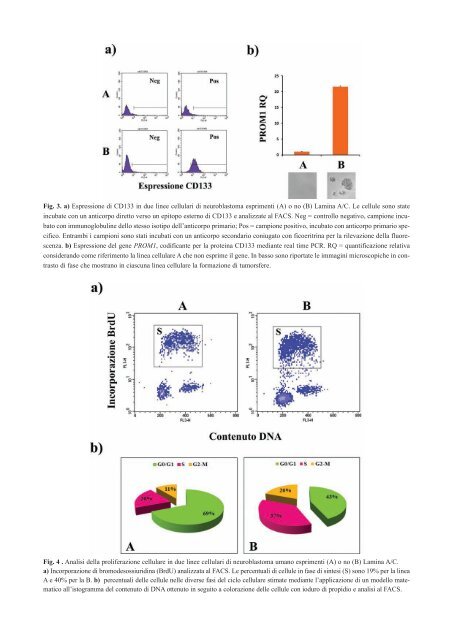
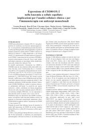
Fig. 3. a) Espressione di CD133 in due linee cellulari di neuroblastoma esprimenti (A) o no (B) Lamina A/C. Le <strong>cellule</strong> sono state incubate con un anticorpo diretto verso un epitopo esterno di CD133 e analizzate al FACS. Neg = controllo negativo, campione incubato con immunoglobuline dello stesso isotipo dell’anticorpo primario; Pos = campione positivo, incubato con anticorpo primario specifico. Entrambi i campioni sono stati incubati con un anticorpo secondario coniugato con ficoeritrina per la rilevazione della fluorescenza. b) Espressione del gene PROM1, codificante per la proteina CD133 mediante real time PCR. RQ = quantificazione relativa considerando come riferimento la linea cellulare A che non esprime il gene. In basso sono riportate le immagini microscopiche in contrasto di fase che mostrano in ciascuna linea cellulare la formazione di tumorsfere. Fig. 4 . Analisi della proliferazione cellulare in due linee cellulari di neuroblastoma umano esprimenti (A) o no (B) Lamina A/C. a) Incorporazione di bromodesossiuridina (BrdU) analizzata al FACS. Le percentuali di <strong>cellule</strong> in fase di sintesi (S) sono 19% per la linea A e 40% per la B. b) percentuali <strong>delle</strong> <strong>cellule</strong> nelle diverse fasi del ciclo cellulare stimate mediante l’applicazione di un modello matematico all’istogramma del contenuto di DNA ottenuto in seguito a colorazione <strong>delle</strong> <strong>cellule</strong> con ioduro di propidio e analisi al FACS.
lamin A/C gene by CpG island promoter hypermethylation in hematologic malignancies, and its association with poor survival in nodal diffuse large B-cell lymphoma. J. Clin. Oncol. 23, 3940-3947. 2) Andres, V. and Gonzalez, J.M. (2009). Role of A-type lamins in signaling, transcription, and chromatin organization. J. Cell Biol. 187, 945-957. 3) Ben,Y.R., Muchir, A., Arimura, T., Massart, C., Demay, L., Richard, P., and Bonne, G. (2005). Genetics of laminopathies. Novartis. Found. Symp. 264, 81-90. 4) Broers, J.L., Ramaekers, F.C., Bonne, G., Yaou, R.B., and Hutchison, C.J. (2006). Nuclear lamins: laminopathies and their role in premature ageing. Physiol Rev. 86, 967-1008. 5) Constantinescu, D., Gray, H.L., Sammak, P.J., Schatten, G.P., and Csoka, A.B. (2006). Lamin A/C expression is a marker of mouse and human embryonic stem cell differentiation. Stem Cells 24, 177-185. 6) Edsjo, A., Holmquist, L., and Pahlman, S. (2007). Neuroblastoma as an experimental model for neuronal differentiation and hypoxia-induced tumor cell dedifferentiation. Semin. Cancer Biol. 17, 248-256. 7) Espada, J., Varela, I., Flores, I., Ugalde, A.P., Cadinanos, J., Pendas, A.M., Stewart, C.L., Tryggvason, K., Blasco, M.A., Freije, J.M., and <strong>Lo</strong>pez-Otin, C. (2008). Nuclear envelope defects cause stem cell dysfunction in premature-aging mice. J. Cell Biol. 181, 27-35. 8) Francastel, C., Schubeler, D., Martin, D.I., and Groudine, M. (2000). Nuclear compartmentalization and gene activity. Nat. Rev. Mol. Cell Biol. 1, 137-143. 9) Frock, R.L., Kudlow, B.A., Evans, A.M., Jameson, S.A., Hauschka, S.D., and Kennedy, B.K. (2006). Lamin A/C and emerin are critical for skeletal muscle satellite cell differentiation. Genes Dev. 20, 486-500. 10) Gatti, G., Maresca, G., Natoli, M., Florenzano, F., Nicolin, A., Felsani, A., and D’Agnano, I. (2009). MYC prevents apoptosis and enhances endoreduplication induced by paclitaxel. P<strong>Lo</strong>S. One. 4, e5442. 11) Houben, F., Ramaekers, F.C., Snoeckx, L.H., and Broers, J.L. (2007). Role of nuclear lamina-cytoskeleton interactions in the maintenance of cellular strength. Biochim. Biophys. Acta 1773, 675-686. 12) Hutchison, C.J. (2002). Lamins: building blocks or regulators of gene expression? Nat. Rev. Mol. Cell Biol. 3, 848- 858. 13) Janaki, R.M. and Parnaik, V.K. (2006). An essential GT motif in the lamin A promoter mediates activation by CREB-binding protein. Biochem. Biophys. Res. Commun. 348, 1132-1137. 14) Ketema, M., Wilhelmsen, K., Kuikman, I., Janssen, H., Hodzic, D., and Sonnenberg, A. (2007). Requirements for the localization of nesprin-3 at the nuclear envelope and its interaction with plectin. J. Cell Sci. 120, 3384-3394. 15) Lehner, C.F., Stick, R., Eppenberger, H.M., and Nigg, E.A. (1987). Differential expression of nuclear lamin proteins during chicken development. J. Cell Biol. 105, 577-587. 16) Lin, F. and Worman, H.J. (1997). Expression of nuclear lamins in human tissues and cancer cell lines and transcription from the promoters of the lamin A/C and B1 genes. Exp. Cell Res. 236, 378-384. 17) Lloyd, D.J., Trembath, R.C., and Shackleton, S. (2002). A novel interaction between lamin A and SREBP1: implica- Lettere GIC Vol. 20, Num. 2 - Agosto 2011 tions for partial lipodystrophy and other laminopathies. Hum. Mol. Genet. 11, 769-777. 18) <strong>Lo</strong>wry, W.E. and Richter, L. (2007). Signaling in adult stem cells. Front Biosci. 12, 3911-3927. 19) Maresca, G., Natoli, M., Arisi, I., Trisciuoglio, D., Brandi, R., D’Aguanno,S., D’Onofrio, M., Urbani, A., Del Bufalo, D., Felsani, A., and D’Agnano, I. Knock-down of Lamin A/C affects differentiation and progression of human neuroblastoma cells. Submitted to Cancer Research. 20) Meshorer, E. and Misteli, T. (2006). Chromatin in pluripotent embryonic stem cells and differentiation. Nat. Rev. Mol. Cell Biol. 7, 540-546. 21) Moir, R.D., Spann, T.P., Herrmann, H., and Goldman, R.D. (2000). Disruption of nuclear lamin organization blocks the elongation phase of DNA replication. J. Cell Biol. 149, 1179-1192. 22) Natoli, M., Leoni, B.D., D’Agnano, I., D’Onofrio, M., Brandi, R., Arisi, I., Zucco, F., and Felsani, A. (2011). Cell growing density affects the structural and functional properties of Caco-2 differentiated monolayer. J. Cell Physiol 226, 1531-1543. 23) Okumura, K., Hosoe, Y., and Nakajima, N. (2004). c-Jun and Sp1 family are critical for retinoic acid induction of the lamin A/C retinoic acid-responsive element. Biochem. Biophys. Res. Commun. 320, 487-492. 24) Prokocimer, M., Davidovich, M., Nissim-Rafinia, M., Wiesel- Motiuk, N., Bar, D.Z., Barkan, R., Meshorer, E., and Gruenbaum,Y. (2009). Nuclear lamins: key regulators of nuclear structure and activities. J. Cell Mol. Med. 13, 1059-1085. 25) Reddy, K.L., Zullo, J.M., Bertolino, E., and Singh, H. (2008). Transcriptional repression mediated by repositioning of genes to the nuclear lamina. Nature 452, 243-247. 26) Riemer, D., Stuurman, N., Berrios, M., Hunter, C., Fisher, P.A., and Weber, K. (1995). Expression of Drosophila lamin C is developmentally regulated: analogies with vertebrate A-type lamins. J. Cell Sci. 108 ( Pt 10), 3189-3198. 27) Rober, R.A., Weber, K., and Osborn, M. (1989). Differential timing of nuclear lamin A/C expression in the various organs of the mouse embryo and the young animal: a developmental study. Development 105, 365-378. 28) Schirmer, E.C. and Foisner, R. (2007). Proteins that associate with lamins: many faces, many functions. Exp. Cell Res. 313, 2167-2179. 29) Spann, T.P., Goldman,A.E., Wang, C., Huang, S., and Goldman, R.D. (2002). Alteration of nuclear lamin organization inhibits RNA polymerase II-dependent transcription. J. Cell Biol. 156, 603-608. 30) Stick, R. and Hausen,P. (1985). Changes in the nuclear lamina composition during early development of Xenopus laevis. Cell 41, 191-200. 31) Takamori, Y., Tamura, Y., Kataoka, Y., Cui, Y., Seo, S., Kanazawa, T., Kurokawa, K., and Yamada, H. (2007). Differential expression of nuclear lamin, the major component of nuclear lamina, during neurogenesis in two germinal regions of adult rat brain. Eur. J. Neurosci. 25, 1653-1662. ATTIVITÀ SCIENTIFICA 19